a study of herniotomy alone in young adults with
TRANSCRIPT
1
A STUDY OF HERNIOTOMY ALONE IN YOUNG ADULTS WITH
UNCOMPLICATED INDIRECT INGUINAL HERNIA IN HOSPITAL
SEBERANG JAYA, PULAU PINANG
By
DR. SYAHRUL ANUAR BIN SALLEH
DISSERTATION SUBMITTED IN PARTIAL FULFILLMENT OF
THE REQUIREMENT FOR THE DEGREE OF MASTERS OF
MEDICINE (GENERAL SURGERY)
UNIVERSITI SAINS MALAYSIA
2016

2
TABLE OF CONTENTS
I. ACKNOWLEDGEMENTS
II. ABSTRAK (BAHASA MELAYU)
III. ABSTRACT
1. INTRODUCTION
1.1 LITERATURE REVIEW
1.2 RATIONALE FOR THE STUDY
2. STUDY PROTOCOL
2.1 DOCUMENT SUBMITTED TO FOR ETHICAL APPROVAL
2.2 ETHICAL APPROVAL LETTER
2.3 AMENDMENT FROM APPROVED OF STUDY PROTOCOL
AND ITS JUSTIFICATION
3. BODY
3.1 TITLE PAGE
3.2 ABSTRACT
3.3 INTRODUCTION
3.4 METHODOLOGY
3.5 RESULTS
3.6 DISCUSSION
3.7 REFERENCES
3.8 TABLES AND FIGURES
4. APPENDICES
4.1 ADDITIONAL TABLES/GRAPHS
3
I. ACKNOWLEDGEMENTS
Alhamdulillah, thank to Allah for giving me an amazing strength from His
bless to finish writing this dissertation. All I have obtained now is by His Grace and
Merciful.
I hereby want to thank my honourable supervisor, Dato‟ Dr Imran Bin Abdul
Khalid, former Head of Department of Surgery, Hospital Seberang Jaya, Pulau Pinang
and my co-supervisor at Hospital Universiti Sains Malaysia, Professor Dato Dr
Mohamad Ziyadi Bin Ghazali for their precious times and guidance at every single step
in completing my dissertation.
Also a special thank you to Head of Department of Surgery, Hospital
Universiti Sains Malaysia, Associate Professor Dr Zaidi Bin Zakaria for giving me such
a great support and encouragement in pushing me forward in completing this study.
I am also thankful to my colleague at Department of Surgery, Hospital
Seberang Jaya, Pulau Pinang, Dr Mohd Aeruan Bin Yusoff for his extra efforts in
facilitating collection of patient‟s data from the Record Office, Hospital Seberang Jaya,
Pulau Pinang.
Above all, my courteous thanks to Associate Professor Wan Mohd
Zahiruddin Bin Wan Mohammad, Department of Community Health, Hospital
Universiti Sains Malaysia, for his expert opinions in guiding me in all statistical matters.
Last but not least, I am very greatly thankful to my beloved wife, Dr Mastura
Binti Mohd Sopian and my daughter, Raissa Sofea Bin Syahrul Anuar for giving me
such a continuous support, opinion and encouragement not only in preparing this

4
dissertation but also in completing throughout my Masters of Medicine (Surgery)
course.
5
II. ABSTRAK
Latar Belakang: Pilihan-pilihan pembedahan masih menjadi kontroversi dalam orang-
orang dewasa muda dengan hernia inguinal tidak langsung yang tidak rumit. Tujuan
kajuian ini ialah untuk menilai hasil-hasil herniotomy dalam orang-orang dewasa muda
dengan hernia inguinal tidak langsung yang tidak rumit.
Kaedah: Ini adalah satu kajian rentas retrospektif rekod kes pesakit berumur antara 15
dan 34 tahun yang didiagnos mempunyai hernia inguinal tidak langsung yang tidak
rumit dan telah menjalani herniotomi di Hospital Seberang Jaya, Pulau Pinang. Terdapat
3 aspek hasil pembedahan iaitu darah beku selepas pembedahan, kesakitan kronik dan
perulangan diambil kira. Keputusan dianalisa menggunakan kaedah analisis
penghuraian.
Keputusan: Sejumlah 117 pesakit berumur antara 15 dan 34 tahun (Purata 23.8 ± 5.5
tahun), merangkumi 108 lelaki dan 9 perempuan dengan nisbah lelaki:perempuan ialah
12:1. Kebanyakan adalah orang-orang Melayu (65.8%) diikuti oleh orang-orang asing
(22.2%). Kebanyakan tiada sejarah perubatan (92.3%) atau sejarah pembedahan (88%)
dan bukan perokok (69.2%). Purata jangkamasa bengkak dan sakit sebelum kedatangan
adalah 1653.5 ± 2263.9 hari dan 94.98 ± 323.79 hari, masing-masing. Kebanyakan
hernia adalah hernia sebelah kanan (59.8%). 79.5% daripada pembedahan dijalankan
dibawah bius setempat dan 80.34% dijalankan sebagai kes pembedahan harian. Purata
jangkamasa pembedahan adalah 46.38 ± 21.5 minit. Kandungan beg hernia adalah
6
kebanyakannya kosong (72.6%). Terdapat darah beku selepas pembedahan dan
perulangan berlaku dalam 5 % and 2% kes, masing-masing. Tidak terdapat kesakitan
kronik.
Rumusan: Herniotomi sahaja adalah cukup dalam merawat orang-orang dewasa muda
dengan hernia inguinal tidak langsung yang tidak rumit dari segi darah beku selepas
pembedahan, sakit kronik dan perulangan berbanding jenis-jenis prosedur lain. Selain itu
ia juga menyediakan kelebihan-kelebihan tambahan dari segi tenaga kerja, jangkamasa
pembedahan, penginapan hospital, jumlah kos dan penyembuhan awal.
Kata Kunci: Herniotomi, Orang-orang dewasa muda, hernia inguinal tidak langsung
yang tidak rumit.
7
III. ABSTRACT
Background: Options of surgery are still controversial in young adults with
uncomplicated indirect inguinal hernia. The purpose of this study is to evaluate
outcomes of herniotomy in young adults with uncomplicated indirect inguinal hernias.
Methods: This is a retrospective cross-sectional study of case record of patients aged
between 15 and 34 years old who were diagnosed to have uncomplicated indirect
inguinal hernia and underwent herniotomy in Hospital Seberang Jaya, Pulau Pinang.
There are 3 aspects of outcome of the surgery in which are postoperative haematoma,
chronic pain and recurrence are taken into account. Results were analyzed using
desriptive analysis method.
Results: A total of 117 patients aged between 15 and 34 years (Mean 23.8 ± 5.5 years),
comprising 108 males and 9 females with a male:female ratio of 12:1. Majority were
Malays (65.8%) followed by foreigners (22.2%). Majority had no medical history
(92.3%) or surgical history (88%) and non-smoker (69.2%). Mean duration of swelling
and pain before presentation was 1653.5 ± 2263.9 days and 94.98 ± 323.79 days,
respectively. Majority of hernia was right-sided hernia (59.8%). 79.5% of surgery was
performed under local anaesthesia and 80.34% was done as day-case surgery. Mean
duration surgery was 46.38 ± 21.5 minutes. Content of hernial sac was mostly empty
(72.6%). There were postoperative haematoma and recurrence occurred in 5 % and 2%
8
of cases, respectively. There was no chronic pain.
Conclusion: Herniotomy alone is sufficient in treating young adults with uncomplicated
indirect inguinal hernia in term of postoperative haematoma, chronic pain and
recurrence compared to other types of procedure. Moreover it can also provide extra
advantages in term of manpower, duration of surgery, hospital stay, total cost of
hospitalization and early recovery.
Keywords: Herniotomy, young adults, uncomplicated indirect inguinal hernia.
9
1.0 INTRODUCTION
In general, a hernia is defined as an abnormal protrusion of a portion of an organ
or tissue through an abnormal opening (defect) in the cavity containing it. Inguinal
hernias represent for about 75% of all abdominal wall hernias, with a lifetime risk of
27% in males and 3% in females (Kingsnorth et al., 2003). The incidence of inguinal
hernias is approximately 3% to 5% in term infants and 13% in infants born at less than
33 weeks of gestational age (Grosfeld JL, 1989). Repair of inguinal hernia is one of the
most common operations performed in general surgery, with rates ranging between 10
per 100 000 of the population in the United Kingdom and 28 per 100 000 in the United
States (Devlin HB, 1995). Ninety five per cent of patients presenting to primary care are
males, and in men the incidence rises from 11 per 10 000 person years aged between 16
and 24 years to 200 per 10 000 person aged 75 years or above (Chow A et al., 2007).
Sixty to seventy per cent of the inguinal hernias occur on the right side (D. Misra et al.,
1994, Charles N.R. et al., 2000, O.D. Osifo et al., 2008, Avinash et al., 2006). Inguinal
hernias are more common in males with reported ratio of 3-14:1 in few literatures
(Charles N.R. et al., 2000, GB Pradhan et al., 2011).
Groin hernias in children are mainly inguinal in nature. Inguinal hernias in
children are indirect in nature in more than 99% of cases. Indirect inguinal hernias occur
when abdominal contents protrude through the deep (internal) inguinal ring and lateral
to the inferior epigastric vessels. The contents may descend downward along the
inguinal sac to the scrotum through the superficial (external) inguinal ring. They occur
as a result of failure of embryonic closure of the processus vaginalis. In about 0.5-1% of
10
cases, inguinal hernias in children may be direct and are said to be due to the weakness
of the floor of the inguinal canal or occur after surgery to correct indirect inguinal
inguinal hernia. Whereas inguinal hernias in the elderly are commonly due to direct
inguinal inguinal hernia which is due to weakness of the posterior wall of the inguinal
canal (Hesselbach‟s or inguinal triangle) which is bounded laterally by the inferior
epigastric vessels, medially by the lateral border of the rectus abdominis muscle and
inferiorly by the inguinal ligament. Although theoretically the incidence of indirect
inguinal hernia in adults is thought to be lower than children as most of them detected
early during childhood it is not surprising to find indirect inguinal hernia as late as 70
years old in certain countries with peak age of between 48 and 54 years old (Akinkuolie
et al., 2011). Occupation seems not to have influence on the incidence of indirect
inguinal hernia in adults (Akinkuolie et al., 2011).
It is crucial to be able to detect type of inguinal hernia in each patient clinically
in order to choose suitable type of surgery for each type although there may be difficult
to assess type of the inguinal hernia especially in patients with both pathologies.
Theoretically patent processus vaginalis is repaired by herniotomy in which involves
high ligation and excision of the hernia sac. Whereas for direct inguinal hernia the
posterior wall of the inguinal canal is repaired by strengthening it either by using
technique of herniorrhaphy or hernioplasty.
Herniotomy, open or laparoscopic is the gold standard treatment for children
with inguinal hernias since vast majority of them have indirect inguinal hernias.
Although older children (above 12 years) & young adults with indirect hernias in many
centres are often treated with herniorrhaphy or hernioplasty the better type of surgery in
11
treating young adults with indirect inguinal hernia is still yet debatable. Many studies
reported herniorrhaphy increased operation time, days off work, postoperative pain,
costs of treatment & requires more skills to perform. Nowadays many countries are
going toward day-care surgery as it can save a lot of money, time and hospital stay with
low risk of complications. Herniotomy was successfully performed as day-care surgery
in many countries (D.C. Obalum et al., 2008, Yeung et al., 2002).
Generally according to few literatures incidence of recurrence in inguinal hernia
ranged between 1% and 25%. Though it is true that true incidence of recurrence can be
assessed only after long term follow up but many studies reinforce that most of the cases
are reported within first year after the surgery. Since this condition is curable & if left
untreated, some of complication such as strangulation and intestinal obstruction may
prove lethal.
In term of type of surgical repair suitable for indirect inguinal hernia in young
adults still remains controversial. Some centres may perform hernioplasty instead of
herniotomy in order to prophylactically repair the posterior wall of the inguinal canal to
encounter difficulty in the future if recurrence or new hernia occur after surgical repair.
The purpose of this study generally is to evaluate outcomes of herniotomy alone in
young adults with uncomplicated indirect inguinal hernias. If this study is able to prove
that herniotomy alone is not inferior to hernioplasty in treating uncomplicated indirect
inguinal hernia in young adults in terms of its three aspects of outcomes which are
postoperative haematoma, chronic pain and recurrence it may have a lot of benefits in
term of patient‟s hospital stay, cost of treatment and so on without compromising their
outcomes.
12
1.1 LITERATURE REVIEW
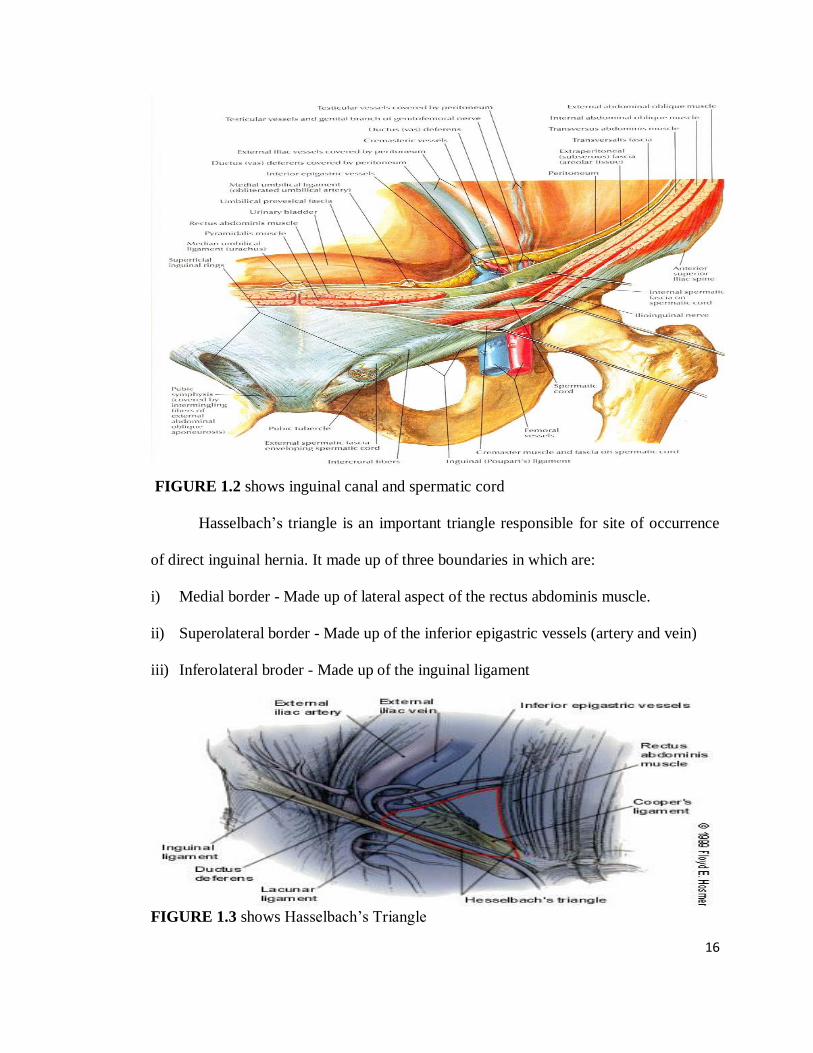
1.1.1 ANATOMY OF GROIN AND INGUINAL CANAL
Groin or inguinal region is an area bounded by
i) Anterior superior iliac spine and inguinal ligament (Poupart‟s ligament) laterally
ii) Pubic tubercle, pubic crest and symphysis pubis inferiorly
iii) Linea alba medially
iv) A perpendicular imaginary line drawn between linea alba and anterior superior iliac
spine superiorly
Layers of anterior abdominal wall:
1) Skin
2) Subcutaneous tissue
- Can be divided into two layers:
i) Superficial fatty layer (Camper‟s fascia)
ii) Deep fibrous layer (Scarpa‟s fascia)
3) Musculo-aponeurotic structures
- Can be divided into three layers of muscle and aponeurosis:
i) External oblique - the outermost layer and it is totally aponeurotic in the groin.
ii) Internal oblique - the intermediate layer.
iii) Transversus abdominis - the innermost layer and the key layer in hernia
repair.
The superficial (external) inguinal ring is not a true ring but a triangular defect in
the external oblique aponeurosis. It is situated above and lateral to the pubic tubercle.

13
Normally the ring will not allow for admission of the tip of the little finger. The deep
(internal) inguinal ring is an oval-shaped defect in the transversalis fascia. Clinically it
lies about half an inch (1.25 cm) above midway between the superior anterior iliac spine
and the pubic tubercle (the midpoint of the inguinal ligament).
Inguinal canal is an oblique, cone-shaped canal connecting between deep and
superficial inguinal rings. The length is about 4 to 6 cm in an adult. The canal is directed
inferomedially. It lies just superior and parallel to medial half of the inguinal ligament.
Both superficial and deep inguinal rings are almost overlapping to each other in a child
and thus make the inguinal canal short. The obliquity of the canal during childhood is
also slight. As we grow older the canal is elongated as both superficial and deep inguinal
rings move away from each other. There are a lot of structures pass through the canal in
the male. In the female the spermatic cord is replaced by the round ligament of the
uterus.
The spermatic cord contains structures running from and to the testis and
suspends the testis in the scrotum. Coverings of the spermatic cord from within are
internal spermatic fascia (transversalis fascia), cremasteric fascia (internal oblique
muscle and fascia), external spermatic fascia (external oblique muscle and fascia) and
processus vaginalis. In the male the contents of the spermatic cord are as follow:
i) Ilioinguinal nerve
- The nerve arises from the first lumbar nerve (L1) and emerges from the lateral border
of the psoas major muscle and passes obliquely across the quadratus lumborum. At a
point just medial to the anterior superior iliac spine, it pierces the transversus and
14
internal oblique muscles to enter the inguinal canal and exits through the superficial
inguinal ring.
- Supplies somatic sensation to the skin of the medial and upper thigh.
ii) Genital branch of the genitofemoral nerve
- Genitofemoral nerve arises from L1–L2, courses along the retroperitoneum, and
emerges on the anterior aspect of the psoas muscle. It then divides into femoral and
genital branches. The genital branch enters the inguinal canal lateral to the inferior
epigastric vessels, and it courses ventral to the iliopubic tract and iliac vessels. In males,
it travels through the superficial ring.
- Supplies the ipsilateral scrotum and cremaster muscle.
iii) Vas deferens - a muscular tube acts as a conduit of sperms from the epididymis to
the ejaculatory duct. It passes through the substance of the prostate to open into the
prostatic urethra.
iv) Testicular artery - arises from the aorta at the vertebral level of L2 and supplying the
epididymis and the testis.
v) Artery of the vas deferens - a branch of the inferior vesical artery.
vi) Cremasteric artery - a branch of the inferior epigastric artery.
vii) Pampiniform venous plexus - a venous plexus formed up by up to twelve veins that
merge superiorly as the right or left testicular veins.
viii) Sympathetic nerve fibers - derived from the pelvic and para-aortic plexuses
ix) Lymphatic vessels - drain the testis and adjacent structures to the lumbar lymph
nodes.
x) Cremasteric nerve
15
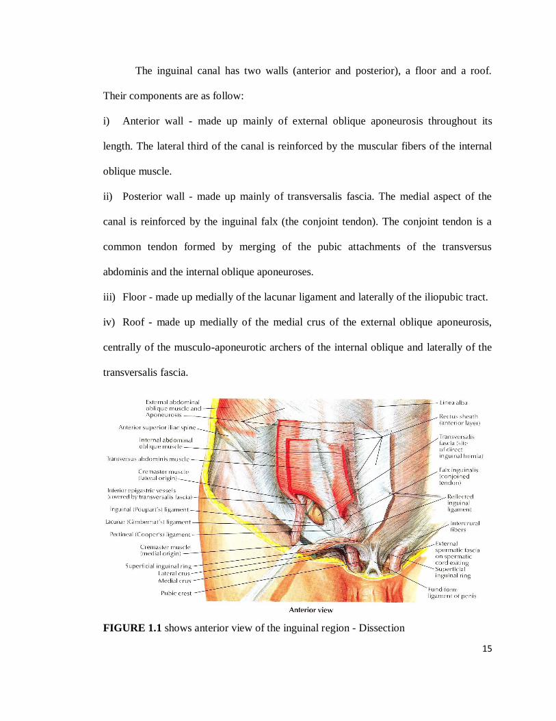
The inguinal canal has two walls (anterior and posterior), a floor and a roof.
Their components are as follow:
i) Anterior wall - made up mainly of external oblique aponeurosis throughout its
length. The lateral third of the canal is reinforced by the muscular fibers of the internal
oblique muscle.
ii) Posterior wall - made up mainly of transversalis fascia. The medial aspect of the
canal is reinforced by the inguinal falx (the conjoint tendon). The conjoint tendon is a
common tendon formed by merging of the pubic attachments of the transversus
abdominis and the internal oblique aponeuroses.
iii) Floor - made up medially of the lacunar ligament and laterally of the iliopubic tract.
iv) Roof - made up medially of the medial crus of the external oblique aponeurosis,
centrally of the musculo-aponeurotic archers of the internal oblique and laterally of the
transversalis fascia.
FIGURE 1.1 shows anterior view of the inguinal region - Dissection
16
FIGURE 1.2 shows inguinal canal and spermatic cord
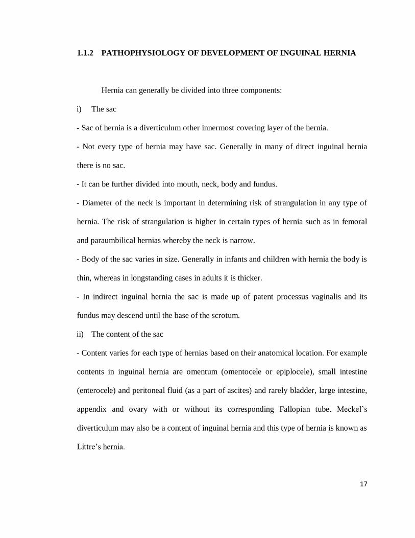
Hasselbach‟s triangle is an important triangle responsible for site of occurrence
of direct inguinal hernia. It made up of three boundaries in which are:
i) Medial border - Made up of lateral aspect of the rectus abdominis muscle.
ii) Superolateral border - Made up of the inferior epigastric vessels (artery and vein)
iii) Inferolateral broder - Made up of the inguinal ligament
FIGURE 1.3 shows Hasselbach‟s Triangle
17
1.1.2 PATHOPHYSIOLOGY OF DEVELOPMENT OF INGUINAL HERNIA
Hernia can generally be divided into three components:
i) The sac
- Sac of hernia is a diverticulum other innermost covering layer of the hernia.
- Not every type of hernia may have sac. Generally in many of direct inguinal hernia
there is no sac.
- It can be further divided into mouth, neck, body and fundus.
- Diameter of the neck is important in determining risk of strangulation in any type of
hernia. The risk of strangulation is higher in certain types of hernia such as in femoral
and paraumbilical hernias whereby the neck is narrow.
- Body of the sac varies in size. Generally in infants and children with hernia the body is
thin, whereas in longstanding cases in adults it is thicker.
- In indirect inguinal hernia the sac is made up of patent processus vaginalis and its
fundus may descend until the base of the scrotum.
ii) The content of the sac
- Content varies for each type of hernias based on their anatomical location. For example
contents in inguinal hernia are omentum (omentocele or epiplocele), small intestine
(enterocele) and peritoneal fluid (as a part of ascites) and rarely bladder, large intestine,
appendix and ovary with or without its corresponding Fallopian tube. Meckel‟s
diverticulum may also be a content of inguinal hernia and this type of hernia is known as
Littre‟s hernia.
18
iii) The covering of the sac
- It is layers of structures through which the sac passes.
An inguinal hernia is an abnormal protrusion of parietal peritoneum and viscera,
such as the small bowel, through a normal or abnormal opening from the abdominal
cavity at the inguinal region. There are many ways of classifying inguinal hernias. They
may be known as either congenital or acquired hernia or traditionally it is classified into
three types, which are indirect, direct and femoral hernia based on the site of herniation
relative to surrounding structures. Femoral hernia protrudes into a small and non-flexible
ring below and lateral to the pubic tubercle which is known as femoral ring. There was a
classification system available to classify inguinal hernia based on its location, type and
site, and the system is known as Nyhus classification system of inguinal hernia. It
classified hernias into five types:
i) Type I
- Indirect hernia with normal internal abdominal ring.
- Typically in infants, children, small adults.
ii) Type II
- Indirect hernia with enlarged internal ring but without impingement on the floor of the
inguinal canal and does not extend to the scrotum.
iii) Type IIIA
- Direct hernia (regardless of size)
iv) Type IIIB
- Indirect hernia that has enlarged enough to encroach upon the posterior inguinal wall
19
OR
- Indirect sliding or scrotal hernias are usually placed in this category because they are
commonly associated with extension to the direct space OR
- Pantaloon hernias (Dual hernia with combined features of both indirect and direct
components; Saddle-bag hernia)
v) Type IIIC
- Femoral hernia
vi) Type IV
- Recurrent hernia
- Modifiers A-D are sometimes used, which correspond to indirect, direct, femoral, and
mixed, respectively.
Nowadays inguinal hernias are better divided into two major types as the femoral
hernia is classified into its own entity. They are indirect and direct inguinal hernias in
which occur due to different pathologies. Overall, there are limited data regarding the
etiology of inguinal hernia development. Generally speaking indirect inguinal hernia is
due to presence of persistent processus vaginalis (complete or at least of the superior
part) and direct inguinal hernia is due to weakness of the Hasselbach‟s triangle due to
many factors. In direct inguinal hernia the weakness of the Hasselbach‟s triangle can
either due to distended superficial ring, narrow conjoint tendon or attenuation of
aponeurosis in males more than 40 years old.
20
Main aetiologic factors of inguinal hernia can be further divided into three main
factor types, which are:
A. Congenital factors
i) Sex
- Males are more prone to develop inguinal hernia than females
ii) Biological factors
a) Biophysical and biochemical factors
b) Pathophysiological factors
iii) Anatomical factors
a) Obliquity of inguinal canal
b) Patent processes vaginalis
c) Subtle variants in the attachment and arrangement of abdominal muscles
iv) Descent of testis
B. Contributing factors
i) Aging
- As aging causes atrophy of aponeuroses and muscles, weakening of posterior wall of
the inguinal canal and deep inguinal ring leading to direct inguinal hernia.
ii) Obesity
- This factor predisposes to hernia formation in two ways. First by increasing the intra-
abdominal pressure due to accumulation of intra-abdominal visceral fat, and second by
infiltration of fat in the muscles. Thus both ways lead to weakness of abdominal
musculature.
iii) Pulmonary disease (such as chronic obstructive pulmonary disease in adults or
21
whooping cough in children)
- These disorders may lead to chronic cough and thus weakens the abdominal muscles
and aponeuroses in long-standing cases.
- In other way long-standing chronic obstructive pulmonary disease may also lead to cor
pulmonale in which there is right-sided cardiac failure as a result of a primary disorder
of the pulmonary system.
iv) Genitourinary disorders (such as prostatism, cystocele, urethrocele and urethral
stricture)
- In order to overcome the distal obstruction the patient may chronically strain in
urinating in which lead to prolonged increase in intra-abdominal pressure.
v) Gastrointestinal disorders (such as chronic constipation and diverticular disease)
- This mechanism is the same as those occur in genitourinary disorders whereby the
patient will be chronically strain in defecating in order to pass out faeces. End result will
be prolonged increased intra-abdominal pressure.
vi) Cardiac disease (such as congestive cardiac failure)
- Contributed by two mechanisms:
a) Formation of ascites especially in the right-sided cardiac failure.
b) Prolonged use of accessory abdominal muscles due to dyspnea.
- Both may subsequently lead to prolonged increase in intra-abdominal pressure
C. Predisposing factors
i) Sudden increase in intra-abdominal pressure (such as occur in straining, vigorous
coughing and lifting heavy objects)
ii) Trauma to abdominal muscles and aponeuroses
22
Indirect inguinal hernia can be further divided into few types based on
anatomical and clinical types. They are:
A. Anatomical types
i) Based on extent of hernia
a) Bubunocele
- When the hernia does not come out of the superficial ring
b) Incomplete hernia
- When it comes out through the superficial ring but fails to reach the base of the
scrotum
c) Complete hernia
- When it descends into the bottom of the scrotum
ii) Based on content of hernia
a) Epiplocele or omentocoele - When it contains omentum.
b) Enterocele - When it contains the small bowel.
c) Cystocele - when it contains urinary bladder.
B. Clinical types
i) Reducible hernia
- Normally an uncomplicated hernia is reducible. That means the contents can be
returned into the abdominal cavity. It either occurs spontaneously or has to be manually
reduced. But the sac remains in its position.
- It presents clinically as an expansile impulse on coughing.
ii) Irreducible hernia
- The contents cannot be returned to the abdomen despite manually reduced but it does
23
not suggest any other complication.
- It is usually due to either overcrowding within the sac or adhesions between the sac
and its contents.
iii) Obstructed hernia (Irreducibility + Intestinal obstruction)
- Here the hernia is associated with intestinal obstruction due to occlusion of the lumen
of the bowel but there is no evidence of interference with the blood supply of the
affected intestine.
- Symptoms such as colicky pain and tenderness over the site of hernia, is less severe
and the onset is more gradual than in strangulated hernias.
- Usually there is no clear difference clinically between obstructed and strangulated
hernias. Thus it will be safer to assume strangulated hernia if in doubt and treat it
accordingly as it carries significant morbidity and mortality.
iv) Incarcerated hernia
- This a loose term used previously as an alternative to reflect obstruction or
strangulation.
- The term is correctly employed only when it is considered that the lumen of that
portion of the colon occupying a hernial sac is blocked with faeces. In this case, the
irreducible content will be felt clinically as a doughy and indentable swelling.
v) Strangulated hernia (Irreducibility + Obstruction + Arrest of blood supply to the
contents)
- Here the contents are so constricted as to interfere with the blood supply and makes the
contents susceptible for ischaemia.
- Gangrenous bowel may occur as soon as 5 to 6 hours after the onset of first symptom.
24
- Paraumbilical and femoral hernias are more prone to develop strangulation than
inguinal hernia as their neck of the hernias is generally narrow and rigid.
vi) Inflamed hernia
- This is very rare situation in which the contents of the sac have become inflamed.
- Occurs as a result of inflammation of the contents itself, such as acute appendicitis or
salpingitis, or from external causes such as the trophic ulcers that develop in the
dependent areas of larger incisional or umbilical hernias.
- Clinical presentation may mimic presentation of strangulated hernia.
- The hernia is usually tender but not tense.
- Overlying skin is edematous and erythematous.
- Treatment is based on treatment of the underlying cause.
C) Rare specific types
i) Hernia–en–glissade (Sliding hernia)
- In this type the caecum on the right side or the pelvic colon on the left side or the
urinary bladder on either side slides down outside the hernial sac forming a part of its
wall, being covered by peritoneum on the hernial aspect only.
ii) Richter‟s Hernia
- In this only a portion of the circumference of the bowel becomes strangulated. This
condition is particularly dangerous as operation is frequently delayed because of the
clinical features resembling gastroenteritis.
iii) Littre‟s hernia
- When it contains Meckel‟s diverticulum.