a strategy of retrograde injection of bone marrow mononuclear cells into the myocardium for the...
TRANSCRIPT
Original article
A strategy of retrograde injection of bone marrow mononuclear cells intothe myocardium for the treatment of ischemic heart disease
Shin-Ichiro Yokoyama a, Noboru Fukuda a,b,*, Yuxin Li a, Kazuhiro Hagikura a,Tadateru Takayama a, Satoshi Kunimoto a, Junko Honye a, Satoshi Saito a, Mika Wada b,
Aya Satomi b, Maiko Kato b, Hideo Mugishima b, Yoshiaki Kusumi c,Masako Mitsumata c, Toyoaki Murohara d
a Department of Medicine, Nihon University School of Medicine, 30-1, Ooyaguchi-kamimachi, Itabas..., Tokyo 173-8610, Japanb Department of Advanced Medicine, Division of Cell Regeneration and Transplantation, Nihon University School of Medicine, Tokyo 173-8610, Japan
c Department of Pathology, Nihon University School of Medicine, Tokyo 173-8610, Japand Department of Cardiology, Nagoya University Graduate School of Medicine, Nagoya 466-8550, Japan
Received 10 February 2005; received in revised form 25 May 2005; accepted 16 June 2005
Available online 04 November 2005
Abstract
Objective. – Bone marrow cells implantation (BMI) has been reported to efficiently improve ischemic heart disease. However, BMIstrategies are generally invasive. To establish a BMI strategy for ischemic heart disease, we performed implantation of autologous cryopre-served mononuclear cells (MNCs) from bone marrow (BM) retrogradely into the myocardium via the coronary vein in pigs with acutemyocardial infarction (AMI) and old myocardial infarction (OMI).
Methods. – BM cells were harvested from the pigs’ fumurs. MNCs were collected by centrifugation and were cryopreserved. Anteriormyocardial infarction was induced by occlusion of the midportion of the left anterior descending coronary artery without surgical interven-tion. Frozen BM cells were quickly thawed and injected retrogradely via the coronary vein into the myocardium through a single ballooninfusion catheter 6 h and 2 weeks after the induction of infarction. Four weeks after implantation, coronary arteriograms were obtained,cardiac function was analyzed with the use of a conductance catheter, and histopathologic anlysis was performed with a confocal lasermicroscope. Plasma levels of natriuretic peptides and angiogenic growth factors were measured after BMI.
Results. – Flow cytometric analysis revealed that 90% of cryopreserved BM cells were viable in vitro. Labeled BM cells were entirelydistributed around in the infarcted area of maycardium in pigs. BMI increased collateral neovascuralization in infarcted hearts. BMI signifi-cantly improved cardiac function in AMI with BMI and OMI with BMI groups. BMI also increased the formation of microcapillary arteriesin infarcted hearts. Levels of natriuretic peptides were significantly decreased, and levels of vascular endothelial growth factor (VEGF) andbasic fibroblast growth factor (FGF2) were significantly increased after BMI. Confocal laser microscopy revealed the presence of prolifera-tive and activated myocardial cells in infarcted hearts after BMI.
Conclusion. – The retrograde infusion of cryopreserved BM cells into myocardium efficiently induced angiogenesis and improved cardiacfunction in pigs with AMI or OMI. These results suggest that the present strategy of BMI will be safe and feasible as an angiogenic cell therapyfor ischemic heart disease.© 2005 Elsevier Ltd. All rights reserved.
Keywords: Ischemic heart disease; Regenerative medicine; Bone marrow cell; Angiogenesis; Retrograde infusion; Cryopreservation
1. Introduction
Current standard treatments for acute myocardial infarc-tion (AMI) are predominantly based on reperfusion of the
obstructive coronary arteries by thrombolysis with tissue plas-minogen activator (tPA) and/or by percutaneous coronaryintervention (PCI). Therapeutic approaches to the manage-ment of myocardial ischemia including angina pectoris andold myocardial infarction (OMI) commonly include manipu-lations designed to reduce myocardial oxygen demand or to
* Corresponding author. Tel.: +81 3 3972 8111; fax: +81 3 3972 8666.E-mail address: [email protected] (N. Fukuda).
Journal of Molecular and Cellular Cardiology 40 (2006) 24–34
www.elsevier.com/locate/yjmcc
0022-2828/$ - see front matter © 2005 Elsevier Ltd. All rights reserved.doi:10.1016/j.yjmcc.2005.06.008
increase blood supply to ischemic area mechanically by coro-nary artery bypass grafting (CABG) or PCI with medication.The recovery of cardiac function after infarction is impor-tant, because the mortality of patients withAMI remains high.Alternative therapies include those of induction the develop-ment of new collateral vessels.
Transfer of angiogenic genes such as those of vascularendothelial growth factor (VEGF) and basic fibroblast growthfactor (FGF2) has been reported to increase collateral bloodflow and to improve cardiac function in cases of chronic myo-cardial ischemia [1,2]. The gene therapies raise a number ofpractical issues for application in human trials, including thedevelopment of adequate delivery systems and safety [3,4].
Autologous cell therapy for induction of angiogenesis isthought to be more progressive regenerative treatment. Bonemarrow (BM) includes abundant stem cells [5] and endothe-lial progenitor cells [6,7]. Therapeutic Angiogenesis usingCell Transplantation (TACT) Study Investigators reported thatBM cell implantation (BMI) improves ischemic ulcers in casesof limb ischemia by improving blood flow [8]. Thus, BMIhas been considered for application in ischemic heart disease[9,10].
Direct injection into the myocardium and intra-coronaryinfusion are methods of delivery of BM cells to the ischemicheart [11,12]. In addition, catheter-based injection of cellsinto the ischemic area has recently been reported [13,14].Because these procedures are invasive, require special tech-niques and are costly, they cannot be performed in generalhospitals. In addition, in occluded coronary arteries with poorcollaterals, transplanted cells may not reach the targetischemic lesion. Less invasive angiogenic cell transplanta-tion into the ischemic heart are therefore necessary. Regen-erative cell therapies for ischemic heart disease have not beenestablished.
The harvest of BM cells can be dangerous for patients withsevere ischemic heart disease because it can cause anemia orhypovolemia. Thus, preservation of BM cells from patientsand the ability of preserved BM cells to induce agniogenesisshould be evaluated.
To establish an efficient strategy of BMI for ischemic heartdisease, we cryopreserved BM cells and delivered the cellsretrogradely via the coronary vein to the myocardium in pigs.We evaluated the ability of this method to induce angiogen-esis and its effect on cardiac function in pigs with AMI andOMI.
2. Materials and methods
This study conformed to the Guide for the Care and Useof Laboratory Animals published by the US National Insti-tutes of Health (NIH Publication No. 85-23, revised 1996).
2.1. Preparation and cryopreservation of BM mononuclearcells (BM-MNCs)
All surgical procedures were performed under generalanesthesia and continuous electrocardiographic (ECG) and
blood pressure monitoring. Adult male pigs (35 kg) were pre-medicated with ketamine (20 mg/kg, im) before induction ofanesthesia with 4% isoflurane. Anesthesia was maintainedwith 1–2.5% isoflurane. A volume of 400 ml of peripheralblood was obtained 2 weeks before BM harvest and we cre-ated a transfusion bag of red cell concentrate in mannnitol-adenine-phosphate solution (RC-MAP, Terumo Medical Co.)from those blood. A half volume of RC-MAP was used forautologous blood transfusion to improve anemia at the har-vest and the rest was used in combination with BM blood atthe isolation of MNCs to be full-filled the collected bag. Allpigs received an infusion of iron (Tontetsu, Pfizer Pharmacy,50 mg/day) for 3. Those with a red blood cell count >3.5 × 10⁶
per ml were included in the experiments. Two weeks later,400 ml of BM cells were harvested bilaterally from the femursof all pigs. During harvest, the pigs were received 200 ml ofautologous blood transfusion for 1 h. After harvest, MNCswere collected by the use of program modifications (MNCsoftware Version 5.1, BMP) of the COBE Spectra ApheresisSystem (Gambro BCT, Inc.). Five milliliters of dimethyl sul-foxide and 10% dextran 40 (Terumo) were added to 20 ml ofthe collected MNCs-enriched blood [15]. For implantationof preserved BM cells, the collected MNCs were cryopre-served with an auto-preserved liquid nitrogen system (Cry-oMed, Futaba Medical) according to the following protocol:pre-freeze rate 10 °C/min, start freeze temperature at –3 °C,end freeze temperature at –10 °C to –15 °C, post-freeze rate2 °C/min and end temperature at –50 °C.
2.2. Flow cytometric analysis and colony forming unit
The thawed BM-MNCs were stained with a green fluores-cent marker PKH2-GL (Sigma Chemical Co.). The PKH-GLis the green fluorescent cell linker kit for general cell mem-brane labeling. At first, we determined the optimal dye/cellconcentration. After the centrifuging collected cells (400 × g)for 5 min, we carefully aspirated the supernatant on the pelletand labeled the half of implanted cells using PKH2-GL label-ing kit. After the labeling, the cells were analyzed by flowcytometry (FACS Calibur, Becton Dickinson). The parts ofthe thawed BM-MNCs (N = 11) were analyzed by flowcytometry before the implantation with the use of 7-amino-actinomycin D (7AAD) (VIA-PROBETM, Becton Dickin-son) to evaluate the cell viability.
Preserved cell content was evaluated by plating 50,000 cellsin 1.1% methylcellulose (Stem Cell Technologies) supple-mented with 30% fetal bovine serum (FBS) and 50 ng/mlinterleukin (IL)-3, or with 30% FBS and a cytokine mixture(2 ng/ml IL-3, 25 ng/ml stem cell factor, 5 ng/ml granulocyte-macrophage colony stimulating factor and 2 U/ml humanerythropoietin). We determine the timing colony forming unitsfrom the following manual (Colony Assays of HematopieticCells Using Methylcellulose Media; Terry Fox LaboratoryMedia Preperation Service) [16]. Fourteen days culture isenough time to make the colony for leukocyte-progenitorcells. Seven days is only enough time to assess CFU-E. Colo-
25S.-I. Yokoyama et al. / Journal of Molecular and Cellular Cardiology 40 (2006) 24–34
nies (>50 cells) were counted after 14 days in culture at 37 °C,5% CO2. Colonies were classified as colony forming unit-granulocyte/monocyte (CFU-GM), burst-forming unit-erythroid (BFU-E) from an early erythroid progenitor or CFU-Mix, a more immature, multipotent type of colony thatcontaining cells from myeloid and erythroid lineages.
2.3. Induction of myocardial infarction
We performed right and left coronary arteriography (CAG)after injection of 2.5 mg of isosorbide-dinitrate (ISDN) intoeach coronary artery under general anesthesia. The pigs weresubjected to acute antero-septal myocardial infarction byimplantation of beads of 2.0 mm in diameter with 0.8 mmholes into the mid portion of the left anterior descending coro-nary artery (LAD) through a guide-wire. After placement,coronary occlusion occurred gradually with increasingthrombi around the beads. Occlusion of the LAD was con-firmed angiographically after 6 h. All procedures were per-formed under fluoroscopic guidance.
2.4. Animal groups and BMI
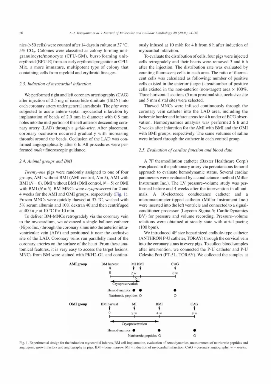
Twenty-one pigs were randomly assigned to one of fourgroups, AMI without BMI (AMI control, N = 5), AMI withBMI (N = 6), OMI without BMI (OMI control, N = 5) or OMIwith BMI (N = 5). BM-MNCs were cryopreserved for 2 and4 weeks for the AMI and OMI groups, respectively (Fig. 1).Frozen MNCs were quickly thawed at 37 °C, washed with5% serum albumin and 10% dextran 40 and then centrifugedat 400 × g at 10 °C for 10 min.
To deliver BM-MNCs retrogradely via the coronary veinto the myocardium, we advanced a single balloon catheter(Nipro Inc.) through the coronary sinus into the anterior intra-ventricular vein (AIV) and positioned it near the occlusivesite of the LAD. Coronary veins run parallelly most of thecoronary arteries on the surface of the heart. From these ana-tomical features, it is very easy to access the target lesions.MNCs from BM were stained with PKH2-GL and continu-
ously infused at 10 ml/h for 4 h from 6 h after induction ofmyocardial infarction.
To evaluate the distribution of cells, four pigs were injectedcells retrogradely and their hearts were removed 3 and 6 hafter the injection. The distribution rate was evaluated bycounting fluorescent cells in each area. The ratio of fluores-cent cells was calculated as following: number of positivecells existed in the anterior (target) area/number of positivecells existed in the non-anterior (non-target) area × 100%.Three horizontal sections (5 mm proximal site, occlusive siteand 5 mm distal site) were selected.
Thawed MNCs were infused continuously through thecoronary vein catheter into the LAD area, including theischemic border and infarct areas for 4 h under of ECG obser-vation. Hemodynamics analysis was performed 6 h and2 weeks after infarction for the AMI with BMI and the OMIwith BMI groups, respectively. The same volumes of salinewere infused through the catheter in each control group.
2.5. Evaluation of cardiac function and blood data
A 7F thermodilution catheter (Baxter Healthcare Corp.)was placed in the pulmonary artery via percutaneous femoralapproach to evaluate hemodynamic status. Several cardiacparameters were evaluated by a conductance method (MillarInstrument Inc.). The LV pressure–volume study was per-formed before and 4 weeks after the intervention in all ani-mals. A 10-electrode conductance catheter and amicromanometer-tipped catheter (Millar Instrument Inc.)were inserted into the left ventricle and connected to a signal-conditioner processor (Leycom Sigma-5; CardioDynamicsBV) for pressure and volume recording. Pressure–volumerelations were obtained at steady state with atrial pacing(100 bpm).
We introduced 4F size heparinized endhole-type catheter(ANTHRON P-U catheter, TORAY) through the cervical veininto the coronary sinus in every pigs. To collect blood samplesafter intervention, we connected the P-U catheter and P-UCelesite Port (PT-5L, TORAY). We collected the samples at
Fig. 1. Experimental design for the induction myocardial infarcts, BM cell implantation, evaluation of hemodynamics, measurement of natriuretic peptides andangiogenic growth factors and angiography in pigs. BM = bone marrow, MI = induction of myocardial infarction, CAG = coronary angiography, w = weeks.
26 S.-I. Yokoyama et al. / Journal of Molecular and Cellular Cardiology 40 (2006) 24–34
3, 7, 14, 28 days after the intervention. Plasma levels of VEGFand FGF2 were measured with an ELISA kit (R&D Sys-tems). Plasma levels of atrial natriuretic peptide (ANP) andbrain natriuretic peptide (BNP) were measured with a radio-immunoassay kit (Yamasa Shoyu) from peripheral blood.Blood chemistry data were evaluated in the same bloodsamples.
2.6. Angiography and evaluation of coronary flow reserve
Selective CAG was performed to detect collateral sources.Collateral indices were assigned as follows: 0 = no visiblecollaterals, 1 = collateral formation visible only with con-trast stain, 2 = partial filling of the main epicardial vessel,3 = complete filling of the distal LAD.
We also measured coronary flow reserve as the function ofvessels. Coronary flow velocities were measured with a Dop-pler Flowire (Cardiometrics, Inc., Mountain View, USA). TheFlowireTM is a 175 cm long, flexible, steerable angioplastyguide wire, 0.014 in. in diameter, with a 12 MHz piezoelec-tric ultrasound transducer at its tip. The velocity data are pro-cessed on-line by fast Fourier transformation and displayedwith a real-time gray scale spectral display. The Flowire wasthen advanced into LAD and was positioned just proximalsite of the occluded lesion to obtain a stable blood flow veloc-ity signal. Baseline flow velocities under ISDN drip wereobtained and arterial pressure and heart rate were recorded.The following intra-coronary Doppler parameters were mea-sured: diastolic to systolic velocity ratio (DSVR), averagepeak velocity (APV) during the procedure. Coronary flowreserve (CFR) was calculated the ratio max APV and base-line APV.
2.7. Histopathology
All pigs were killed with overdoses of intravenous injec-tions of pentobarbital and potassium chloride, and their heartswere removed after CAG. PKH2-GL labeled tissues obtainedfrom the ischemic region were snap-frozen. Each heart wasfixed in 20% formaldehyde, and 5-µm-thick sections, includ-ing the whole area at risk were obtained (Leica RM2145 mi-crotome, Leica Instruments) and treated with hematoxylin andeosin (HE) and Masson trichrome stain.
To confirm the proliferative activity of cells, we stainedselected specimens from control and treated groups simulta-neously with anti-MIB-1 antibody. MIB-1 (Ki67) antigen isexpressed throughout the cell cycle and is a reliably distin-guishes proliferation. Fluorescence detection of the labeledproliferative cells through staining with fluorochromated avi-din [avidin-Alexa Fluor 488™ (green fluorescence), DAKOJapan Corp., dilution 1:500]. Tissues were also stained fora-sarcomeric actin as a marker of myocardial cells. Simulta-neous immunofluorescence detection of myocardial markersusing the appropriate primary antibodies and secondary anti-bodies conjugated to the fluorochromes [Alexa Fluor 633™(infrared fluorescence), DAKO, dilution 1:500]. Fluorescent
signals were detected by optical sectioning using a LeicaTCS-NT confocal laser-scanning microscope (Leica Instru-ments). Vessel walls were stained for a-smooth muscle actin(a-SMA) (Becton Dickinson), antibody to evaluate vasculardevelopment. Because of the lack of appropriate markers forpig endothelium, we evaluated myocardial microvessel den-sity (microvessels per mm2), microvessel mean area mea-sured from a-SMA immunostained sections atzb 200 magni-fications from five fields of each section. Hansen-Smith et al.[17] reported the increase in the number of microvessels onlypartially covered by a-SMA suggests arteriolization of cap-illaries.
To quantify coronary artery, the vessel number and the totalvessel area including the adventitial layer of the epicardialcoronary artery, was measured in sections 5 mm distal theocclusive site. Myocardial microvessel area (cm2) in sectionsof 5 mm distal the occlusive site were also measured ina-SMA immunostained sections [18].
We determined the fibrosis content of the myocardium withan automatic image analysis system, by planimetric analysis(NIH Image 1.61/ppc). We evaluated the entire area of eachhistologic section 10 mm distal the occlusive site. MassonTrichrome stain sections were used for detection the fibroticarea. The infarct area was calculated according to fibrosisgrade (fibrosis area/total area × 100%).
2.8. Statistical analysis
All results are presented as mean ± S.D. The significanceof differences between mean values was evaluated by Stu-dent’s t-test for unpaired data and by two-way analysis ofvariance (ANOVA) followed by Duncan’s multiple rangetests. P values < 0.05 were considered statistically signifi-cant.
3. Results
3.1. Characteristics of isolated BM cells
FACS analysis of BM cells stained with PKH2-GL showedthat the separated cells included many type of cells includingMNCs, lymphocytes, granulocytes and red blood cells, thepopulation of MNCs was the largest (86 ± 8.8% of BM cells)(data not shown).
The number of MNCs in 400 ml of femoral BM used forimplantation into the myocardium was 3.2 ± 1.2 × 10⁹
(N = 11) after separation. Recovery of viable BM cells fromcryopreservation was 87.5 ± 6.9% (N = 11). This reagent(7AAD) is used as a viability probe for methods of dead cellexclusion. Thus 7AAD was used for evaluation of cell viabil-ity. Myeloid and erythroid cells were assessed by colony assaybefore and after cryopreservation of separated MNCs andprior to implantation. The recovery of colony forming pro-genitor cells after cryopreservation of separated MNCs isshown in Table 1. Colony forming progenitor cells catego-
27S.-I. Yokoyama et al. / Journal of Molecular and Cellular Cardiology 40 (2006) 24–34

rized as CFU-GM, BFU-E or CFU-Mix were completelyrecovered after cryopreservation, indicating that cryopreser-vation after separation caused no significant loss of theenriched progenitor cells from the myeloid or erythroid lin-eages which are vital for transplant engraftment.
3.2. Distribution of BM-MNCs via coronary vein catheter
The distribution of BM-MNCs stained with PKH2-GL andinjected retrogradely via the coronary vein into the infarctedheart is shown in Fig. 2. Over 90% of labeled MNCs werefound around in the infarcted area, and the remaining cellswere found only in the left circumflex coronary artery (LCX)area 3 h after injection. Labeled MNCs were not identified inthe injured myocardium after intra-coronary infusion in heartsfrom pigs withAMI (data not shown). Fig. 2A shows a cinean-giogram of contras medium injection to AIV. The injectedmedium distributed apex lesion in pig heart. It is clearly vis-ible green color indicates fluorescence of cells in infractedheart (Fig. 2C–E). Injected MNCs were attached to vesselwalls (Fig. 2C) and were present in the myocardium (Fig. 2D,E) 6 h after the injection.
3.3. Angiogenesis after BMI
Collateral vessels were observed in all groups. However,antegrade flow due to bridge collaterals was observed only inthe BMI groups (Fig. 3A). Collateral index of the BMI groupswere significantly higher than that in the control groups(Fig. 3B). However, the collateral flow findings existed onlyaround areas of infarction. These collateral vessels were toosmall size to be visible in cineangiograms. Thus, the retro-grade administration of BM-MNCs via the coronary veinresulted in a directional increase in collateral circulation thatwas regionally restricted to the left coronary system.
Data from a Doppler Flowire show BMI therapy increasedcoronary flow reserve both in AMI and OMI (but not signifi-cantly). Increasing coronary flow reserve equal to increasingbet of functional micro vessels (data not shown).
3.4. Cardiac function after BMI
Changes in cardiac function before and 6 h or 2 weeksafter induction of the myocardial infarction in the AMI andOMI groups with or without BMI are shown in Fig. 4. End-diastolic volume (EDV) was significantly lower in AMI andOMI groups than the control groups 4 weeks after BMI. Ejec-tion fraction (EF) was significantly higher in the AMI andOMI groups than the control groups 4 weeks after BMI. End-diastolic pressure (EDP) was significantly lower in the AMIgroup but not in the OMI group than in the control groups at4 weeks after BMI.
3.5. Levels of natriuretic peptides and angiogenic growthfactors after BMI
ANP levels in peripheral blood were significantly increasedin the AMI control but not in the OMI groups. ANP levels4 weeks after BMI were significantly lower in the AMI groupthan in the control group. BNP levels in peripheral blood weresignificantly increased in the OMI groups but not in the AMI
Table 1Recovery of colony forming progenitor cells after cryopreservation of theseparation of MNCs
CFU-GM BFU-E CFU-MixTotalFresh MNCs 38 ± 6 10 ± 4 26 ± 3 76 ± 10Cryopreserved MNCs 35 ± 6 9 ± 2 28 ± 5 72 ± 9
Colony forming unit (CFU); fresh (prior to preservation) and preserved cellcontent was evaluated by plating 50,000 cells in 1.1% methylcellulose sup-plemented with 30% FBS and 50 ng/ml IL-3, or with 30% FBS and a cyto-kine mixture (2 ng/ml IL-3, 25 ng/ml SCF, 5 ng/ml GM-CSF and 2 U/mlhuman erythropoietin). Colonies (>50 cells) were counted after 14 days inculture at 37 °C, 5% CO2. Colonies were classified into the groups fol-lowing CFU-granulocyte/monocyte (CFU-GM), burst-forming unit-erythroid(BFU-E) from an early erythroid progenitor or CFU-Mix. Data are themean ± S.D. There are no significant differences between fresh and cryopre-served MNCs.
Fig. 2. Distribution of bone marrow mononuclear cells (BM-MNCs) injected retrogradely into infarcted heart. Cells were stained with a green fluorescentmarker, PKH2-GL, and injected via the coronary vein 6 h after induction of myocardial infarction. (A) A cineangiogram of contras medium injection to AIV.The injected medium distributed apex lesion of pig heart. (B) Distribution of fluorescent cells in all areas of the section 3 h after injection. Presence offluorescent cells (C) in vessel walls and (D, E) in the myocardium 6 h after injection.
28 S.-I. Yokoyama et al. / Journal of Molecular and Cellular Cardiology 40 (2006) 24–34

groups. BNP levels 4 weeks after BMI were significantlylower in the AMI and OMI groups with BMI than that in thecontrol groups (Table 2).
Levels of FGF2 and VEGF were significantly increased inthe AMI and OMI groups 3 days after BMI in comparison tolevels in the control groups (data not shown).
Fig. 3. Coronary angiography in pigs with acute or old myocardial infarcts with BM cell implantation (AMI with BMI or OMI with BMI). (A) Selectivecoronary angiographies were recorded from an RAO 30 view. Arrows indicate antegrade flow due to bridge collaterals. (B) Collateral indices. Collateral indexwere rated according to the following: 0 = no visible collaterals, 1 = collateral formation only with contrast stain, 2 = partial filling of the main epicardial vessel,3 = complete filling of the distal LAD. Data are the mean ± S.D. (N = 5). *P < 0.05 vs. each control.
Fig. 4. Changes in cardiac function after BMI before and 6 h or 2 weeks after induction of myocardial infarction (MI) in the AMI group or the OMI group, and4 weeks after BMI in each group. Several LV parameters, EDP, EDV, and EF were evaluated with the conductance method. Data are the mean ± S.D. *P < 0.05 vs.each control.
29S.-I. Yokoyama et al. / Journal of Molecular and Cellular Cardiology 40 (2006) 24–34

3.6. Histopathologic findings after BMI
Four weeks after BMI in the AMI group, PKH2-GL fluo-rescence was observed by confocal laser scopy in mature ves-sels walls (Fig. 5A) and in the microvascular system in thepreserved myocardium around the infarcted area (Fig. 5B).
We counted the number of a-SMA positive microvesselsin the infarcted areas 5 mm distal to the occlusive site. Thenumber of microvessels as capillary artery under 100 µm indiameter was significantly higher in infarcted hearts from theAMI with BMI and OMI with BMI groups than in heartsfrom the control groups. There were no significant differ-ences in the number of microvessels with multilayer smoothmuscle over 100 µm in diameter between the BMI groupsand the control groups (Fig. 6A). a-SMA-positive cells werepredominantly identified in the epicardium. In particular, therewere more microvascular areas in the epicardium close to theocclusive site of the LAD in the BMI groups (Fig. 6B). Somesections from implanted hearts showed growing cardiac
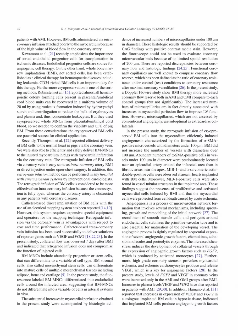
muscle fibers around vessels (Fig. 7A). By confocal lasermicroscopy, MIB-1-positive cells (green) and a-sarcomericactin-positive cells (red) were observed around microvesselsand were located around the infarct area implanted withBM-MNCs, and double-positive cells (yellow) were observedonly in hearts from the AMI with BMI group (Fig. 7B1–B3).In the OMI with BMI it was rare for us to detect the double-positive cells. The number of double-positive cells was muchless or none in the control groups than BMI group (Fig. 7B4–B6).
With respect to the effect of BMI on cardiac fibrosis, fibro-sis contents were smaller, but not significantly, in hearts fromthe BMI groups than those from control groups (AMI controlvs. AMI with BMI = 23.5 ± 8.4% vs. 17.5 ± 5.1%, OMI con-trol vs. OMI with BMI = 24.6 ± 9.5% vs. 19.8 ± 7.8%).
4. Discussion
To establish an efficient BMI strategy for ischemic heartdisease, we implanted cryopreserved BM-MNCs via the coro-nary vein into the myocardium of pigs with AMI or OMI. Weobserved good recovery of viable cells from cryopreserva-tion and improved cardiac function with antegrade collateralneovascuralization and increased numbers of microvessels inpigs with AMI as well as those with OMI.
BMI is reported to increase regional blood flow andimprove cardiac function in pigs with myocardial infarction[19,20]. However, BM cells were injected into the heart byopen-chest surgery in these studies. Cell delivery into the myo-cardium can occur via the intra-coronary artery or intra-coronary vein and by direct injection under open-chest orclosed-chest surgery. Catheter-based cell delivery is an easierand less invasive method. Assmus et al. [11] recently reportedthat autologous BM-MNCs via balloon catheter, which placedinto the coronary artery, repair the infarcted myocardium in
Table 2Levels in natriuretic peptides in peripheral blood after BMI
6 h 4 weeksANP AMI control 89 ± 26 154 ± 48 b
(pg/ml) AMI with BMI 105 ± 38 72 ± 25 a
BNP AMI control 232 ± 56 200 ± 56(pg/ml) AMI with BMI 225 ± 49 129 ± 34 a
2 weeks 6 weeksANP OMI control 181 ± 31 189 ± 29(pg/ml) OMI with BMI 175 ± 28 174 ± 17BNP OMI control 39 ± 6 151 ± 15 b
(pg/ml) OMI with BMI 42 ± 5 105 ± 18 a,b
Plasma levels of ANP and BNP in peripheral blood in pigs 6 h and 4 weeksafter induction of myocardial infarction in the AMI group and OMI groupand 4 weeks after BMI in each group were measured by radioimmunoassay.Data are the mean ± S.D.
a P < 0.05 vs. each control.b P < 0.05 vs. 6 h in the AMI group and 2 weeks in the OMI group.
Fig. 5. Histologic findings of ischemic areas in hearts from pigs with AMI 4 weeks after BMI. PKH2-GL-labeled tissues obtained from the ischemic region weresnap-frozen. (A) Angiography during cell infusion. (B) Preserved myocardium around infarcted area stained with hematoxylin and eosin (original magnifica-tion × 100). Lower panel indicates green fluorescence observed in mature vessels walls. (C–E) Confocal laser microscopic findings (original magnificationC × 40. D, E × 400).
30 S.-I. Yokoyama et al. / Journal of Molecular and Cellular Cardiology 40 (2006) 24–34
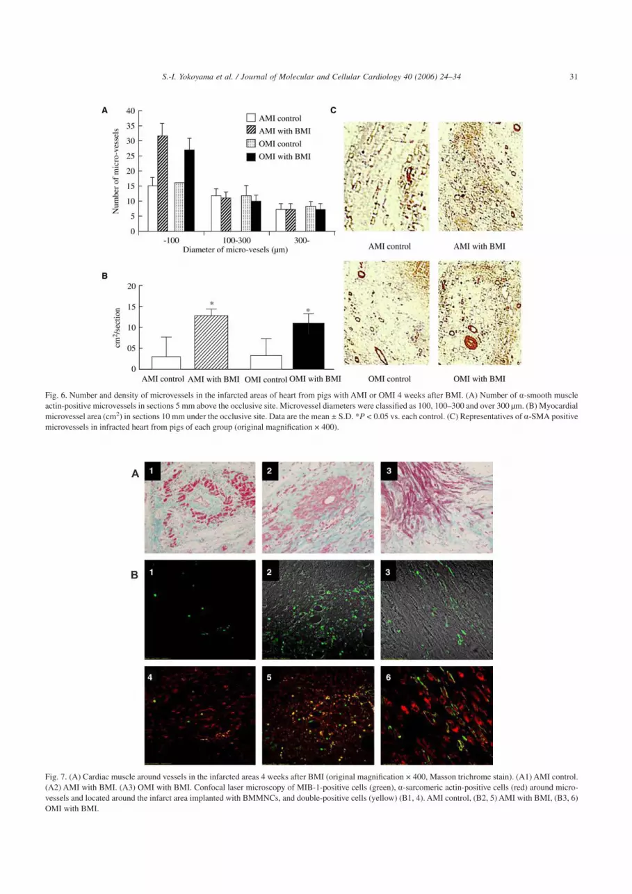
Fig. 6. Number and density of microvessels in the infarcted areas of heart from pigs with AMI or OMI 4 weeks after BMI. (A) Number of a-smooth muscleactin-positive microvessels in sections 5 mm above the occlusive site. Microvessel diameters were classified as 100, 100–300 and over 300 µm. (B) Myocardialmicrovessel area (cm2) in sections 10 mm under the occlusive site. Data are the mean ± S.D. *P < 0.05 vs. each control. (C) Representatives of a-SMA positivemicrovessels in infracted heart from pigs of each group (original magnification × 400).
Fig. 7. (A) Cardiac muscle around vessels in the infarcted areas 4 weeks after BMI (original magnification × 400, Masson trichrome stain). (A1) AMI control.(A2) AMI with BMI. (A3) OMI with BMI. Confocal laser microscopy of MIB-1-positive cells (green), a-sarcomeric actin-positive cells (red) around micro-vessels and located around the infarct area implanted with BMMNCs, and double-positive cells (yellow) (B1, 4). AMI control, (B2, 5) AMI with BMI, (B3, 6)OMI with BMI.
31S.-I. Yokoyama et al. / Journal of Molecular and Cellular Cardiology 40 (2006) 24–34
patients withAMI. However, BM cells administered via intra-coronary infusion attached poorly to the myocardium becauseof the high value of blood flow in the coronary artery.
Kawamoto et al. [21] recently emphasizes the importanceof sorted endothelial progenitor cells for transplantation inischemic diseases. Endothelial progenitor cells are source forangiogenic cell therapy. On the other hand, whole bone mar-row implantation (BMI), not sorted cells, has been estab-lished as a clinical therapy for hematopoetic diseases includ-ing leukemia. CD34-riched BM cells is an important key forthis therapy. Furthermore cryopreservation is one of the sort-ing methods. Rubinstein et al. [15] reported almost all hemato-poietic colony forming cells present in placental/umbilicalcord blood units can be recovered in a uniform volume of20 ml by using rouleaux formation induced by hydroxyethylstarch and centrifugation to reduce the bulk of erythrocytesand plasma and, thus, concentrate leukocytes. But they usedcryopreserved whole MNCs from placental/umbilical cordblood, so we needed to confirm the viability and CFU of pigBM. From these considerations the cryopreserved BM cellsare powerful source for clinical application.
Recently, Thompson et al. [22] reported efficient deliveryof BM cells to the normal heart in pigs via the coronary vein.We were also able to efficiently and safely deliver BM-MNCsto the injured myocardium in pigs with myocardial infarctionvia the coronary vein. The retrograde infusion of BM cellsvia coronary vein is easy same as intra-coronary artery BMIor direct injection under open-chest surgery. In addition, thisretrograde infusion method can be performed in any hospitalwith cineangiogram systems by interventional cardiologists.The retrograde infusion of BM cells is considered to be moreeffective than intra-coronary infusion because the venous sys-tem is fully open, whereas the coronary artery is obstructedin any patients with coronary diseases.
Catheter-based direct implantation of BM cells with theuse of an electrical mapping system has been reported [14,19].However, this system requires expensive special equipmentand operators for the mapping technique. Retrograde infu-sion via the coronary vein is advantageous with respect tocost and time performance. Catheter-based trans-coronaryvein infusion has been used successfully to deliver solutionsof reporter genes such as VEGF and FGF2 [18,22,23]. In thepresent study, collateral flow was observed 7 days after BMIand indicated that retrograde infusion does not compromisethe function of injected cells.
BM-MNCs include abundantly progenitor or stem cells,that can differentiate to a variable of cell type. BM stromalcells, also called mesenchymal stem cells, can differentiateinto mature cells of multiple mesenchymal tissues includingadipose, bone and cartilage [5]. In the present study, the fluo-rescence labeled BM-MNCs differentiated into endothelialcells around the infarcted area, suggesting that BM-MNCsdo not differentiate into a variable of cells in arterial systemsin vivo.
The substantial increases in myocardial perfusion obtainedin the present study were accompanied by histologic evi-
dence of increased numbers of microcapillaries under 100 µmin diameter. These histologic results should be supported byCAG findings with positive contrast media stain. However,the fluoroscope could not be used to evaluate coronarymicrovascular beds because of its limited spatial resolutionof 200 µm. There are reported discrepancies between coro-nary flow and histologic findings [24,25]. Functional coro-nary capillaries are well known to comprise coronary flowreserve, which has been defined as the ratio of coronary resis-tance under control (rest) conditions to coronary resistanceafter maximal coronary vasodilation [26]. In the present study,a Doppler Flowire study show BMI therapy more increasedcoronary flow reserve both in AMI and OMI compare to eachcontrol groups (but not significantly). The increased num-bers of microcapillaries are in fact directly associated withincreases in myocardial perfusion flow to improve LV func-tion. However, microcapillaries, which are not assessed byconventional angiography, are suboptimal as extracardiac col-laterals.
In the present study, the retrograde infusion of cryopre-served BM cells into the myocardium efficiently inducedangiogenesis characterized by the formation of a-SMA-positive microvessels with diameters under 100 µm. BMI didnot increase the number of vessels with diameters over100 µm. Abundant numbers of a-SMA-positive cells in ves-sels under 100 µm in diameter were predominantly locatednear an epicardial artery around the infarcted area than infibrotic areas near the apex. MIB-1- and a-sarcomeric actin-double-positive cells were observed at area in hearts implantedwith BM cells. Moreover, MIB-1-positive cells were alsofound in vessel tubular structures in the implanted area. Thesefindings suggest the presence of proliferative and activatedmyocardial cells induced by BMI and that the myocardialcells were protected from cell death caused by acute ischemia.
Angiogenesis is a process of microvascular network for-mation that involves several mechanisms, including sprout-ing, growth and remodeling of the initial network [27]. Therecruitment of smooth muscle cells and pericytes aroundendothelial cells stabilizes the primitive vascular tube and isalso essential for maturation of the developing vessel. Theangiogenic process is tightly regulated by sequential expres-sion of several angiogenic growth factors, chemokines, adhe-sion molecules and proteolytic enzymes. The increased shearstress induces the development of collateral vessels throughthe expression of angiogenic growth factors such as FGF2,which is produced by activated monocytes [27]. Further-more, high-grade coronary stenosis provokes myocardialischemia, and ischemic cardiomyocytes produce and releaseVEGF, which is a key for angiogenic factors [28]. In thepresent study, levels of FGF2 and VEGF in coronary veinswere increased only in the AMI and OMI groups after BMI.Increases in plasma levelsVEGF and FGF2 have also reportedin patients with AMI [29,30]. In addition, Hamano et al. [31]reported that increases in expression of VEGF and FGF2 inautologous implanted BM cells in hypoxic tissue, indicatedthat implanted BM cells produce angiogenic growth factors
32 S.-I. Yokoyama et al. / Journal of Molecular and Cellular Cardiology 40 (2006) 24–34
under hypoxic conditions to enhance angiogenesis in theischemic heart. Since blood samples for cytokine measure-ment were selectively collected from coronary vein not fromperipheral blood, they directly reflect myocardial cytokines.Because increased levels of VEGF and FGF2 were identifiedonly in our BMI groups, these angiogenic cytokines are likelyrelated to the process of a-SMA-positive microvascular net-work formation and proliferation.
In the present study, we confirmed that cryopreservedBM-MNCs are viable and that they considerably improve car-diac function in pigs with AMI or OMI. These findings indi-cated that cryopreserved stem cells or progenitor cells couldbe made available as regenerative treatment for ischemic heartdisease. Patients with prior myocardial infarction are morethan three times as likely to have infarcts as those withoutprior myocardial infarction [32]. It is possible that infusionof preservation of BM cells from those patients will be aneffective treatment for subsequent myocardial infarction.
Refractory angina and ischemic cardiomyopathy are mul-tivessel diseases that are resistant to treatment with CABG,PCI and medicines and are suitable target diseases for BMI.We induced AMI in pigs as a model AMI and OMI and ofischemic heart conditions such as human ischemic cardiomy-opathy and refractory angina pectoris. However, since all ofpigs had no coronary diseases before creation of myocardialinfarction, establishment of such animal models in pigs ispractically difficult. It has been reported the ischemic andinjured hearts produce abundant angiogenic cytokines, andthat the conditions of injured tissues produce potential fac-tors to repair these tissues [33–35]. In the present study, onlynon-necrotic myocardium was preserved, and the amount ofmyogenesis was less than the amount of angiogenesis. Thepathologic examination showed that the volume of theischemic myocardium determined the recovery of LV func-tion. The amount of preserved and recovered myocardiumafter BMI was greater in hearts with AMI than in hearts withOMI. Thus the baseline condition of the patient determinesthe amount of recovery after cell therapy. We believe that themost suitable target ischemic heart disease for BMI is AMI.It is very difficult to recover from poor cardiac functionaccompanied by decreased LV volume, these hearts requireadditional regenerative therapy.
In conclusion, retrograde infusion of cryopreserved BMcells into the myocardium efficiently induced angiogenesisand improved cardiac function in pigs with AMI or OMI, sug-gesting that the present BMI strategy will be a clinically safeand feasible angiogenic cell therapy for ischemic heart dis-ease.
Acknowledgements
This work was supported in part by a Grant-in Aid for theHigh-Tech Research Center from the Japanese Ministry ofEducation, Science, Sports, and Culture to Nihon Universityand by a grant from New Energy and Industrial TechnologyDevelopment Organization (NEDO).
References
[1] Symes JF, Losordo DW, Vale PR, Lathi KG, Esakof DD, Mayskiy M,et al. Gene therapy with vascular endothelial growth factor for inop-erable coronary artery disease. Ann Thorac Surg 1999;68:830–6.
[2] Grines CL, Watkins MW, Helmer G, Penny W, Brinker J, Marmur JD,et al. Angiogenic gene therapy (AGENT) trial in patients with stableangina pectoris. Circulation 2002;105:1291–7.
[3] Losordo DW, Vale PR, Hendel RC, Milliken CE, Fortuin FD, Cum-mings N, et al. Phase 1/2 placebo-controlled, double-blind, dose-escalating trial of myocardial vascular endothelial growth factor2 gene transfer by catheter delivery in patients with chronic myocar-dial ischemia. Circulation 2002;105:2012–8.
[4] Vale PR, Losordo DW, Milliken CE, McDonald MC, Gravelin LM,Curry CM, et al. Randomized, single-blind, placebo-controlled pilotstudy of catheter-based myocardial gene transfer for therapeuticangiogenesis using left ventricular electromechanical mapping inpatients with chronic myocardial ischemia. Circulation 2001;103:2138–43.
[5] Herzog EL, Chai L, Krause DS. Plasticity of marrow-derived stemcells. Blood 2003;102:3483–93.
[6] Asahara T, Murohara T, Sullivan A, Silver M, van der Zee R, Li T,et al. Isolation of putative progenitor endothelial cells for angiogen-esis. Science 1997;275:964–7.
[7] Asahara T, Masuda H, Takahashi T, Kalka C, Pastore C, Silver M,et al. Bone marrow origin of endothelial progenitor cells responsiblefor postnatal vasculogenesis in physiological and pathologicalneovascularization. Circ Res 1999;85:221–8.
[8] Tateishi-Yuyama E, Matsubara H, Murohara T, Ikeda U, Shintani S,Masaki H, et al. Therapeutic Angiogenesis using Cell Transplantation(TACT) Study Investigators. Therapeutic angiogenesis for patientswith limb ischaemia by autologous transplantation of bone-marrowcells: a pilot study and a randomised controlled trial. Lancet 2002;360:427–35.
[9] Orlic D, Kajstura J, Chimenti S, Bodine DM, Leri A, Anversa P.Transplanted adult bone marrow cells repair myocardial infarcts inmice. Ann N Y Acad Sci 2001;938:221–9.
[10] Orlic D, Kajstura J, Chimenti S, Jakoniuk I, Anderson SM, Li B, et al.Bone marrow cells regenerate infarcted myocardium. Nature 2001;410(6829):701–5.
[11] Assmus B, Schachinger V, Teupe C, Britten M, Lehmann R, Dobert N,et al. Transplantation of progenitor cells and regeneration enhance-ment in acute myocardial infarction (TOPCARE-AMI). Circulation2002;106:3009–17.
[12] Strauer BE, Brehm M, Zeus T, Kostering M, Hernandez A, Sorg RV,et al. Repair of infarcted myocardium by autologous intracoronarymononuclear bone marrow cell transplantation in humans. Circulation2002;106:1913–8.
[13] Tse HF, Kwong YL, Chan JK, Lo G, Ho CL, Lau CP. Angiogenesis inischaemic myocardium by intramyocardial autologous bone marrowmononuclear cell implantation. Lancet 2003;361:47–9.
[14] Fuchs S, Satler LF, Kornowski, Okubagzi P, Weisz G, Baffour R, et al.Catheter-based autologous bone marrow myocardial injection inno-option patients with advanced coronary artery disease: a feasibilitystudy. J Am Coll Cardiol 2003;41:1721–4.
[15] Rubinstein P, Dobrila L, Rosenfield RE, Adamson JW, Migliaccio G,Migliaccio AR, et al. Processing and cryopreservation ofplacental/umbilical cord blood for unrelated bone marrow reconstitu-tion. Proc Natl Acad Sci USA 1995;92:10119–22.
[16] Eaves CJ, Eaves AC. Fundamental control of hematopoiesis. In:Fissher J, editor. Biochemical pharmacology of blood and bloodform-ing organs, handbook of experimental pharmacology, (vol. 101).Berlin: Springer Verlag; 1992. p. 5–31.
[17] Hansen-Smith F, Egginton S, Hudlicka O. Growth of arterioles inchronically stimulated adult rat skeletal muscle. Microcirculation1998;5:49–59.
33S.-I. Yokoyama et al. / Journal of Molecular and Cellular Cardiology 40 (2006) 24–34
[18] Rutanen J, Rissanen TT, Markkanen JE, Gruchala M, Silvennoinen P,Kivela A, et al. Adenoviral catheter-mediated intramyocardial genetransfer using the mature form of vascular endothelial growth factor-Dinduces transmural angiogenesis in porcine heart. Circulation 2004;109:1029–35.
[19] Kamihata H, Matsubara H, Nishiue T, Fujiyama S, Tsutsumi Y,Ozono R, et al. Implantation of bone marrow mononuclear cells intoischemic myocardium enhances collateral perfusion and regionalfunction via side supply of angioblasts, angiogenic ligands, and cytok-ines. Circulation 2001;104:1046–52.
[20] Tomita S, Mickle DA, Weisel RD, Jia ZQ, Tumiati LC, Allidina Y,et al. Improved heart function with myogenesis and angiogenesis afterautologous porcine bone marrow stromal cell transplantation. J Tho-rac Cardiovasc Surg 2002;123:1132–40.
[21] Kawamoto A, Tkebuchava T, Yamaguchi J, Nishimura H, Yoon YS,Milliken C, et al. Intramyocardial transplantation of autologousendothelial progenitor cells for therapeutic neovascularization ofmyocardial ischemia. Circulation 2003;107:461–8.
[22] Thompson CA, Nasseri BA, Makower J, Houser S, McGarry M,Lamson T, et al. Percutaneous transvenous cellular cardiomyoplasty.A novel nonsurgical approach for myocardial cell transplantation. JAm Coll Cardiol 2003;41:1964–71.
[23] von Degenfeld G, Raake P, Kupatt C, Lebherz C, Hinkel R, Gild-ehaus FJ, et al. Selective pressure-regulated retroinfusion of fibroblastgrowth factor-2 into the coronary vein enhances regional myocardialblood flow and function in pigs with chronic myocardial ischemia. JAm Coll Cardiol 2003;42:1120–8.
[24] Lund GK, Watzinger N, Saeed M, Reddy GP,Yang M, Araoz PA, et al.Chronic heart failure: global left ventricular perfusion and coronaryflow reserve with velocity-encoded cine MR imaging: initial results.Radiology 2003;227:209–15.
[25] Thornburg KL, Reller MD. Coronary flow regulation in the fetalsheep. Am J Physiol 1999;277:R1249–R1260.
[26] Strauer BE. The concept of coronary flow reserve. J CardiovascPharmacol 1992;19(Suppl 5):S67–80.
[27] Yancopoulos GD, Davis S, Gale NW, Rudge JS, Wiegand SJ,Holash J. Vascular-specific growth factors and blood vessel formation.Nature 2000;407:242–8.
[28] Simons M, Bonow RO, Chronos NA, Cohen DJ, Giordano FJ, Ham-mond HK, et al. Clinical trials in coronary angiogenesis: issues,problems, consensus: an expert panel summary. Circulation 2000;102:E73–E86.
[29] Fujita M, Ikemoto M, Kishishita M, Otani H, Nohara R, Tanaka T,et al. Elevated basic fibroblast growth factor in pericardial fluid ofpatients with unstable angina. Circulation 1996;94:610–3.
[30] Tamura K, Nakajima H, Rakue H, Sasame A, Naito Y, Nagai Y, et al.Elevated circulating levels of basic fibroblast growth factor and vas-cular endothelial growth factor in patients with acute myocardialinfarction. Jpn Circ J 1999;63:357–61.
[31] Hamano K, Li TS, Kobayashi T, Kobayashi S, Matsuzaki M, Esato K.Angiogenesis induced by the implantation of self-bone marrow cells:a new material for therapeutic angiogenesis. Cell Transplant 2000;9:439–43.
[32] Haffner SM, Lehto S, Ronnemaa T, Pyorala K, Laakso M. Mortalityfrom coronary heart disease in subjects with type 2 diabetes and innondiabetic subjects with and without prior myocardial infarction. NEngl J Med 1998;339:229–34.
[33] Waltenberger J. Modulation of growth factor action: implications forthe treatment of cardiovascular diseases. Circulation 1997;96:4083–94.
[34] Post MJ, Laham R, Sellke FW, Simons M. Therapeutic angiogenesisin cardiology using protein formulations. Cardiovasc Res 2001;49:522–31.
[35] Gustafsson T, Bodin K, Sylven C, Gordon A, Tyni-Lenne R, Jans-son E. Increased expression of VEGF following exercise training inpatients with heart failure. Eur J Clin Invest 2001;31:362–6.
34 S.-I. Yokoyama et al. / Journal of Molecular and Cellular Cardiology 40 (2006) 24–34