a proinflammatory role for il18 in rheumatoid arthritis
TRANSCRIPT
IntroductionA critical recent advance in cellular immunology hasbeen the discovery of functionally distinct T-cell subsets,Th1 and Th2, separated on the basis of their cytokineexpression. That such functional dichotomy has directrelevance to autoimmune disease has been recognizedwith the demonstration of distinct roles for Th1 andTh2 responses in several autoimmune models, includingcollagen-induced arthritis (CIA) and nonobese diabetic(NOD) mice (1–3). Reversal of such T-cell polarizationlikely confers therapeutic advantage, as immunopathol-ogy can be ameliorated or abrogated by administrationof cytokines of reciprocal activity (2, 4).
Several data suggest that rheumatoid arthritis (RA) isa “Th1-associated” disease (5–8). However, the factorsthat initiate and sustain Th1 responses in RA synoviumare unclear. Animal and human in vitro studies havedemonstrated a critical role for macrophage-derived IL-12 in Th1 cell development (9). Although IL-12 may bedetected in RA synovium, the absolute levels presentremain unclear (9–11). Recently, a novel cytokine, IL-18,has been cloned that exhibits powerful Th1-promotingactivities in synergy with IL-12 (12). Pro–IL-18 is cleavedby IL-1β–converting enzyme (ICE, caspase 1) to yield anactive 18-kDa glycoprotein with significant structural
similarity to IL-1 (13). IL-18 induces proliferation,upregulates IL-2Rα expression, and promotes IFN-γ,TNF-α, and GM-CSF production by Th1 clones (14, 15).IL-18 enhances T-cell and natural killer–cell (NK-cell)cytotoxicity and directly induces IFN-γ production byNK cells (16). Synthesis of IL-18 has been described inmacrophages, Kupffer cells, keratinocytes, articularchondrocytes, and osteoblasts (15, 17, 18). IL-18 mRNAis upregulated in NOD mice and the murine IL-18 genemaps to the Idd2 susceptibility locus, suggesting a poten-tial role in predisposition to autoimmunity (19). More-over protective Th1 responses during murine Cryptococ-cus neoformans or yersinia infections may be abrogated orenhanced by manipulation of IL-18 expression (20, 21).
We therefore sought to identify IL-18 expression in tis-sues from patients with RA, to establish its potential func-tional effects within synovial membrane, and to determinethose factors that regulate its expression therein. Wereport here that IL-18 is present in significantly elevatedlevels in the synovium of patients with RA but not in thatof those with osteoarthritis (OA). Together with IL-12 andIL-15, IL-18 induced and sustained articular Th1 cellresponses, manifest as IFN-γproduction. Furthermore, IL-18 independently promoted monokine production,through a direct effect on synovial macrophages.
The Journal of Clinical Investigation | November 1999 | Volume 104 | Number 10 1393
A proinflammatory role for IL-18 in rheumatoid arthritis
J. Alastair Gracie,1 Rosalyn J. Forsey,1 Woon Ling Chan,2 Ashley Gilmour,1
Bernard P. Leung,3 Morag R. Greer,1 Kirsty Kennedy,1 Robert Carter,2 Xiao-Qing Wei,3
Damo Xu,3 Max Field,1 Alan Foulis,4 Foo Y. Liew,3 and Iain B. McInnes1
1Centre for Rheumatic Diseases, University of Glasgow, Glasgow G31 2ER, United Kingdom2Department of Virology, St. Bartholomew and Royal London School of Medicine, University of London, London EC1A 7BE, United Kingdom
3Department of Immunology, and4Department of Pathology, University of Glasgow, Glasgow G11 6NT, United Kingdom
Address correspondence to: Iain B. McInnes, Centre for Rheumatic Diseases, University of Glasgow Department of Medicine, 10 Alexandra Parade, Glasgow G31 2ER, United Kingdom. Phone: 44-141-211-4687; Fax: 44-141-211-4878; E-mail: [email protected].
Received for publication May 12, 1999, and accepted in revised form September 22, 1999.
IL-18 is a novel cytokine with pleiotropic activities critical to the development of T-helper 1 (Th1)responses. We detected IL-18 mRNA and protein within rheumatoid arthritis (RA) synovial tissues insignificantly higher levels than in osteoarthritis controls. Similarly, IL-18 receptor expression was detect-ed on synovial lymphocytes and macrophages. Together with IL-12 or IL-15, IL-18 induced significantIFN-γproduction by synovial tissues in vitro. IL-18 independently promoted GM-CSF and nitric oxideproduction, and it induced significant TNF-α synthesis by CD14+ macrophages in synovial cultures; thelatter effect was potentiated by IL-12 or IL-15. TNF-α and IFN-γsynthesis was suppressed by IL-10 andTGF-β. IL-18 production in primary synovial cultures and purified synovial fibroblasts was, in turn,upregulated by TNF-α and IL-1β, suggesting that monokine expression can feed back to promote Th1cell development in synovial membrane. Finally, IL-18 administration to collagen/incomplete Freund’sadjuvant–immunized DBA/1 mice facilitated the development of an erosive, inflammatory arthritis,suggesting that IL-18 can be proinflammatory in vivo. Together, these data indicate that synergisticcombinations of IL-18, IL-12, and IL-15 may be of importance in sustaining both Th1 responses andmonokine production in RA.
J. Clin. Invest. 104:1393–1401 (1999).
MethodsReagents. DMEM, RPMI 1640, glutamine, penicillin/strep-tomycin, FCS, LPS, and trypsin/EDTA were obtainedfrom Sigma (Poole, United Kingdom), and collagenasewas obtained from Worthington Scientific (Lakewood,New Jersey, USA). Recombinant human TNF-α and IFN-γ were gifts from G.R Adolf (Bender Wien, Vienna, Aus-tria). Recombinant human IL-1β, IL-10, IL-12, TGF-β,and murine IL-18 were from R&D Systems (Abingdon,United Kingdom).
Cloning, expression, purification, and biologic activity ofhuman IL-18. Total RNA was extracted from THP-1cells (stimulated for 18 hours with human IFN-γ [100U/mL] and LPS [1 µg/mL]) using the TRIzol Reagent(Life Technologies, Paisley, United Kingdom). RNAwas transcribed into cDNA using SuperScript II RT(Life Technologies) according to a standard protocol.Primer set pairs designed from human IL-18 sequencedata were used to clone hIL-18 from the cDNA: sense5′ATCAGGATCCTTTGGCAAGCTTGAATCTAAATTATC3′antisense 5′ATAGGTCGACTTCGTTTTGAACAGTGAA-CATTATAG3′ (product 487 bp). The PCR product wasconfirmed by sequencing, cloned into the pQE-30expression vector (QIAGEN, Dorking, United King-dom), and expressed in Escherichia coli M15 (QIAGEN).After induction with isopropyl-D-thiogalactoside(BioLine, London, United Kingdom), IL-18 wasextracted under native conditions and purified as a 6×histidine-tagged fusion protein using a nickel agarose
purification system (QIAGEN) according to the man-ufacturer’s recommendations. Purity was analyzed bySDS-PAGE and Coomassie blue staining, whichshowed a single-protein band at approximately23,000. Biologic activity was determined by IFN-γinduction from PBMC cultures (12). Cytokines usedfor in vitro assays were free of LPS as assessed by thelimulus amoebocyte assay (Sigma).
Generation of anti–IL-18 antibodies. Monoclonal anti-human IL-18 antibodies were generated as described pre-viously (22). Briefly, after immunization and boosting ofBALB/c mice with recombinant IL-18, spleen cells wereremoved for fusion with NS0 cells. Positive clones wereselected and checked for specificity by ELISA. Clone D3B6(IgG1) was chosen for routine use. A polyclonal sheepanti-human IL-18 was raised by standard methods (SAPU,Carluke, United Kingdom). Confirmatory monoclonalanti-human IL-18 mAb’s (clones 1.51E3E1, 3.51D3D4,and 6.42D4D1) were a gift from A. Jackson (Imperial Can-cer Research Fund, Leeds, United Kingdom). Antibodyspecificity was confirmed by Western blotting. Affinity-purified material electrophoresed in 15% SDS-PAGE wastransferred onto nitrocellulose (Sigma). After blockingovernight, the membrane was incubated with D3B6, fol-lowed by sheep anti-mouse IgG-HRP (Amersham Life Sci-ences, Little Chalfont, United Kingdom), and developedby enhanced chemiluminescence (Amersham Interna-tional, United Kingdom). D3B6 bound IL-18 at molecu-lar weight 23,000, but not IL-1β (data not shown).
Clinical materials and cell preparation. Synovial mem-branes (arthroplasty specimens), fluids, and blood sam-ples were obtained from patients with RA and patientswith OA satisfying American College of Rheumatologycriteria (23). Single-cell suspensions were prepared frommembranes as described previously (24). Briefly, tissuefragments were digested with collagenase (2.5 mg/mL)at 37°C for 2 hour, filtered through gauze, and washed.If required, synovial fibroblasts (SFBs) were grown outof primary cell suspensions in 25-cm2 flasks and usedfrom third to fifth passages (>99% AS02+ by flow cytom-etry; Binding Site, Birmingham, United Kingdom).Blood and synovial fluid (SF) mononuclear cell fractionswere obtained by density gradient centrifugationthrough Lymphoprep (Nycomed, Oslo, Norway). Cul-ture was performed at 37°C/5%CO2 using Nunclon tis-sue culture plastics (Life Technologies) in RPMI-1640supplemented with 10% FCS, 2 mM glutamine, 100IU/mL penicillin, 100 mg/mL streptomycin, and 1.25µg/mL amphotericin B. Cells at 1 × 10⁶/mL were cul-tured for 48 hours unless otherwise stated. Supernatantswere stored at –70°C. SF mononuclear cells for intracel-lular TNF-α expression were cultured for 4 hours with10 µg/mL Brefeldin A (Sigma).
Cytokine and cytokine receptor estimation. IL-18 was meas-ured by ELISA, using either antibodies from R&D Sys-tems or using the mAb D3B6 in conjunction with sheepanti-human IL-18 raised in our laboratories. Briefly,Immulon4 microtiter plates (Dynex, Billingshurst, Unit-ed Kingdom) were coated with murine monoclonal anti-human IL-18 antibody (R&D Systems or D3B6). Recom-binant IL-18 or samples were incubated and detectedwith either biotin-conjugated goat anti-human IL-18
1394 The Journal of Clinical Investigation | November 1999 | Volume 104 | Number 10
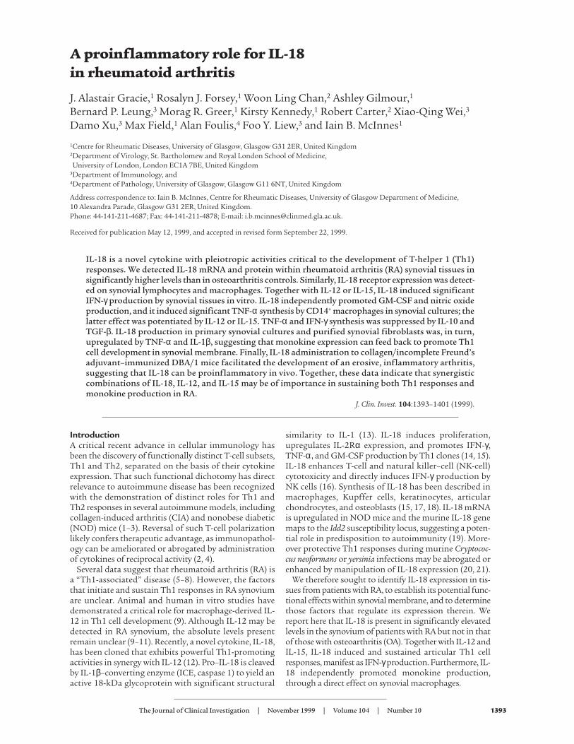
Figure 1(a) IL-18 mRNA was detected in RA and OA synovial membranes by RT-PCR (representative of 20 RA and 10 OA tissues). (b) Significantly high-er levels of IL-18 were detected in RA SF than in sera (18 matched sam-ples assayed after removal of rheumatoid factor).
(R&D Systems) or biotin-conjugated sheep anti-humanIL-18 and developed with Streptavidin-HRP (SAPU) andTMB substrate (Dynex). Rheumatoid factor (RF) wasremoved from SF by prior incubation with RapiTex beads(Behring Diagnostics, Marburg, Germany). As an addi-tional specificity control and to minimize RF contami-nation, monoclonal murine anti–IL-1β (R&D Systems)was used as an irrelevant capture antibody, followed bysample, secondary antibodies, and detection system asbefore. IFN-γwas measured by ELISA as already describedhere, using paired anti-human IFN-γ antibodies (gift ofT. Meager, National Institute for Biological Standardsand Control, Potters Bar, United Kingdom), detectionwith horseradish peroxidase–conju-gated donkey anti-rabbit IgG (Jack-son ImmunoResearch LaboratoriesInc., West Grove, Pennsylvania,USA), and TMB substrate. GM-CSFwas measured by ELISA (Genzyme,Cambridge, Massachusetts, USA).Intracellular TNF-α expression wasidentified in permeabilized synovialmononuclear populations by FACSanalysis (Becton Dickinson, Cowley,United Kingdom) using PE-conju-gated monoclonal anti–TNF-α anti-body (R&D Systems), together withFITC anti-CD3 or FITC anti-CD14antibodies (Becton Dickinson). IL-18 receptor expression was deter-mined by FACS analysis using mon-oclonal anti-human IL-18 receptorantibody (R&D Systems).
Expression of IL-18 and IL-18RmRNA in synovial tissue. cDNA wasprepared from synovial tissue andfibroblasts using TRIzol extraction(Life Technologies) and RT. IL-18mRNA was detected using primersdescribed previously for cloning IL-18. Primers for IL-18R were: sense5′CCCAACGATAAAGAAGAACGC C3′antisense 5′TGTCTGTGCCTCCCGT-GCTGGC3′ (product 419 bp), andfor β-actin: sense 5′GTGGGGCGCC-CCAGGCACCA3′ antisense 5′CTC-CTTAATGTCAC GCACG ATTTC3′(product 548 bp).
Nitric oxide estimation. Nitric oxide(NO) was measured as its oxidizedproduct, nitrite, using the Griessreaction (24).
Immunohistochemistry. Formalde-hyde-fixed paraffin sections (3 µm)were stained by a standard Strepta-vidin-HRP protocol. Product wasviewed with 3,3′-dia-minobenzidinetetrachloride (DAB; Sigma) andcounterstained with hematoxylin(Sigma). IL-18 expression was quan-tified by 2 independent observers bylight microscopy. Staining was
quantified from 0 to 4 as follows. Lining layer (LL)/inter-stitial (IS) areas: 0, no staining; 1, <5% positivecells/nuclei per high power field (HPF; ×400); 2, 5–30%;3, 30–90%; 4, >90%. Lymphocytic aggregate (LA) area: 0,no staining; 1, <5%; 2, 5–20%; 3, 20–40%; 4, >40%. Posi-tive vessels were expressed as percent of total vessels perHPF. For double staining, sections were first stainedwith anti–IL-18 and developed with SG peroxidase sub-strate (blue; Vector Laboratories Ltd., Peterborough,United Kingdom). After hydrogen peroxide (Sigma)treatment to quench residual HRP activity, sections werefurther stained with anti-CD68 or anti-CD3 antibody(DAKO, Ely, United Kingdom) and developed with DAB
The Journal of Clinical Investigation | November 1999 | Volume 104 | Number 10 1395
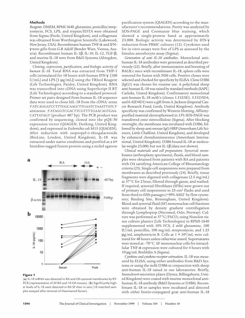
Figure 2(a) IL-18 was detected immunochemically (brown) in synovial membrane using anti–IL-18 anti-body (D3B6). (b) CD3 localization (brown) in parallel sections indicated IL-18 expression in Tcell–rich aggregates. Double staining with anti–IL-18 (blue/black) and (c) anti-CD3 (brown) or(d) anti-CD68 (brown) antibodies, without nuclear counterstain, localized IL-18 mainly to CD68+
macrophages. Black arrow, double positive; white arrow, IL-18 single positive.
(brown), without nuclear counterstain. Negative con-trols, including the omission of 1 or both primary anti-bodies or the addition of isotype-matched control anti-bodies of irrelevant specificity, confirmed the absence ofcross-reactivity between anti–IL-18, anti-CD3, or anti-CD68 antibodies. SFBs were formaldehyde fixed in situin Lab-Tek chamber slides (Nunclon) and stained asalready described here.
Induction and assessment of CIA. CIA was induced asdescribed previously (25). Briefly, male DBA/1 mice (Har-lan Olac, Bicester, United Kingdom) were immunized(intradermally) with 200 µg of type II bovine collagen(CII; Sigma) in CFA or incomplete adjuvant (IFA; DifcoLaboratories, Detroit, Michigan, USA). After intraperi-toneal CII (200 µg in PBS) challenge on day 21, arthritisdevelopment was scored by erythema, swelling, or lossfunction per paw (scale 0–3; maximum, 12 per mouse) bya treatment-blind observer. Mice were sacrificed, hindlimbs were fixed in 10% neutral-buffered formalin, and 5-µm sections were stained with hematoxylin and eosin(Sigma). Mice immunized with CII/IFA also receivedintraperitoneal injections of either murine IL-18 (100 ng)in PBS/0.1% BSA or PBS/0.1% BSA from day –1 to day 4and from day 20 to day 24.
Statistical analysis. This was performed on Minitab soft-ware, (State College, Pennsylvania, USA) using paired t
test and Mann-Whitney analyses as appro-priate.
ResultsIL-18 and IL-18 receptor expression in RA syn-ovial membrane. We sought to identify IL-18mRNA and protein expression within RAsynovial tissues. Using RT-PCR, we demon-strated constitutive IL-18 mRNA expres-sion in 20 of 20 RA synovial samples (Fig-ure 1a) and in 8 of 10 OA samples
examined. IL-18 was detected by ELISA in 12 of 18 SFsobtained from patients with RA (Figure 1b), whereasmatched sera typically contained low levels of IL-18 only(SF vs. serum: P < 0.005). Prior removal of rheumatoidfactor did not alter the levels of IL-18 detected. More-over, no significant signal was obtained when 8 SFs wereassayed using anti–IL-1β antibody as a capture antibodyin an otherwise identical ELISA protocol. Thus, neithercross-reactivity with IL-1 nor RF contamination is likelyto explain our observations. The median concentrationdetected in positive samples was 342 pg/mL (intraquar-tile range, 210–1,606), comparable to TNF-α concentra-tions normally detected in SF (11). IL-12 (p40) concen-trations in the SF (median, 82 pg/mL; IQ range, 0–313)did not correlate with those of IL-18. These data clearlyindicate that IL-18 is expressed in RA synovial mem-brane, and, because secretion of IL-18 requires precursorcleavage (13), they strongly suggest that mature IL-18 ispresent in the synovial compartment.
To determine the distribution of IL-18 expression, wegenerated an mAb to human IL-18 (clone D3B6) thatexhibited a cytoplasmic staining pattern in RA synovialtissues (Figure 2a). Staining was neutralized by priorincubation with recombinant IL-18, but not with thestructurally related cytokine IL-1β (data not shown). IL-18 staining was detected in 16 of 18 RA synovia exam-ined. IL-18 expression was most prominent within andadjacent to lymphocytic aggregates (Table 1) and wasdemonstrated using double staining to reside primarilyin CD68+ macrophages. A small population of CD68–, IL-18+ cells were also detected, likely representing fibroblasts(Figure 2d). These appearances were similar to those ingerminal centers in inflamed human tonsil (data notshown). IL-18 staining was also detected within and adja-cent to the lining layer, albeit at lower levels (Table 1; Fig-ure 2a). Double staining indicated that this IL-18 expres-sion was predominantly, but not exclusively, foundwithin CD68+ macrophages. CD3+ lymphocytes wereconsistently negative (Figure 2, c and d). This distributionis in contrast to that of other monokines such as IL-1β,TNF-α, or IL-12, which are usually prominent in liningand sublining layer macrophages (10, 11). Expression ofIL-18 in endothelial cells was present in 9 of 18 tissuesexamined at low levels only (5 ± 7% vessels per HPF). Lit-tle IL-18 protein was detected in OA synovial membranes(Table 1). Confirmatory immunohistology was per-formed using a further series of 3 anti-human IL-18mABs, raised from discrete clones, which revealed anidentical, neutralizable staining pattern in parallel RAsynovial tissues (data not shown).
We next sought evidence for IL-18R expression using
1396 The Journal of Clinical Investigation | November 1999 | Volume 104 | Number 10
Table 1Immunolocalization of IL-18 in synovial membraneA
Lining layer Aggregate Interstitium Endothelium
Mean ± SD histology score Percent positive vessels/HPF
RA (n = 18) 0.5 ± 0.3B 1.7 ± 0.8C 0.6 ± 0.5C 5OA (n = 7) 0.1 ± 0.1 0 0.03 ± 0.07 0.4
AIL-18 was detected using monoclonal anti-human IL-18 antibody (D3B6). Scores (range, 0–4) were allo-cated as described in Methods (HPF, ×400). BP < 0.004, CP < 0.001(Mann-Whitney), RA vs. OA tissues.
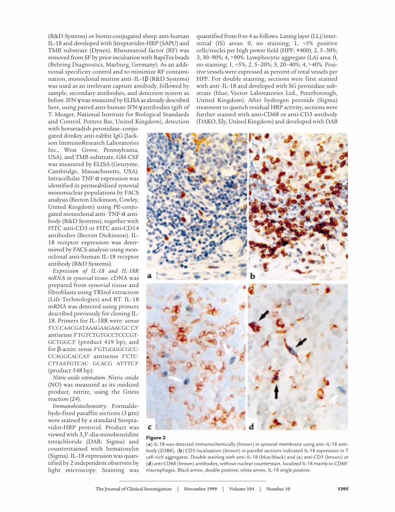
Figure 3(a) IL-18 receptor mRNA expression was detected by RT-PCR in 10 of 10RA synovial membranes (5 representative data are shown in lanes 1–5).LPS-stimulated THP-1 cells served as positive control. (b) Pooled data(mean ± SEM) to characterize IL-18R+ cells in RA synovial fluid mononu-clear cells (n = 8 patients).
reagents specific for its unique α chain. IL-18Rα mRNAwas detected by RT-PCR in 10 of 10 RA synovial mem-branes examined (Figure 3), with product specificityconfirmed by sequencing. Cellular expression of IL-18Rα protein was characterized by FACS analysis ofnonpermeabilized SF mononuclear cells. Significantsubpopulations of CD3+ SF lymphocytes and CD14+
macrophages expressed IL-18R (Figure 3).IL-18 induced IFN-γ production in RA synovial membrane.
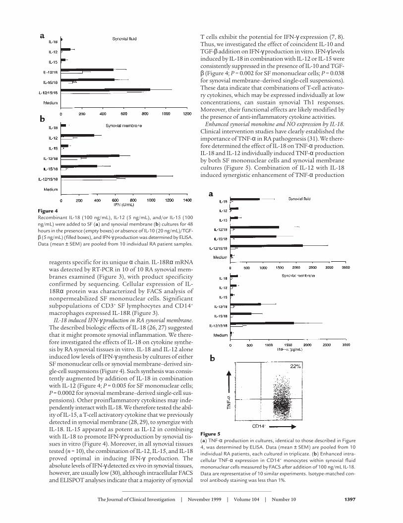
The described biologic effects of IL-18 (26, 27) suggestedthat it might promote synovial inflammation. We there-fore investigated the effects of IL-18 on cytokine synthe-sis by RA synovial tissues in vitro. IL-18 and IL-12 aloneinduced low levels of IFN-γsynthesis by cultures of eitherSF mononuclear cells or synovial membrane–derived sin-gle-cell suspensions (Figure 4). Such synthesis was consis-tently augmented by addition of IL-18 in combinationwith IL-12 (Figure 4; P = 0.005 for SF mononuclear cells;P = 0.0002 for synovial membrane–derived single-cell sus-pensions). Other proinflammatory cytokines may inde-pendently interact with IL-18. We therefore tested the abil-ity of IL-15, a T-cell activatory cytokine that we previouslydetected in synovial membrane (28, 29), to synergize withIL-18. IL-15 appeared as potent as IL-12 in combiningwith IL-18 to promote IFN-γproduction by synovial tis-sues in vitro (Figure 4). Moreover, in all synovial tissuestested (n = 10), the combination of IL-12, IL-15, and IL-18proved optimal in inducing IFN-γ production. Theabsolute levels of IFN-γdetected ex vivo in synovial tissues,however, are usually low (30), although intracellular FACSand ELISPOT analyses indicate that a majority of synovial
T cells exhibit the potential for IFN-γ expression (7, 8).Thus, we investigated the effect of coincident IL-10 andTGF-β addition on IFN-γproduction in vitro. IFN-γlevelsinduced by IL-18 in combination with IL-12 or IL-15 wereconsistently suppressed in the presence of IL-10 and TGF-β (Figure 4; P = 0.002 for SF mononuclear cells; P = 0.038for synovial membrane–derived single-cell suspensions).These data indicate that combinations of T-cell activato-ry cytokines, which may be expressed individually at lowconcentrations, can sustain synovial Th1 responses.Moreover, their functional effects are likely modified bythe presence of anti-inflammatory cytokine activities.
Enhanced synovial monokine and NO expression by IL-18.Clinical intervention studies have clearly established theimportance of TNF-α in RA pathogenesis (31). We there-fore determined the effect of IL-18 on TNF-α production.IL-18 and IL-12 individually induced TNF-α productionby both SF mononuclear cells and synovial membranecultures (Figure 5). Combination of IL-12 with IL-18induced synergistic enhancement of TNF-α production
The Journal of Clinical Investigation | November 1999 | Volume 104 | Number 10 1397
Figure 4Recombinant IL-18 (100 ng/mL), IL-12 (5 ng/mL), and/or IL-15 (100ng/mL) were added to SF (a) and synovial membrane (b) cultures for 48hours in the presence (empty boxes) or absence of IL-10 (20 ng/mL)/TGF-β (5 ng/mL) (filled boxes), and IFN-γproduction was determined by ELISA.Data (mean ± SEM) are pooled from 10 individual RA patient samples.
Figure 5(a) TNF-α production in cultures, identical to those described in Figure4, was determined by ELISA. Data (mean ± SEM) are pooled from 10individual RA patients, each cultured in triplicate. (b) Enhanced intra-cellular TNF-α expression in CD14+ monocytes within synovial fluidmononuclear cells measured by FACS after addition of 100 ng/mL IL-18.Data are representative of 10 similar experiments. Isotype-matched con-trol antibody staining was less than 1%.
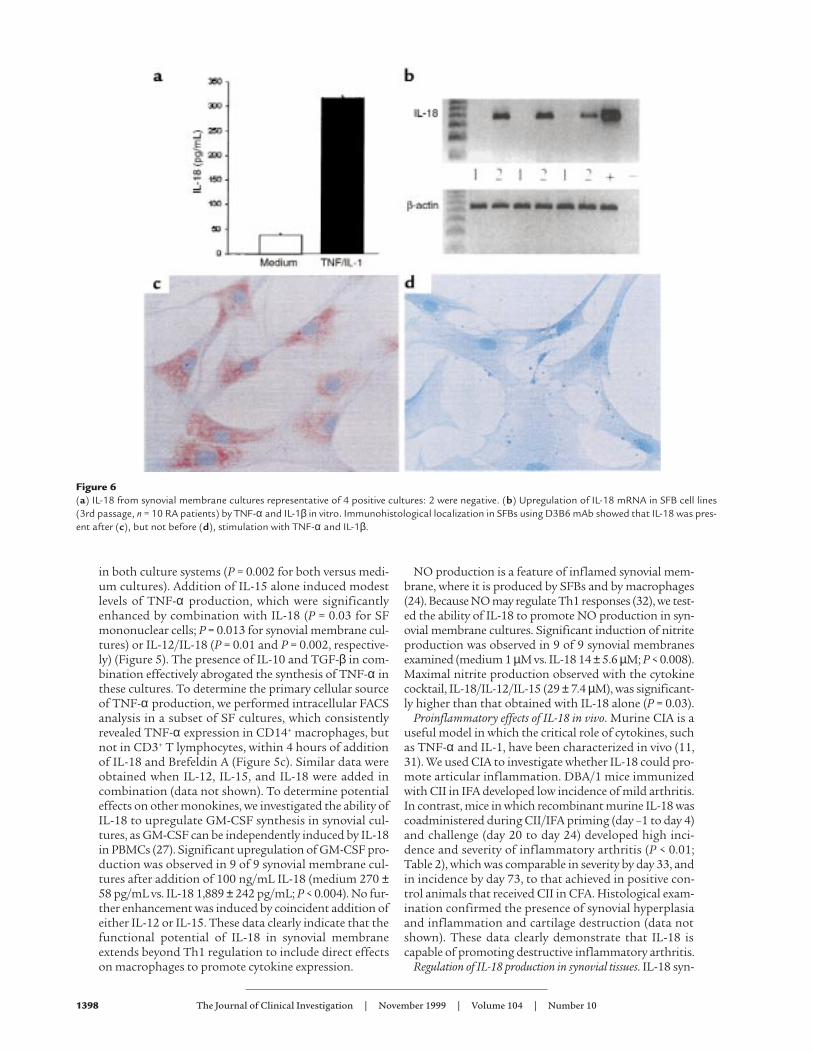
in both culture systems (P = 0.002 for both versus medi-um cultures). Addition of IL-15 alone induced modestlevels of TNF-α production, which were significantlyenhanced by combination with IL-18 (P = 0.03 for SFmononuclear cells; P = 0.013 for synovial membrane cul-tures) or IL-12/IL-18 (P = 0.01 and P = 0.002, respective-ly) (Figure 5). The presence of IL-10 and TGF-β in com-bination effectively abrogated the synthesis of TNF-α inthese cultures. To determine the primary cellular sourceof TNF-α production, we performed intracellular FACSanalysis in a subset of SF cultures, which consistentlyrevealed TNF-α expression in CD14+ macrophages, butnot in CD3+ T lymphocytes, within 4 hours of additionof IL-18 and Brefeldin A (Figure 5c). Similar data wereobtained when IL-12, IL-15, and IL-18 were added incombination (data not shown). To determine potentialeffects on other monokines, we investigated the ability ofIL-18 to upregulate GM-CSF synthesis in synovial cul-tures, as GM-CSF can be independently induced by IL-18in PBMCs (27). Significant upregulation of GM-CSF pro-duction was observed in 9 of 9 synovial membrane cul-tures after addition of 100 ng/mL IL-18 (medium 270 ±58 pg/mL vs. IL-18 1,889 ± 242 pg/mL; P < 0.004). No fur-ther enhancement was induced by coincident addition ofeither IL-12 or IL-15. These data clearly indicate that thefunctional potential of IL-18 in synovial membraneextends beyond Th1 regulation to include direct effectson macrophages to promote cytokine expression.
NO production is a feature of inflamed synovial mem-brane, where it is produced by SFBs and by macrophages(24). Because NO may regulate Th1 responses (32), we test-ed the ability of IL-18 to promote NO production in syn-ovial membrane cultures. Significant induction of nitriteproduction was observed in 9 of 9 synovial membranesexamined (medium 1 µM vs. IL-18 14 ± 5.6 µM; P < 0.008).Maximal nitrite production observed with the cytokinecocktail, IL-18/IL-12/IL-15 (29 ± 7.4 µM), was significant-ly higher than that obtained with IL-18 alone (P = 0.03).
Proinflammatory effects of IL-18 in vivo. Murine CIA is auseful model in which the critical role of cytokines, suchas TNF-α and IL-1, have been characterized in vivo (11,31). We used CIA to investigate whether IL-18 could pro-mote articular inflammation. DBA/1 mice immunizedwith CII in IFA developed low incidence of mild arthritis.In contrast, mice in which recombinant murine IL-18 wascoadministered during CII/IFA priming (day –1 to day 4)and challenge (day 20 to day 24) developed high inci-dence and severity of inflammatory arthritis (P < 0.01;Table 2), which was comparable in severity by day 33, andin incidence by day 73, to that achieved in positive con-trol animals that received CII in CFA. Histological exam-ination confirmed the presence of synovial hyperplasiaand inflammation and cartilage destruction (data notshown). These data clearly demonstrate that IL-18 iscapable of promoting destructive inflammatory arthritis.
Regulation of IL-18 production in synovial tissues. IL-18 syn-
1398 The Journal of Clinical Investigation | November 1999 | Volume 104 | Number 10
Figure 6(a) IL-18 from synovial membrane cultures representative of 4 positive cultures: 2 were negative. (b) Upregulation of IL-18 mRNA in SFB cell lines(3rd passage, n = 10 RA patients) by TNF-α and IL-1β in vitro. Immunohistological localization in SFBs using D3B6 mAb showed that IL-18 was pres-ent after (c), but not before (d), stimulation with TNF-α and IL-1β.
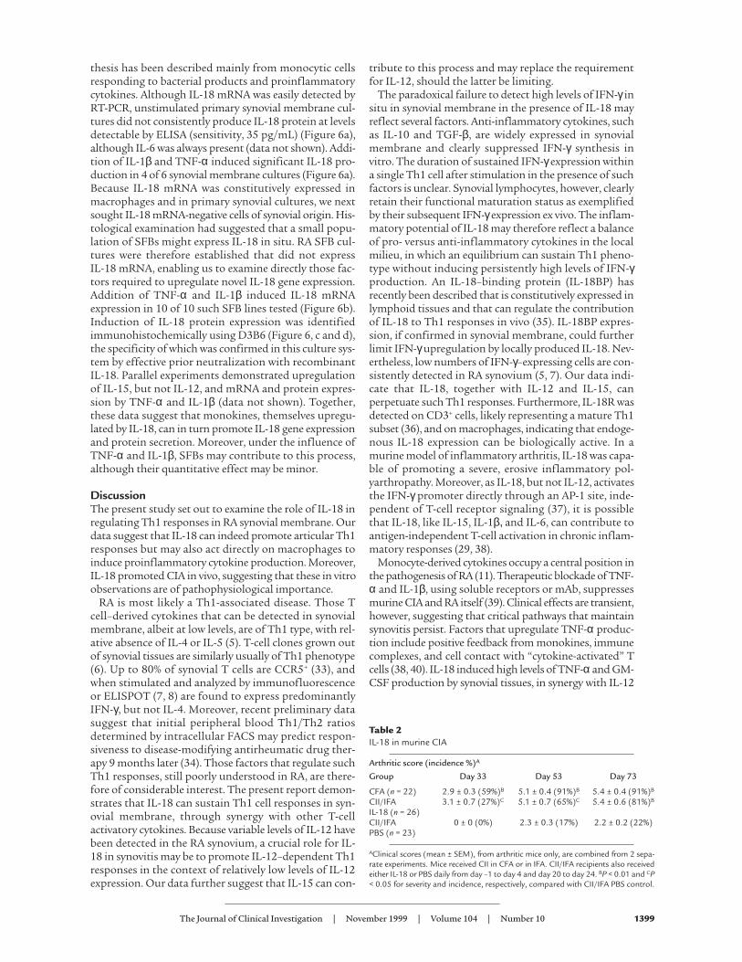
thesis has been described mainly from monocytic cellsresponding to bacterial products and proinflammatorycytokines. Although IL-18 mRNA was easily detected byRT-PCR, unstimulated primary synovial membrane cul-tures did not consistently produce IL-18 protein at levelsdetectable by ELISA (sensitivity, 35 pg/mL) (Figure 6a),although IL-6 was always present (data not shown). Addi-tion of IL-1β and TNF-α induced significant IL-18 pro-duction in 4 of 6 synovial membrane cultures (Figure 6a).Because IL-18 mRNA was constitutively expressed inmacrophages and in primary synovial cultures, we nextsought IL-18 mRNA-negative cells of synovial origin. His-tological examination had suggested that a small popu-lation of SFBs might express IL-18 in situ. RA SFB cul-tures were therefore established that did not expressIL-18 mRNA, enabling us to examine directly those fac-tors required to upregulate novel IL-18 gene expression.Addition of TNF-α and IL-1β induced IL-18 mRNAexpression in 10 of 10 such SFB lines tested (Figure 6b).Induction of IL-18 protein expression was identifiedimmunohistochemically using D3B6 (Figure 6, c and d),the specificity of which was confirmed in this culture sys-tem by effective prior neutralization with recombinantIL-18. Parallel experiments demonstrated upregulationof IL-15, but not IL-12, and mRNA and protein expres-sion by TNF-α and IL-1β (data not shown). Together,these data suggest that monokines, themselves upregu-lated by IL-18, can in turn promote IL-18 gene expressionand protein secretion. Moreover, under the influence ofTNF-α and IL-1β, SFBs may contribute to this process,although their quantitative effect may be minor.
DiscussionThe present study set out to examine the role of IL-18 inregulating Th1 responses in RA synovial membrane. Ourdata suggest that IL-18 can indeed promote articular Th1responses but may also act directly on macrophages toinduce proinflammatory cytokine production. Moreover,IL-18 promoted CIA in vivo, suggesting that these in vitroobservations are of pathophysiological importance.
RA is most likely a Th1-associated disease. Those Tcell–derived cytokines that can be detected in synovialmembrane, albeit at low levels, are of Th1 type, with rel-ative absence of IL-4 or IL-5 (5). T-cell clones grown outof synovial tissues are similarly usually of Th1 phenotype(6). Up to 80% of synovial T cells are CCR5+ (33), andwhen stimulated and analyzed by immunofluorescenceor ELISPOT (7, 8) are found to express predominantlyIFN-γ, but not IL-4. Moreover, recent preliminary datasuggest that initial peripheral blood Th1/Th2 ratiosdetermined by intracellular FACS may predict respon-siveness to disease-modifying antirheumatic drug ther-apy 9 months later (34). Those factors that regulate suchTh1 responses, still poorly understood in RA, are there-fore of considerable interest. The present report demon-strates that IL-18 can sustain Th1 cell responses in syn-ovial membrane, through synergy with other T-cellactivatory cytokines. Because variable levels of IL-12 havebeen detected in the RA synovium, a crucial role for IL-18 in synovitis may be to promote IL-12–dependent Th1responses in the context of relatively low levels of IL-12expression. Our data further suggest that IL-15 can con-
tribute to this process and may replace the requirementfor IL-12, should the latter be limiting.
The paradoxical failure to detect high levels of IFN-γ insitu in synovial membrane in the presence of IL-18 mayreflect several factors. Anti-inflammatory cytokines, suchas IL-10 and TGF-β, are widely expressed in synovialmembrane and clearly suppressed IFN-γ synthesis invitro. The duration of sustained IFN-γexpression withina single Th1 cell after stimulation in the presence of suchfactors is unclear. Synovial lymphocytes, however, clearlyretain their functional maturation status as exemplifiedby their subsequent IFN-γexpression ex vivo. The inflam-matory potential of IL-18 may therefore reflect a balanceof pro- versus anti-inflammatory cytokines in the localmilieu, in which an equilibrium can sustain Th1 pheno-type without inducing persistently high levels of IFN-γproduction. An IL-18–binding protein (IL-18BP) hasrecently been described that is constitutively expressed inlymphoid tissues and that can regulate the contributionof IL-18 to Th1 responses in vivo (35). IL-18BP expres-sion, if confirmed in synovial membrane, could furtherlimit IFN-γupregulation by locally produced IL-18. Nev-ertheless, low numbers of IFN-γ–expressing cells are con-sistently detected in RA synovium (5, 7). Our data indi-cate that IL-18, together with IL-12 and IL-15, canperpetuate such Th1 responses. Furthermore, IL-18R wasdetected on CD3+ cells, likely representing a mature Th1subset (36), and on macrophages, indicating that endoge-nous IL-18 expression can be biologically active. In amurine model of inflammatory arthritis, IL-18 was capa-ble of promoting a severe, erosive inflammatory pol-yarthropathy. Moreover, as IL-18, but not IL-12, activatesthe IFN-γ promoter directly through an AP-1 site, inde-pendent of T-cell receptor signaling (37), it is possiblethat IL-18, like IL-15, IL-1β, and IL-6, can contribute toantigen-independent T-cell activation in chronic inflam-matory responses (29, 38).
Monocyte-derived cytokines occupy a central position inthe pathogenesis of RA (11). Therapeutic blockade of TNF-α and IL-1β, using soluble receptors or mAb, suppressesmurine CIA and RA itself (39). Clinical effects are transient,however, suggesting that critical pathways that maintainsynovitis persist. Factors that upregulate TNF-α produc-tion include positive feedback from monokines, immunecomplexes, and cell contact with “cytokine-activated” Tcells (38, 40). IL-18 induced high levels of TNF-α and GM-CSF production by synovial tissues, in synergy with IL-12
The Journal of Clinical Investigation | November 1999 | Volume 104 | Number 10 1399
Table 2IL-18 in murine CIA
Arthritic score (incidence %)A
Group Day 33 Day 53 Day 73
CFA (n = 22) 2.9 ± 0.3 (59%)B 5.1 ± 0.4 (91%)B 5.4 ± 0.4 (91%)B
CII/IFA 3.1 ± 0.7 (27%)C 5.1 ± 0.7 (65%)C 5.4 ± 0.6 (81%)B
IL-18 (n = 26)CII/IFA 0 ± 0 (0%) 2.3 ± 0.3 (17%) 2.2 ± 0.2 (22%)PBS (n = 23)
AClinical scores (mean ± SEM), from arthritic mice only, are combined from 2 sepa-rate experiments. Mice received CII in CFA or in IFA. CII/IFA recipients also receivedeither IL-18 or PBS daily from day –1 to day 4 and day 20 to day 24. BP < 0.01 and CP< 0.05 for severity and incidence, respectively, compared with CII/IFA PBS control.
and IL-15. Single-cell staining revealed TNF-α protein inCD14+ macrophages after IL-18 addition to BrefeldinA–treated synovial cells. Because no IFN-γ secretionoccurred in these cultures, a direct effect of IL-18 on TNF-α production by synovial macrophages is probable. IL-18induces IL-1β mRNA expression directly in purifiedmacrophages, whereas TNF-α production by PBMCs hasbeen attributed to CD3/CD4+ cells (27, 41). Whether thisminor discrepancy is a function of the activation/matura-tion status of synovial versus blood macrophages requiresclarification. Together these data clearly indicate that theproinflammatory activities of IL-18 in synovial membraneextend beyond Th1 cells to include macrophages.
IL-18 expression in synovial tissues in vitro was enhancedby TNF-α and IL-1β, raising the possibility of a positivefeedback loop that could lead to reciprocal amplification ofTh1 responses and monocyte production. Moreover, IL-18inhibited TGF-β–induced proliferation, induced matrixmetalloproteinase and inducible NO synthase (NOS) geneexpression, and enhanced glycosaminoglycan release inchondrocytes in vitro (18). IL-18 could therefore contributeto cartilage degradation, although whether synovium-derived IL-18 can contribute locally to this process remainsto be determined. We have now also shown that IL-18induced nitrite synthesis in synovial membrane cultures.Given that NO reduces IL-18 cleavage through inhibitionof ICE (42), upregulation of NO release by IL-18, IL-12, orIL-15 in synovial cultures may represent an endogenousregulatory pathway for IL-18 (and IL-1β) production. Com-patible with this, we previously demonstrated dysregulatedTh1 responses in iNOS-deficient mice (32).
IL-18 is a member of the IL-1 cytokine family that iswidely expressed in RA synovium. IL-1β and IL-18 geneexpression is differentially regulated (26). The pattern ofdistribution of IL-18 in synovial membrane appears dis-tinct from that of IL-1β. Whereas IL-1β is highly expressedthroughout the lining layer, interstitium, and vasculature,IL-18 is localized primarily to lymphocytic aggregates.Because ICE regulates the processing and secretion fromintracellular precursors of both IL-1β and IL-18 (26), itseems likely that other distinct mechanisms will con-tribute to differential cytokine expression in discrete partsof the synovium. ICE inhibition remains an enticing ther-apeutic target. ICE inhibitors effectively suppress murineCIA (43). Whether such effects are associated with sup-pression of IL-18–dependent effects remains unclear.
A further implication of this study is that detection of acytokine and determination of its absolute concentrationin SF is unlikely to correlate directly to its relative func-tional importance, but should rather reflect its potentialfor synergy. We have identified an important role for IL-18, together with IL-15 and IL-12, in such a network with-in the synovial membrane. Although the mechanism ofsuch synergy is yet to be defined, the present study pro-vides rationale for identifying functional cytokine “cas-settes” that together may be amenable to inhibition.
AcknowledgmentsThis work received financial support from the ArthritisResearch Campaign, United Kingdom; The MedicalResearch Council; The Wellcome Trust; The NuffieldFoundation; and the MacFeat Bequest, University of
Glasgow. Clinical samples were kindly provided by J.Hunter, A. Kinninmonth, I. Stother, R.D. Sturrock, H.A.Capell, and R. Madhok.
1. Mauri, C., Williams, R.O., Walmsley, M., and Feldmann, M. 1996. Rela-tionship between Th1/Th2 cytokine patterns and the arthritogenicresponse in collagen induced arthritis. Eur. J. Immunol. 26:1511–1518.
2. Racke, M.K., et al. 1994. Cytokine-induced immune deviation as a ther-apy for inflammatory autoimmune disease. J. Exp. Med. 180:1961–1966.
3. Katz, J.D., Benoist, C., and Mathis, D. 1995. T helper cell subsets ininsulin dependent diabetes. Science. 268:1185–1188.
4. Mosmann, T.R., and Sad, S. 1996. The expanding universe of T cell sub-sets: Th1, Th2 and more. Immunol. Today. 17:13–17.
5. Ulfgren, A.-K., Lindblad, S., Klareskog, L., Andersson, J., and Andersson,U. 1995. Detection of cytokine producing cells in the synovial membranefrom patients with rheumatoid arthritis. Ann. Rheum. Dis. 54:654–661.
6. Miltenburg, A.M.M., van Laar, J.M, De Kuiper, R., Daha, M.R., and Breed-veld, F.C. 1992. T cells cloned from human rheumatoid synovial mem-brane functionally represent the Th1 subset. Scand. J. Immunol.35:603–610.
7. Dolhain, R.J.E.M., Heiden, A.N., Haar, N.T., Breedveld, F.C., and Mil-tenburg, A.M.M. 1996. Shift towards T lymphocytes with a T helper 1cytokine secretion profile in the joints of patients with rheumatoidarthritis. Arthritis Rheum. 39:1961–1969.
8. Ronnelid, J., et al. 1998. Production of T cell cytokines at the single celllevel in patients with inflammatory arthritides: enhanced activity in syn-ovial fluid compared to blood. Br. J. Rheum. 37:7–14.
9. Gately, M.K., et al. 1998. The interleukin-12/interleukin-12 receptor sys-tem. Ann. Rev. Immunol. 16:495–521.
10. Morita, Y., et al. 1998. Expression of interleukin-12 in synovial tissuefrom patients with rheumatoid arthritis. Arthritis Rheum. 41:306–314.
11. Feldmann, F., Brennan, F.M., and Maini, R.N. 1996. Role of cytokines inrheumatoid arthritis. Ann. Rev. Immunol. 14:397–440.
12. Ushio, S., et al. 1996. Cloning of the cDNA for human IFNγ-inducingfactor, expression in Escherichia coli, and studies on the biologic activitiesof the protein. J. Immunol. 156:4274–4279.
13. Gu, Y., et al. 1997. Activation of interferon-gamma inducing factor medi-ated by interleukin-1β converting enzyme. Science. 275:206–209.
14. Okamura, H., Kashiwamura, S., Tsutsui, H., Yoshimoto, T., and Nakan-ishi, K. 1998. Regulation of interferon-γproduction by IL-12 and IL-18.Curr. Opin. Immunol. 10:259–264.
15. Kohno, K., and Kurimoto, M. 1998. Interleukin 18, a cytokine whichresembles IL-1 structurally and IL-12 functionally but exerts its effectindependently of both. Clin. Immunol. Immunopathol. 86:11–15.
16. Takeda, K., et al. 1998. Defective NK cell activity and Th1 response in IL-18-deficient mice. Immunity. 8:383–390.
17. Udagawa, N., et al. 1997. Interleukin-18 (interferon-gamma–inducingfactor) is produced by osteoblasts and acts via granulocyte/macrophagecolony-stimulating factor and not via interferon-gamma to inhibitosteoclast formation. J. Exp. Med. 185:1005–1012.
18. Olee, T., Hashimoto, S., Quach, J., and Lotz, M. 1999. IL-18 is producedby articular chondrocytes and induces proinflammatory and catabolicresponses. J Immunol. 162:1096–1100.
19. Rothe, H., Jenkins, N., Copeland, N., and Kolb, H. 1997. Active stage ofautoimmune diabetes is associated with the expression of a novelcytokine, IGIF, which is located near Idd2. J. Clin. Invest. 99:469–474.
20. Kawakami, K., et al. 1997. IL-18 protects mice against pulmonary anddisseminated infection with Cryptococcus neoformans by inducing IFN-γproduction. J. Immunol. 159:5528–5534.
21. Bohn, E., et al. 1998. IL-18 (IFN-gamma–inducing factor) regulates earlycytokine production in, and promotes resolution of, bacterial infectionin mice. J. Immunol. 160:299–307.
22. Chan, W.L., and Mitchison, N.A. 1982. Use of somatic cell hybrids forproduction of monospecific viral antibodies. Lab. Res. Methods Biol. Med.15:125–141.
23. Arnett, F.C., Edworthy, S.M., and Block, D.A. 1988. The AmericanRheumatism Association 1987 revised criteria for the classification ofrheumatoid arthritis. Arthritis Rheum. 31:315–324.
24. McInnes, I.B., et al. 1997. Production of nitric oxide in the synovial mem-brane of rheumatoid and osteoarthritis patients. J. Exp. Med.184:1519–1524.
25. Ruchatz, H., Leung, B.P., Wei, X.-Q., McInnes, I.B., Liew, F.Y. 1998. Sol-uble IL-15 receptor alpha-chain administration prevents murine colla-gen-induced arthritis: a role for IL-15 in development of antigen-induced immunopathology. J. Immunol. 160:5654–5660.
26. Puren, A.J., Fantuzzi, G., and Dinarello, C.A. 1999. Gene expression, syn-thesis, and secretion of interleukin 18 and interleukin 1β are differen-tially regulated in human blood mononuclear cells and mouse spleencells. Proc. Natl. Acad. Sci. USA. 96:2256–2261.
27. Dinarello, C.A., et al. 1998. Overview of interleukin-18: more than aninterferon-gamma inducing factor. J. Leukoc. Biol. 63:658–664.
1400 The Journal of Clinical Investigation | November 1999 | Volume 104 | Number 10
28. McInnes, I.B., et al. 1996. The role of interleukin-15 in T-cell migrationand activation in rheumatoid arthritis. Nat. Med. 2:175–182.
29. McInnes, I.B., and Liew, F.Y. 1998. Interleukin 15: a proinflammatoryrole in rheumatoid arthritis synovitis. Immunol. Today. 19:75–79.
30. Firestein, G.S., and Zvaifler, N.J. 1987. Peripheral blood and synovialfluid monocyte activation in inflammatory arthritis. II. Low levels ofsynovial fluid and synovial tissue interferon suggest that gamma-inter-feron is not the primary macrophage activating factor. Arthritis Rheum.30:864–871.
31. Feldmann, M., Charles, P., Taylor, P., and Maini, R.N. 1998. Biologicalinsights from clinical trials with anti-TNF therapy. Springer Semin.Immunopathol. 20:21–28.
32. McInnes, I.B., and Liew, F.Y. 1998. Nitric oxide and immune response.In Nitric oxide and bone disease. M. Hukkanen, J.M. Polak, and S.P.F. Hugh-es, editors. Cambridge University Press. Cambridge, United Kingdom.8–20.
33. Loetscher, P., et al. 1998 CCR5 is characteristic of Th1 lymphocytes.Nature. 391:344–345.
34. van der Graaff, W.L., Prins, A.P., Dijkmans, B.A., and van Lier, R.A. 1998.Prognostic value of Th1/Th2 ratio in rheumatoid arthritis. Lancet.351:1931.
35. Novick, D., et al. 1999. Interleukin-18 binding protein: a novel modula-tor of the Th1 cytokine response. Immunity. 10:127–136.
36. Xu, D., et al. 1998. Selective expression and functions of interleukin 18 recep-
tor on T helper (Th) type 1 but not Th2 cells. J. Exp. Med. 188:1485–1492.37. Barbulescu, K., et al. 1998. IL-12 and IL-18 differentially regulate the
transcriptional activity of the human IFN-gamma promoter in primaryCD4+ T lymphocytes. J. Immunol. 160:3642–3647.
38. McInnes, I.B., Leung, B.P., Sturrock, R.D., Field, M., and Liew, F.Y. 1997.Interleukin-15 mediates T cell–dependent regulation of tumour necro-sis factor-alpha production in rheumatoid arthritis. Nat. Med. 3:189–195.
39. Williams, R.O., Feldmann, M., and Maini, R.N. 1994. Anti-tumournecrosis factor ameliorates joint disease in murine collagen inducedarthritis. Proc. Natl. Acad. Sci. USA. 89:9784–9788.
40. Vey, E., Zhang, J., and Dayer, J.-M. 1992. IFNγ and 1,25(OH) D3 induceon THP-1 cells distinct patterns of cell surface antigen expression,cytokine production and responsiveness to contact with activated Tcells. J. Immunol. 149:2040–2046.
41. Puren, A.J., Fantuzzi, G., Gu, Y., Su, M.S., and Dinarello, C.A. 1998. Inter-leukin-18 (IFNγ-inducing factor) induces IL-8 and IL-1β via TNF-α pro-duction from non-CD14+ human blood mononuclear cells. J. Clin. Invest.101:711–721.
42. Kim, Y.M., Talanian, R.V., Li, J., and Billiar, T.R. 1998. Nitric oxide preventsIL-1β and IFN-—inducing factor (IL-18) release from macrophages byinhibiting caspase-1 (IL-1β-converting enzyme). J. Immunol. 161:122–128.
43. Ku, G., Faust, T., Lauffer, L.L., Livingston, D.J., and Harding, M.W. 1996.Interleukin-1β converting enzyme inhibition blocks progression of typeII collagen–induced arthritis in mice. Cytokine. 8:377–386.
The Journal of Clinical Investigation | November 1999 | Volume 104 | Number 10 1401