a pathological late pleistocene badger from san sidero (apulia, southern italy): implications for...
TRANSCRIPT
Forum communication
A pathological Late Pleistocene badger from San Sidero (Apulia,Southern Italy): Implications for developmental pathology andfeeding behaviour
Dawid A. Iurino a, *, Rosario Fico b, Raffaele Sardella a
a Dipartimento di Scienze della Terra, Sapienza Universit!a di Roma, Piazzale Aldo Moro 5, 00185, Rome, Italyb Centro di Referenza Nazionale per la Medicina Forense Veterinaria Istituto Zoopro!lattico Sperimentale del Lazio e della Toscana, Sezione di Grosseto,Viale Europa, 30, 58100, Grosseto, Italy
a r t i c l e i n f o
Article history:Available online xxx
Keywords:PaleopathologyChronic suppurative osteomyelitisMustelidaeMeles melesLate Pleistocene
a b s t r a c t
Among fossil vertebrates, oral pathologies are of particular interest, because of their considerable effecton teeth and maxillary/dentary bones and, as a consequence, on mastication and feeding behaviour. Thisstudy focused on a pathological left hemimandible referred to the mustelidMeles meles unearthed from aLate Pleistocene karst !lling deposit at San Sidero (Apulia, South Italy). This fossil shows unusual markedabnormalities related to a rare case of nonodontogenic chronic suppurative osteomyelitis. Clinicaldiagnosis of the disease and the timing of its development have been de!ned on the basis of a veterinaryapproach and X-ray analyses. Such a pathological condition can be explained as a consequence of awound due to a porcupine quill. The analysis of the injury also provides information about the biome-chanics of the bite and on the feeding behaviour. The study case con!rms how palaeopathological an-alyses can be considered valuable tools to reconstruct the physiology of animals that lived in the past andto depict in detail the interactions among Late Pleistocene mammals, thus allowing a more accuratereconstruction of the ecology in fossil mammals.
© 2014 Elsevier Ltd and INQUA. All rights reserved.
1. Introduction
The study of palaeopathological traces on fossil bones is avaluable tool in reconstructing care conditions in extinct animals.Some pathologies and abnormalities may reveal interesting aspectsof physiological and anatomical features in fossil animals and, insome cases, also important information about the behaviour andthe past interaction between animals (Siegel, 1976; Palmqvist et al.,1999; Iurino et al., 2013; Iurino and Sardella, 2014; Iurino, 2014). In2006, during a !eld survey in the quarries of San Sidero (Lecce,Apulia), a research team of the Earth Sciences Department ofSapienza University of Rome collected large mammal remains fromthe Late Pleistocene karst !lling deposits exposed in that area.Among them three carnivores showed clear evidence of pathol-ogies or injuries. Iurino et al. (2013) described a case of chronicperiodontitis on a partial skull and mandible referable to the
hypercarniveore canid Cuon alpinus, while a process of alveolarreossi!cation after canine loss in Crocuta crocuta has beendescribed by Iurino and Sardella (2014). The third carnivore is thefocus of the present paper. The fossil is a hemimandible of Melesmeles (SS 2006/1) with marked abnormalities related to a rare caseof nonodontogenic chronic suppurative osteomyelitis. Such apathological condition may be explained as a consequence of awound due to a porcupine quill. The morphological abnormalitiesof the left hemimandible have been investigated using tomographictechniques and veterinary diagnostics to reconstruct the develop-ment process of the pathology and its inferences on badger feedingbehaviour and care condition.
2. Geological setting
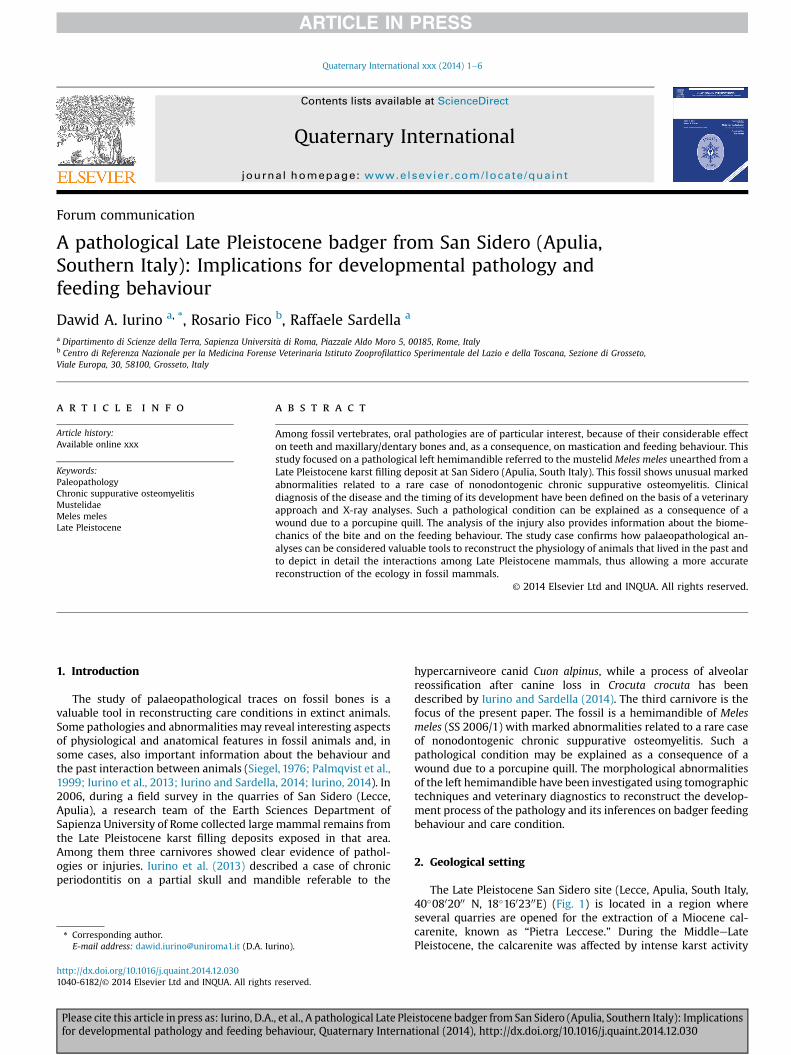
The Late Pleistocene San Sidero site (Lecce, Apulia, South Italy,40!0802000 N, 18!1602300E) (Fig. 1) is located in a region whereseveral quarries are opened for the extraction of a Miocene cal-carenite, known as “Pietra Leccese.” During the MiddleeLatePleistocene, the calcarenite was affected by intense karst activity
* Corresponding author.E-mail address: [email protected] (D.A. Iurino).
Contents lists available at ScienceDirect
Quaternary International
journal homepage: www.elsevier .com/locate/quaint
http://dx.doi.org/10.1016/j.quaint.2014.12.0301040-6182/© 2014 Elsevier Ltd and INQUA. All rights reserved.
Quaternary International xxx (2014) 1e6
Please cite this article in press as: Iurino, D.A., et al., A pathological Late Pleistocene badger from San Sidero (Apulia, Southern Italy): Implicationsfor developmental pathology and feeding behaviour, Quaternary International (2014), http://dx.doi.org/10.1016/j.quaint.2014.12.030
that formed an articulated !ssure network that the quarry activitieshave since exposed (Selleri et al., 2003; Selleri, 2007). The verticallyfunnel-shaped karst !ssures, locally named “ventarole,”were !lledwith “terre rosse,” residual reddish clays rich with iron oxides,including, in some cases, vertebrate fossil bones. The fossil remains,carried by surface water run-off in the !ssures, are generally wellpreserved, and articulated bones can be found. The faunal assem-blage recorded into the “terre rosse” deposit includes Equus ferus,Equus hydruntinus, Stephanorhinus hemitoechus, Dama dama, Cervuselaphus, Capreolus capreolus, Bos primigenius, Bison priscus, Susscrofa, Canis lupus, Canis mosbachensis, Cuon alpinus, Vulpes vulpes,Panthera pardus, Felis silvestris, Lynx lynx, Crocuta crocuta, Melesmeles, Lepus europaeus, Oryctolagus cuniculus, Erinaceus cf.E. europaeus, Microtus (Terricola) savii, and Apodemus sylvaticus,Eliomys quercinus and has been biochronologically referred to thebeginning of the Late Pleistocene (De Giuli, 1983; Bologna et al.,1994; Bedetti et al., 2004).
3. Materials and methods
3.1. Radiography and CT
Radiographs were taken at the Veterinary Clinic “La ClinicaVeterinaria Borghesiana” (Roma), using digital Radiological Unit RxFoschi x1 425 HF. It consists of an X-ray tube of KW 16e32, withrotating anode of 2800 rpm (HZ 50) and 60 mm in diameter. Themaximum anode voltage is 130 kV. Radiographs have beenmade inprojection L-L (latero-lateralis) 42 kV; mAs 20, projection M-L(medio-lateralis) 42 kV; mAs 20, with dimensions of 3072 " 3072pixels. Tomographic imagesweremade at M. G. Vannini Hospital byPhilips Brilliance CT 64-channel scanner. The fossil of M. meles wasscanned in its entirety in the coronal (i.e. transverse of some au-thors) slice plane from front to back. The scanning resulted in 137slices (i.e. images) with dimensions of 768 " 596 pixels. The slicesare 0.9 mm thick with an interslice space (the space betweenconsecutive slices) of 0.45 mm. Segmentation and 3D rendering ofthe hemimaxilla were computed using the Open-Source softwareOsiriX 5.6, 32-bit for Mac.
3.2. Referred material
SS 2006/1: left hemimandible with M1 and M2“SS” refers to San Sidero. The specimen is stored at the Earth
Science Department of Sapienza University of Rome.
3.2.1. DescriptionThe fossil remain of Meles meles from San Sidero (SS 2006/1) is
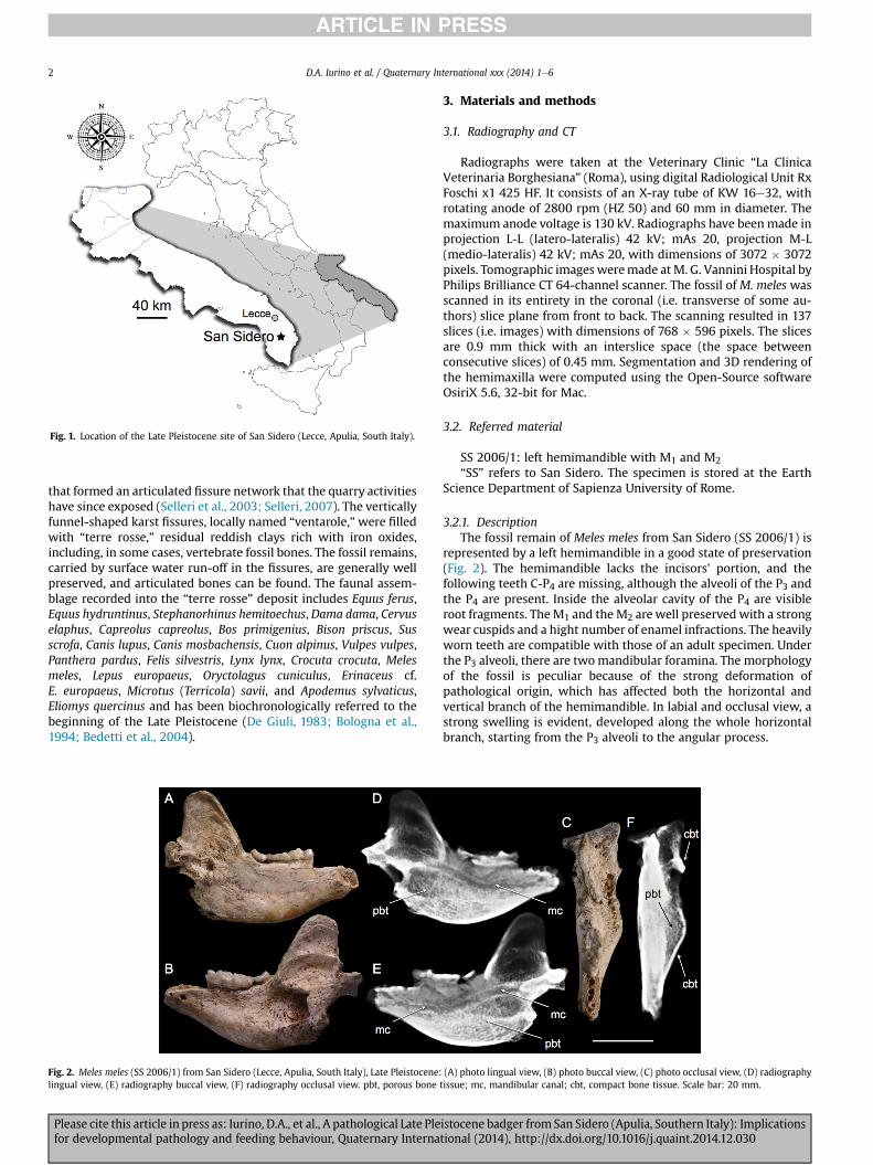
represented by a left hemimandible in a good state of preservation(Fig. 2). The hemimandible lacks the incisors' portion, and thefollowing teeth C-P4 are missing, although the alveoli of the P3 andthe P4 are present. Inside the alveolar cavity of the P4 are visibleroot fragments. TheM1 and the M2 are well preserved with a strongwear cuspids and a hight number of enamel infractions. The heavilyworn teeth are compatible with those of an adult specimen. Underthe P3 alveoli, there are twomandibular foramina. The morphologyof the fossil is peculiar because of the strong deformation ofpathological origin, which has affected both the horizontal andvertical branch of the hemimandible. In labial and occlusal view, astrong swelling is evident, developed along the whole horizontalbranch, starting from the P3 alveoli to the angular process.
Fig. 1. Location of the Late Pleistocene site of San Sidero (Lecce, Apulia, South Italy).
Fig. 2. Meles meles (SS 2006/1) from San Sidero (Lecce, Apulia, South Italy), Late Pleistocene: (A) photo lingual view, (B) photo buccal view, (C) photo occlusal view, (D) radiographylingual view, (E) radiography buccal view, (F) radiography occlusal view. pbt, porous bone tissue; mc, mandibular canal; cbt, compact bone tissue. Scale bar: 20 mm.
D.A. Iurino et al. / Quaternary International xxx (2014) 1e62
Please cite this article in press as: Iurino, D.A., et al., A pathological Late Pleistocene badger from San Sidero (Apulia, Southern Italy): Implicationsfor developmental pathology and feeding behaviour, Quaternary International (2014), http://dx.doi.org/10.1016/j.quaint.2014.12.030
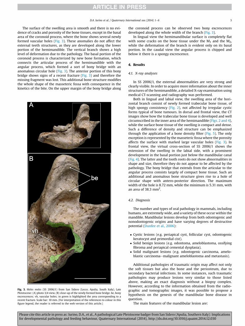
The surface of the swelling area is smooth and there is no evi-dence of cracks and porosity of the bone tissues, except in the basalarea of the coronoid process, where the bone shows several newlyformed vascular holes (Fig. 3). These anomalies do not affect theexternal teeth structures, as they are developed along the lowerportion of the hemimandible. The vertical branch shows a highlevel of deformation due to the pathology. The basal portion of thecoronoid process is characterized by new bone formation, whichconnects the articular process of the hemimandible with theangular process, which formed a sort of bony bridge with ananomalous circular hole (Fig. 3). The anterior portion of this bonybridge shows signs of a recent fracture (Fig. 3) and therefore themissing fragment was lost. This additional bone structure modi!esthe whole shape of the masseteric fossa with consequence in thekinetics of the bite. On the upper margin of the bony bridge along
the coronoid process can be observed two bony excrescencesdeveloped along the whole width of the branch (Fig. 3).
In lingual view the hemimandibular surface is completely "atwith some cracks on the bone tissue under the M1 and the M2,while the deformation of the branch is evident only on its basalportion. In the caudal view the angular process is chipped andbelow it there is a spongy excrescence.
4. Results
4.1. X-ray analyses
In SS 2006/1, the external abnormalities are very strong andclearly visible. In order to acquiremore information about the innerstructures of the hemimandible, a detailed X-ray examination usingmedical CT-scanning and radiography was performed.
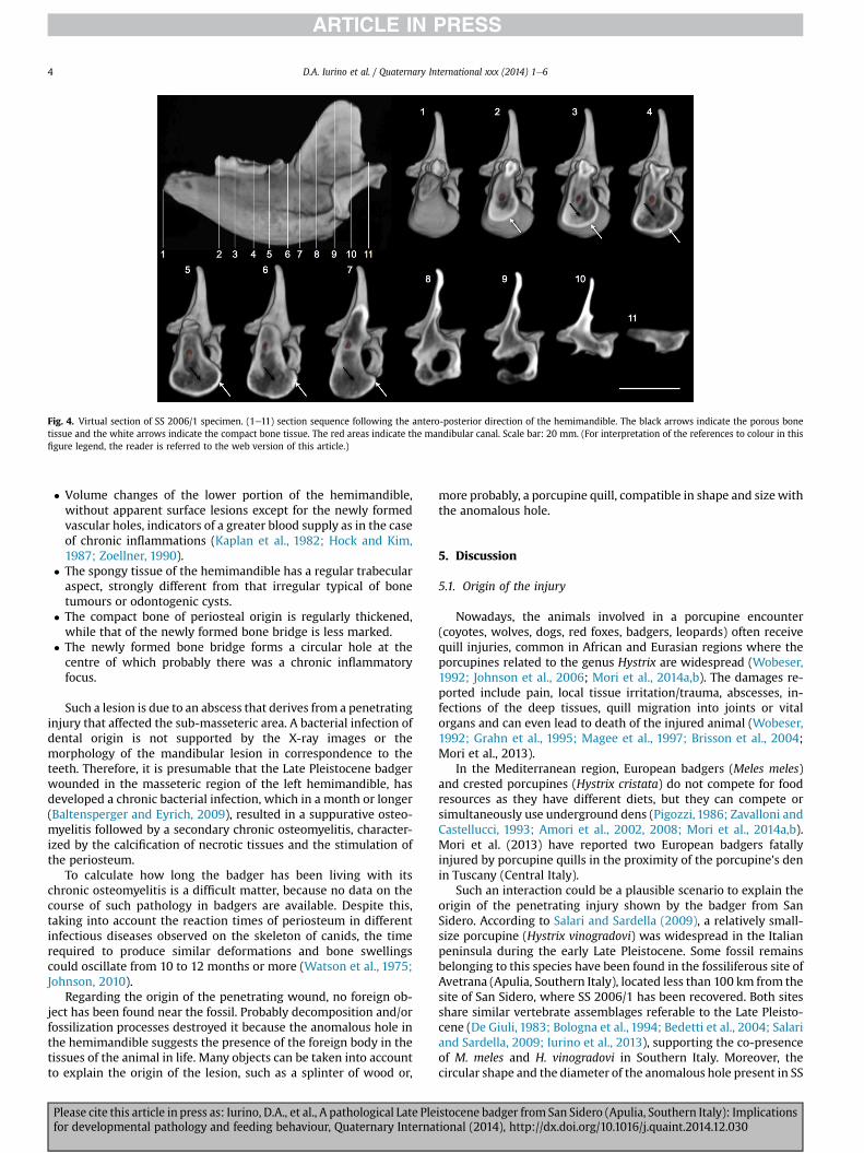
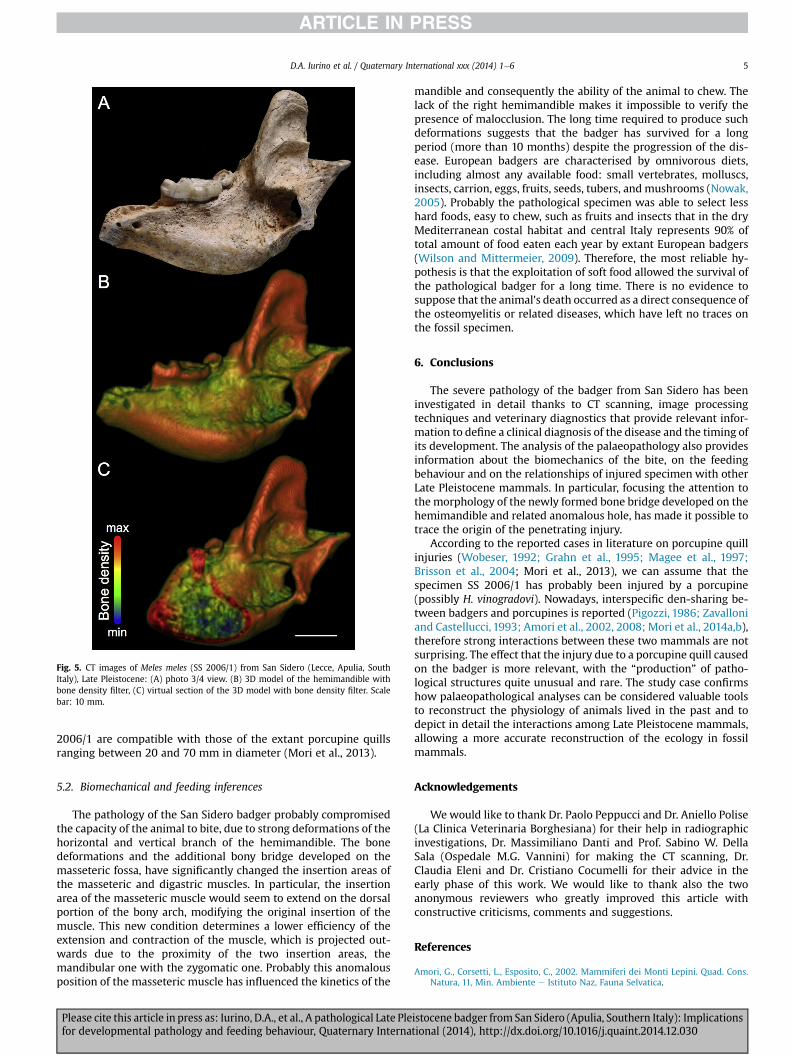
Both in lingual and labial view, the swelling area of the hori-zontal branch consist of newly formed trabecular bone tissue, ofhigh spongy consistency (Fig. 2), not affected by irregular cysticforms typical of bone tumours. In dorsal and frontal view, the CTimages show how the trabecular bone tissue is developed and wellcircumscribed in the inner area of the hemimandible (Figs. 2 and 4),while the surface bone tissue of the swelling is compact and dense.Such a difference of density and structure can be emphasizedthrough the application of a bone density !lter (Fig. 5). The onlyexception is represented by themasseteric fossawhere the porosityaffects the surface with marked large vascular holes (Fig. 3). Infrontal view, the virtual cross-section of SS 2006/1 shows theextension of the swelling in the labial side, with a prominentdevelopment in the basal portion just below the mandibular canal(Fig. 4). The latter and the tooth roots do not show abnormalities inshape and size, therefore they do not appear to be affected by thepathology. The bony bridge that extends from the articular to theangular process consists largely of compact bone tissue. Such anadditional and anomalous bone structure gives rise to a hole ofcircular shape with antero-posterior direction. The maximumwidth of the hole is 8.72 mm, while the minimum is 5.31 mm, withan area of 38.3 mm2.
4.2. Diagnosis
The number and types of oral pathology in mammals, includinghumans, are extremely wide, and a variety of these occurwithin themandible. Mandibular lesions develop from both odontogenic andnonodontogenic origins and have varying degrees of destructivepotential (Dunfee et al., 2006):
# Cystic lesions (e.g. periapical cyst, follicular cyst, odontogenickeratocyst and primordial cist).
# Solid benign lesions (e.g. odontoma, ameloblastoma, ossifying!broma and periapical cemental dysplasia).
# Solid malignant lesions (e.g. odontogenic carcinoma, amelo-blastic carcinomaemalignant ameloblastoma and metastasis).
Additional pathologies of traumatic origin may affect not onlythe soft tissues but also the bone and the periosteum, due tosecondary bacterial infections. In some instances, such traumaticpathologies may produce lesions very similar to those listedabove, making an exact diagnosis without a biopsy complex.However, according to the information obtained from the radio-graphic and tomographic images, it was possible to propose ahypothesis on the genesis of the mandibular bone disease inquestion.
The main features of the mandibular lesion are:
Fig. 3. Meles meles (SS 2006/1) from San Sidero (Lecce, Apulia, South Italy), LatePleistocene: (A) photo 3/4 view, (B) close-up of the newly formed bone bridge. be, bonyexcrescences; vh, vascular holes; in green is highlighted the area corresponding to arecent fracture. Scale bar: 10 mm. (For interpretation of the references to colour in this!gure legend, the reader is referred to the web version of this article.)
D.A. Iurino et al. / Quaternary International xxx (2014) 1e6 3
Please cite this article in press as: Iurino, D.A., et al., A pathological Late Pleistocene badger from San Sidero (Apulia, Southern Italy): Implicationsfor developmental pathology and feeding behaviour, Quaternary International (2014), http://dx.doi.org/10.1016/j.quaint.2014.12.030
# Volume changes of the lower portion of the hemimandible,without apparent surface lesions except for the newly formedvascular holes, indicators of a greater blood supply as in the caseof chronic in"ammations (Kaplan et al., 1982; Hock and Kim,1987; Zoellner, 1990).
# The spongy tissue of the hemimandible has a regular trabecularaspect, strongly different from that irregular typical of bonetumours or odontogenic cysts.
# The compact bone of periosteal origin is regularly thickened,while that of the newly formed bone bridge is less marked.
# The newly formed bone bridge forms a circular hole at thecentre of which probably there was a chronic in"ammatoryfocus.
Such a lesion is due to an abscess that derives from a penetratinginjury that affected the sub-masseteric area. A bacterial infection ofdental origin is not supported by the X-ray images or themorphology of the mandibular lesion in correspondence to theteeth. Therefore, it is presumable that the Late Pleistocene badgerwounded in the masseteric region of the left hemimandible, hasdeveloped a chronic bacterial infection, which in a month or longer(Baltensperger and Eyrich, 2009), resulted in a suppurative osteo-myelitis followed by a secondary chronic osteomyelitis, character-ized by the calci!cation of necrotic tissues and the stimulation ofthe periosteum.
To calculate how long the badger has been living with itschronic osteomyelitis is a dif!cult matter, because no data on thecourse of such pathology in badgers are available. Despite this,taking into account the reaction times of periosteum in differentinfectious diseases observed on the skeleton of canids, the timerequired to produce similar deformations and bone swellingscould oscillate from 10 to 12 months or more (Watson et al., 1975;Johnson, 2010).
Regarding the origin of the penetrating wound, no foreign ob-ject has been found near the fossil. Probably decomposition and/orfossilization processes destroyed it because the anomalous hole inthe hemimandible suggests the presence of the foreign body in thetissues of the animal in life. Many objects can be taken into accountto explain the origin of the lesion, such as a splinter of wood or,
more probably, a porcupine quill, compatible in shape and sizewiththe anomalous hole.
5. Discussion
5.1. Origin of the injury
Nowadays, the animals involved in a porcupine encounter(coyotes, wolves, dogs, red foxes, badgers, leopards) often receivequill injuries, common in African and Eurasian regions where theporcupines related to the genus Hystrix are widespread (Wobeser,1992; Johnson et al., 2006; Mori et al., 2014a,b). The damages re-ported include pain, local tissue irritation/trauma, abscesses, in-fections of the deep tissues, quill migration into joints or vitalorgans and can even lead to death of the injured animal (Wobeser,1992; Grahn et al., 1995; Magee et al., 1997; Brisson et al., 2004;Mori et al., 2013).
In the Mediterranean region, European badgers (Meles meles)and crested porcupines (Hystrix cristata) do not compete for foodresources as they have different diets, but they can compete orsimultaneously use underground dens (Pigozzi, 1986; Zavalloni andCastellucci, 1993; Amori et al., 2002, 2008; Mori et al., 2014a,b).Mori et al. (2013) have reported two European badgers fatallyinjured by porcupine quills in the proximity of the porcupine's denin Tuscany (Central Italy).
Such an interaction could be a plausible scenario to explain theorigin of the penetrating injury shown by the badger from SanSidero. According to Salari and Sardella (2009), a relatively small-size porcupine (Hystrix vinogradovi) was widespread in the Italianpeninsula during the early Late Pleistocene. Some fossil remainsbelonging to this species have been found in the fossiliferous site ofAvetrana (Apulia, Southern Italy), located less than 100 km from thesite of San Sidero, where SS 2006/1 has been recovered. Both sitesshare similar vertebrate assemblages referable to the Late Pleisto-cene (De Giuli, 1983; Bologna et al., 1994; Bedetti et al., 2004; Salariand Sardella, 2009; Iurino et al., 2013), supporting the co-presenceof M. meles and H. vinogradovi in Southern Italy. Moreover, thecircular shape and the diameter of the anomalous hole present in SS
Fig. 4. Virtual section of SS 2006/1 specimen. (1e11) section sequence following the antero-posterior direction of the hemimandible. The black arrows indicate the porous bonetissue and the white arrows indicate the compact bone tissue. The red areas indicate the mandibular canal. Scale bar: 20 mm. (For interpretation of the references to colour in this!gure legend, the reader is referred to the web version of this article.)
D.A. Iurino et al. / Quaternary International xxx (2014) 1e64
Please cite this article in press as: Iurino, D.A., et al., A pathological Late Pleistocene badger from San Sidero (Apulia, Southern Italy): Implicationsfor developmental pathology and feeding behaviour, Quaternary International (2014), http://dx.doi.org/10.1016/j.quaint.2014.12.030
2006/1 are compatible with those of the extant porcupine quillsranging between 20 and 70 mm in diameter (Mori et al., 2013).
5.2. Biomechanical and feeding inferences
The pathology of the San Sidero badger probably compromisedthe capacity of the animal to bite, due to strong deformations of thehorizontal and vertical branch of the hemimandible. The bonedeformations and the additional bony bridge developed on themasseteric fossa, have signi!cantly changed the insertion areas ofthe masseteric and digastric muscles. In particular, the insertionarea of the masseteric muscle would seem to extend on the dorsalportion of the bony arch, modifying the original insertion of themuscle. This new condition determines a lower ef!ciency of theextension and contraction of the muscle, which is projected out-wards due to the proximity of the two insertion areas, themandibular one with the zygomatic one. Probably this anomalousposition of the masseteric muscle has in"uenced the kinetics of the
mandible and consequently the ability of the animal to chew. Thelack of the right hemimandible makes it impossible to verify thepresence of malocclusion. The long time required to produce suchdeformations suggests that the badger has survived for a longperiod (more than 10 months) despite the progression of the dis-ease. European badgers are characterised by omnivorous diets,including almost any available food: small vertebrates, molluscs,insects, carrion, eggs, fruits, seeds, tubers, and mushrooms (Nowak,2005). Probably the pathological specimen was able to select lesshard foods, easy to chew, such as fruits and insects that in the dryMediterranean costal habitat and central Italy represents 90% oftotal amount of food eaten each year by extant European badgers(Wilson and Mittermeier, 2009). Therefore, the most reliable hy-pothesis is that the exploitation of soft food allowed the survival ofthe pathological badger for a long time. There is no evidence tosuppose that the animal's death occurred as a direct consequence ofthe osteomyelitis or related diseases, which have left no traces onthe fossil specimen.
6. Conclusions
The severe pathology of the badger from San Sidero has beeninvestigated in detail thanks to CT scanning, image processingtechniques and veterinary diagnostics that provide relevant infor-mation to de!ne a clinical diagnosis of the disease and the timing ofits development. The analysis of the palaeopathology also providesinformation about the biomechanics of the bite, on the feedingbehaviour and on the relationships of injured specimen with otherLate Pleistocene mammals. In particular, focusing the attention tothe morphology of the newly formed bone bridge developed on thehemimandible and related anomalous hole, has made it possible totrace the origin of the penetrating injury.
According to the reported cases in literature on porcupine quillinjuries (Wobeser, 1992; Grahn et al., 1995; Magee et al., 1997;Brisson et al., 2004; Mori et al., 2013), we can assume that thespecimen SS 2006/1 has probably been injured by a porcupine(possibly H. vinogradovi). Nowadays, interspeci!c den-sharing be-tween badgers and porcupines is reported (Pigozzi, 1986; Zavalloniand Castellucci, 1993; Amori et al., 2002, 2008; Mori et al., 2014a,b),therefore strong interactions between these two mammals are notsurprising. The effect that the injury due to a porcupine quill causedon the badger is more relevant, with the “production” of patho-logical structures quite unusual and rare. The study case con!rmshow palaeopathological analyses can be considered valuable toolsto reconstruct the physiology of animals lived in the past and todepict in detail the interactions among Late Pleistocene mammals,allowing a more accurate reconstruction of the ecology in fossilmammals.
Acknowledgements
Wewould like to thank Dr. Paolo Peppucci and Dr. Aniello Polise(La Clinica Veterinaria Borghesiana) for their help in radiographicinvestigations, Dr. Massimiliano Danti and Prof. Sabino W. DellaSala (Ospedale M.G. Vannini) for making the CT scanning, Dr.Claudia Eleni and Dr. Cristiano Cocumelli for their advice in theearly phase of this work. We would like to thank also the twoanonymous reviewers who greatly improved this article withconstructive criticisms, comments and suggestions.
References
Amori, G., Corsetti, L., Esposito, C., 2002. Mammiferi dei Monti Lepini. Quad. Cons.Natura, 11, Min. Ambiente e Istituto Naz, Fauna Selvatica.
Fig. 5. CT images of Meles meles (SS 2006/1) from San Sidero (Lecce, Apulia, SouthItaly), Late Pleistocene: (A) photo 3/4 view. (B) 3D model of the hemimandible withbone density !lter, (C) virtual section of the 3D model with bone density !lter. Scalebar: 10 mm.
D.A. Iurino et al. / Quaternary International xxx (2014) 1e6 5
Please cite this article in press as: Iurino, D.A., et al., A pathological Late Pleistocene badger from San Sidero (Apulia, Southern Italy): Implicationsfor developmental pathology and feeding behaviour, Quaternary International (2014), http://dx.doi.org/10.1016/j.quaint.2014.12.030
Amori, G., Contoli, L., Nappi, A., 2008. Fauna d'Italia, Mammalia II, vol. XLIV, p. 736.Edizioni Calderini del Sole 24 ORE.
Baltensperger, M., Eyrich, G., 2009. Osteomyelitis of the jaws: de!nition and clas-si!cation. In: Baltensperger, M. (Ed.), Osteomyelitis of the Jaws. Springer BerlinHeidelberg, pp. 5e56.
Bedetti, C., Pavia, M., Sardella, R., 2004. Nuovi dati sull'associazione a vertebratifossili del Pleistocene superiore di San Sidero, Maglie (Puglia, SE Italia). In: IVgiornate di Paleontologia, pp. 21e23 maggio 2004, Bolzano.
Bologna, P., Di Stefano, G., Manzi, G., Petronio, C., Sardella, R., Squazzini, E., 1994.Late Pleistocene mammals from the Melpignano (LE) “Ventarole”: preliminaryanalysis and correlations. Bollettino della Societ!a Paleontologica Italiana 33,265e274.
Brisson, B.A., Bersenas, A., Etue, S.M., 2004. Ultrasonographic diagnosis of septicarthritis secondary to porcupine quill migration in a dog. Journal, AmericanVeterinary Medicine Association 224, 1467e1470.
De Giuli, C., 1983. Le faune pleistoceniche del Salento. 1dLa fauna di S. Sidero 3.I Quaderni del Museo di Maglie 1, 45e84.
Dunfee, B.L., Sakai, O., Pistey, R., Gohel, A., 2006. Radiologic and pathologic char-acteristics of benign and malignant lesions of the mandible. Radiographics 26(6), 1751e1768.
Grahn, B.H., Szentimery, D., Pharr, J.W., Farrow, C.S., Fowler, D., 1995. Ocular andorbital porcupine quills in the dog: a review and case series. Canadian Veteri-nary Journal 36, 488e493.
Hock, J.M., Kim, S., 1987. Blood "ow in healed and in"amed periodontal tissues ofdogs. Journal of Periodontal Research 22 (1), 1e5.
Iurino, D.A., 2014. Body size reduction and tooth agenesis in Late Pleistocene Melesmeles (Carnivora, Mammalia) from Ingarano (Southern Italy). Rivista Societ!aPaleontologica Italiana 120 (1), 109e118.
Iurino, D.A., Sardella, R., 2014. Medical CT scanning and the study of hidden oralpathologies in fossil carnivores. Pal"aontologische Zeitschrift 1e9. http://dx.doi.org/10.1007/s12542-013-0220-2.
Iurino, D.A., Fico, R., Petrucci, M., Sardella, R., 2013. A Pathological Late Pleistocenecanid from San Sidero (Italy): implications for social-and feeding-behaviour.Naturwissenschaften 100 (3), 235e243.
Johnson, K.A., 2010. Skeletal disease. In: Ettinger, S.J., Feldman, E.C. (Eds.), Textbookof Veterinary Internal Medicine. Saunders, pp. 2301e2356.
Johnson, M.D., Magnusson, K.D., Shmon, C.L., Waldner, C., 2006. Porcupine quillinjuries in dogs: a retrospective of 296 cases (1998e2002). Canadian VeterinaryJournal 47 (7), 677e682.
Kaplan, M.L., Jeffcoat, M.K., Goldhaber, P., 1982. Blood "ow in gingiva and alveolarbone in beagles with periodontal disease. Journal of Periodontal Research 17(4), 384e389.
Magee, A.A., Ragle, C.A., Howlett, M.R., 1997. Use of tenoscopy for management ofseptic tenosynovitis caused by a penetrating porcupine quill in the synovialsheath surrounding the digital "exor tendons of a horse. Journal, AmericanVeterinary Medicine Association 210, 1768e1770.
Mori, E., Maggini, I., Menchetti, M., 2014a. When quills kill: the defense strategy ofthe crested porcupine Hystrix cristata L., 1758. Mammalia 78 (2), 229e234.
Mori, E., Menchetti, M., Balestrieri, A., 2014b. Interspeci!c den-sharing: a study onEuropean badger setts using camera traps. Acta Ethologica. http://dx.doi.org/10.1007/s10211-014-0197-1.
Nowak, R.M., 2005. Walker's Carnivores of the World. The Johns Hopkins UniversityPress, Baltimore and London.
Palmqvist, P., Arribas, A., Martínez-Navarro, B., 1999. Ecomorphological study oflarge canids from lower Pleistocene of southeastern Spain. Lethaia 32, 75e88.
Pigozzi, G., 1986. Crested porcupines Hystrix cristatawithin Badger setts Meles melesin the Maremma Natural Park, Italy. Saugetierkundliche Mitteilungen Band 33,261e263.
Salari, L., Sardella, R., 2009. The Pleistocene porcupine Hystrix vinogradovi Argyr-opulo, 1941 in Italy. Bollettino della Societ!a Paleontologica Italiana 48 (2),123e127.
Selleri, G., 2007. Karstic landscape evolution of southern Apulia fore- land duringthe Pleistocene. Geogra!a Fisica e Dinamica Quaternaria 30, 77e86.
Selleri, G., Sanso, P., Walsh, N., 2003. The karst of Salento region (Apulia, southernItaly): constraints for management. Acta Carsologica 32, 19e28.
Siegel, J., 1976. Animal palaeopathology: possibilities and problems. Journal ofArchaeological Science 3 (4), 349e384.
Watson, A.D.J., Huxtable, C.R.R., Farrow, B.R.H., 1975. Craniomandibular osteopathyin Doberman Pinschers. Journal of Small Animal Practice 16, 11e19.
Wilson, D.E., Mittermeier, R.A., 2009. The Handbook of the Mammals of the World.In: Carnivores, vol. 1. Lynx Editions, Barcelona.
Wobeser, G., 1992. Traumatic, degenerative, and developmental lesions in wolvesand coyotes from Saskatchewan. Journal of Wildlife Diseases 28 (2), 268e275.
Zavalloni, D., Castellucci, M., 1993. Analisi dell'areale dell'istrice (Hystrix cristataLinnaeus, 1758) in Romagna. Hystrix 5, 53e62.
Zoellner, H., 1990. The Vascular Response in Chronic Periodontitis (PhD thesis).Department of Preventive Dentistry, University of Sydney.
D.A. Iurino et al. / Quaternary International xxx (2014) 1e66
Please cite this article in press as: Iurino, D.A., et al., A pathological Late Pleistocene badger from San Sidero (Apulia, Southern Italy): Implicationsfor developmental pathology and feeding behaviour, Quaternary International (2014), http://dx.doi.org/10.1016/j.quaint.2014.12.030