7-tesla magnetic resonance imaging precisely and noninvasively reflects inflammation and remodeling...
TRANSCRIPT

Research Article7-Tesla Magnetic Resonance Imaging Preciselyand Noninvasively Reflects Inflammation and Remodeling of theSkeletal Muscle in a Mouse Model of Antisynthetase Syndrome
Clara Sciorati1 Antonio Esposito23 Lara Campana1 Tamara Canu2
Antonella Monno1 Anna Palmisano2 Francesco De Cobelli23 Alessandro Del Maschio23
Dana P Ascheman4 Angelo A Manfredi13 and Patrizia Rovere-Querini13
1 Division of Regenerative Medicine Stem Cells amp Gene Therapy San Raffaele Scientific Institute Via Olgettina 60 20132 Milan Italy2 Department of Radiology and Preclinical Imaging Facility San Raffaele Scientific Institute Via Olgettina 60 20132 Milan Italy3 School of Medicine Vita-Salute San Raffaele University Via Olgettina 58 Milan Italy4University of Miami Miller School of Medicine Miami FL 33136 USA
Correspondence should be addressed to Patrizia Rovere-Querini roverepatriziahsrit
Received 25 January 2014 Accepted 4 April 2014 Published 5 May 2014
Academic Editor Dario Coletti
Copyright copy 2014 Clara Sciorati et al This is an open access article distributed under the Creative Commons Attribution Licensewhich permits unrestricted use distribution and reproduction in any medium provided the original work is properly cited
Inflammatory myopathies comprise heterogeneous disorders Their etiopathogenesis is poorly understood because of the paucityof informative experimental models and of approaches for the noninvasive study of inflamed tissues Magnetic resonance imaging(MRI) provides information about the state of the skeletal muscle that reflects various facets of inflammation and remodelingThistechnique has been scarcely used in experimental models of inflammatory myopathies We characterized the performance of MRIin a well-established mouse model of myositis and the antisynthetase syndrome based on the immunization of wild-type micewith the amino-terminal fragment of histidyl-tRNA synthetase (HisRS) Over an eight-week period following myositis inductionMRI enabled precise identification of pathological events taking place in muscle tissue Areas of edema and of active inflammationidentified by histopathology paralleledmusclemodifications detected noninvasively byMRIMuscles changes were chronologicallyassociated with the establishment of autoimmunity as reflected by the development of anti-HisRS antibodies in the blood ofimmunized mice MR imaging easily appreciated muscle damage and remodeling even if actual disruption of myofiber integrity(as assessed by serum concentrations of creatinine phosphokinase) was limited Thus MR imaging represents an informative andnoninvasive analytical tool for studying in vivo immune-mediated muscle involvement
1 Introduction
Inflammatory myopathies (IM) comprise a group of het-erogeneous muscle diseases that share key common char-acteristics including in particular muscle weakness lowendurance [1] tissue infiltration by inflammatory cells [2ndash4] and myofiber necrosisregeneration with an increase ofcreatine phosphokinase (CPK) serum levels during acutephases of the disease [5] The presence of autoantibodiestargeting ubiquitous intracellular proteins involved in genetranscription or protein synthesis and translocation [6] high-lights an autoimmune origin of the disease Autoantibodies
against histidyl-tRNA synthetase (HisRS also called Jo-1)are particularly well-studied [7 8] and their serum levelcorrelates with various measures of disease activity [9]
The pathogenesis of IM is jet poorly understood Animalmodels that fully reproduce the various features of humandisease are needed [10] Myositis induced upon immuniza-tion with HisRS appears particularly informative since itreproduces both the break of tolerance towards selectedautoantigens and specific combined inflammatory involve-ment of the skeletal muscle and lung that are hallmarks of thehuman antisynthetase syndrome [11 12] Despite the insightprovided by such models the application of noninvasive
Hindawi Publishing CorporationBioMed Research InternationalVolume 2014 Article ID 879703 8 pageshttpdxdoiorg1011552014879703
2 BioMed Research International
methods for in vivo monitoring of disease activity such asmagnetic resonance imaging (MRI) has been lacking
Magnetic resonance imaging (MRI) is a powerful andinformative technique to investigate soft tissues with theability to noninvasively characterize parenchymal changesoccurring in patients withmyositis Imaging has traditionallyhad an ancillary role in the diagnosis of myositis and inflam-matory myopathies Routinely the MRI protocol includesT1-weighted images and fluid-sensitive sequences such asshort tau inversion recovery (STIR) providing qualitativeinformation about muscle tone fat infiltration and muscleedema [13] Transaxial and coronal sections of the shouldersand thighs are usually obtained on each patient T1-weightedimages allow to assess the muscle thicknessmass and toscore the degree of fatty infiltration fluid-sensitive sequencesdetect the presence of edema [13] Routine MRI performedwith T1-weighted and STIR sequences is more sensitive butless specific than biopsy in diagnosis it is advantageous foroptimizing efficacy of classical diagnostic procedures [14]Its importance is progressively growing since it allows tononinvasively characterize the distribution and pattern ofparenchymal changes and tomonitor the disease progressionwhich has important implications for treatment [13 15]
In the mouse MRI has been used after tissue injuryinduced by maximal lengthening contractions [16] and inexperimental models of skeletal muscle dystrophy (mdxand dysferlin-deficient mice) to assess disease progression[17] Foci of high intensity signal in T2-weighted imagescorrespond to dystrophic lesions in mdx mice [18 19]while changes in gadofluorine enhancement were identifiedin dysferlin-deficient animals [20] Recently MRI has alsobeen used in C57BL6 mice to assess the specific featuresof the homeostatic response of healthy muscles to acutesterile injury induced by cardiotoxin (CTX) [21] SpecificallyT2 mapping and diffusion-tensor imaging (DTI) provideuseful information on the extent of myofibril necrosis and ofleukocyte infiltration aswell as on the kinetics of regeneration[21] Herewe show thatMRI including advanced quantitativetechniques as T2 mapping and DTI is a useful tool toassess the inflammatory changes and the tissue remodelingassociated with autoimmune myositis in an experimentalmodel of HisRS-induced myositis Architectural changes inthe tissue organization were temporally linked to muscledamage and to the establishment of the specific autoantibodyresponse
2 Materials and Methods
21 Animal Model and Study Design Sterile injury by CTXinjection and immunization experiments was carried outusing C57BL6J wild-type mice (Charles River WilmingtonMA USA) aged 8ndash10 weeks Animals were housed in thepathogen-free facility at our institution and treated in accor-dance with the European Community guidelines and withthe approval of the Institutional Ethical Committee (IACUCnumber 512) For sterile injury animals were anesthetizedby intraperitoneal injection of tribromoethanol (Avertin)at a dose of 250mgKg and tibialis anterior (TA) muscleswere injected with CTX (50 120583L 10 120583M final concentration
Naja mossambica mossambica Sigma-Aldrich) in phosphatebuffer solution (PBS) using an insulin needle (310 cc InsulinSyringe from Becton-Dickinson Franklin Lakes NJ USA)Vehicle-treated mice received PBS alone (50120583L) For HisRSimmunization gastrocnemius (GS) muscles were injectedwith a recombinant protein fragment corresponding to theamino-terminal portion (residues 1ndash151) of the murine HisRSmolecule produced as a maltose binding protein and char-acterized as described in [11] Immunization was carried outusing a mixture of the antigen (4mgmL) and incompleteFreundrsquos adjuvant (IFA 1 1 vol vol 100 120583Lmouse) Vehicle-treated mice received a mixture of PBS and IFA Independentcohorts (6micegroup)weremonitored byMRI aftermyositisinduction sham treatment (IFAPBS) or sterile CTX injuryat various time points times 0 and 7 14 28 or 56 days afterHisRS immunization and times 0 1 3 7 10 15 and 30 daysafter CTX injection Table 1 shows experimental timing forthe two different models of damage
22 MRI MRI studies were performed on a 7T preclinicalmagnetic resonance scanner (Bruker BioSpec 7030USRParavision 50 Germany) equipped with 450675mTmgradients (slow rate 3400ndash4500 Tms rise time 140 120583s) Aphased-array rat-heart coil with four internal preamplifierswas used as receiver coupledwith a 72mm linear-volume coilas transmitter Mice were under general anesthesia obtainedby 15ndash2 isoflurane (Forane Abbott) vaporized in 100oxy-gen (flow 1 lmin) in prone position with the right leg fixedin the center of the coil Breathing and body temperatureweremonitored during MRI (SA Instruments Inc Stony BrookNY USA) and maintained around 30 breaths per minuteand 37∘C respectively After positioning in the magnetisocenter a fieldmap based shimming (MAPSHIM softwarepackage Paravision-50 Bruker Germany) was performed tooptimize B0 field homogeneity MRI parameters were usedto assess skeletal muscle that included T2-relaxation time(T2-rt) evaluated by T2 mapping and fractional anisotropy(FA) evaluated by diffusion tensor imaging (DTI) Mus-cle T2 maps were obtained using a multislice-multiecho(MSME) sequence with fat suppression (repetition time =1938ms 16 echo times = 107317168ms field-of-view =20 times 20mm matrix = 256 times 256 spatial resolution =0078 times 0078mmpixel NSA = 4) acquired on axial plane(10 slices thickness = 1mm gap = 0mm) DTI data wereobtained using a SpinEcho-EPI sequence (DTI-Epi) with30 diffusion gradient directions (repetition time = 3750msecho time = 33ms b-values for direction = 0 secmm2ndash700 secmm2 diffusion gradient duration = 4ms diffusiongradient separation = 20ms NSA = 2) DTI-Epi sequenceshared the same geometrical features (field-of-view =30 times 30mm matrix = 128 times 128 spatial resolution = 0234 times0234mmpixel 10 slices slice thickness = 1mm gap =0mm)
23 Image Analysis MRI postprocessingwas performedwithParavision-50 software (Bruker) AverageADC FA andT2-rt values were obtained from the regions of interest (ROIs)of five subsequent slices placed both on TA (CTX-injured
BioMed Research International 3
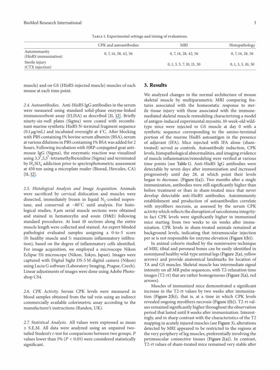
Table 1 Experimental settings and timing of evaluations
CPK and autoantibodies MRI HistopathologyAutoimmunity(HisRS immunization) 0 7 14 28 42 56 0 7 14 28 42 56 0 7 14 28 56
Sterile injury(CTX injection) 0 1 3 5 7 10 15 30 0 1 3 5 10 30
muscle) and on GS (HIsRS-injected muscle) muscles of eachmouse at each time point
24 Autoantibodies Anti-HisRS IgG antibodies in the serumwere measured using standard solid-phase enzyme-linkedimmunosorbent assay (ELISA) as described [11 12] Brieflyninety-six-well plates (Sigma) were coated with recombi-nant murine synthetic HisRS N-terminal fragment sequence(01 120583gmL) and incubated overnight at 4∘C After blockingwith PBS containing 1 bovine serum albumin (BSA) serumat various dilutions in PBS containing 1BSAwas added for 2hours Following incubation with HRP-conjugated goat anti-mouse IgG (Sigma) the enzymatic reaction was visualizedusing 331015840551015840-tetramethylbenzidine (Sigma) and terminatedby H2SO4addiction prior to spectrophotometric assessment
at 450 nm using a microplate reader (Biorad Hercules CA)[11 12]
25 Histological Analysis and Image Acquisition Animalswere sacrificed by cervical dislocation and muscles weredissected immediately frozen in liquid N
2-cooled isopen-
tane and conserved at minus80∘C until analysis For histo-logical studies 8120583m serial muscle sections were obtainedand stained in hematoxylin and eosin (HampE) followingstandard procedures At least 10 sections along the entiremuscle length were collected and stained An expert blindedpathologist evaluated samples assigning a 0-to-5 score(0 healthy muscle and 5 maximum inflammatory infiltra-tion) based on the degree of inflammatory cells identifiedFor image acquisition we employed a microscope NikonEclipse 55i microscope (Nikon Tokyo Japan) Images werecaptured with Digital Sight DS-5M digital camera (Nikon)using Lucia G software (Laboratory Imaging Prague Czech)Linear adjustments of images were done using Adobe Photo-shop CS4
26 CPK Activity Serum CPK levels were measured inblood samples obtained from the tail vein using an indirectcommercially available colorimetric assay according to themanufacturerrsquos instructions (Randox UK)
27 Statistical Analysis All values were expressed as meanplusmn SEM All data were analyzed using an unpaired two-tailed Studentrsquos t-test for comparisons between two groups 119875values lower than 5 (119875 lt 005) were considered statisticallysignificant
3 Results
We analyzed changes in the normal architecture of mouseskeletal muscle by multiparametric MRI comparing fea-tures associated with the homeostatic response to ster-ile tissue injury with those associated with the immune-mediated skeletal muscle remodeling characterizing a modelof antigen-induced experimental myositis 10-week-old wild-type mice were injected in GS muscle at day 0 with asynthetic sequence corresponding to the amino-terminalportion of the murine HisRS autoantigen in the presenceof adjuvant (IFA) Mice injected with IFA alone (sham-treated) served as controls Autoantibody induction CPKlevels histopathological abnormalities and imaging evidenceof muscle inflammationremodeling were verified at varioustime points (see Table 1) Anti-HisRS IgG antibodies weredetectable by seven days after immunization and increasedprogressively until day 28 at which point their levelsbegan to decrease (Figure 1(a)) Two months after a singleimmunization antibodies were still significantly higher thanbefore treatment or than in sham-treated mice that neverdevelop detectable anti-HisRS antibodies Autoimmunityestablishment and production of autoantibodies correlatewith myofibers necrosis as assessed by the serum CPKactivity which reflects the disruption of sarcolemma integrityin fact CPK levels were significantly higher in immunizedmice starting from two weeks to six weeks after immu-nization CPK levels in sham-treated animals remained atbackground levels indicating that intramuscular injectionper se is not responsible for enzyme elevation (Figure 1(b))
In animal cohorts studied by the noninvasive techniqueof MRI tibial and peroneal bones can be easily identified innoninjured healthy wild-type animal legs (Figure 2(a) yellowarrows) and provide anatomical landmarks for location ofTA and GS muscles Skeletal muscle has intermediate signalintensity on all MR pulse sequences with T2-relaxation timeimages (T2-rt) that are rather homogeneous (Figure 2(a) redarrows)
Muscles of immunized mice demonstrated a significantincrease in the T2-rt values by two weeks after immuniza-tion (Figure 2(b)) that is at a time in which CPK levelsrevealed ongoing myofibers necrosis (Figure 1(b)) T2-rt val-ues remained significantly higher throughout the observationperiod that lasted until 8 weeks after immunization Interest-ingly and in sharp contrast with the characteristics of the T2mapping in acutely injuredmuscles (see Figure 3) alterationsdetected by MRI appeared to be restricted to the regions atthe very periphery of leg muscles preferentially involving theperimuscular connective tissues (Figure 2(a)) In contrastT2-rt values of sham-treated mice remained very stable after
4 BioMed Research International
3
2
1
0
0 14 28 42 56
Time (days)
Ant
i-HisR
S an
tibod
ies (
OD
)
lowast
lowast
lowast
lowast
lowast
HisRSSham
(a)
200
150
100
50
0
0 14 28 42 56
Time (days)
CPK
activ
ity (U
I)
lowast lowast
lowast
HisRSSham
(b)
Figure 1 Anti-HisRS autoantibody induction is associated with sustained muscle damage Assessment of anti-HisRS IgG antibodies (a) andCPK activity (b) in serum retrieved before treatment and at various times after immunization 119899 ge 6 lowaststatistically different from sham-treatedmice 119875 lt 005
intramuscular injection Of interest DTI assessment revealedthat FA values were substantially stable after immunizationand did not differ from those of sham-treated animals(Figure 2(d)) We then verified the histological correlatesof the results observing that tissues of immunized animalsundergo a preferential fragmentation of perimysial connec-tive tissue which is characterized from intense infiltration bymononuclear cells Edema with limited evidence of necrosisand myofiber atrophy most frequently occurred in proximityto inflamed connective tissue In contrast inflammatoryinvolvement of endomysial and perivascular regions wasuncommon with only some degree of muscle regeneration(Figure 2(a)) Extending these observations we assessed thelink between T2-rt values of immunized mice at varioustime points and a tissue histological score for inflammation(see the Materials and Methods section for details) T2-rtvalues and the histological damageinfiltration score weresignificantly correlated (119903 = 0959 119875 lt 0005 Figure 2(c))suggesting that T2-rt values accurately reflect perimysialinflammation
As an internal control for ongoing muscle remodelingwe studied in parallel the homeostatic response to sterileinjury by CTX an agent that induces transient musclenecrosis associated with well-characterized reversible alter-ations detectable bymultiparametricMRI ([21] and Figure 3)T2-rt values 1 day after sterile injury were significantlyhigher than those of sham-treated mice and similar to thoseobserved in mice with autoimmune myositis at later timepoints (compare Figure 3(b) with Figure 2(b)) Kinetic wasremarkably different since in CTX-injured mice T2-rt valuesdropped to background levels ten days after treatment andremained stable thereafter (Figure 3(b)) Parallel histopatho-logical study of muscle tissue obtained from mice sacrificedafter imaging revealed that damage resulted in rapid and
massive death of myofibers by day 1 with subsequent infil-tration of the tissue by inflammatory cells and appearanceof small regeneratingcentronucleated fibers by day 5 afterinjection At day 10 necrosis and inflammation were hardlyapparent while extensive regeneration was taking place(Figure 3(a)) In contrast FA values dropped 24 hours afterinjury possibly because of the simultaneous necrosis ofmyofibers that abruptly lose their architecture in responseto CTX (Figure 3(c)) With time FA values returned tonormal in conjunction with the successful regeneration ofthe tissue Similarly FA values remained relatively stable inHRS-induced myositis that is associated with fairly limitednecrosis and regeneration (Figure 2(d)) further supportingthe relationship between FA values and the state of musclerepair
4 Discussion
IM are heterogeneous disorders histologically characterizedby muscle inflammation with the presence of autoreactivelymphocytes fiber degeneration and overexpression ofMHCclass I Common clinical features are muscle weaknessserum autoantibodies and elevated muscle enzyme [7 8]Recent data suggest not only that both innate immunityand adaptive immunity are involved in the IM pathogenesisbut also that intrinsic muscle defects (such as metabolicdefects and endoplasmic reticulum stress and autophagy andhypoxia) may contribute to damage [1] The link betweenremodeling of skeletal muscle and autoimmune responseswhich typically target ubiquitous expressed autoantigens isextremely difficult to study as the events underlying reducedmuscle strength and endurance [8 22] The availability ofinformative animal models is thus crucial [23] Traditionallythe assessment of ongoingmuscle inflammatory involvement
BioMed Research International 5
0 14 28 56
(days)
(a)
0
10
20
30
40
50
0 14 28 42 56
T2-r
t (m
s)
Time (days)HisRSSham
lowast
lowastlowast
lowast
(b)
50
40
30
20
10
0
0 2 4 6
Histological score (au)
T2-rt
(ms)
r2= 09590
P = 00036
(c)
HisRSSham
0
01
02
03
04
05
0 14 28 42 56
FA (
au)
Time (days)
(d)
Figure 2 T2MRI mapping reflects inflammation and remodeling in experimental HisRS-induced myositis Representative T2-weighted imagesof immunizedmice ((a) upper panels) obtained before (time 0) and 14 28 or 56 days after treatment reveal ongoing inflammatory involvementand are well compatible with results obtained at the same time points by histopathology in selected animals ((a) lower panels HampE) Yellowarrows indicate the fibula (upper) and tibia (lower) bones while red arrows show skeletal muscles (GS left and TA right)The graphs show thedynamic trends of T2-rt ((b) y-axis) and FA ((d) y-axis) in the longitudinal study of immunized and sham-treatedmice before and at varioustimes after treatment (x-axis days) 119899 ge 6 lowaststatistically different from sham-treated mice 119875 lt 005 The graph in (c) depicts correlationanalysis between T2-rt and the histological score that reflects muscle damage and infiltration (see Methods) 119899 = 3 for each time point Thetime points considered for the correlation analysis were days 0 14 28 42 and 56
6 BioMed Research International
(days)0 1 5 10
(a)
0
5
10
15
20
25
30
35
40
45
50
0 5 10 15 20 25 30
T2-r
t
Time (days)
CTXSham
lowastlowast
lowast
lowastlowast
lowast
(b)
CTXSham
0
01
02
03
04
05
0 5 10 15 20 25 30
FA (
au)
Time (days)
lowast
lowast
lowast
lowastlowast
(c)
Figure 3 T2 and DTI MRI mapping reflect independent inflammatory events in acutely injured muscles Representative T2-weighted imagesbefore (time 0) and at various times (1 5 or 10 days) after intramuscular CTX injection ((a) upper images) Results are compatible with thoseobtained at the same time points by histopathology in selected animals ((a) lower panels HampE) The graphs show the dynamic trends ofT2-rt ((b) y-axis) and FA ((c) y-axis) in the longitudinal study of damaged (CTX) and sham-treated mice before and at various times aftertreatment (x-axis days) 119899 ge 6 lowaststatistically different from sham-treated mice 119875 lt 005
has been rather cumbersome since by definition it requireshistopathological studies and animal sacrifice
While replacement is difficult to achieve in this fieldof research given the complexity of events involved inmyositis pathogenesis that can be only reproduced in invivo systems the availability of noninvasive tools to monitormuscle inflammation will permit reductions in the numberof animals necessary for experimental studies and increasethe amount of relevant information that can be derived fromsingle animals followed over time At the same time thisimagingmodality will complement information derived fromhistopathological studies that encompass only limited areas oftissue and therefore suffer from potential sampling bias
Here we have focused onMRI-based imaging techniquesthat have been previously shown to represent noninvasive andquantitative tools for the assessment of muscle inflammationand remodeling in experimental animals [13 16 21 24 25]
We utilized a biologically relevant model of experimentalmyositis which can be elicited by immunization of wild-type mice with a synthetic sequence corresponding to a well-characterized autoantigen HisRS [11 12]This model triggersanti-HisRS autoantibodies as well as self-sustaining injuryand leukocyte infiltration of skeletal muscle reproducingkey features of spontaneous human disease that can also beassociated with fever arthritis mechanicrsquos hands Raynaudrsquosphenomenon and interstitial lung disease as part of the anti-synthetase syndrome [26] Reference [27] shows that muscleinflammation has specific features in these patients with theantisynthetase syndrome with predominant involvement ofperimysial connective tissue and less significant peripheralatrophy or microvascular injury [28 29]
As shown by our results MRI is a powerful tool toinvestigate muscle inflammation occurring in the mousemodel of HisRS-inducedmyositis Most significantly cohorts
BioMed Research International 7
of mice can be longitudinally followed with this techniquethat accurately detects specific histopathological features ofthe events taking place in the tissue upon autoimmunity Inour study MRI proved extremely sensitive detecting earlytissue changes associated with relatively minor increasesin the extent of muscle necrosis revealed by changes inserumCPK levels ImportantlyMRI of sham-treated animalsremained stable over the 8 weeks of the study effectivelyruling out major effects related to the immunization vehicleIFA
Overall our study provides strong evidence of the linkbetween HisRS-induced immune responses and myositisthat occurs as early as fourteen days after immunizationMoreover noninvasive T2 mapping reveals the involvementof perimysial connective tissue that is a specific feature of thehuman antisynthetase syndrome [28] clearly differentiatingHisRS-induced myositis from the CTX model of acute self-limited muscle necrosis As such the T2-rt values effectivelymirror the extent and distribution of muscle inflammationnot only in the setting of homeostatic responses to steriletoxic injury [21] but also in a more complex and long-lasting model of immune-mediated muscle remodeling Ofimportance the degree of anisotropy of water diffusivityremained substantially stable in the mice that we havestudied upon myositis induction contrasting sharply withMRI results obtained in CTX-treated mice Because thisparameter largely reflects the diffusion of water in intracel-lular spaces [24 30] these results are in agreement with thepreferential involvement of the perimysial tissue (reflectedby clear histological evidence of edema in the perifascicularspace) and the substantial conservation of tissue architecturein HisRS-induced myositis
Based on these observations our findings indicate thatMRI represents a versatile tool that can sensitively detectinflammatory processes associated with various types ofmuscle injury At the same time this imaging modalityaffords the opportunity to noninvasivelymonitormore subtlemodifications in the architecture and inflammatory stateof skeletal muscle including those changes associated withinitial age related decline in muscle mass and simultaneousincreases of both T2-rt and FA Further studies are warrantedto verify whether specific MRI codes are also associated withother conditions characterized by persistent inflammationand wasting of the skeletal muscle
5 Conclusions
In conclusion our study was designed to characterize theability of MR imaging to reveal specific inflammatory eventsin the skeletal muscle of mice with HisRS-induced myositisOur results provide an in-depth noninvasive characteriza-tion of this myositis model confirming the possibility tolink HisRS-induced immune responses and skeletal muscleinflammation to MRI T2-rt changes This technique allowsincreasing the amount of relevant information that can bederived from single animals followed over time and maystrongly improve and facilitate the study of new therapeuticstrategies for human disease as well as of the immunological
and local events involved in myositis pathogenesis At thesame time this imaging modality can help histopathologicalstudies encompassing their potential sampling biaswith com-plement quantitative information The possibility to followthe same animal from the beginning to the end of treatmentand to analyze more parameters at ones can allow betterstatistically evaluation of data and underlining the conceptthat strong efforts should be dedicated to the refinement ofexperimental procedures that limit the number of animalsneeded for experimental models of human disease
Conflict of Interests
The authors declare that there is no conflict of interestsregarding the publication of this paper
Acknowledgments
This work was supported by the Italian Ministry of Health(Fondo per gli Investimenti della Ricerca di Base-IDEASto Patrizia Rovere-Querini and Ricerca Finalizzata 2011 toPatrizia Rovere-Querini and Angelo A Manfredi) by theAssociazione Italiana Ricerca sul Cancro (AIRC IG11761to Angelo A Manfredi) and by the Italian Ministry ofUniversity and Research (PRIN 2010 to Angelo AManfredi)
References
[1] S Rayavarapu W Coley T B Kinder and K NagarajuldquoIdiopathic inflammatory myopathies pathogenic mechanismsof muscle weaknessrdquo Skelet Muscle vol 3 p 13 2013
[2] A Bender N Ernst A Iglesias K Dornmair H Wekerleand R Hohlfeld ldquoT cell receptor repertoire in polymyositisclonal expansion of autoaggressive CD8+ T cellsrdquo Journal ofExperimental Medicine vol 181 no 5 pp 1863ndash1868 1995
[3] R Hohlfeld and A G Engel ldquoCoculture with autologousmyotubes of cytotoxic T cells isolated frommuscle in inflamma-tory myopathiesrdquo Annals of Neurology vol 29 no 5 pp 498ndash507 1991
[4] J M Pandya A E R Fasth M Zong et al ldquoExpanded Tcell receptor V120573-restricted T cells from patients with sporadicinclusion body myositis are proinflammatory and cytotoxicCD28null T cellsrdquo Arthritis and Rheumatism vol 62 no 11 pp3457ndash3466 2010
[5] J H Choi Y E Park S I Kim et al ldquoDifferential Immunohisto-logical features of inflammatory myopathies and dysferlinopa-thyrdquo Journal of Korean Medical Science vol 24 no 6 pp 1015ndash1023 2009
[6] A Ghirardello N Bassi L Palma et al ldquoAutoantibodiesin polymyositis and dermatomyositisrdquo Current RheumatologyReports vol 15 p 335 2013
[7] A L Mammen ldquoAutoimmune myopathies autoantibodiesphenotypes and pathogenesisrdquo Nature Reviews Neurology vol7 no 6 pp 343ndash354 2011
[8] P Venalis and I E Lundberg ldquoImmune mechanisms inpolymyositis and dermatomyositis and potential targets fortherapyrdquo Rheumatology (Oxford) vol 53 no 3 pp 397ndash4052014
[9] K B Stone C V Oddis N Fertig et al ldquoAnti-Jo-1 antibodylevels correlate with disease activity in idiopathic inflammatory
8 BioMed Research International
myopathyrdquo Arthritis and Rheumatism vol 56 no 9 pp 3125ndash3131 2007
[10] Y Katsumata and D P Ascherman ldquoAnimal models in myosi-tisrdquo Current Opinion in Rheumatology vol 20 no 6 pp 681ndash685 2008
[11] Y Katsumata W M Ridgway T Oriss et al ldquoSpecies-specificimmune responses generated by histidyl-tRNA synthetaseimmunization are associated with muscle and lung inflamma-tionrdquo Journal of Autoimmunity vol 29 no 2-3 pp 174ndash1862007
[12] M Soejima EH Kang X Gu Y Katsumata P R Clemens andD P Ascherman ldquoRole of innate immunity in a murine modelof histidyl-transfer RNA synthetase (Jo-1)-mediated myositisrdquoArthritis and Rheumatism vol 63 no 2 pp 479ndash487 2011
[13] G P Kuo and J A Carrino ldquoSkeletal muscle imaging andinflammatory myopathiesrdquo Current Opinion in Rheumatologyvol 19 no 6 pp 530ndash535 2007
[14] P O Carstens and J Schmidt ldquoDiagnosis pathogenesis andtreatment ofmyositis recent advancesrdquoClinicalampExperimentalImmunology vol 175 no 3 pp 349ndash358 2014
[15] R V Curiel R Jones and K Brindle ldquoMagnetic resonanceimaging of the idiopathic inflammatory myopathies structuraland clinical aspectsrdquo Annals of the New York Academy ofSciences vol 1154 pp 101ndash114 2009
[16] A B McMillan D Shi S J P Pratt and R M LoveringldquoDiffusion tensor MRI to assess damage in healthy and dys-trophic skeletal muscle after lengthening contractionsrdquo Journalof Biomedicine and Biotechnology vol 2011 Article ID 97072610 pages 2011
[17] L M McIntosh R E Baker and J E Anderson ldquoMagnetic res-onance imaging of regenerating and dystrophic mouse musclerdquoBiochemistry and Cell Biology vol 76 no 2-3 pp 532ndash541 1998
[18] S J Pratt S Xu R J Mullins and R M Lovering ldquoTemporalchanges in magnetic resonance imaging in the mdx mouserdquoBMC Research Notes vol 6 p 262 2013
[19] S Tardif-de Gery J Vilquin P Carlier et al ldquoMusculartransverse relaxation timemeasurement bymagnetic resonanceimaging at 4 Tesla in normal and dystrophic dydy anddy(2j)dy(2j) micerdquo Neuromuscular Disorders vol 10 no 7 pp507ndash513 2000
[20] S Schmidt A Vieweger M Obst et al ldquoDysferlin-deficientmuscular dystrophy gadofluorine M suitability at MR imagingin a mouse modelrdquo Radiology vol 250 no 1 pp 87ndash94 2009
[21] A Esposito L Campana A Palmisano et al ldquoMagneticresonance imaging at 7T reveals common events in age-relatedsarcopenia and in the homeostatic response to muscle sterileinjuryrdquo PLoS One vol 8 Article ID e59308 2013
[22] L Casciola-Rosen K Nagaraju P Plotz et al ldquoEnhancedautoantigen expression in regenerating muscle cells inidiopathic inflammatory myopathyrdquo Journal of ExperimentalMedicine vol 201 no 4 pp 591ndash601 2005
[23] D P Ascherman ldquoAnimal models of inflammatory myopathyrdquoCurrent Rheumatology Reports vol 14 no 3 pp 257ndash263 2012
[24] B M Damon Z Ding A W Anderson A S Freyer and J CGore ldquoValidation of diffusion tensor MRI-based muscle fibertrackingrdquoMagnetic Resonance inMedicine vol 48 no 1 pp 97ndash104 2002
[25] E Amarteifio A M Nagel H U Kauczor and M A WeberldquoFunctional imaging in muscular diseasesrdquo Insights Imagingvol 2 no 5 pp 609ndash619 2011
[26] I N Targoff ldquoAutoantibodies and their significance inmyositisrdquoCurrent Rheumatology Reports vol 10 no 4 pp 333ndash340 2008
[27] L Casciola-Rosen ldquoHistidyl-transfer RNA synthetase a keyparticipant in idiopathic inflammatory myopathiesrdquo Arthritisand Rheumatism vol 63 no 2 pp 331ndash333 2011
[28] TMozaffar andA Pestronk ldquoMyopathy with anti-Jo-1 antibod-ies pathology in perimysium and neighbouring muscle fibresrdquoJournal of Neurology Neurosurgery and Psychiatry vol 68 no 4pp 472ndash478 2000
[29] W Stenzel H H Goebel and E Aronica ldquoReview immune-mediated necrotizing myopathiesmdasha heterogeneous group ofdiseases with specific myopathological featuresrdquo Neuropathol-ogy and Applied Neurobiology vol 38 no 7 pp 632ndash646 2012
[30] B M Damon D M Wigmore Z Ding J C Gore and J AKent-Braun ldquoCluster analysis of muscle functional MRI datardquoJournal of Applied Physiology vol 95 no 3 pp 1287ndash1296 2003

2 BioMed Research International
methods for in vivo monitoring of disease activity such asmagnetic resonance imaging (MRI) has been lacking
Magnetic resonance imaging (MRI) is a powerful andinformative technique to investigate soft tissues with theability to noninvasively characterize parenchymal changesoccurring in patients withmyositis Imaging has traditionallyhad an ancillary role in the diagnosis of myositis and inflam-matory myopathies Routinely the MRI protocol includesT1-weighted images and fluid-sensitive sequences such asshort tau inversion recovery (STIR) providing qualitativeinformation about muscle tone fat infiltration and muscleedema [13] Transaxial and coronal sections of the shouldersand thighs are usually obtained on each patient T1-weightedimages allow to assess the muscle thicknessmass and toscore the degree of fatty infiltration fluid-sensitive sequencesdetect the presence of edema [13] Routine MRI performedwith T1-weighted and STIR sequences is more sensitive butless specific than biopsy in diagnosis it is advantageous foroptimizing efficacy of classical diagnostic procedures [14]Its importance is progressively growing since it allows tononinvasively characterize the distribution and pattern ofparenchymal changes and tomonitor the disease progressionwhich has important implications for treatment [13 15]
In the mouse MRI has been used after tissue injuryinduced by maximal lengthening contractions [16] and inexperimental models of skeletal muscle dystrophy (mdxand dysferlin-deficient mice) to assess disease progression[17] Foci of high intensity signal in T2-weighted imagescorrespond to dystrophic lesions in mdx mice [18 19]while changes in gadofluorine enhancement were identifiedin dysferlin-deficient animals [20] Recently MRI has alsobeen used in C57BL6 mice to assess the specific featuresof the homeostatic response of healthy muscles to acutesterile injury induced by cardiotoxin (CTX) [21] SpecificallyT2 mapping and diffusion-tensor imaging (DTI) provideuseful information on the extent of myofibril necrosis and ofleukocyte infiltration aswell as on the kinetics of regeneration[21] Herewe show thatMRI including advanced quantitativetechniques as T2 mapping and DTI is a useful tool toassess the inflammatory changes and the tissue remodelingassociated with autoimmune myositis in an experimentalmodel of HisRS-induced myositis Architectural changes inthe tissue organization were temporally linked to muscledamage and to the establishment of the specific autoantibodyresponse
2 Materials and Methods
21 Animal Model and Study Design Sterile injury by CTXinjection and immunization experiments was carried outusing C57BL6J wild-type mice (Charles River WilmingtonMA USA) aged 8ndash10 weeks Animals were housed in thepathogen-free facility at our institution and treated in accor-dance with the European Community guidelines and withthe approval of the Institutional Ethical Committee (IACUCnumber 512) For sterile injury animals were anesthetizedby intraperitoneal injection of tribromoethanol (Avertin)at a dose of 250mgKg and tibialis anterior (TA) muscleswere injected with CTX (50 120583L 10 120583M final concentration
Naja mossambica mossambica Sigma-Aldrich) in phosphatebuffer solution (PBS) using an insulin needle (310 cc InsulinSyringe from Becton-Dickinson Franklin Lakes NJ USA)Vehicle-treated mice received PBS alone (50120583L) For HisRSimmunization gastrocnemius (GS) muscles were injectedwith a recombinant protein fragment corresponding to theamino-terminal portion (residues 1ndash151) of the murine HisRSmolecule produced as a maltose binding protein and char-acterized as described in [11] Immunization was carried outusing a mixture of the antigen (4mgmL) and incompleteFreundrsquos adjuvant (IFA 1 1 vol vol 100 120583Lmouse) Vehicle-treated mice received a mixture of PBS and IFA Independentcohorts (6micegroup)weremonitored byMRI aftermyositisinduction sham treatment (IFAPBS) or sterile CTX injuryat various time points times 0 and 7 14 28 or 56 days afterHisRS immunization and times 0 1 3 7 10 15 and 30 daysafter CTX injection Table 1 shows experimental timing forthe two different models of damage
22 MRI MRI studies were performed on a 7T preclinicalmagnetic resonance scanner (Bruker BioSpec 7030USRParavision 50 Germany) equipped with 450675mTmgradients (slow rate 3400ndash4500 Tms rise time 140 120583s) Aphased-array rat-heart coil with four internal preamplifierswas used as receiver coupledwith a 72mm linear-volume coilas transmitter Mice were under general anesthesia obtainedby 15ndash2 isoflurane (Forane Abbott) vaporized in 100oxy-gen (flow 1 lmin) in prone position with the right leg fixedin the center of the coil Breathing and body temperatureweremonitored during MRI (SA Instruments Inc Stony BrookNY USA) and maintained around 30 breaths per minuteand 37∘C respectively After positioning in the magnetisocenter a fieldmap based shimming (MAPSHIM softwarepackage Paravision-50 Bruker Germany) was performed tooptimize B0 field homogeneity MRI parameters were usedto assess skeletal muscle that included T2-relaxation time(T2-rt) evaluated by T2 mapping and fractional anisotropy(FA) evaluated by diffusion tensor imaging (DTI) Mus-cle T2 maps were obtained using a multislice-multiecho(MSME) sequence with fat suppression (repetition time =1938ms 16 echo times = 107317168ms field-of-view =20 times 20mm matrix = 256 times 256 spatial resolution =0078 times 0078mmpixel NSA = 4) acquired on axial plane(10 slices thickness = 1mm gap = 0mm) DTI data wereobtained using a SpinEcho-EPI sequence (DTI-Epi) with30 diffusion gradient directions (repetition time = 3750msecho time = 33ms b-values for direction = 0 secmm2ndash700 secmm2 diffusion gradient duration = 4ms diffusiongradient separation = 20ms NSA = 2) DTI-Epi sequenceshared the same geometrical features (field-of-view =30 times 30mm matrix = 128 times 128 spatial resolution = 0234 times0234mmpixel 10 slices slice thickness = 1mm gap =0mm)
23 Image Analysis MRI postprocessingwas performedwithParavision-50 software (Bruker) AverageADC FA andT2-rt values were obtained from the regions of interest (ROIs)of five subsequent slices placed both on TA (CTX-injured
BioMed Research International 3
Table 1 Experimental settings and timing of evaluations
CPK and autoantibodies MRI HistopathologyAutoimmunity(HisRS immunization) 0 7 14 28 42 56 0 7 14 28 42 56 0 7 14 28 56
Sterile injury(CTX injection) 0 1 3 5 7 10 15 30 0 1 3 5 10 30
muscle) and on GS (HIsRS-injected muscle) muscles of eachmouse at each time point
24 Autoantibodies Anti-HisRS IgG antibodies in the serumwere measured using standard solid-phase enzyme-linkedimmunosorbent assay (ELISA) as described [11 12] Brieflyninety-six-well plates (Sigma) were coated with recombi-nant murine synthetic HisRS N-terminal fragment sequence(01 120583gmL) and incubated overnight at 4∘C After blockingwith PBS containing 1 bovine serum albumin (BSA) serumat various dilutions in PBS containing 1BSAwas added for 2hours Following incubation with HRP-conjugated goat anti-mouse IgG (Sigma) the enzymatic reaction was visualizedusing 331015840551015840-tetramethylbenzidine (Sigma) and terminatedby H2SO4addiction prior to spectrophotometric assessment
at 450 nm using a microplate reader (Biorad Hercules CA)[11 12]
25 Histological Analysis and Image Acquisition Animalswere sacrificed by cervical dislocation and muscles weredissected immediately frozen in liquid N
2-cooled isopen-
tane and conserved at minus80∘C until analysis For histo-logical studies 8120583m serial muscle sections were obtainedand stained in hematoxylin and eosin (HampE) followingstandard procedures At least 10 sections along the entiremuscle length were collected and stained An expert blindedpathologist evaluated samples assigning a 0-to-5 score(0 healthy muscle and 5 maximum inflammatory infiltra-tion) based on the degree of inflammatory cells identifiedFor image acquisition we employed a microscope NikonEclipse 55i microscope (Nikon Tokyo Japan) Images werecaptured with Digital Sight DS-5M digital camera (Nikon)using Lucia G software (Laboratory Imaging Prague Czech)Linear adjustments of images were done using Adobe Photo-shop CS4
26 CPK Activity Serum CPK levels were measured inblood samples obtained from the tail vein using an indirectcommercially available colorimetric assay according to themanufacturerrsquos instructions (Randox UK)
27 Statistical Analysis All values were expressed as meanplusmn SEM All data were analyzed using an unpaired two-tailed Studentrsquos t-test for comparisons between two groups 119875values lower than 5 (119875 lt 005) were considered statisticallysignificant
3 Results
We analyzed changes in the normal architecture of mouseskeletal muscle by multiparametric MRI comparing fea-tures associated with the homeostatic response to ster-ile tissue injury with those associated with the immune-mediated skeletal muscle remodeling characterizing a modelof antigen-induced experimental myositis 10-week-old wild-type mice were injected in GS muscle at day 0 with asynthetic sequence corresponding to the amino-terminalportion of the murine HisRS autoantigen in the presenceof adjuvant (IFA) Mice injected with IFA alone (sham-treated) served as controls Autoantibody induction CPKlevels histopathological abnormalities and imaging evidenceof muscle inflammationremodeling were verified at varioustime points (see Table 1) Anti-HisRS IgG antibodies weredetectable by seven days after immunization and increasedprogressively until day 28 at which point their levelsbegan to decrease (Figure 1(a)) Two months after a singleimmunization antibodies were still significantly higher thanbefore treatment or than in sham-treated mice that neverdevelop detectable anti-HisRS antibodies Autoimmunityestablishment and production of autoantibodies correlatewith myofibers necrosis as assessed by the serum CPKactivity which reflects the disruption of sarcolemma integrityin fact CPK levels were significantly higher in immunizedmice starting from two weeks to six weeks after immu-nization CPK levels in sham-treated animals remained atbackground levels indicating that intramuscular injectionper se is not responsible for enzyme elevation (Figure 1(b))
In animal cohorts studied by the noninvasive techniqueof MRI tibial and peroneal bones can be easily identified innoninjured healthy wild-type animal legs (Figure 2(a) yellowarrows) and provide anatomical landmarks for location ofTA and GS muscles Skeletal muscle has intermediate signalintensity on all MR pulse sequences with T2-relaxation timeimages (T2-rt) that are rather homogeneous (Figure 2(a) redarrows)
Muscles of immunized mice demonstrated a significantincrease in the T2-rt values by two weeks after immuniza-tion (Figure 2(b)) that is at a time in which CPK levelsrevealed ongoing myofibers necrosis (Figure 1(b)) T2-rt val-ues remained significantly higher throughout the observationperiod that lasted until 8 weeks after immunization Interest-ingly and in sharp contrast with the characteristics of the T2mapping in acutely injuredmuscles (see Figure 3) alterationsdetected by MRI appeared to be restricted to the regions atthe very periphery of leg muscles preferentially involving theperimuscular connective tissues (Figure 2(a)) In contrastT2-rt values of sham-treated mice remained very stable after
4 BioMed Research International
3
2
1
0
0 14 28 42 56
Time (days)
Ant
i-HisR
S an
tibod
ies (
OD
)
lowast
lowast
lowast
lowast
lowast
HisRSSham
(a)
200
150
100
50
0
0 14 28 42 56
Time (days)
CPK
activ
ity (U
I)
lowast lowast
lowast
HisRSSham
(b)
Figure 1 Anti-HisRS autoantibody induction is associated with sustained muscle damage Assessment of anti-HisRS IgG antibodies (a) andCPK activity (b) in serum retrieved before treatment and at various times after immunization 119899 ge 6 lowaststatistically different from sham-treatedmice 119875 lt 005
intramuscular injection Of interest DTI assessment revealedthat FA values were substantially stable after immunizationand did not differ from those of sham-treated animals(Figure 2(d)) We then verified the histological correlatesof the results observing that tissues of immunized animalsundergo a preferential fragmentation of perimysial connec-tive tissue which is characterized from intense infiltration bymononuclear cells Edema with limited evidence of necrosisand myofiber atrophy most frequently occurred in proximityto inflamed connective tissue In contrast inflammatoryinvolvement of endomysial and perivascular regions wasuncommon with only some degree of muscle regeneration(Figure 2(a)) Extending these observations we assessed thelink between T2-rt values of immunized mice at varioustime points and a tissue histological score for inflammation(see the Materials and Methods section for details) T2-rtvalues and the histological damageinfiltration score weresignificantly correlated (119903 = 0959 119875 lt 0005 Figure 2(c))suggesting that T2-rt values accurately reflect perimysialinflammation
As an internal control for ongoing muscle remodelingwe studied in parallel the homeostatic response to sterileinjury by CTX an agent that induces transient musclenecrosis associated with well-characterized reversible alter-ations detectable bymultiparametricMRI ([21] and Figure 3)T2-rt values 1 day after sterile injury were significantlyhigher than those of sham-treated mice and similar to thoseobserved in mice with autoimmune myositis at later timepoints (compare Figure 3(b) with Figure 2(b)) Kinetic wasremarkably different since in CTX-injured mice T2-rt valuesdropped to background levels ten days after treatment andremained stable thereafter (Figure 3(b)) Parallel histopatho-logical study of muscle tissue obtained from mice sacrificedafter imaging revealed that damage resulted in rapid and
massive death of myofibers by day 1 with subsequent infil-tration of the tissue by inflammatory cells and appearanceof small regeneratingcentronucleated fibers by day 5 afterinjection At day 10 necrosis and inflammation were hardlyapparent while extensive regeneration was taking place(Figure 3(a)) In contrast FA values dropped 24 hours afterinjury possibly because of the simultaneous necrosis ofmyofibers that abruptly lose their architecture in responseto CTX (Figure 3(c)) With time FA values returned tonormal in conjunction with the successful regeneration ofthe tissue Similarly FA values remained relatively stable inHRS-induced myositis that is associated with fairly limitednecrosis and regeneration (Figure 2(d)) further supportingthe relationship between FA values and the state of musclerepair
4 Discussion
IM are heterogeneous disorders histologically characterizedby muscle inflammation with the presence of autoreactivelymphocytes fiber degeneration and overexpression ofMHCclass I Common clinical features are muscle weaknessserum autoantibodies and elevated muscle enzyme [7 8]Recent data suggest not only that both innate immunityand adaptive immunity are involved in the IM pathogenesisbut also that intrinsic muscle defects (such as metabolicdefects and endoplasmic reticulum stress and autophagy andhypoxia) may contribute to damage [1] The link betweenremodeling of skeletal muscle and autoimmune responseswhich typically target ubiquitous expressed autoantigens isextremely difficult to study as the events underlying reducedmuscle strength and endurance [8 22] The availability ofinformative animal models is thus crucial [23] Traditionallythe assessment of ongoingmuscle inflammatory involvement
BioMed Research International 5
0 14 28 56
(days)
(a)
0
10
20
30
40
50
0 14 28 42 56
T2-r
t (m
s)
Time (days)HisRSSham
lowast
lowastlowast
lowast
(b)
50
40
30
20
10
0
0 2 4 6
Histological score (au)
T2-rt
(ms)
r2= 09590
P = 00036
(c)
HisRSSham
0
01
02
03
04
05
0 14 28 42 56
FA (
au)
Time (days)
(d)
Figure 2 T2MRI mapping reflects inflammation and remodeling in experimental HisRS-induced myositis Representative T2-weighted imagesof immunizedmice ((a) upper panels) obtained before (time 0) and 14 28 or 56 days after treatment reveal ongoing inflammatory involvementand are well compatible with results obtained at the same time points by histopathology in selected animals ((a) lower panels HampE) Yellowarrows indicate the fibula (upper) and tibia (lower) bones while red arrows show skeletal muscles (GS left and TA right)The graphs show thedynamic trends of T2-rt ((b) y-axis) and FA ((d) y-axis) in the longitudinal study of immunized and sham-treatedmice before and at varioustimes after treatment (x-axis days) 119899 ge 6 lowaststatistically different from sham-treated mice 119875 lt 005 The graph in (c) depicts correlationanalysis between T2-rt and the histological score that reflects muscle damage and infiltration (see Methods) 119899 = 3 for each time point Thetime points considered for the correlation analysis were days 0 14 28 42 and 56
6 BioMed Research International
(days)0 1 5 10
(a)
0
5
10
15
20
25
30
35
40
45
50
0 5 10 15 20 25 30
T2-r
t
Time (days)
CTXSham
lowastlowast
lowast
lowastlowast
lowast
(b)
CTXSham
0
01
02
03
04
05
0 5 10 15 20 25 30
FA (
au)
Time (days)
lowast
lowast
lowast
lowastlowast
(c)
Figure 3 T2 and DTI MRI mapping reflect independent inflammatory events in acutely injured muscles Representative T2-weighted imagesbefore (time 0) and at various times (1 5 or 10 days) after intramuscular CTX injection ((a) upper images) Results are compatible with thoseobtained at the same time points by histopathology in selected animals ((a) lower panels HampE) The graphs show the dynamic trends ofT2-rt ((b) y-axis) and FA ((c) y-axis) in the longitudinal study of damaged (CTX) and sham-treated mice before and at various times aftertreatment (x-axis days) 119899 ge 6 lowaststatistically different from sham-treated mice 119875 lt 005
has been rather cumbersome since by definition it requireshistopathological studies and animal sacrifice
While replacement is difficult to achieve in this fieldof research given the complexity of events involved inmyositis pathogenesis that can be only reproduced in invivo systems the availability of noninvasive tools to monitormuscle inflammation will permit reductions in the numberof animals necessary for experimental studies and increasethe amount of relevant information that can be derived fromsingle animals followed over time At the same time thisimagingmodality will complement information derived fromhistopathological studies that encompass only limited areas oftissue and therefore suffer from potential sampling bias
Here we have focused onMRI-based imaging techniquesthat have been previously shown to represent noninvasive andquantitative tools for the assessment of muscle inflammationand remodeling in experimental animals [13 16 21 24 25]
We utilized a biologically relevant model of experimentalmyositis which can be elicited by immunization of wild-type mice with a synthetic sequence corresponding to a well-characterized autoantigen HisRS [11 12]This model triggersanti-HisRS autoantibodies as well as self-sustaining injuryand leukocyte infiltration of skeletal muscle reproducingkey features of spontaneous human disease that can also beassociated with fever arthritis mechanicrsquos hands Raynaudrsquosphenomenon and interstitial lung disease as part of the anti-synthetase syndrome [26] Reference [27] shows that muscleinflammation has specific features in these patients with theantisynthetase syndrome with predominant involvement ofperimysial connective tissue and less significant peripheralatrophy or microvascular injury [28 29]
As shown by our results MRI is a powerful tool toinvestigate muscle inflammation occurring in the mousemodel of HisRS-inducedmyositis Most significantly cohorts
BioMed Research International 7
of mice can be longitudinally followed with this techniquethat accurately detects specific histopathological features ofthe events taking place in the tissue upon autoimmunity Inour study MRI proved extremely sensitive detecting earlytissue changes associated with relatively minor increasesin the extent of muscle necrosis revealed by changes inserumCPK levels ImportantlyMRI of sham-treated animalsremained stable over the 8 weeks of the study effectivelyruling out major effects related to the immunization vehicleIFA
Overall our study provides strong evidence of the linkbetween HisRS-induced immune responses and myositisthat occurs as early as fourteen days after immunizationMoreover noninvasive T2 mapping reveals the involvementof perimysial connective tissue that is a specific feature of thehuman antisynthetase syndrome [28] clearly differentiatingHisRS-induced myositis from the CTX model of acute self-limited muscle necrosis As such the T2-rt values effectivelymirror the extent and distribution of muscle inflammationnot only in the setting of homeostatic responses to steriletoxic injury [21] but also in a more complex and long-lasting model of immune-mediated muscle remodeling Ofimportance the degree of anisotropy of water diffusivityremained substantially stable in the mice that we havestudied upon myositis induction contrasting sharply withMRI results obtained in CTX-treated mice Because thisparameter largely reflects the diffusion of water in intracel-lular spaces [24 30] these results are in agreement with thepreferential involvement of the perimysial tissue (reflectedby clear histological evidence of edema in the perifascicularspace) and the substantial conservation of tissue architecturein HisRS-induced myositis
Based on these observations our findings indicate thatMRI represents a versatile tool that can sensitively detectinflammatory processes associated with various types ofmuscle injury At the same time this imaging modalityaffords the opportunity to noninvasivelymonitormore subtlemodifications in the architecture and inflammatory stateof skeletal muscle including those changes associated withinitial age related decline in muscle mass and simultaneousincreases of both T2-rt and FA Further studies are warrantedto verify whether specific MRI codes are also associated withother conditions characterized by persistent inflammationand wasting of the skeletal muscle
5 Conclusions
In conclusion our study was designed to characterize theability of MR imaging to reveal specific inflammatory eventsin the skeletal muscle of mice with HisRS-induced myositisOur results provide an in-depth noninvasive characteriza-tion of this myositis model confirming the possibility tolink HisRS-induced immune responses and skeletal muscleinflammation to MRI T2-rt changes This technique allowsincreasing the amount of relevant information that can bederived from single animals followed over time and maystrongly improve and facilitate the study of new therapeuticstrategies for human disease as well as of the immunological
and local events involved in myositis pathogenesis At thesame time this imaging modality can help histopathologicalstudies encompassing their potential sampling biaswith com-plement quantitative information The possibility to followthe same animal from the beginning to the end of treatmentand to analyze more parameters at ones can allow betterstatistically evaluation of data and underlining the conceptthat strong efforts should be dedicated to the refinement ofexperimental procedures that limit the number of animalsneeded for experimental models of human disease
Conflict of Interests
The authors declare that there is no conflict of interestsregarding the publication of this paper
Acknowledgments
This work was supported by the Italian Ministry of Health(Fondo per gli Investimenti della Ricerca di Base-IDEASto Patrizia Rovere-Querini and Ricerca Finalizzata 2011 toPatrizia Rovere-Querini and Angelo A Manfredi) by theAssociazione Italiana Ricerca sul Cancro (AIRC IG11761to Angelo A Manfredi) and by the Italian Ministry ofUniversity and Research (PRIN 2010 to Angelo AManfredi)
References
[1] S Rayavarapu W Coley T B Kinder and K NagarajuldquoIdiopathic inflammatory myopathies pathogenic mechanismsof muscle weaknessrdquo Skelet Muscle vol 3 p 13 2013
[2] A Bender N Ernst A Iglesias K Dornmair H Wekerleand R Hohlfeld ldquoT cell receptor repertoire in polymyositisclonal expansion of autoaggressive CD8+ T cellsrdquo Journal ofExperimental Medicine vol 181 no 5 pp 1863ndash1868 1995
[3] R Hohlfeld and A G Engel ldquoCoculture with autologousmyotubes of cytotoxic T cells isolated frommuscle in inflamma-tory myopathiesrdquo Annals of Neurology vol 29 no 5 pp 498ndash507 1991
[4] J M Pandya A E R Fasth M Zong et al ldquoExpanded Tcell receptor V120573-restricted T cells from patients with sporadicinclusion body myositis are proinflammatory and cytotoxicCD28null T cellsrdquo Arthritis and Rheumatism vol 62 no 11 pp3457ndash3466 2010
[5] J H Choi Y E Park S I Kim et al ldquoDifferential Immunohisto-logical features of inflammatory myopathies and dysferlinopa-thyrdquo Journal of Korean Medical Science vol 24 no 6 pp 1015ndash1023 2009
[6] A Ghirardello N Bassi L Palma et al ldquoAutoantibodiesin polymyositis and dermatomyositisrdquo Current RheumatologyReports vol 15 p 335 2013
[7] A L Mammen ldquoAutoimmune myopathies autoantibodiesphenotypes and pathogenesisrdquo Nature Reviews Neurology vol7 no 6 pp 343ndash354 2011
[8] P Venalis and I E Lundberg ldquoImmune mechanisms inpolymyositis and dermatomyositis and potential targets fortherapyrdquo Rheumatology (Oxford) vol 53 no 3 pp 397ndash4052014
[9] K B Stone C V Oddis N Fertig et al ldquoAnti-Jo-1 antibodylevels correlate with disease activity in idiopathic inflammatory
8 BioMed Research International
myopathyrdquo Arthritis and Rheumatism vol 56 no 9 pp 3125ndash3131 2007
[10] Y Katsumata and D P Ascherman ldquoAnimal models in myosi-tisrdquo Current Opinion in Rheumatology vol 20 no 6 pp 681ndash685 2008
[11] Y Katsumata W M Ridgway T Oriss et al ldquoSpecies-specificimmune responses generated by histidyl-tRNA synthetaseimmunization are associated with muscle and lung inflamma-tionrdquo Journal of Autoimmunity vol 29 no 2-3 pp 174ndash1862007
[12] M Soejima EH Kang X Gu Y Katsumata P R Clemens andD P Ascherman ldquoRole of innate immunity in a murine modelof histidyl-transfer RNA synthetase (Jo-1)-mediated myositisrdquoArthritis and Rheumatism vol 63 no 2 pp 479ndash487 2011
[13] G P Kuo and J A Carrino ldquoSkeletal muscle imaging andinflammatory myopathiesrdquo Current Opinion in Rheumatologyvol 19 no 6 pp 530ndash535 2007
[14] P O Carstens and J Schmidt ldquoDiagnosis pathogenesis andtreatment ofmyositis recent advancesrdquoClinicalampExperimentalImmunology vol 175 no 3 pp 349ndash358 2014
[15] R V Curiel R Jones and K Brindle ldquoMagnetic resonanceimaging of the idiopathic inflammatory myopathies structuraland clinical aspectsrdquo Annals of the New York Academy ofSciences vol 1154 pp 101ndash114 2009
[16] A B McMillan D Shi S J P Pratt and R M LoveringldquoDiffusion tensor MRI to assess damage in healthy and dys-trophic skeletal muscle after lengthening contractionsrdquo Journalof Biomedicine and Biotechnology vol 2011 Article ID 97072610 pages 2011
[17] L M McIntosh R E Baker and J E Anderson ldquoMagnetic res-onance imaging of regenerating and dystrophic mouse musclerdquoBiochemistry and Cell Biology vol 76 no 2-3 pp 532ndash541 1998
[18] S J Pratt S Xu R J Mullins and R M Lovering ldquoTemporalchanges in magnetic resonance imaging in the mdx mouserdquoBMC Research Notes vol 6 p 262 2013
[19] S Tardif-de Gery J Vilquin P Carlier et al ldquoMusculartransverse relaxation timemeasurement bymagnetic resonanceimaging at 4 Tesla in normal and dystrophic dydy anddy(2j)dy(2j) micerdquo Neuromuscular Disorders vol 10 no 7 pp507ndash513 2000
[20] S Schmidt A Vieweger M Obst et al ldquoDysferlin-deficientmuscular dystrophy gadofluorine M suitability at MR imagingin a mouse modelrdquo Radiology vol 250 no 1 pp 87ndash94 2009
[21] A Esposito L Campana A Palmisano et al ldquoMagneticresonance imaging at 7T reveals common events in age-relatedsarcopenia and in the homeostatic response to muscle sterileinjuryrdquo PLoS One vol 8 Article ID e59308 2013
[22] L Casciola-Rosen K Nagaraju P Plotz et al ldquoEnhancedautoantigen expression in regenerating muscle cells inidiopathic inflammatory myopathyrdquo Journal of ExperimentalMedicine vol 201 no 4 pp 591ndash601 2005
[23] D P Ascherman ldquoAnimal models of inflammatory myopathyrdquoCurrent Rheumatology Reports vol 14 no 3 pp 257ndash263 2012
[24] B M Damon Z Ding A W Anderson A S Freyer and J CGore ldquoValidation of diffusion tensor MRI-based muscle fibertrackingrdquoMagnetic Resonance inMedicine vol 48 no 1 pp 97ndash104 2002
[25] E Amarteifio A M Nagel H U Kauczor and M A WeberldquoFunctional imaging in muscular diseasesrdquo Insights Imagingvol 2 no 5 pp 609ndash619 2011
[26] I N Targoff ldquoAutoantibodies and their significance inmyositisrdquoCurrent Rheumatology Reports vol 10 no 4 pp 333ndash340 2008
[27] L Casciola-Rosen ldquoHistidyl-transfer RNA synthetase a keyparticipant in idiopathic inflammatory myopathiesrdquo Arthritisand Rheumatism vol 63 no 2 pp 331ndash333 2011
[28] TMozaffar andA Pestronk ldquoMyopathy with anti-Jo-1 antibod-ies pathology in perimysium and neighbouring muscle fibresrdquoJournal of Neurology Neurosurgery and Psychiatry vol 68 no 4pp 472ndash478 2000
[29] W Stenzel H H Goebel and E Aronica ldquoReview immune-mediated necrotizing myopathiesmdasha heterogeneous group ofdiseases with specific myopathological featuresrdquo Neuropathol-ogy and Applied Neurobiology vol 38 no 7 pp 632ndash646 2012
[30] B M Damon D M Wigmore Z Ding J C Gore and J AKent-Braun ldquoCluster analysis of muscle functional MRI datardquoJournal of Applied Physiology vol 95 no 3 pp 1287ndash1296 2003
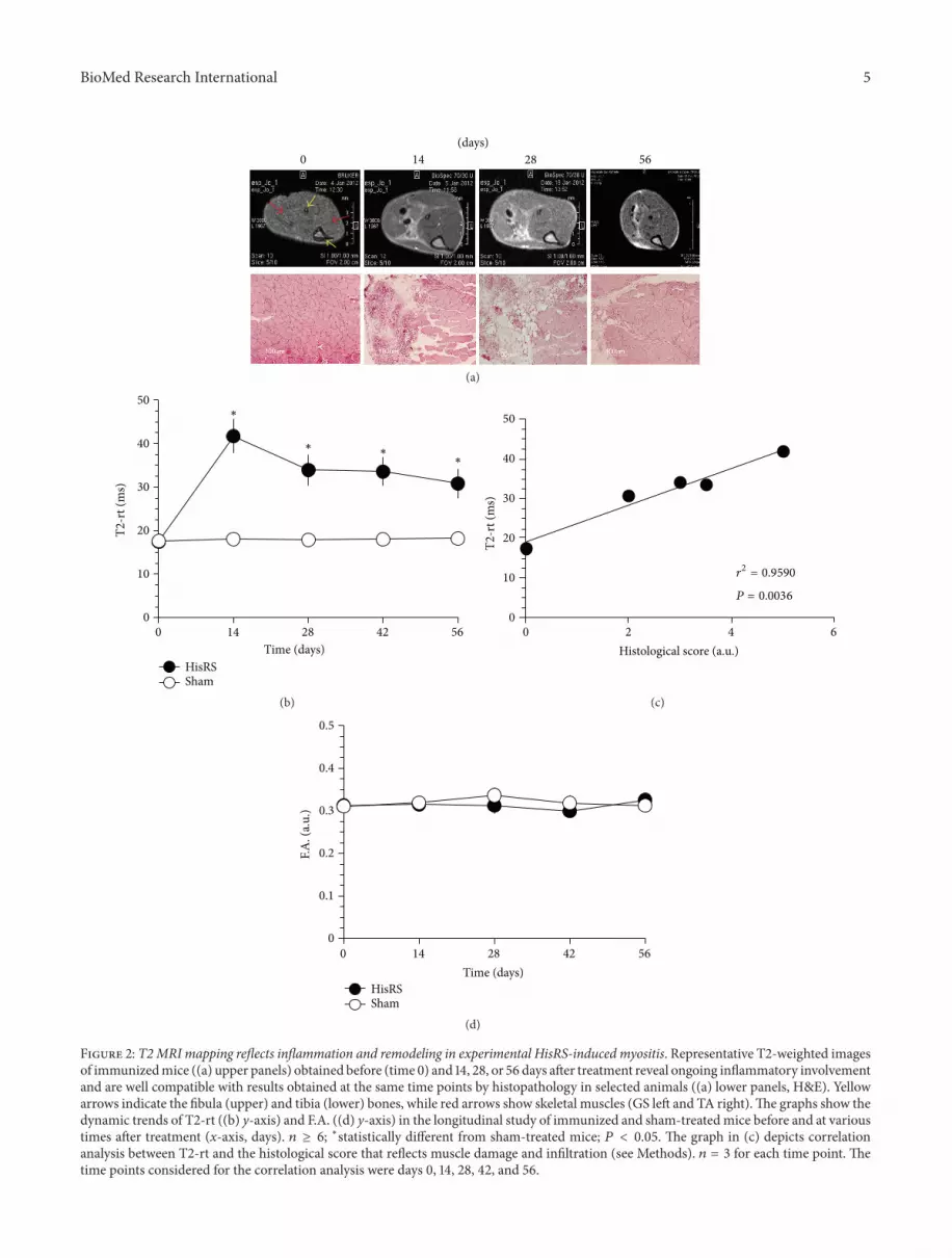
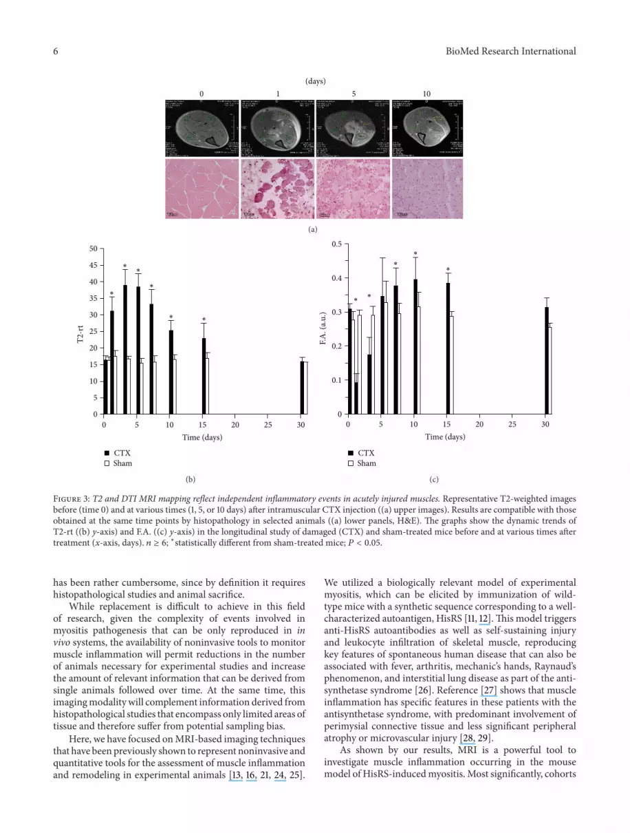
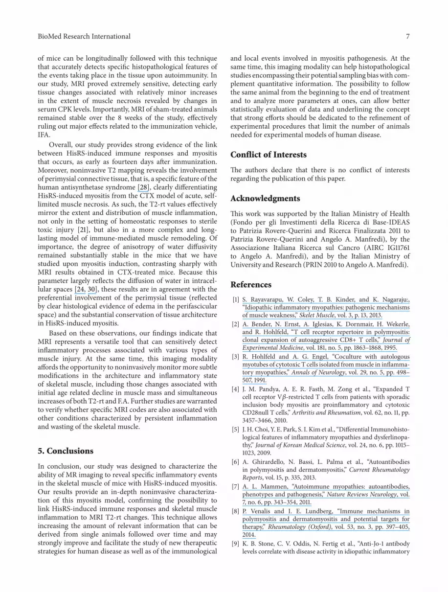