52. pcr alternatives-murad
TRANSCRIPT
ISSN : 0976-4879
Journal ofPharmacy &
BioAllied SciencesAn Official Publication of
Organization of Pharmaceutical Unity with BioAllied Sciences (OPUBS)
Vol 5/ Issue 4/ October-December 2013
al�Uict nu ite yc �Wa im tr ha �Bh iP o�f Ao� llin eo dit �Sa cz i ien na cg er sO
OPUBS
Journ
al o
f Ph
arm
acy &
Bio
Allie
d S
cie
nc
es
• Vo
lum
e 5
• Issue
4 • O
cto
be
r-Dec
em
ber 2
01
3 • P
ages 2
45-?
??
Journal of Pharmacy and Bioallied Sciences October-December 2013 Vol 5 Issue 4 245
Review Article
N ucleic acid amplification is a pivotal process in biotechnology and molecular biology and has
been widely used in research, medicine, agriculture and forensics. Polymerase chain reaction (PCR) was the first nucleic acid amplification method developed and until now has been the method of choice since its invention by Mullis.[1] PCR is the preferred method for application oriented fields involving nucleic acid amplification for
its simplicity, easier methodology, extensively validated standard operating procedure and availability of reagents and equipments. However, PCR has a good no of limitations, including high cost of equipment, contamination chances, sensitivity to certain classes of contaminants and inhibitors, requirement of thermal cycling etc.[2] These limitations gave birth to alternative methods such as loop mediated isothermal amplification (LAMP), nucleic acid sequence based amplification (NASBA), self‑sustained sequence replication (3SR), rolling circle amplification (RCA) etc., most of which are isothermal nucleic acid amplification methods obviating the need of thermal cycler. These methods offer potential advantages over PCR for speed, cost, scale or portability. In this review, most potential alternative methods of PCR are discussed in terms of their principles and applications.
Nucleic acid amplification: Alternative methods of polymerase chain reactionMd. Fakruddin, Khanjada Shahnewaj Bin Mannan1, Abhijit Chowdhury, Reaz Mohammad Mazumdar2, Md. Nur Hossain, Sumaiya Islam, Md. Alimuddin Chowdhury3
ABSTRACTNucleic acid amplification is a valuable molecular tool not only in basic research but also in application oriented fields, such as clinical medicine development, infectious diseases diagnosis, gene cloning and industrial quality control. A comperehensive review of the literature on the principles, applications, challenges and prospects of different alternative methods of polymerase chain reaction (PCR) was performed. PCR was the first nucleic acid amplification method. With the advancement of research, a no of alternative nucleic acid amplification methods has been developed such as loop mediated isothermal amplification, nucleic acid sequence based amplification, strand displacement amplification, multiple displacement amplification. Most of the alternative methods are isothermal obviating the need for thermal cyclers. Though principles of most of the alternate methods are relatively complex than that of PCR, they offer better applicability and sensitivity in cases where PCR has limitations. Most of the alternate methods still have to prove themselves through extensive validation studies and are not available in commercial form; they pose the potentiality to be used as replacements of PCR. Continuous research is going on in different parts of the world to make these methods viable technically and economically.
KEY WORDS: Amplification methods, ligase chain reaction, loop mediated isothermal amplification, multiple displacement amplification, nucleic acid sequence based amplification, polymerase chain reaction alternatives
Industrial Microbiology Laboratory, Institute of Food Science and Technology, Bangladesh Council of Scientific and Industrial Research, Dhaka, 1Center for Food and Waterborne Diseases, Icddr, b, Dhaka, 2BCSIR Laboratories Chittagong, Chittagong, 3Department of Microbiology, University of Chittagong, Chittagong, Bangladesh
Address for correspondence: Mr. Md. Fakruddin E‑mail: [email protected]
Received : 03-02-13Review completed : 24-02-13Accepted : 16-08-13
How to cite this article: Fakruddin M, Mannan KS, Chowdhury A, Mazumdar RM, Hossain MN, Islam S, et al. Nucleic acid amplification: Alternative methods of polymerase chain reaction. J Pharm Bioall Sci 2013;5:245-52.
Access this article onlineQuick Response Code:
Website:
www.jpbsonline.org
DOI:
10.4103/0975-7406.120066
Fakruddin, et al.: Alternative methods of PCR
246 Journal of Pharmacy and Bioallied Sciences October-December 2013 Vol 5 Issue 4
Alternative Methods of Polymerase Chain Reaction
Several alternative amplification methods have been developed already, such as LAMP,[3] 3SR[4] or NASBA,[5] strand displacement amplification (SDA)[6] and RCA,[7] ligase chain reaction (LCR).[8]
Alternative methods are described here in brief.
Loop Mediated Isothermal Amplification
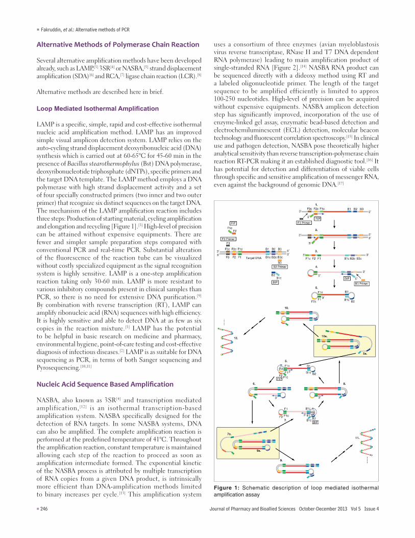
LAMP is a specific, simple, rapid and cost‑effective isothermal nucleic acid amplification method. LAMP has an improved simple visual amplicon detection system. LAMP relies on the auto‑cycling strand displacement deoxyribonucleic acid (DNA) synthesis which is carried out at 60‑65°C for 45‑60 min in the presence of Bacillus stearothermophylus (Bst) DNA polymerase, deoxyribonucleotide triphosphate (dNTPs), specific primers and the target DNA template. The LAMP method employs a DNA polymerase with high strand displacement activity and a set of four specially constructed primers (two inner and two outer primer) that recognize six distinct sequences on the target DNA. The mechanism of the LAMP amplification reaction includes three steps: Production of starting material, cycling amplification and elongation and recycling [Figure 1].[3] High‑level of precision can be attained without expensive equipments. There are fewer and simpler sample preparation steps compared with conventional PCR and real‑time PCR. Substantial alteration of the fluorescence of the reaction tube can be visualized without costly specialized equipment as the signal recognition system is highly sensitive. LAMP is a one‑step amplification reaction taking only 30‑60 min. LAMP is more resistant to various inhibitory compounds present in clinical samples than PCR, so there is no need for extensive DNA purification.[9] By combination with reverse transcription (RT), LAMP can amplify ribonucleic acid (RNA) sequences with high efficiency. It is highly sensitive and able to detect DNA at as few as six copies in the reaction mixture.[3] LAMP has the potential to be helpful in basic research on medicine and pharmacy, environmental hygiene, point‑of‑care testing and cost‑effective diagnosis of infectious diseases.[2] LAMP is as suitable for DNA sequencing as PCR, in terms of both Sanger sequencing and Pyrosequencing.[10,11]
Nucleic Acid Sequence Based Amplification
NASBA, also known as 3SR[4] and transcription mediated amplification,[12] is an isothermal transcription‑based amplification system. NASBA specifically designed for the detection of RNA targets. In some NASBA systems, DNA can also be amplified. The complete amplification reaction is performed at the predefined temperature of 41°C. Throughout the amplification reaction, constant temperature is maintained allowing each step of the reaction to proceed as soon as amplification intermediate formed. The exponential kinetic of the NASBA process is attributed by multiple transcription of RNA copies from a given DNA product, is intrinsically more efficient than DNA‑amplification methods limited to binary increases per cycle.[13] This amplification system
uses a consortium of three enzymes (avian myeloblastosis virus reverse transcriptase, RNase H and T7 DNA dependent RNA polymerase) leading to main amplification product of single‑stranded RNA [Figure 2].[14] NASBA RNA product can be sequenced directly with a dideoxy method using RT and a labeled oligonucleotide primer. The length of the target sequence to be amplified efficiently is limited to approx 100‑250 nucleotides. High‑level of precision can be acquired without expensive equipments. NASBA amplicon detection step has significantly improved, incorporation of the use of enzyme‑linked gel assay, enzymatic bead‑based detection and electrochemiluminescent (ECL) detection, molecular beacon technology and fluorescent correlation spectroscopy.[15] In clinical use and pathogen detection, NASBA pose theoretically higher analytical sensitivity than reverse transcription‑polymerase chain reaction RT‑PCR making it an established diagnostic tool.[16] It has potential for detection and differentiation of viable cells through specific and sensitive amplification of messenger RNA, even against the background of genomic DNA.[17]
Figure 1: Schematic description of loop mediated isothermal amplification assay
Fakruddin, et al.: Alternative methods of PCR
Journal of Pharmacy and Bioallied Sciences October-December 2013 Vol 5 Issue 4 247
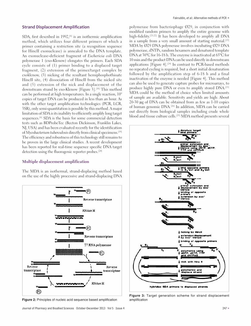
Strand Displacement Amplification
SDA, first described in 1992,[6] is an isothermic amplification method, which utilizes four different primers of which a primer containing a restriction site (a recognition sequence for HincII exonuclease) is annealed to the DNA template. An exonuclease‑deficient fragment of Eschericia coli DNA polymerase 1 (exo‑Klenow) elongates the primers. Each SDA cycle consists of (1) primer binding to a displaced target fragment, (2) extension of the primer/target complex by exoklenow, (3) nicking of the resultant hemiphosphothioate HincII site, (4) dissociation of HincII from the nicked site and (5) extension of the nick and displacement of the downstream strand by exo‑Klenow [Figure 3].[18] This method can be performed at high temperatures. In a single reaction, 109 copies of target DNA can be produced in less than an hour. As with the other target amplification technologies (PCR, LCR, 3SR), only semi‑quantitation is possible by this method. A major limitation of SDA is its inability to efficiently amplify long target sequences.[6] SDA is the basis for some commercial detection tests such as BDProbeTec (Becton Dickinson, Franklin Lakes, NJ, USA) and has been evaluated recently for the identification of Mycobacterium tuberculosis directly from clinical specimens.[19] The efficiency and robustness of this technology still remains to be proven in the large clinical studies. A recent development has been reported for real‑time sequence specific DNA target detection using the fluorogenic reporter probes.[20]
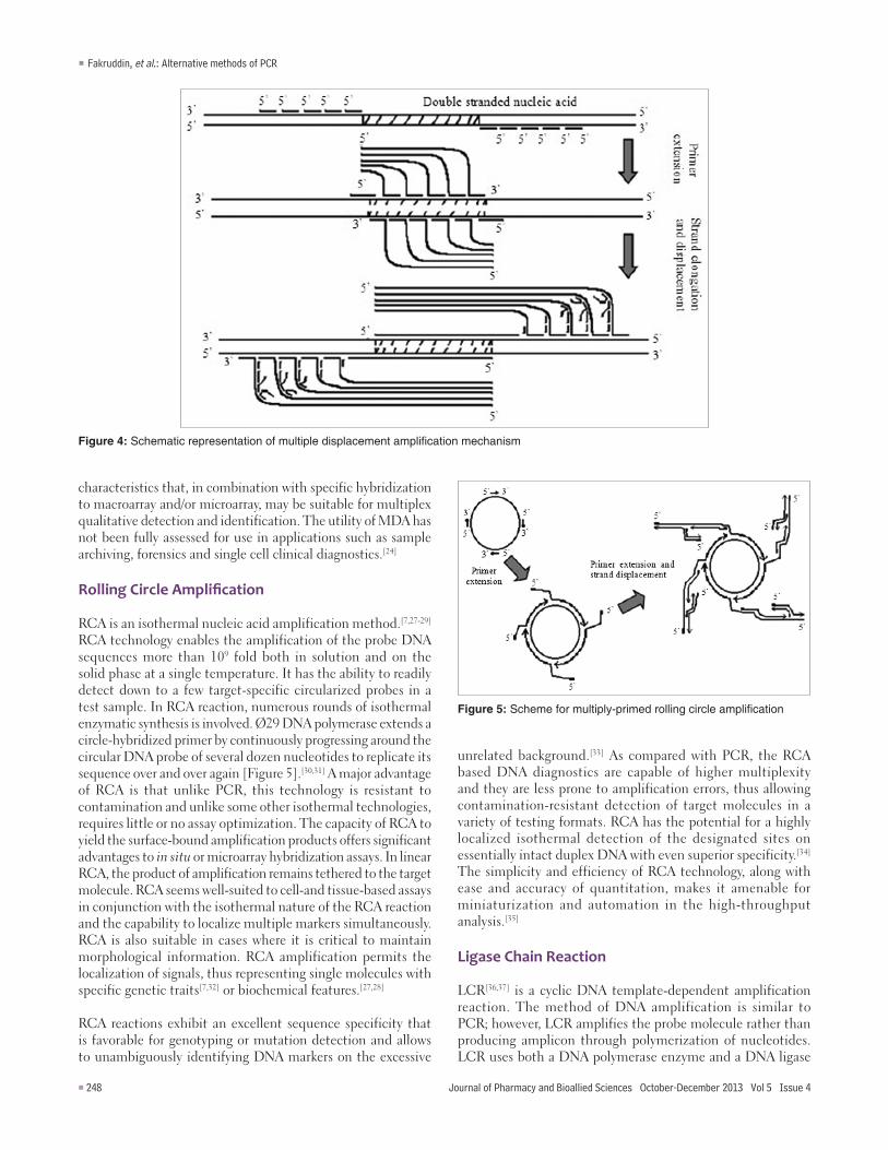
Multiple displacement amplification
The MDA is an isothermal, strand‑displacing method based on the use of the highly processive and strand‑displacing DNA
polymerase from bacteriophage Ø29, in conjunction with modified random primers to amplify the entire genome with high‑fidelity.[21,22] It has been developed to amplify all DNA in a sample from a very small amount of starting material.[23] MDA by Ø29 DNA polymerase involves incubating Ø29 DNA polymerase, dNTPs, random hexamers and denatured template DNA at 30°C for 16‑18 h. The enzyme is inactivated at 65°C for 10 min and the product DNA can be used directly in downstream applications [Figure 4].[24] In contrast to PCR‑based methods no repeated cycling is required, but a short initial denaturation followed by the amplification step of 6‑18 h and a final inactivation of the enzyme is needed [Figure 4]. This method can also be used to generate capture probes for microarrays, to produce highly pure DNA or even to amplify stored DNA.[25] MDA could be the method of choice when limited amounts of sample are available. Sensitivity and yields are high: About 20‑30 µg of DNA can be obtained from as few as 1‑10 copies of human genomic DNA.[26] In addition, MDA can be carried out directly from biological samples including crude whole blood and tissue culture cells.[25] MDA method presents several
Figure 2: Principles of nucleic acid sequence based amplificationFigure 3: Target generation scheme for strand displacement amplification
Fakruddin, et al.: Alternative methods of PCR
248 Journal of Pharmacy and Bioallied Sciences October-December 2013 Vol 5 Issue 4
characteristics that, in combination with specific hybridization to macroarray and/or microarray, may be suitable for multiplex qualitative detection and identification. The utility of MDA has not been fully assessed for use in applications such as sample archiving, forensics and single cell clinical diagnostics.[24]
Rolling Circle Amplification
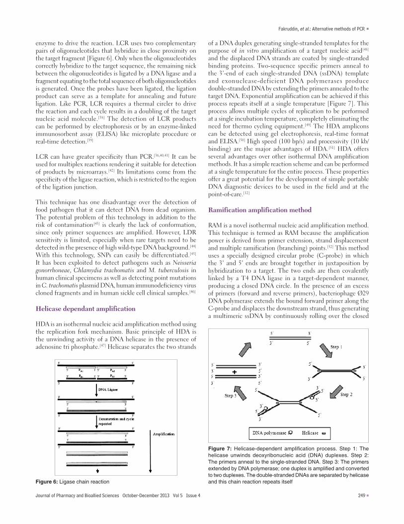
RCA is an isothermal nucleic acid amplification method.[7,27‑29] RCA technology enables the amplification of the probe DNA sequences more than 109 fold both in solution and on the solid phase at a single temperature. It has the ability to readily detect down to a few target‑specific circularized probes in a test sample. In RCA reaction, numerous rounds of isothermal enzymatic synthesis is involved. Ø29 DNA polymerase extends a circle‑hybridized primer by continuously progressing around the circular DNA probe of several dozen nucleotides to replicate its sequence over and over again [Figure 5].[30,31] A major advantage of RCA is that unlike PCR, this technology is resistant to contamination and unlike some other isothermal technologies, requires little or no assay optimization. The capacity of RCA to yield the surface‑bound amplification products offers significant advantages to in situ or microarray hybridization assays. In linear RCA, the product of amplification remains tethered to the target molecule. RCA seems well‑suited to cell‑and tissue‑based assays in conjunction with the isothermal nature of the RCA reaction and the capability to localize multiple markers simultaneously. RCA is also suitable in cases where it is critical to maintain morphological information. RCA amplification permits the localization of signals, thus representing single molecules with specific genetic traits[7,32] or biochemical features.[27,28]
RCA reactions exhibit an excellent sequence specificity that is favorable for genotyping or mutation detection and allows to unambiguously identifying DNA markers on the excessive
unrelated background.[33] As compared with PCR, the RCA based DNA diagnostics are capable of higher multiplexity and they are less prone to amplification errors, thus allowing contamination‑resistant detection of target molecules in a variety of testing formats. RCA has the potential for a highly localized isothermal detection of the designated sites on essentially intact duplex DNA with even superior specificity.[34] The simplicity and efficiency of RCA technology, along with ease and accuracy of quantitation, makes it amenable for miniaturization and automation in the high‑throughput analysis.[35]
Ligase Chain Reaction
LCR[36,37] is a cyclic DNA template‑dependent amplification reaction. The method of DNA amplification is similar to PCR; however, LCR amplifies the probe molecule rather than producing amplicon through polymerization of nucleotides. LCR uses both a DNA polymerase enzyme and a DNA ligase
Figure 4: Schematic representation of multiple displacement amplification mechanism
Figure 5: Scheme for multiply-primed rolling circle amplification
Fakruddin, et al.: Alternative methods of PCR
Journal of Pharmacy and Bioallied Sciences October-December 2013 Vol 5 Issue 4 249
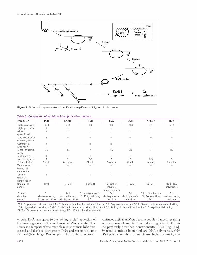
enzyme to drive the reaction. LCR uses two complementary pairs of oligonucleotides that hybridize in close proximity on the target fragment [Figure 6]. Only when the oligonucleotides correctly hybridize to the target sequence, the remaining nick between the oligonucleotides is ligated by a DNA ligase and a fragment equating to the total sequence of both oligonucleotides is generated. Once the probes have been ligated, the ligation product can serve as a template for annealing and future ligation. Like PCR, LCR requires a thermal circler to drive the reaction and each cycle results in a doubling of the target nucleic acid molecule.[38] The detection of LCR products can be performed by electrophoresis or by an enzyme‑linked immunosorbent assay (ELISA) like microplate procedure or real‑time detection.[39]
LCR can have greater specificity than PCR.[36,40,41] It can be used for multiplex reactions rendering it suitable for detection of products by microarrays.[42] Its limitations come from the specificity of the ligase reaction, which is restricted to the region of the ligation junction.
This technique has one disadvantage over the detection of food pathogen that it can detect DNA from dead organism. The potential problem of this technology in addition to the risk of contamination[43] is clearly the lack of conformation, since only primer sequences are amplified. However, LDR sensitivity is limited, especially when rare targets need to be detected in the presence of high wild‑type DNA background.[44] With this technology, SNPs can easily be differentiated.[45] It has been exploited to detect pathogens such as Neisseria gonorrhoneae, Chlamydia trachomatis and M. tuberculosis in human clinical specimens as well as detecting point mutations in C. trachomatis plasmid DNA, human immunodeficiency virus cloned fragments and in human sickle cell clinical samples.[46]
Helicase dependant amplification
HDA is an isothermal nucleic acid amplification method using the replication fork mechanism. Basic principle of HDA is the unwinding activity of a DNA helicase in the presence of adenosine tri phosphate.[47] Helicase separates the two strands
of a DNA duplex generating single‑stranded templates for the purpose of in vitro amplification of a target nucleic acid[48] and the displaced DNA strands are coated by single‑stranded binding proteins. Two‑sequence specific primers anneal to the 3’‑end of each single‑stranded DNA (ssDNA) template and exonuclease‑deficient DNA polymerases produce double‑stranded DNA by extending the primers annealed to the target DNA. Exponential amplification can be achieved if this process repeats itself at a single temperature [Figure 7]. This process allows multiple cycles of replication to be performed at a single incubation temperature, completely eliminating the need for thermo cycling equipment.[49] The HDA amplicons can be detected using gel electrophoresis, real‑time format and ELISA.[50] High speed (100 bp/s) and processivity (10 kb/binding) are the major advantages of HDA.[51] HDA offers several advantages over other isothermal DNA amplification methods. It has a simple reaction scheme and can be performed at a single temperature for the entire process. These properties offer a great potential for the development of simple portable DNA diagnostic devices to be used in the field and at the point‑of‑care.[12]
Ramification amplification method
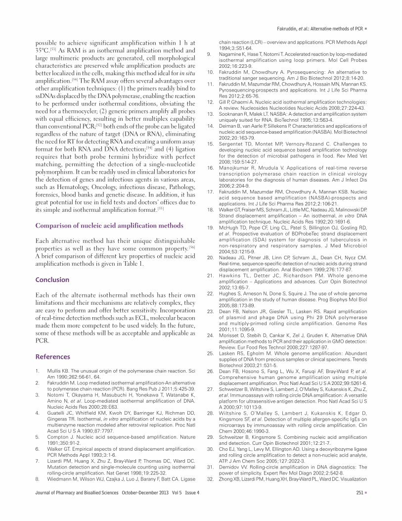
RAM is a novel isothermal nucleic acid amplification method. This technique is termed as RAM because the amplification power is derived from primer extension, strand displacement and multiple ramification (branching) points.[52] This method uses a specially designed circular probe (C‑probe) in which the 3’ and 5’ ends are brought together in juxtaposition by hybridization to a target. The two ends are then covalently linked by a T4 DNA ligase in a target‑dependent manner, producing a closed DNA circle. In the presence of an excess of primers (forward and reverse primers), bacteriophage Ø29 DNA polymerase extends the bound forward primer along the C‑probe and displaces the downstream strand, thus generating a multimeric ssDNA by continuously rolling over the closed
Figure 6: Ligase chain reaction
Figure 7: Helicase-dependent amplification process. Step 1: The helicase unwinds deoxyribonucleic acid (DNA) duplexes. Step 2: The primers anneal to the single-stranded DNA. Step 3: The primers extended by DNA polymerase; one duplex is amplified and converted to two duplexes. The double-stranded DNAs are separated by helicase and this chain reaction repeats itself
Fakruddin, et al.: Alternative methods of PCR
250 Journal of Pharmacy and Bioallied Sciences October-December 2013 Vol 5 Issue 4
Figure 8: Schematic representation of ramification amplification of ligated circular probe
circular DNA, analogous to the “rolling circle” replication of bacteriophages in vivo. The multimeric ssDNA generated then serves as a template where multiple reverse primers hybridize, extend and displace downstream DNA and generate a large ramified (branching) DNA complex. This ramification process
continues until all ssDNAs become double‑stranded, resulting in an exponential amplification that distinguishes itself from the previously described nonexponential RCA [Figure 8]. By using a unique bacteriophage DNA polymerase, Ø29 DNA polymerase, that has an intrinsic high processivity, it is
Table 1: Comparison of nucleic acid amplification methodsParameter PCR LAMP 3SR SDA LCR NASBA RCA
High sensitivity <10 <10 10 10 <10 10 <10High specificity + + + + + + +Allow quantification
+ + ± ± − ± −
Live versus dead microorganisms
+ + + − − ± −
Commercial availability
+ − ± ± − − −
Linear dynamic range
6‑7 6 7 ND ND 7 ND
Multiplexity + − + − + + +No. of enzymes 1 1 2‑3 2 2 2‑3 1Primer design Simple Complex Simple Complex Simple Simple ComplexTolerance to biological compounds
− + − − − − −
Need to template denaturation
+ − + + + + −
Denaturing agents
Heat Betaine Rnase H Restriction enzymes;
bumper primers
Helicase Rnase H Ø29 DNA polymerase
Product detection method
Gel electrophoresis,
ELISA, real time
Gel electrophoresis,
turbidity, real time
Gel electrophoresis, ELISA, real time,
ECL
Gel electrophoresis,
real time
Gel electrophoresis,
real time
Gel electrophoresis, ELISA, real time,
ECL
Gel electrophoresis,
real time
PCR: Polymerase chain reaction, LAMP: Loop‑mediated isothermal amplification, SR: Sequence replication, SDA: Strand displacement amplification, LCR: Ligase chain reaction, NASBA: Nucleic acid sequence based amplification, RCA: Rolling circle amplification, DNA: Deoxyribonucleic acid, ELISA: Enzyme‑linked immunosorbent assay, ECL: Electrochemiluminescent
Fakruddin, et al.: Alternative methods of PCR
Journal of Pharmacy and Bioallied Sciences October-December 2013 Vol 5 Issue 4 251
possible to achieve significant amplification within 1 h at 35°C.[53] As RAM is an isothermal amplification method and large multimeric products are generated, cell morphological characteristics are preserved while amplification products are better localized in the cells, making this method ideal for in situ amplification.[54] The RAM assay offers several advantages over other amplification techniques: (1) the primers readily bind to ssDNAs displaced by the DNA polymerase, enabling the reaction to be performed under isothermal conditions, obviating the need for a thermocycler; (2) generic primers amplify all probes with equal efficiency, resulting in better multiplex capability than conventional PCR;[52] both ends of the probe can be ligated regardless of the nature of target (DNA or RNA), eliminating the need for RT for detecting RNA and creating a uniform assay format for both RNA and DNA detection;[54] and (4) ligation requires that both probe termini hybridize with perfect matching, permitting the detection of a single‑nucleotide polymorphism. It can be readily used in clinical laboratories for the detection of genes and infectious agents in various areas, such as Hematology, Oncology, infectious disease, Pathology, forensics, blood banks and genetic disease. In addition, it has great potential for use in field tests and doctors’ offices due to its simple and isothermal amplification format.[55]
Comparison of nucleic acid amplification methods
Each alternative method has their unique distinguishable properties as well as they have some common property.[56] A brief comparison of different key properties of nucleic acid amplification methods is given in Table 1.
Conclusion
Each of the alternate isothermal methods has their own limitations and their mechanisms are relatively complex, they are easy to perform and offer better sensitivity. Incorporation of real‑time detection methods such as ECL, molecular beacon made them more competent to be used widely. In the future, some of these methods will be as acceptable and applicable as PCR.
References
1. Mullis KB. The unusual origin of the polymerase chain reaction. Sci Am 1990;262:56‑61, 64.
2. Fakruddin M. Loop mediated isothermal amplification‑An alternative to polymerase chain reaction (PCR). Bang Res Pub J 2011;5:425‑39.
3. Notomi T, Okayama H, Masubuchi H, Yonekawa T, Watanabe K, Amino N, et al. Loop‑mediated isothermal amplification of DNA. Nucleic Acids Res 2000;28:E63.
4. Guatelli JC, Whitfield KM, Kwoh DY, Barringer KJ, Richman DD, Gingeras TR. Isothermal, in vitro amplification of nucleic acids by a multienzyme reaction modeled after retroviral replication. Proc Natl Acad Sci U S A 1990;87:7797.
5. Compton J. Nucleic acid sequence‑based amplification. Nature 1991;350:91‑2.
6. Walker GT. Empirical aspects of strand displacement amplification. PCR Methods Appl 1993;3:1‑6.
7. Lizardi PM, Huang X, Zhu Z, Bray‑Ward P, Thomas DC, Ward DC. Mutation detection and single‑molecule counting using isothermal rolling‑circle amplification. Nat Genet 1998;19:225‑32.
8. Wiedmann M, Wilson WJ, Czajka J, Luo J, Barany F, Batt CA. Ligase
chain reaction (LCR) – overview and applications. PCR Methods Appl 1994;3:S51‑64.
9. Nagamine K, Hase T, Notomi T. Accelerated reaction by loop‑mediated isothermal amplification using loop primers. Mol Cell Probes 2002;16:223‑9.
10. Fakruddin M, Chowdhury A. Pyrosequencing: An alternative to traditional sanger sequencing. Am J Bio Biotechnol 2012;8:14‑20.
11. Fakruddin M, Mazumdar RM, Chowdhury A, Hossain MN, Mannan KS. Pyrosequencing‑prospects and applications. Int J Life Sci Pharma Res 2012;2:65‑76.
12. Gill P, Ghaemi A. Nucleic acid isothermal amplification technologies: A review. Nucleosides Nucleotides Nucleic Acids 2008;27:224‑43.
13. Sooknanan R, Malek LT. NASBA: A detection and amplification system uniquely suited for RNA. BioTechnol 1995;13:563‑4.
14. Deiman B, van Aarle P, Sillekens P. Characteristics and applications of nucleic acid sequence‑based amplification (NASBA). Mol Biotechnol 2002;20:163‑79.
15. Sergentet TD, Montet MP, Vernozy‑Rozand C. Challenges to developing nucleic acid sequence based amplification technology for the detection of microbial pathogens in food. Rev Med Vet 2008;159:514‑27.
16. Manojkumar R, Mrudula V. Applications of real‑time reverse transcription polymerase chain reaction in clinical virology laboratories for the diagnosis of human diseases. Am J Infect Dis 2006;2:204‑9.
17. Fakruddin M, Mazumdar RM, Chowdhury A, Mannan KSB. Nucleic acid sequence based amplification (NASBA)‑prospects and applications. Int J Life Sci Pharma Res 2012;2:106‑21.
18. Walker GT, Fraiser MS, Schram JL, Little MC, Nadeau JG, Malinowski DP. Strand displacement amplification – An isothermal, in vitro DNA amplification technique. Nucleic Acids Res 1992;20:1691‑6.
19. McHugh TD, Pope CF, Ling CL, Patel S, Billington OJ, Gosling RD, et al. Prospective evaluation of BDProbeTec strand displacement amplification (SDA) system for diagnosis of tuberculosis in non‑respiratory and respiratory samples. J Med Microbiol 2004;53:1215‑9.
20. Nadeau JG, Pitner JB, Linn CP, Schram JL, Dean CH, Nycz CM. Real‑time, sequence‑specific detection of nucleic acids during strand displacement amplification. Anal Biochem 1999;276:177‑87.
21. Hawkins TL, Detter JC, Richardson PM. Whole genome amplification – Applications and advances. Curr Opin Biotechnol 2002;13:65‑7.
22. Hughes S, Arneson N, Done S, Squire J. The use of whole genome amplification in the study of human disease. Prog Biophys Mol Biol 2005;88:173‑89.
23. Dean FB, Nelson JR, Giesler TL, Lasken RS. Rapid amplification of plasmid and phage DNA using Phi 29 DNA polymerase and multiply‑primed rolling circle amplification. Genome Res 2001;11:1095‑9.
24. Morisset D, Stebih D, Cankar K, Zel J, Gruden K. Alternative DNA amplification methods to PCR and their application in GMO detection: Review. Eur Food Res Technol 2008;227:1287‑97.
25. Lasken RS, Egholm M. Whole genome amplification: Abundant supplies of DNA from precious samples or clinical specimens. Trends Biotechnol 2003;21:531‑5.
26. Dean FB, Hosono S, Fang L, Wu X, Faruqi AF, Bray‑Ward P, et al. Comprehensive human genome amplification using multiple displacement amplification. Proc Natl Acad Sci U S A 2002;99:5261‑6.
27. Schweitzer B, Wiltshire S, Lambert J, O’Malley S, Kukanskis K, Zhu Z, et al. Immunoassays with rolling circle DNA amplification: A versatile platform for ultrasensitive antigen detection. Proc Natl Acad Sci U S A 2000;97:10113‑9.
28. Wiltshire S, O’Malley S, Lambert J, Kukanskis K, Edgar D, Kingsmore SF, et al. Detection of multiple allergen‑specific IgEs on microarrays by immunoassay with rolling circle amplification. Clin Chem 2000;46:1990‑3.
29. Schweitzer B, Kingsmore S. Combining nucleic acid amplification and detection. Curr Opin Biotechnol 2001;12:21‑7.
30. Cho EJ, Yang L, Levy M, Ellington AD. Using a deoxyribozyme ligase and rolling circle amplification to detect a non‑nucleic acid analyte, ATP. J Am Chem Soc 2005;127:2022‑3.
31. Demidov VV. Rolling‑circle amplification in DNA diagnostics: The power of simplicity. Expert Rev Mol Diagn 2002;2:542‑8.
32. Zhong XB, Lizardi PM, Huang XH, Bray‑Ward PL, Ward DC. Visualization
Fakruddin, et al.: Alternative methods of PCR
252 Journal of Pharmacy and Bioallied Sciences October-December 2013 Vol 5 Issue 4
of oligonucleotide probes and point mutations in interphase nuclei and DNA fibers using rolling circle DNA amplification. Proc Natl Acad Sci U S A 2001;98:3940‑5.
33. Mothershed EA, Whitney AM. Nucleic acid‑based methods for the detection of bacterial pathogens: Present and future considerations for the clinical laboratory. Clin Chim Acta 2006;363:206‑20.
34. Lievens B, Grauwet TJ, Cammue BP, Thomma PH. Recent developments in diagnostics of plant pathogens: A review. Rec Res Dev Microbiol 2005;9:1‑23.
35. Gusev Y, Sparkowski J, Raghunathan A, Ferguson H Jr, Montano J, Bogdan N, et al. Rolling circle amplification: A new approach to increase sensitivity for immunohistochemistry and flow cytometry. Am J Pathol 2001;159:63‑9.
36. Wu DY, Wallace RB. The l igation amplif ication reaction (LAR) – Amplification of specific DNA sequences using sequential rounds of template‑dependent ligation. Genomics 1989;4:560‑9.
37. Barany F. Genetic disease detection and DNA amplification using cloned thermostable ligase. Proc Natl Acad Sci U S A 1991;88:189‑93.
38. Lisby G. Application of nucleic acid amplification in clinical microbiology. Mol Biotechnol 1999;12:75‑99.
39. Csako G. Present and future of rapid and/or high‑throughput methods for nucleic acid testing. Clin Chim Acta 2006;363:6‑31.
40. Barany F. The ligase chain reaction in a PCR world. PCR Methods Appl 1991;1:5‑16.
41. Khanna M, Cao W, Zirvi M, Paty P, Barany F. Ligase detection reaction for identification of low abundance mutations. Clin Biochem 1999;32:287‑90.
42. Gerry NP, Witowski NE, Day J, Hammer RP, Barany G, Barany F. Universal DNA microarray method for multiplex detection of low abundance point mutations. J Mol Biol 1999;292:251‑62.
43. Dean D, Ferrero D, McCarthy M. Comparison of performance and cost‑effectiveness of direct fluorescent‑antibody, ligase chain reaction, and PCR assays for verification of chlamydial enzyme immunoassay results for populations with a low to moderate prevalence of chlamydia trachomatis infection. J Clin Microbiol 1998;36:94‑9.
44. Prasad D, Vidyarthi AS. DNA based methods used for characterization and detection of food borne bacterial pathogens
with special consideration to recent rapid methods. Afr J Biotechnol 2009;8:1768‑75.
45. Kolbehdari D, Robinson JA. QTL mapping using multiple markers simultaneously. Am J Agric Biol Sci 2007;2:195‑201.
46. Andras SC, Power JB, Cocking EC, Davey MR. Strategies for signal amplification in nucleic acid detection. Mol Biotechnol 2001;19:29‑44.
47. Vincent M, Xu Y, Kong H. Helicase‑dependent isothermal DNA amplification. EMBO Rep 2004;5:795‑800.
48. An L, Tang W, Ranalli TA, Kim HJ, Wytiaz J, Kong H. Characterization of a thermostable UvrD helicase and its participation in helicase‑dependent amplification. J Biol Chem 2005;280:28952‑8.
49. Eisenstein M. DNA cloning and amplification; Breaking the cycle. Nat Methods 2004;1:1‑2.
50. Gill P, Abdul‑Tehrani H, Ghaemi A, Hashempour T, Amiri VP. Molecular detection of mycobacterium tuberculosis by tHDA‑ELISA DIG detection system. Int J Antimicrob Agents 2007;29:570‑1.
51. Jeong YJ, Park K, Kim DE. Isothermal DNA amplification in vitro: The helicase‑dependent amplification system. Cell Mol Life Sci 2009;66:3325‑36.
52. Zhang DY, Brandwein M, Hsuih TC, Li H. Amplification of target‑specific, ligation‑dependent circular probe. Gene 1998;211:277‑85.
53. Beals TP, Smith JH, Nietupski RM, Lane DJ. A mechanism for ramified rolling circle amplification. BMC Mol Biol 2010;11:94.
54. Hsuih TC, Park YN, Zaretsky C, Wu F, Tyagi S, Kramer FR, et al. Novel, ligation‑dependent PCR assay for detection of hepatitis C in serum. J Clin Microbiol 1996;34:501‑7.
55. Zhang DY, Brandwein M, Hsuih T, Li HB. Ramification amplification: A novel isothermal DNA amplification method. Mol Diagn 2001;6:141‑50.
56. Fakruddin M, Chowdhury A, Hossain Z. Competitiveness of PCR to alternate amplification methods. Am J Biochem Mol Biol 2013;3:71‑80.
Source of Support: Nil, Conflict of Interest: None declared.