188re radiopharmaceuticals for radiosynovectomy: evaluation and comparison of tin colloid,...
TRANSCRIPT

BioMed CentralBMC Nuclear Medicine
ss
Open AcceResearch article188Re radiopharmaceuticals for radiosynovectomy: evaluation and comparison of tin colloid, hydroxyapatite and tin-ferric hydroxide macroaggregatesEduardo Savio*1, María Cristina Ures1, Patricia Zeledón2, Victoria Trindade1, Andrea Paolino1, Virginia Mockford1, Antonio Malanga3, Marcelo Fernández1 and Javier Gaudiano4Address: 1Cátedra de Radioquímica. Facultad de Química. Universidad de la República. Montevideo, Uruguay, 2Servicio de Medicina Nuclear. Hospital San Juan de Dios. San José de Costa Rica, Costa Rica, 3Cátedra de Farmacotecnia. Facultad de Química. Universidad de la República. Montevideo, Uruguay and 4Centro de Medicina Nuclear. Hospital de Clínicas. Facultad de Medicina. Universidad de la República. Montevideo, Uruguay
Email: Eduardo Savio* - [email protected]; María Cristina Ures - [email protected]; Patricia Zeledón - [email protected]; Victoria Trindade - [email protected]; Andrea Paolino - [email protected]; Virginia Mockford - [email protected]; Antonio Malanga - [email protected]; Marcelo Fernández - [email protected]; Javier Gaudiano - [email protected]
* Corresponding author
AbstractBackground: Radiosynovectomy is a therapy used to relieve pain and inflammation from rheumatoid arthritis and relateddiseases. In this study three 188Re particulate compounds were characterized according to their physico-chemical properties andtheir biological behavior in rabbits. The results were compared in order to establish which was the radiopharmaceutical thatbetter fits the requirements of this kind of radiotherapy.
Methods: Three radiopharmaceutical formulations, tin colloid, hydroxyapatite particles (HA) and ferric hydroxidemacroaggregates coated with tin colloid (FHMA), were physically characterized (number, volume and surface of the particles).For this purpose laser diffraction methodology was used. To evaluate cavity leakage of activity the following studies in NewZealand rabbits were performed: scintigraphic images for 48 hr after intraarticular injection of each radiopharmaceutical,biodistribution at 48 hr and urine samples collection during the first 24 hr post-radiopharmaceutical administration.
Results: Labeling procedures for 188Re-HA and 188Re-Sn-FHMA were labour intensive while 188Re-Sn was easily prepared.Furthermore, 188Re-Sn colloid offered the greatest surface area in the 2–10 microm range and was obtained with a radiochemicalpurity over 95%, while percentage of bound activity for 188Re-HA and 188Re-Sn-FHMA were 55% and 92% respectively. Stabilitywas verified for the three radiopharmaceuticals for 24 hr. Scintigraphic studies and biodistribution in rabbits after intraarticularadministration of the radiopharmaceuticals showed relevant activity only in the knee, this being over 90% of the residual activityin the whole body at 48 hr in every case. Renal elimination of 188Re-Sn colloid and 188Re-Sn-FHMA was detected by activitymeasurements in urine samples, during the first 12 hr post-radiopharmaceutical injection.
The percentage of activity retained in the knee was 69.1% for 188Re-Sn colloid, 55.1% for 188Re-Sn-FHMA and 33.6% for 188Re-HA.
Conclusion: The 188Re-Sn colloid was easy to prepare, minimum facilities were required, was stable for 24 hr and showedminimal leakage from the joint after intraarticular injection into the rabbit's knee. Furthermore, 188Re-Sn colloid has greaterretention in the knee when it is compared with the other radiopharmaceuticals, so it could provide the best therapeutic effect/absorbed dose ratio for the patient.
Published: 30 January 2004
BMC Nuclear Medicine 2004, 4:1
Received: 07 November 2003Accepted: 30 January 2004
This article is available from: http://www.biomedcentral.com/1471-2385/4/1
© 2004 Savio et al; licensee BioMed Central Ltd. This is an Open Access article: verbatim copying and redistribution of this article are permitted in all media for any purpose, provided this notice is preserved along with the article's original URL.
Page 1 of 10(page number not for citation purposes)
BMC Nuclear Medicine 2004, 4 http://www.biomedcentral.com/1471-2385/4/1
BackgroundThree principal diseases associated with synovia hypertro-phy are rheumatoid arthritis, hemophilic joint disease(principally related to bleeding into the joint), and somecases of traumatic joint disorders. The number of hemo-philic patients is relatively small. In contrast, rheumatoidarthritis affects 1–2% of the world's population with apreponderance of men over women [1,2]. Cellular recruit-ment and proliferation, with subsequent formation ofsynovial granulation tissue (pannus) and an increasedsecretion of synovial fluid characterize the inflammatoryprocess. Progression of the disease leads to the destructionof the joint or loss of function. Surgery is the last optionas a conventional technique for treating intractable jointdisease after pharmacological therapies have failed, butremoval of all the tissue is not frequently achieved and sothere is high recurrence after 3 to 5 years [2]. Radiationsynovectomy provides an interesting alternative becausecartilage is relatively radioresistent, so a radioactive agentwith effective soft tissue penetration can be administereddirectly into the joint, causing no harm to the adjacentcartilage. As beta radiation can penetrate only a few hun-dred cell diameters, microparticles labeled with beta emit-ting radionuclides are effective in treating the disease byradiation in confined spaces without endangering nearbynormal tissues [2]. Radiopharmaceutical particle sizemust be small enough to be phagocyted by the superficialcells of the sinovium, but not so small as to facilitate a fastbiological clearance by diffusion from the joint [2,3].
Many beta emitters, and many other particulate chemicalcompounds have been used as radiosynovectomy agents[4-6]. In this study 188Re is the chosen radionuclide as it isreadily available on routine basis from the 188W/188Regenerator. The radionuclide 188Re, has a β-ray emission ofsufficient energy (2.11 MeV) to penetrate 5–10 mm ofthickened synovial membrane, and its low-level γ-rayemission (155 keV) makes scintigraphic monitoring pos-sible, without harming patients or practitioners. Its half-life (16.9 hr) is adequate in terms of obtaining an appro-priate therapeutic effect or for handling of the agent,avoiding hazardous residual effects.
In this study, particulate chemical compounds such as tincolloid, hydroxyapatite particles and ferric hydroxidemacroaggregates were compared from the physico-chemi-cal and biological point of view.
The most important criteria of therapeutically useful radi-olabeled microparticles are their physico-chemical charac-teristics such as size range, surface area and volume,insolubility in aqueous media and irreversible attachmentof radionuclide to the particles [1,2,7]. The best methodof particle size determination is electron microscopy andmore recently laser scattering, both of which provide
information on particle number, size and size distribu-tion. The last of these is used in this study.
Radiopharmaceutical leakage from the knee was evalu-ated by the acquisition of scintigraphic images over 48 hrafter intraarticular administration of the radiolabeledpreparations to New Zealand rabbits. Biodistributionstudies were also performed.
The results were compared in order to establish which wasthe radiopharmaceutical that best fits the requirements ofthis kind of therapy.
MethodsRadiopharmaceutical compositionFour radiopharmaceutical kits were prepared according tothe following formulations:
188Re-Tin colloid [8]
• 15 mg SnCl2.2H2O
• 0.5 mL HCl 0.1 N
• N2 atmosphere
188Re-Hydroxyapatite particles (Indirect method) [1]
First step: preparation of 188Re-HEDP kit
• 10 mg HEDP (free acid)
• 3 mg gentisic acid
• 0.3 mg KReO4
• 3.8 mg SnCl2.2H2O
Second step: preparation of 188Re-Hydroxyapatite (HA)particles kit:
• 40 mg Hydroxyapatite (Ceramed, Type II, 20 um, CATN° 157-2000)
• 650 µL 0.9% saline solution
• 50 µL of 20% Tween 80 (in water)
• 100 µL of SnCl2.2H2O solution (4 mg/mL)
• N2 atmosphere
188Re-Hydroxyapatite particles (Direct method) [9]
Page 2 of 10(page number not for citation purposes)
BMC Nuclear Medicine 2004, 4 http://www.biomedcentral.com/1471-2385/4/1
• 40 mg Hydroxyapatite (Ceramed, Type II, 20 µm, CATN° 157-2000)
• 20 mg SnCl2.2H2O
• 13.7 mg K2C2O4.H2O
pH was adjusted to 1.5 with HCl 0.75 N
188Re-Ferric Hydroxide Macroaggregates, modified fromCastro M, Portilla A. [10]
First step: preparation of 188Re-Tin colloid:
• 15 mg SnCl2.2H2O
• 0.5 mL HCl 0.1 N
• N2 atmosphere
Second step: preparation of 188Re-Ferric Hydroxide Mac-roaggregates (FHMA):
• 1.8 mL FeSO4.7H2O (7.36 mg/mL)
• 0.7 mL NaOH 0.1 N
• 1.1 mL 0.9% saline solution
• 0.6 mL PVP K30 (16 mg/mL)
• 3 mL PVP K30 (16 mg/mL) pH 8.5
All reagents used were analytical grade.
Equipment• Dose calibrator system: Capintec Radioisotope Calibra-tor CRC 5
• Solid scintillation counter NaI(Tl) 3 × 3": EG&G ORTECMultichannel Analyzer
• Particle size analyzer: Particle Size Analyzer Coulter ®
• Gammacamera: Sophy Camera DSX
Labeling procedure and quality controlThe 188Re used in all formulations were eluted from a188W/188Re generator (Oak Ridge Laboratories, UnitedStates).
188Re-Tin colloidThe 188Re-Sn colloid was labeled by the addition of 500µCi (18.5 MBq) of 188ReO4
- to the kit formulation
described above, then it was autoclaved for 1 hour. pHwas adjusted to 6.0 with the addition of NaOH 1.0 N.
Radiochemical purity was determined by paper chroma-tography (Whatmann 1) using 0.9% saline solution as themobile phase. Radioactivity was measured with a NaI(Tl)solid scintillation counter.
188Re-Hydroxyapatite particles (Direct method)40 mg of hydroxyapatite particles were mixed in a centri-fuge tube with 13.7 mg K2C2O4.H2O and 20 mgSnCl2.2H2O with 0.3 mL of distilled water. 3 mCi (111MBq) of 188ReO4
- (contained in 0.4 mL) was added to thetube. The pH of the reaction mixture was made acidic (pH1.5) by slow addition of HCl 0.75 N. The suspension wasvortexed and incubated at room temperature for 1 hour. 1mL of ascorbic acid (10 mg/mL) was added to the suspen-sion at the end of the hour and was centrifuged at 2000rpm for 2 minutes. Activity in supernatant and particleswas measured in a dose calibrator system. The particleswere washed twice with 2 mL of ascorbic acid. The activityof the particles and washings was also measured. The finalsuspension was made in ascorbic acid (10 mg/mL, pH 5).
The percentage of bound activity was determined bymeasuring the activity of both particles and supernatantsolution in a dose calibrator. From these data the percent-age of labeled particles yield was calculated.
188Re-Hydroxyapatite particles (Indirect method)First step: preparation of 188Re-HEDP. 188Re-HEDP was pre-pared by adding 1 mL of a 188ReO4
- solution (11 mCi /414 MBq) to a vial containing the lyophilized HEDP kitformulation described above (pH ≈ 1). The solution washeated in a water bath for 10 minutes at 100°C.
Radiochemical purity of 188Re-HEDP was determined bythe paper chromatography method using a solution ofHEDP 0.01 M in saline/Whatmann 3 MM and acetone/Whatmann 1 to determine 188ReO4-, 188Re-HEDP andreduced hydrolyzed 188Re species respectively.
Second step: preparation of 188Re-HA. 188Re-Hydroxyapatiteparticles were prepared by sequential addition of the fol-lowing materials to a centrifuge tube containing 40 mg ofhydroxyapatite particles: 650 µL of N2-purged saline, 200µL of 188Re-HEDP (4 mCi / 148 MBq), 50 µL of 20%Tween 80 in water and 100 µL of a N2-purged SnCl2.2H2Osolution (4 mg/mL). The mixture was incubated for 1hour at room temperature with occasional stirring. 4 mLof saline solution was added to the contents of the tubeand then centrifuged at 2000 rpm for 4 minutes. Thesupernatant and the particles were separated. The 188Re-Hydroxyapatite particles were resuspended with 3 mL ofsaline solution.
Page 3 of 10(page number not for citation purposes)

BMC Nuclear Medicine 2004, 4 http://www.biomedcentral.com/1471-2385/4/1
The percentage of bound activity was determined bymeasuring the activity of both particles and supernatantsolution in a dose calibrator. From these data the percent-age of labeled particles yield was calculated.
188Re-Sn-Ferric Hydroxide MacroaggregatesFirst step: preparation of 188Re-Sn colloid: The 188Re-Sn col-loid was labeled by the addition of 500 µCi (18.5 MBq) of188ReO4
- to the kit formulation described above, then itwas autoclaved for 1 hour.
Radiochemical purity was determined by paper chroma-tography (Whatmann 1) using 0.9% saline solution as themobile phase. Radioactivity was measured with a NaI(Tl)solid scintillation counter.
pH was adjusted to 7 with NaOH 1.0 N.
Second step: preparation of 188Re-FHMA: 1 mL of 188Re-Sncolloid and 1.8 mL of ferrous sulfate solution (7.36 mg/mL) were mixed in a centrifuge tube, 0.7 mL of NaOH 0.1N and 1.1 mL of saline solution were added. The contentsof the tube were vortexed for 10 seconds. 0.6 mL of Poliv-inylpirrolidone (PVP) solution (16 mg/mL) were added,vortexed and centrifuged at 1400 rpm for 4 minutes.
The precipitate particles were separated and the labeledFHMA were washed as follows.
188Re-FHMA were mixed with 3 mL of a PVP solution (16mg/mL, pH 8.5), vortexed and centrifuged at 1400 rpmfor 4 minutes. The supernatant and the macroaggregatewere separated and the activity of both was measured in adose calibrator.
The 188Re-FHMA was resuspended in 1.5 mL of salinesolution and 1.0 mL of phosphate buffer 0.2 M (pH 7.5).
The percentage of bound activity was determined bymeasuring the activity of both precipitate and supernatantsolution in a dose calibrator system. From these data thepercentage of FHMA labeled yield was calculated.
Physical characterization of the radiopharmaceuticalsNon-radioactive forms of the radiopharmaceuticals wereprepared using tracer quantities of potassium perrhenate
in saline solution in volumes and concentrations corre-sponding to those of generator eluate.
The number, volume and area of the radiopharmaceuti-cals particles were analyzed with a laser diffraction particlesize analyzer. Particles size were grouped in the followingranges, <2 µm, 2–10 µm, 10–40 µm and >40 µm.
Stability studiesIn vitro and in vivo stability studies were performed forthe 188Re-Sn colloid, 188Re-HA particles (Direct method)and for the 188Re-Sn-FHMA.
In vitro stability studiesEach radiopharmaceutical was kept at room temperatureor at 37°C for 2 and 24 hr after labeling. Stability of 188Re-Sn colloid, 188Re-HA particles and 188Re-Sn-FHMA wasassessed in saline solution, ascorbic acid (pH 5)/humanserum and saline solution/human serum, respectively. Inevery case the percentage of bound activity was measured.
In vivo stability studiesUrine samples were collected during the first 24 hr post-radiopharmaceutical administration.
Biodistribution in New Zealand adult male rabbits (4 kgweight) was performed. The animals were sacrificed withan overdose of sodium thiopenthal after 48 hr of intraar-ticular administration of 188Re-Sn colloid, 188Re-HA directmethod and 188Re-Sn-FHMA (1 mCi/mL).
Knee joint, thyroid, heart, urinary bladder, gall bladder,liver, spleen, lungs, stomach, intestines, kidney, muscle,bone, blood and urine samples were collected and radio-activity was measured in a scintillation counter.
Scintigraphic studiesThe New Zealand rabbits were anesthetized by intramus-cular administration of 50 mg/kg of ketamine and 10 mg/mL of xilazine. Scintigraphic images were acquired with aSophy Camera DSX, with medium energy collimator, at 0,24 and 48 hr after intraarticular administration of eachradiopharmaceutical.
Table 1: Radiopharmaceutical stability during 24 hr after labeling as percentage
188Re-Sn 188Re-Sn-FHMA 188Re-HA
Saline solution 100 90 .....Ascorbic acid ..... ...... 65Human serum ..... 86 69
Page 4 of 10(page number not for citation purposes)

BMC Nuclear Medicine 2004, 4 http://www.biomedcentral.com/1471-2385/4/1
ResultsLabeling procedures188Re-Sn colloid was obtained with a radiochemical purityover 95% and was stable for 24 hr, as was previouslyreported [11].
188Re-HA was labeled according to Chinol M. procedure[1]. The first step which consisted in 188Re-HEDP prepara-tion, was successfully achieved (radiochemical purity over99%). However in the second step the percentage ofbound 188Re-HA was not higher than 5%. In consequencea second technique called "direct method" was adopted,similar to that described by Kothari et al [9] with minormodifications. In this case the percentage of bound activ-ity was 55% (n = 3).
188Re-Sn-FHMA was first labeled according to the proce-dure used with 195Dy described by Castro M and PortillaA [10]. The percentage of bound activity obtained withthis procedure was 1.2%. A technique in two steps intro-ducing tin as a reducing agent improved the labelingyields to 91.8% (n = 3).
Physical characterization of the radiopharmaceuticalsThere are no significant differences between the radiop-harmaceuticals formulated taking into account numberand volume of the particles that are in the critical range(less than 2 micrometers and over 40 micrometers) as canbe seen in Table 2. However differences arise when westudy the surface area of each formulation analyzed.
Stability studiesIn vitro stability studiesStability studies for 188Re-Sn were carried out in salinesolution, showing that the radiopharmaceutical was sta-ble for 24 hr at room temperature (Table 1).
For 188Re-HA the stability studies were carried out inhuman serum and ascorbic acid, showing that the radiop-harmaceutical was stable for 24 hr at 37°C (Table 1).
Stability studies for 188Re-Sn-FHMA in saline solution andhuman serum were performed, showing that the radiop-harmaceutical was stable for 24 hr at 37°C (Table 1).
In vivo stability studiesRenal elimination of 188Re-Sn colloid and 188Re-Sn-FHMAwas detected by activity measurements in urine samplesduring the first 12 hr post-radiopharmaceutical injection.
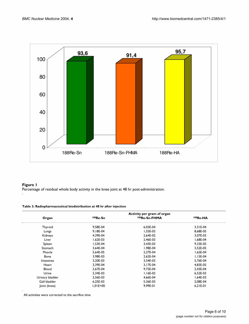
Activities per gram of selected tissue/organs including theknee joint after 48 hr of the radiopharmaceutical intraar-ticular administration to New Zealand rabbits are shownin Table 3.
The percentage of activity retained in the knee joint foreach radiopharmaceutical at 48 hr post injection was over90% of residual activity in the whole body (Figure 1).
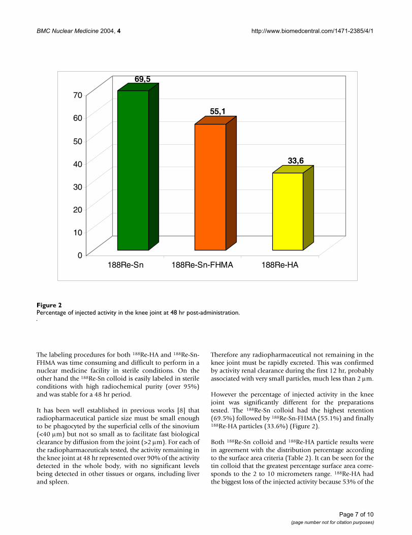
Nevertheless the percentage of injected activity in the kneejoint after 48 hr for each radiopharmaceutical was signifi-cantly different. The 188Re-Sn colloid had the highest per-centage of retention in the knee joint (Figure 2).
Scintigraphic studiesThe scintigraphic images of the rabbits, at 0, 24 and 48 hrpost administration, indicate relevant activity only in theknee, and negligible activity in the rest of the organism foreach formulation (Figure 3).
DiscussionThe labeling procedures for 188Re-HA and 188Re-Sn-FHMAincluded multiple centrifugation and separation steps. Inboth cases modifications from previous techniques had tobe adopted to improve the percentage of bound activity tothe particles. For 188Re-HA a two step procedure (used forSamarium-153 and Rhenium-186 labeled hydroxyapa-tite) [1] was discarded and a direct labeling technique wastried [9], that improved the percentage of bound activityfrom less than 5% to over 55%.
For the ferric hydroxide macroaggregates labeling a proce-dure originally developed for 195Dy was followed. Thismethod did not succeed for 188Re (less than 2% of boundactivity) because of the differences in reduction potentialsof the two radionuclides. Therefore several reducingagents were tested and the best results were obtained withthe two step procedure using tin to reduce the 188ReO4
-
prior to the labeling (coating) of the ferric hydroxide mac-roaggregate (over 90% of bound activity was obtained).
Table 2: Radiopharmaceutical particles distribution according to size, volume and surface
188Re-Sn 188Re-Sn-FHMA 188Re-HAParticle size Number Volume Surface Number Volume Surface Number Volume Surface
< 2 um 90 8 31 95 2 22 98 7 532 to 10 um 9,9 63 61 4 34 42 1,8 10 1910 to 40 um 0,1 29 8 1 64 36 0,2 83 28
>40 um 0 1 0 0 0 0 0 0 0
Page 5 of 10(page number not for citation purposes)
BMC Nuclear Medicine 2004, 4 http://www.biomedcentral.com/1471-2385/4/1
Percentage of residual whole body activity in the knee joint at 48 hr post-administrationFigure 1Percentage of residual whole body activity in the knee joint at 48 hr post-administration.
Table 3: Radiopharmaceutical biodistribution at 48 hr after injection
Activity per gram of organOrgan 188Re-Sn 188Re-Sn-FHMA 188Re-HA
Thyroid 9,58E-04 6,03E-04 3,31E-04Lungs 9,18E-04 1,55E-03 8,68E-05
Kidneys 4,39E-04 2,64E-02 3,07E-03Liver 1,62E-03 2,46E-03 1,68E-04
Spleen 1,53E-04 3,43E-03 9,33E-05Stomach 3,64E-04 1,98E-04 3,52E-05Muscle 5,64E-05 2,27E-04 1,62E-04Bone 3,98E-03 2,62E-04 1,15E-04
Intestines 3,20E-03 3,54E-03 5,76E-04Heart 3,39E-04 3,17E-04 4,83E-05Blood 2,67E-04 9,75E-04 2,43E-04Urine 2,34E-03 1,16E-02 6,52E-03
Urinary bladder 2,56E-03 4,66E-04 1,64E-03Gall bladder 6,25E-03 5,36E-03 2,08E-04Joint (knee) 1,01E+00 9,99E-01 6,21E-01
All activities were corrected to the sacrifice time
93,6 91,495,7
0
20
40
60
80
100
188Re-Sn 188Re-Sn-FHMA 188Re-HA
Page 6 of 10(page number not for citation purposes)
BMC Nuclear Medicine 2004, 4 http://www.biomedcentral.com/1471-2385/4/1
The labeling procedures for both 188Re-HA and 188Re-Sn-FHMA was time consuming and difficult to perform in anuclear medicine facility in sterile conditions. On theother hand the 188Re-Sn colloid is easily labeled in sterileconditions with high radiochemical purity (over 95%)and was stable for a 48 hr period.
It has been well established in previous works [8] thatradiopharmaceutical particle size must be small enoughto be phagocyted by the superficial cells of the sinovium(<40 µm) but not so small as to facilitate fast biologicalclearance by diffusion from the joint (>2 µm). For each ofthe radiopharmaceuticals tested, the activity remaining inthe knee joint at 48 hr represented over 90% of the activitydetected in the whole body, with no significant levelsbeing detected in other tissues or organs, including liverand spleen.
Therefore any radiopharmaceutical not remaining in theknee joint must be rapidly excreted. This was confirmedby activity renal clearance during the first 12 hr, probablyassociated with very small particles, much less than 2 µm.
However the percentage of injected activity in the kneejoint was significantly different for the preparationstested. The 188Re-Sn colloid had the highest retention(69.5%) followed by 188Re-Sn-FHMA (55.1%) and finally188Re-HA particles (33.6%) (Figure 2).
Both 188Re-Sn colloid and 188Re-HA particle results werein agreement with the distribution percentage accordingto the surface area criteria (Table 2). It can be seen for thetin colloid that the greatest percentage surface area corre-sponds to the 2 to 10 micrometers range. 188Re-HA hadthe biggest loss of the injected activity because 53% of the
Percentage of injected activity in the knee joint at 48 hr post-administrationFigure 2Percentage of injected activity in the knee joint at 48 hr post-administration.
69,5
55,1
33,6
0
10
20
30
40
50
60
70
188Re-Sn 188Re-Sn-FHMA 188Re-HA
Page 7 of 10(page number not for citation purposes)

BMC Nuclear Medicine 2004, 4 http://www.biomedcentral.com/1471-2385/4/1
Scintigraphic images at 0, 24 and 48 hr for 188Re-Sn, 188Re-Sn-FHMA and 188Re-HAFigure 3Scintigraphic images at 0, 24 and 48 hr for 188Re-Sn, 188Re-Sn-FHMA and 188Re-HA.
Page 8 of 10(page number not for citation purposes)
BMC Nuclear Medicine 2004, 4 http://www.biomedcentral.com/1471-2385/4/1
surface area corresponds to particles of less than 2 µmwhich could leak from the joint.
Nevertheless apparently anomalous values were obtainedfor 188Re-Sn-FHMA distribution (surface area criteria,Table 2) where only 22% of the particle surface corre-sponds to the range lower than 2 µm and the residualactivity in the knee was 55% instead of 80%. The ferrichydroxide radiopharmaceutical is actually a macroaggre-gate coated by a tin colloid as it was observed by electronicmicroscopy of HSA microspheres labeled with 188Re [12].The macroaggregate labeling (coating) may be achievedby a combination of the reduction of 188ReO4
- by tin anda particle surface related co precipitation effect of a ferrichydroxide macroaggregate with a tin colloid. The dissoci-ation of 188Re-Sn from the ferric hydroxide macroaggre-gate could happen in a certain range, and as aconsequence we postulated that the fraction smaller than2 µm could leak from the joint. So actually the leakage of188Re-Sn-FHMA would be by ferric hydroxide macroaggre-gates smaller than 2 micrometers and the 188Re-Sn colloiddissociated with a size of less than 2 µm.
We found that the range from 2 to 10 micrometers was theoptimum because the particles were phagocyted andremained in the target area for at least 48 hr.
From the point of view of the radiological security of thepatient, the best radiopharmaceutical is that one with agreater percentage of the injected dose retained in thejoint because a smaller activity amount had to be injectedto obtain the desired radiation dose. This fact takes intoaccount radioprotection principles for the patient in orderto minimize absorbed dose. The three formulations testedhad rapid renal clearance and showed negligible retentionin other tissues or organs other than the knee. Therefore188Re-Sn which had the greatest retention in the knee(69.1%) was the radiopharmaceutical that could give thedesired effect with the lowest absorbed dose in thepatient's whole body.
Conclusions188Re-Sn could be selected as the best formulation for syn-ovectomy therapy taking into account ease of labelingprocedure, kit formulation, minimum facilities required,suitable physical and biological characteristics and thelowest absorbed dose for the patient. Because of this thehighest benefit/risk relation was found for 188Re-Sn incomparison with 188Re-Sn-FHMA and 188Re-HA.
Competing interestsNone declared.
Authors' contributionsES planned the study, coordinated it and drafted themanuscript.
MCU participated in the design of the radiopharmaceuti-cal, carried out pharmaceutical experiments and draftedthe manuscript.
PZ carried out pharmaceutical experiments and alsodrafted the manuscript.
VT carried out pharmaceutical experiments.
AP carried out the scintigraphic studies.
VM carried out pharmaceutical experiments.
AM performed the Coulter Analizer determinations.
MF performed the experimental animal studies.
JG was responsible for scintigraphic studies.
All authors read and approved the final manuscript.
AcknowledgementsThe authors wish to thank:
- PEDECIBA Química for its financial support.
- Dr. Mercedes Mendoza de García (Fundación Cardioinfantil, Instituto de Cardiología. Bogotá, Colombia) for her collaboration.
References1. Chinol M, Vallabhajosula S, Goldsmith SJ, Klein MJ, Deutsch KF,
Chinen LK, Brodack JW, Deutsch EA, Watson BA, Tofe AJ: Chem-istry and biological behavior of Samarium-153 and Rhenium-186-labeled hydroxyapatite particles: potential radiophar-maceuticals for radiation synovectomy. J Nucl Med 1993,34:1536-1542.
2. Shortkroff S, Sledge CB: Radiation synovectomy. In: Principles ofNuclear Medicine 2nd edition. Edited by: Wagner HN Jr, Szabo Z, Bucha-nan JW. Philadelphia: WB Saunders Co; 1995:1021-1028.
3. Noble J, Jones AG, Davies MA, Sledge CB, Kramer RI, Livni E: Leak-age of radioactive practice system from synovial joint sudiedwith a gamma camera; its application to radiationsynovectomy. J Bone Joint Surg [Am] 1983, 65:381-389.
4. Murray T, Erskine Hilditch T: Therapeutic applications of radi-opharmaceuticals. In: Textbook of Radiopharmacy. Theory and Prac-tice 3rd edition. Edited by: Sampson CB. Cambridge: Gordon and BreachScience Publishers; 1999:380.
5. Clunie G, Ell PJ: A survey of radiation synovectomy in Europe,1991–1993. Eur J Nucl Med 1995, 22:970-976.
6. Lee EB, Shin KC, Lee YJ, Lee YJ, Cheon GJ, Jeong JM, Son MW, SongYW: 188Re-tin colloid as a new therapeutic agent for rheuma-toid arthritis. Nucl Med Commun 2003, 24:689-696.
7. Clunie G, Lui D, Cullum I, Edwards JCW, Ell PJ: Samarium-153-particulate hydroxyapatite radiation synovectomy: biodistri-bution data for chronic knee synovitis. J Nucl Med 1995,36:51-57.
8. Jeong JM, Lee YJ, Kim YJ, Chang YS, Lee DS, Chung J-K, Song YW, LeeMC: Preparation of rhenium-188-tin colloid as a radiationsynovectomy agent and comparison with rhenium-188-sul-fur colloid. Appl Radiat Isot 2000, 52:851-855.
Page 9 of 10(page number not for citation purposes)

BMC Nuclear Medicine 2004, 4 http://www.biomedcentral.com/1471-2385/4/1
Publish with BioMed Central and every scientist can read your work free of charge
"BioMed Central will be the most significant development for disseminating the results of biomedical research in our lifetime."
Sir Paul Nurse, Cancer Research UK
Your research papers will be:
available free of charge to the entire biomedical community
peer reviewed and published immediately upon acceptance
cited in PubMed and archived on PubMed Central
yours — you keep the copyright
Submit your manuscript here:http://www.biomedcentral.com/info/publishing_adv.asp
BioMedcentral
9. Kothari K, Suresh S, Sarma HD, Meera V, Pillai MRA: 188Re-labeledhydroxyapatite particles for radiation synovectomy. ApplRadiat Isot 2003, 58:463-468.
10. Castro M, Portilla A: Production of Dysprosium-165 labeledFHMA. In Regional workshop about ''Production of radionuclides andtherapeutical radiopharmaceuticals'', OIEA-ARCAL XV: 4-15 November1997; Lima-Peru .
11. Ures MC, Savio E, Malanga A, Fernández M, Paolino A, Gaudiano J:Physico-chemical characterization and biological evaluationof 188-Rhenium colloids for radio-synovectomy. BMC NuclMed 2002, 2:1.
12. Wunderlich G, Pinkert J, Andreeff M, Stintz M, Knapp FF Jr, Kropp J,Franke W-G: Preparation and biodistribution of rhenium-188labeled albumin microspheres B 20: a promising new agentfor radiotherapy. Appl Radiat Isot 2000, 52:63-68.
Pre-publication historyThe pre-publication history for this paper can be accessedhere:
http://www.biomedcentral.com/1471-2385/4/1/prepub
Page 10 of 10(page number not for citation purposes)