1-s2 0-s1381514811001581-main
TRANSCRIPT
Reactive & Functional Polymers 71 (2011) 1071–1076
Contents lists available at SciVerse ScienceDirect
Reactive & Functional Polymers
journal homepage: www.elsevier .com/ locate/ react
Ag/AgCl coated polyacrylonitrile nanofiber membranes: Synthesisand photocatalytic properties
Junyu Lei a,1, Wei Wang a,1, Mingxin Song a, Bo Dong a, Zhenyu Li a,⇑, Ce Wang a,⇑, Lijuan Li b
a Alan G. MacDiarmid Institute, Jilin University, Changchun 130012, PR Chinab Department of Chemistry and Biochemistry, California State University, Long Beach, CA 90840, USA
a r t i c l e i n f o
Article history:Received 17 May 2011Received in revised form 12 July 2011Accepted 2 August 2011Available online 9 August 2011
Keywords:ElectrospinningNanofiber membranesCore shell structurePhotocatalysisElectroless plating
1381-5148/$ - see front matter � 2011 Elsevier Ltd. Adoi:10.1016/j.reactfunctpolym.2011.08.002
⇑ Corresponding authors. Tel./fax: +86 431 8516829E-mail addresses: [email protected] (Z. Li), cwa
1 These authors contributed equally to this work.
a b s t r a c t
A membrane based photocatalyst consisting of Ag/AgCl coated PAN nanofibers was synthesized in largequantities by electrospinning technique combining electroless plating method and subsequent in situoxidation strategy. Electrospinning was firstly used to fabricate PAN/AgNO3 composite nanofibers. Afterreduction, Ag nanoparticles dispersed along the nanofibers act as seeds in the following metal electrolessplating step for the growth of continuous Ag shell. Then an in situ oxidation reaction between Ag shellsand FeCl3 solution was carried on to prepare Ag/AgCl coated PAN nanofiber membranes. The as-preparedmaterials exhibited excellent photocatalytic activity under visible-light, long-term stability, flexibility, aswell as easy separation from the liquid. The present work can open a new and effective route forpreparing high-performance membrane based photocatalysts for practical application.
� 2011 Elsevier Ltd. All rights reserved.
1. Introduction
Growing concerns over the threat of chemical warfare agentsand exposure to toxic industrial chemicals have drawn muchattention to the challenge of developing new harmless treatmentmethods for the toxic organic materials. Photocatalytic degrada-tion of harmful organic pollutants in water using semiconductormaterials is one of the most widely studied methods [1–3]. How-ever, semiconductor materials as photocatalysts present severalmajor problems when used in the form of powder slurries: (a)the complicated separation or filtration steps, (b) the problematicuse in continuous flow systems, and (c) the particles aggregationor loss, especially at high concentrations [4]. To overcome thesedrawbacks, considerable efforts have been oriented towards thephotocatalysts immobilization on solid supports as bound particlesor thin solid films in recent years [5–7]. However, in the case ofsuch membranes, the exposed area for photocatalytic activity ismuch lower than that of the highly dispersed slurry’s surfaceresulting in a decreased photocatalytic performance of the thinfilm compared with the slurry solution [8]. A proficient solutionto enhance the photocatalytic reaction rate is to immobilize much
ll rights reserved.
[email protected] (C. Wang).
more efficient semiconductor photocatalysts in the form of films.To date, modified TiO2 has attracted considerable researchinterests in this field [9–12]. Despite some success, the degradationefficiency of film based photocatalysts needs further improvementto meet academic and commercial demands.
Recently, by taking the advantages of plasmon resonance of sil-ver nanoparticles, silver/silver halide, a novel and highly efficientvisible-light photocatalyst, has been synthesized by Huang andco-workers [13]. Hitherto, various silver/silver halide photocata-lysts have been investigated [13–15]. However, these reports wereall focused on powder based silver/silver halide, which has the ten-dency to agglomerate into larger particles. This created an obstaclefor practical applications during the complicated solid–liquid sep-aration process as the damages caused by the intrinsic brittlenessof the inorganic structures.
Electrospun polymer nanofiber membranes have attractedincreasing attention to serve as photocatalysts supports due tothe three-dimensional open structure, large specific surface area,and flexibility in operation. These properties make them quitesuitable for loading photocatalysts [16,17]. Thus in the presentwork, we combined electrospinning technique with subsequentelectroless plating method and in situ oxidation strategy [15]and fabricated a novel photocatalyst: Ag/AgCl coated polyacrylo-nitrile (PAN) nanofiber membranes. Our products exhibit a highsurface area and with the flexibility of PAN nanofibers and thehigh efficient visible-light photocatalytic ability of the Ag/AgClnanoshells.
1072 J. Lei et al. / Reactive & Functional Polymers 71 (2011) 1071–1076
2. Experimental
2.1. Chemicals
Polyacrylonitrile (PAN, Mw = 80,000) was purchased from JilinCarbon Group. N,N-dimethylformamide (DMF), ammonia, ethyleneglycol, ferric chloride, potassium sodium tartrate and methyl or-ange were purchased from Tiantai Chemical Corporation. Silver ni-trate (AgNO3) was obtained from Beijing Chemical Corporation. Allthe reagents were used without further purification.
2.2. Preparation of Ag/AgCl coated PAN nanofiber membranes
The preparation of Ag/AgCl coated PAN nanofiber membranes isas follows: (1) 8 wt.% of polyacrylonitrile and 1.6 wt.% of AgNO3
were dissolved in DMF, under vigorous stirring at 50 �C in darknessfor 2 h. After cooled to room temperature, the mixture was loadedinto a glass syringe and connected to high-voltage power supply.12 kV was provided between the cathode and anode at a distanceof 15 cm to prepare PAN/AgNO3 nanofibers. After the reductionof AgNO3 in ethylene glycol under microwave irradiation, yellowmembranes of PAN/Ag nanofibers were obtained. (2) PAN nanofi-bers containing Ag nanoparticles were put into a beaker containing30 mL of water, 0.6 g of AgNO3 and 1.5 mL of ammonia (solutionA). In another beaker, 30 mL of water and 3 g of potassium sodiumtartrate were mixed as solution B. After solution B added into solu-tion A, electroless plating of Ag was carried out under vigorous stir-ring. After 4 h, the silver gray membranes were taken out from theelectroless plating bath, washed with deionized water and dried.(3) Ag/AgCl coated PAN nanofiber membranes were synthesizedby an in situ oxidation strategy in 0.006 M FeCl3 solution at roomtemperature (FeCl3 + Ag = AgCl + FeCl2). The sample oxidized inFeCl3 for 15 min, 40 min and 60 min are named NMa (nanofibermembrane a), NMb, and NMc, respectively.
For comparison, Ag/AgCl coated PAN solid films were preparedby spin-coating of the DMF solution containing PAN and AgNO3, in-stead of electrospinning. The other steps were the same with thatof Ag/AgCl coated PAN nanofiber membranes.
2.3. Characterization
The morphology and composition of the products werecharacterized by scanning electron microscopy (SEM; SSX-550,Shimadzu), transmission electron microscopy (TEM; HitachiS-570), X-ray diffraction (XRD; Scintag XDS 2000 diffractometerwith a Cu Ka radiation), X-ray photoelectron spectra (XPS;performed on an ESCLABMKII using Mg as excitation source), andUV–Vis absorption spectrometer (Shimadzu UV-3101 PCSpectrometer).
Scheme 1. Schematic illustration of the preparation of Ag/AgCl coated PANnanofibers.
2.4. Photocatalytic tests
Photodegradation studies of methyl orange (MO) were carriedout in a homemade quartz photochemical reactor. For each condi-tion, 60 mg of Ag/AgCl coated PAN nanofiber membranes (cut intosmall pieces) were dispersed in MO aqueous solution (20 mg/L,40 mL) and magnetically stirred in the dark for 60 min to ensurethe adsorption/desorption equilibrium of MO with the catalyst.The visible-light source was a 500 W Xenon lamp (CHFXQ500 W)with a UV filter that is capable of cutting off the UV wavelengthsshorter than 420 nm. The remaining MO concentration afteradsorption/desorption equilibrium (C0) and photodegradation (C)was detected by UV–Vis absorption spectra, and the degradationefficiency was expressed as C/C0.
3. Results and discussion
3.1. Preparation and characterization of Ag/AgCl coated PAN nanofibermembranes
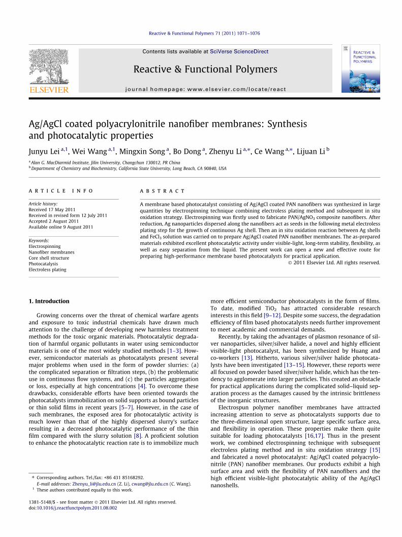
The synthesis of Ag/AgCl coated PAN nanofibers is describedschematically in Scheme 1. Electrospinning is firstly used to fabri-cate PAN/AgNO3 composite nanofibers [18]. Then Ag nanoparticlesdispersed along PAN nanofibers are obtained through the reduc-tion of the as-spun nanofibers. With the Ag nanoparticles as seeds,continuous Ag shells can be electroless plated on the PAN nanofi-bers. After an in situ oxidation strategy in FeCl3 solution, Ag/AgClcoated PAN nanofiber membranes are formed. A scanning electronmicroscopy (SEM) image of the electrospun PAN nanofibers con-taining Ag nanoparticles after reduction is shown in Fig. 1a. As typ-ical for electrospun fibers, the fibers are randomly oriented andthere is a distribution of fiber diameters, ranging from 100 to150 nm. After the electroless plating of Ag on the surface of PANnanofibers, as shown in Fig. 1b, the average diameters of nanofi-bers increases, indicating the formation of Ag shells. Fig. 1c–etracks the morphology changes of the samples after reactions withFeCl3 at various times. As the reaction time increases, the diame-ters of the fibers enlarge and the surface turns coarser. This phe-nomenon occurs as the Ag nanoshells are gradually etched intoAg+ (by Fe3+), which immediately combine with Cl� to form AgClnanoparticles on the Ag nanoshells, resulting in coarser and thickersurface of Ag/AgCl shells coating on PAN nanofibers.
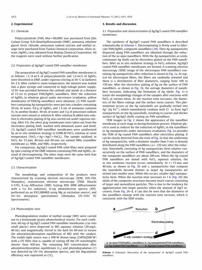
TEM images in Fig. 2 shows the appearance of the nanofibermembrane at each stage in during formation process. Ethylene gly-col is used as reducer for the reduction of AgNO3 in the nanofibersto Ag nanoparticles under microwave irradiation. Fig. 2a providesthe TEM of Ag coated PAN nanofibers after electroless plating. Itcan be clearly detected from the inset of Fig. 2a that the uniformityof Ag nanoparticles, with a diameter smaller than 5 nm, is denselydistributed along the PAN nanofibers (a.v. 120 nm) after the reduc-tion. Nanoshells consisting of Ag nanoparticles form relative con-secutively on the surface of PAN nanofibers, and the diameters ofthe composite nanofibers are about 200 nm. When the Ag coatedPAN nanofibers are mixed with FeCl3 aqueous solution, thein situ oxidation reaction occurs immediately. At t = 15 min and40 min, as shown in Fig. 2b and c, nanoparticles that composethe nanoshells become thinner as larger Ag nanoparticles areetched into smaller ones. While this occurs, smaller AgCl nanopar-ticles form. When the reaction time increases to 1 h (Fig. 2d) theshells of the composite structures become much coarser consistingof larger and nonuniform particles. This is due to the tendency foragglomeration into larger particles when the amount of AgCl in-creases. From Fig. 2b–d, it can also be seen that the diameters ofthe nanofibers enlarge with the reaction time increase, which isconsistent with the SEM results.
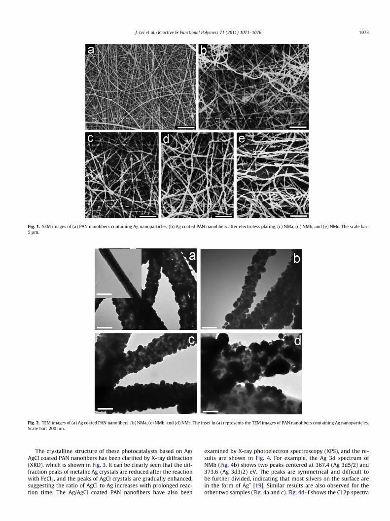
Fig. 1. SEM images of (a) PAN nanofibers containing Ag nanoparticles, (b) Ag coated PAN nanofibers after electroless plating, (c) NMa, (d) NMb, and (e) NMc. The scale bar:5 lm.
Fig. 2. TEM images of (a) Ag coated PAN nanofibers, (b) NMa, (c) NMb, and (d) NMc. The inset in (a) represents the TEM images of PAN nanofibers containing Ag nanoparticles.Scale bar: 200 nm.
J. Lei et al. / Reactive & Functional Polymers 71 (2011) 1071–1076 1073
The crystalline structure of these photocatalysts based on Ag/AgCl coated PAN nanofibers has been clarified by X-ray diffraction(XRD), which is shown in Fig. 3. It can be clearly seen that the dif-fraction peaks of metallic Ag crystals are reduced after the reactionwith FeCl3, and the peaks of AgCl crystals are gradually enhanced,suggesting the ratio of AgCl to Ag increases with prolonged reac-tion time. The Ag/AgCl coated PAN nanofibers have also been
examined by X-ray photoelectron spectroscopy (XPS), and the re-sults are shown in Fig. 4. For example, the Ag 3d spectrum ofNMb (Fig. 4b) shows two peaks centered at 367.4 (Ag 3d5/2) and373.6 (Ag 3d3/2) eV. The peaks are symmetrical and difficult tobe further divided, indicating that most silvers on the surface arein the form of Ag+ [19]. Similar results are also observed for theother two samples (Fig. 4a and c). Fig. 4d–f shows the Cl 2p spectra
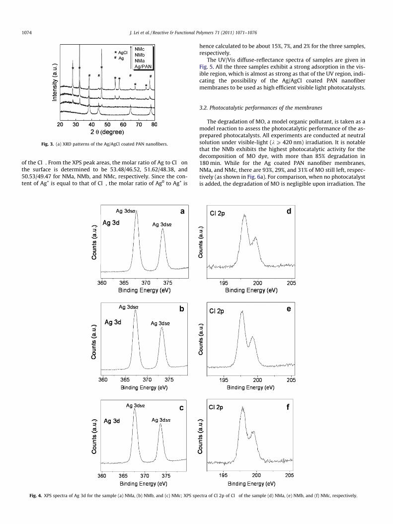
Fig. 3. (a) XRD patterns of the Ag/AgCl coated PAN nanofibers.
1074 J. Lei et al. / Reactive & Functional Polymers 71 (2011) 1071–1076
of the Cl�. From the XPS peak areas, the molar ratio of Ag to Cl� onthe surface is determined to be 53.48/46.52, 51.62/48.38, and50.53/49.47 for NMa, NMb, and NMc, respectively. Since the con-tent of Ag+ is equal to that of Cl�, the molar ratio of Ag0 to Ag+ is
Fig. 4. XPS spectra of Ag 3d for the sample (a) NMa, (b) NMb, and (c) NMc; XPS spe
hence calculated to be about 15%, 7%, and 2% for the three samples,respectively.
The UV/Vis diffuse-reflectance spectra of samples are given inFig. 5. All the three samples exhibit a strong adsorption in the vis-ible region, which is almost as strong as that of the UV region, indi-cating the possibility of the Ag/AgCl coated PAN nanofibermembranes to be used as high efficient visible light photocatalysts.
3.2. Photocatalytic performances of the membranes
The degradation of MO, a model organic pollutant, is taken as amodel reaction to assess the photocatalytic performance of the as-prepared photocatalysts. All experiments are conducted at neutralsolution under visible-light (k P 420 nm) irradiation. It is notablethat the NMb exhibits the highest photocatalytic activity for thedecomposition of MO dye, with more than 85% degradation in180 min. While for the Ag coated PAN nanofiber membranes,NMa, and NMc, there are 93%, 29%, and 31% of MO still left, respec-tively (as shown in Fig. 6a). For comparison, when no photocatalystis added, the degradation of MO is negligible upon irradiation. The
ctra of Cl 2p of Cl� of the sample (d) NMa, (e) NMb, and (f) NMc, respectively.
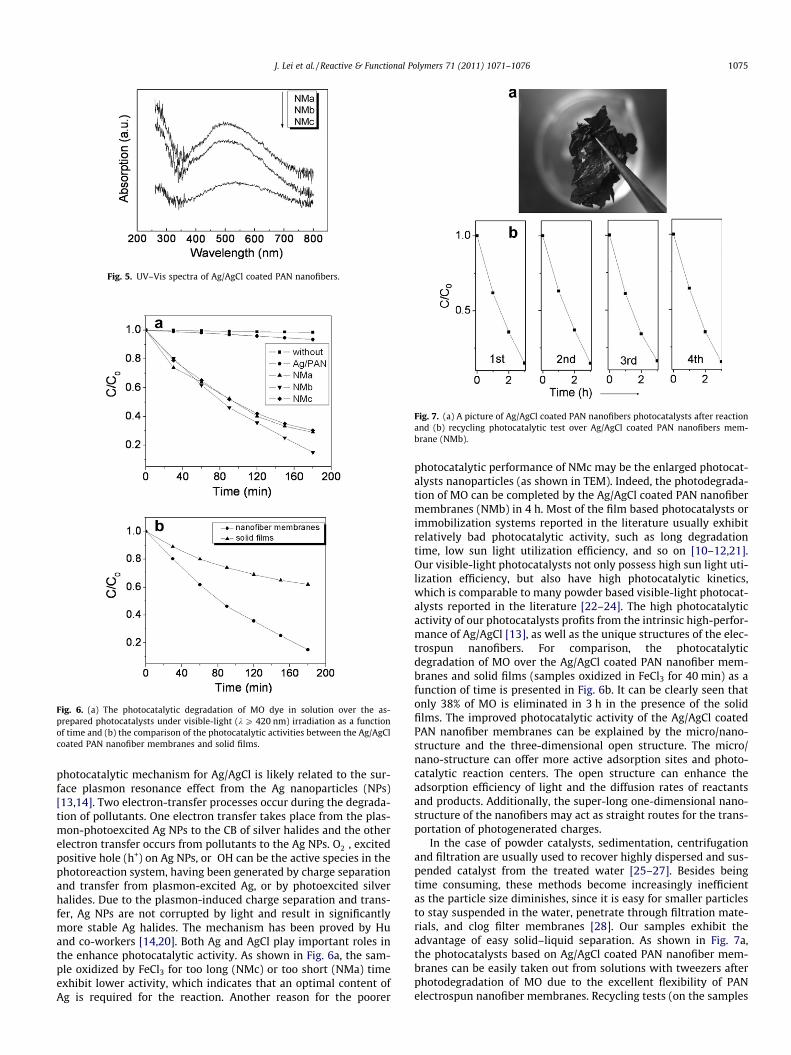
Fig. 5. UV–Vis spectra of Ag/AgCl coated PAN nanofibers.
Fig. 6. (a) The photocatalytic degradation of MO dye in solution over the as-prepared photocatalysts under visible-light (k P 420 nm) irradiation as a functionof time and (b) the comparison of the photocatalytic activities between the Ag/AgClcoated PAN nanofiber membranes and solid films.
Fig. 7. (a) A picture of Ag/AgCl coated PAN nanofibers photocatalysts after reactionand (b) recycling photocatalytic test over Ag/AgCl coated PAN nanofibers mem-brane (NMb).
J. Lei et al. / Reactive & Functional Polymers 71 (2011) 1071–1076 1075
photocatalytic mechanism for Ag/AgCl is likely related to the sur-face plasmon resonance effect from the Ag nanoparticles (NPs)[13,14]. Two electron-transfer processes occur during the degrada-tion of pollutants. One electron transfer takes place from the plas-mon-photoexcited Ag NPs to the CB of silver halides and the otherelectron transfer occurs from pollutants to the Ag NPs. O��2 , excitedpositive hole (h+) on Ag NPs, or �OH can be the active species in thephotoreaction system, having been generated by charge separationand transfer from plasmon-excited Ag, or by photoexcited silverhalides. Due to the plasmon-induced charge separation and trans-fer, Ag NPs are not corrupted by light and result in significantlymore stable Ag halides. The mechanism has been proved by Huand co-workers [14,20]. Both Ag and AgCl play important roles inthe enhance photocatalytic activity. As shown in Fig. 6a, the sam-ple oxidized by FeCl3 for too long (NMc) or too short (NMa) timeexhibit lower activity, which indicates that an optimal content ofAg is required for the reaction. Another reason for the poorer
photocatalytic performance of NMc may be the enlarged photocat-alysts nanoparticles (as shown in TEM). Indeed, the photodegrada-tion of MO can be completed by the Ag/AgCl coated PAN nanofibermembranes (NMb) in 4 h. Most of the film based photocatalysts orimmobilization systems reported in the literature usually exhibitrelatively bad photocatalytic activity, such as long degradationtime, low sun light utilization efficiency, and so on [10–12,21].Our visible-light photocatalysts not only possess high sun light uti-lization efficiency, but also have high photocatalytic kinetics,which is comparable to many powder based visible-light photocat-alysts reported in the literature [22–24]. The high photocatalyticactivity of our photocatalysts profits from the intrinsic high-perfor-mance of Ag/AgCl [13], as well as the unique structures of the elec-trospun nanofibers. For comparison, the photocatalyticdegradation of MO over the Ag/AgCl coated PAN nanofiber mem-branes and solid films (samples oxidized in FeCl3 for 40 min) as afunction of time is presented in Fig. 6b. It can be clearly seen thatonly 38% of MO is eliminated in 3 h in the presence of the solidfilms. The improved photocatalytic activity of the Ag/AgCl coatedPAN nanofiber membranes can be explained by the micro/nano-structure and the three-dimensional open structure. The micro/nano-structure can offer more active adsorption sites and photo-catalytic reaction centers. The open structure can enhance theadsorption efficiency of light and the diffusion rates of reactantsand products. Additionally, the super-long one-dimensional nano-structure of the nanofibers may act as straight routes for the trans-portation of photogenerated charges.
In the case of powder catalysts, sedimentation, centrifugationand filtration are usually used to recover highly dispersed and sus-pended catalyst from the treated water [25–27]. Besides beingtime consuming, these methods become increasingly inefficientas the particle size diminishes, since it is easy for smaller particlesto stay suspended in the water, penetrate through filtration mate-rials, and clog filter membranes [28]. Our samples exhibit theadvantage of easy solid–liquid separation. As shown in Fig. 7a,the photocatalysts based on Ag/AgCl coated PAN nanofiber mem-branes can be easily taken out from solutions with tweezers afterphotodegradation of MO due to the excellent flexibility of PANelectrospun nanofiber membranes. Recycling tests (on the samples

Fig. 8. SEM image of Ag/AgCl coated PAN nanofiber membranes after four timesdegradation of MO solution under visible-light irradiation for 3 h each. The scalebar: 5 lm.
1076 J. Lei et al. / Reactive & Functional Polymers 71 (2011) 1071–1076
NMb) are measured at regular intervals of 1 week to demonstratethe excellent stability of the photocatalysts (Fig. 7b). After 4 weeks,there is almost no loss on the photocatalytic activity. The SEM im-age of the Ag/AgCl coated PAN nanofibers after four times degrada-tion of MO solution is shown in Fig. 8. The photocatalysts shells arestill tightly immobilized on the surface of PAN nanofibers after thedegradation tests, and there is no obvious change compared withFig. 2c. As a conclusion, the Ag/AgCl coated PAN nanofibers possesshigh photocatalytic stability for the photodegradation of organicpollutants.
4. Conclusions
In summary, we have successfully developed a facile and effec-tive approach for coating Ag/AgCl nanoshells on PAN electrospunnanofibers by electroless plating method and subsequent in situoxidation strategy. The as-prepared nanofiber membranes exhibitexcellent photocatalytic activity and long-term stability undervisible-light, as well as easy separation from the liquid. The presentwork can open a new and effective route for preparing high-performance photocatalysts in practical applications and will
provide a baseline for the design and development of efficientcontinuous flow photocatalytic reactors in the future.
Acknowledgments
The work has been supported by National 973 Project (Nos.2007CB936203 and S2009061009), NSF China (No. 50973038),and National 863 Project (No. 2007AA03Z324).
References
[1] D.D. Lin, H. Wu, R. Zhang, W. Pan, Chem. Mater. 21 (2009) 3479.[2] M. Fujihira, Y. Satoh, T. Osa, Nature 293 (1981) 206.[3] H.Y. Li, D.J. Wang, P. Wang, H.M. Fan, T.F. Xie, Chem. Eur. J. 15 (2009) 12521.[4] A. Fujishima, T.N. Rao, D.A. Tryk, J. Photochem. Photobiol. C 1 (2000) 1.[5] S. Fukahori, H. Ichiura, T. Kitaoka, H. Tanaka, Environ. Sci. Technol. 37 (2003)
1048.[6] M. Karches, M. Morstein, P. Rudolf von Rohr, R.L. Pozzo, J.L. Giombi, M.A.
Baltanas, Catal. Today 72 (2002) 267.[7] F. Han, V.S.R. Kambala, M. Srinivasan, D. Rajarathnam, R. Naidu, Appl. Catal. A
359 (2009) 25.[8] I.M. Arabatzis, T. Stergiopoulos, M.C. Bernard, D. Labou, S.G. Neophytides, P.
Falaras, Appl. Catal. B 42 (2003) 187.[9] J. Yang, H.Z. Bai, Q. Jiang, J.S. Lian, Thin Solid Films 516 (2008) 1736.
[10] L.F. Cui, Y.S. Wang, M.T. Niu, G.X. Chen, Y. Cheng, J. Solid State Chem. 182(2009) 2785.
[11] J.G. Yu, H.G. Yu, C.H. Ao, S.C. Lee, J.C. Yu, W.K. Ho, Thin Solid Films 496 (2006)273.
[12] Y. Zhu, S. Xu, D. Yi, React. Funct. Polym. 70 (2010) 282.[13] P. Wang, B.B. Huang, X.Y. Qin, X.Y. Zhuang, Y. Dai, J.Y. Wei, M.H. Whangbo,
Angew. Chem. Int. Ed. 47 (2008) 7931.[14] C. Hu, T.W. Peng, X.X. Hu, Y.L. Nie, X.F. Zhou, J.H. Qu, H. He, J. Am. Chem. Soc.
132 (2010) 857.[15] Y.P. Bi, J.H. Ye, Chem. Commun. 43 (2009) 6551.[16] T.S. He, Z.F. Zhou, W.B. Xu, F.M. Ren, H.H. Ma, J. Wang, Polymer 50 (2009) 3031.[17] J.A. Lee, K.C. Krogman, M.L. Ma, R.M. Hill, P.T. Hammond, G.C. Rutledge, Adv.
Mater. 21 (2009) 1252.[18] Z.Y. Li, H.M. Huang, T.C. Shang, F. Yang, W. Zheng, C. Wang, Nanotechnology 17
(2006) 917.[19] Md. SASShah, M. Nag, T. Kalagara, S. Singh, S.V. Manorama, Chem. Mater. 20
(2008) 2455.[20] X.F. Zhou, C. Hu, X.X. Hu, T.W. Peng, J.H. Qu, J. Phys. Chem. C 114 (2010) 2746.[21] J.S. Im, M.I. Kim, Y.S. Lee, Mater. Lett. 62 (2008) 3652.[22] J. Fang, F.C. Shi, J. Bu, J.J. Ding, S.T. Xu, J. Bao, Y.S. Ma, Z.Q. Jiang, W.P. Zhang, C.
Gao, W.X. Huang, J. Phys. Chem. C 114 (2010) 7940.[23] Z. Jiang, F. Yang, G.D. Yang, L. Kong, M.O. Jones, T.C. Xiao, P.P. Edwards, J.
Photochem. Photobiol. A: Chem. 212 (2010) 8.[24] Z.S. Shan, X.P. Lin, M.L. Liu, H.M. Ding, F.Q. Huang, Solid State Sci. 11 (2009)
1163.[25] Z.Y. Liu, D.D. Sun, P. Guo, J.O. Leckie, Nano Lett. 7 (2007) 1081.[26] Y.Y. Li, Y. Ding, J. Phys. Chem. C 114 (2010) 3175.[27] K. Maeda, K. Domen, Chem. Mater. 22 (2010) 612.[28] N. Bao, X. Feng, Z. Yang, L. Shen, X. Lu, Environ. Sci. Technol. 38 (2004) 2729.