dobutamineincreases cardiac output of...
TRANSCRIPT

1210
Dobutamine Increases Cardiac Outputof the Total Artificial Heart
Implications for Vascular Contribution of InotropicAgents to Augmented Ventricular Function
Philip F. Binkley, MD; Kevin D. Murray, MD; Kim M. Watson, BS;P. David Myerowitz, MD; and Carl V. Leier, MD
Background. The synthetic catecholamine dobutamine increases stroke volume in normalsubjects and in patients with congestive heart failure. In addition to its direct influence on
myocardial contractility, dobutamine may significantly modulate vascular tone because of its a'
and ,B-adrenergic agonist activity.Methods and Results. To test the hypothesis that such vasoactive properties significantly
contribute to the improved ventricular performance noted with this agent, hemodynamicparameters were measured during stepped ascension infusion of dobutamine in a model that isinsensitive to positive inotropic stimulation. Administration of dobutamine in nine calves thatunderwent replacement of the native right and left ventricles with pneumatically driven totalartificial hearts resulted in a significant (p=0.0001) increase in cardiac output from 7.0±+1.8to 8.2±1.8 1/min and a significant (p=0.0001) decrease in total peripheral vascular resistancefrom 1,224± 559 to 745 ±317 dyne-sec/cm5. A less marked influence was noted on the pulmonaryvasculature, with pulmonary vascular resistance exhibiting a significant (p<O.OS) decreasefrom its baseline value only at the peak infusion. Consistent with an increase in venous return,both left and right atrial pressures increased significantly (p<0.005) with dobutamineadministration.
Conclusions. These data demonstrate that the vasoactive properties of dobutamine signifi-cantly contribute to improved ventricular performance independent of direct myocardialstimulation. This effect appears to result in part from a direct modulation of arterial andvenous tones rather than from a reflex response to primary changes in contractility.(Circulation 1991;84:1210-1215)
D obutamine is a synthetic catecholamine thatis known to increase cardiac output innormal subjects and in patients with con-
gestive heart failure.'-3 Although this effect is attrib-uted in large part to direct stimulation of the myo-cardium and consequent increases in ventricularcontractility, dobutamine may influence the arterialand venous vasculature because of a- and ,B-adren-ergic agonist activity.3-10 Dobutamine exists as aracemic mixture of two stereoisomers in which the
From the Divisions of Cardiology and Thoracic Surgery, TheOhio State University Hospital, Columbus, Ohio.
Supported by a Grant-in-Aid from the American Heart Associ-ation, Central Ohio Affiliate, and Eli Lilly and Co., Indianapolis,Ind. P.F.B. is the recipient of a Clinical Associate Physician Awardfrom the National Institutes of Health.Address for reprints: Philip F. Binkley, MD, The Ohio State
University Hospital, Division of Cardiology, 1654 Upham Drive,627 Means Hall, Columbus, OH 43210.
Received December 12, 1989; revision accepted April 9, 1991.
a-adrenergic activity is found to reside in the levoisomer and 13-adrenergic activity is expressed by thedextro isomer.'0 Varying degrees of peripheral vaso-dilation have been reported that have been ascribedto either direct or reflex effects on the vasculature,but the mechanism of this vascular response remainsincompletely described.6-9 The present investigationused a model that is insensitive to the positiveinotropic influence of dobutamine to test the hypoth-esis that the vasoactive properties of this agentsignificantly contribute to improved ventricular per-formance. The results indicate that dobutamine isassociated with a significant reduction of systemicvascular resistance coincident with a significant in-crease in cardiac output that appears to be indepen-dent of its effects on myocardial contractility and maybe ascribed to its direct effects on the vasculature.
MethodsAll investigations and procedures were reviewed
and approved by the animal use committee of the
by guest on June 4, 2018http://circ.ahajournals.org/
Dow
nloaded from
Binkley et al Augmented Function of Artificial Heart 1211
Ohio State University. The model consisted of ninecalves that underwent replacement of the native rightand left ventricles with pneumatically driven totalartificial right and left ventricles (Jarvik 7, Symbion,Inc., Salt Lake City, or Utah 100, University of Utah,Salt Lake City). The procedure for implantation ofthe artificial ventricles has been previously de-scribed.11-'3 Briefly, general anesthesia was main-tained with halothane after pentobarbital inductionand endotracheal intubation. A right lateral thorac-otomy was performed, and the animal was placed oncardiopulmonary bypass. The native right and leftventricles were excised along the atrioventricularvalve rings and above the semilunar valves. Atrialquick-connect cuffs were sewn into the right and leftatria, and vascular grafts were anastomosed to thepulmonary artery and aorta. The artificial right andleft ventricles were then positioned and securedusing the respective atrial quick-connect cuffs andvascular grafts. The right and left ventricles wereconnected via drive line tubing to the respectivepneumatic chambers of the heart driver. Controlparameters for each ventricle consisted of heart rate,drive pressure, and percent time in systole. Withactivation of the artificial ventricles, the animal wasweaned from total cardiopulmonary bypass. Deviceparameters were adjusted to maintain physiologicalpressures and ensure adequate cardiac output. Thewound was closed with running sutures, and drainagetubes were inserted in the pleural space. The animalwas transported to a specially constructed cage thatoptimized animal mobility while ensuring the integ-rity of the drive line connection to the heart driver.All animals were extubated and pleural drainagetubes were removed within 24 to 48 hours. A 4-dayrecovery period preceded the study protocol.
All drug infusions were performed with the animalin the sternal recumbent posture. Aortic, pulmonaryarterial, and left and right atrial pressures and car-diac outputs were monitored for a total of 20 minutesto ensure hemodynamic equilibration, which wasdefined as less than 10% variation in any of theseparameters. Device parameters were adjusted so thatthe artificial ventricles were not completely filling atbaseline. Specifically, heart rate for the nine animalsranged from 100 to 120 beats/min (mean+SD, 109 +7beats/min), and percent time in systole ranged from44% to 50% (mean+SD, 47+3%). Right ventriculardrive pressures ranged from 75 to 120 mm Hg(mean±SD, 103 + 17 mm Hg), and left ventriculardrive pressures ranged from 235 to 285 mm Hg(mean+SD, 262+16 mm Hg). These initial deviceparameters were maintained constant throughout thesubsequent dobutamine infusions. Cardiac outputswere measured using the cardiac output monitoringand diagnostic unit (COMDU, Symbion) of the heartdriver. This device integrates diastolic exhaust airflow and applies a volume conversion factor to cal-culate stroke volume on a beat-by-beat basis. Valida-tion of this technique has been performed by com-
parison with simultaneously measured cardiacoutputs using turbine flowmeters.14
After establishing hemodynamic equilibration,dobutamine was administered for three consecutive10-minute infusions at rates of 6,12, and 24 jig/kg/min using a percutaneously inserted external jugularline. Minute averages of cardiac output were printedcontinuously by the COMDU, and atrial, pulmonaryarterial, and aortic pressures were recorded at thepeak of each infusion. After measurements wereacquired at peak dosing, the infusion was discontin-ued, and the animal was allowed to return to baselinehemodynamic status.
Derived Hemodynamic Parameters andStatistical Analysis
Cardiac output was averaged during the last 5minutes of the baseline period and of each infusionperiod and was used for assessment of dobutamineeffects on stroke volume. Total peripheral vascularresistance (dyne.sec/cm5) was calculated as meansystemic pressure (mm Hg) multiplied by 80 and thendivided by cardiac output (1/min). Similarly, totalpulmonary vascular resistance was calculated accord-ing to the same formula using mean pulmonary arterypressure and cardiac output. Analysis of variance forrepeated measures was used to test for significantchanges in hemodynamic parameters associated withdobutamine administration. Posttest comparisonswere performed to identify specific points that dif-fered significantly from baseline. Statistical signifi-cance was defined as a probability of less than 0.05.
ResultsSystemic Vascular Response
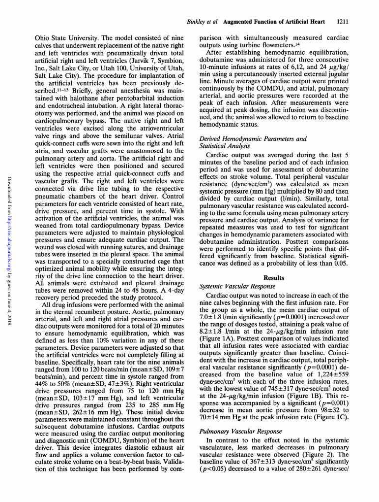
Cardiac output was noted to increase in each of thenine calves beginning with the first infusion rate. Forthe group as a whole, the mean cardiac output of7.0±1.8 1/min significantly (p= 0.0001) increased overthe range of dosages tested, attaining a peak value of8.2±1.8 1/min at the 24-,mg/kg/min infusion rate(Figure 1A). Posttest comparison of values indicatedthat all infusion rates were associated with cardiacoutputs significantly greater than baseline. Coinci-dent with the increase in cardiac output, total periph-eral vascular resistance significantly (p=0.0001) de-creased from the baseline value of 1,224±559dyne.sec/cm5 with each of the three infusion rates,with the lowest value of 745 ±317 dyne-sec/cm5 notedat the 24-,g/kg/min infusion (Figure 1B). This re-sponse was accompanied by a significant (p=0.001)decrease in mean aortic pressure from 98±32 to70±14 mm Hg at the peak infusion rate (Figure 1C).
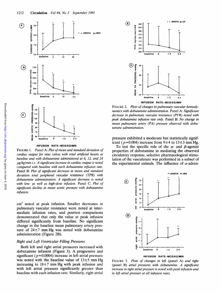
Pulmonary Vascular ResponseIn contrast to the effect noted in the systemic
vasculature, less marked decreases in pulmonaryvascular resistance were observed (Figure 2). Thebaseline value of 367±313 dyne-sec/cm' significantly(p<0.05) decreased to a value of 280±261 dyne.sec/
by guest on June 4, 2018http://circ.ahajournals.org/
Dow
nloaded from
1212 Circulation Vol 84, No 3 September 1991
* = ANOVA p<.00018.5_
7.5-
6.5 ,baseline 6 12 24
1700 ^
E 1500-
1300
c 1100-
:900-
700-
0)
E
cn
U)
z
4c
IE
E 125
cc 115
60 105u,
r: 85
075
65W baseline 6 1 2 2 4
INFUSION RATE--MCG/K/GMINFIGURE 1. Panel A: Plot ofmean and standard deviation ofcardiac output for nine calves with total artificial hearts atbaseline and with dobutamine administered at 6, 12, and 24pg/kg/min i.v. A significant increase in cardiac output is notedcompared with baseline with each dobutamine infusion rate.Panel B: Plot of significant decrease in mean and standarddeviation total peripheral vascular resistance (TPR) withdobutamine administration. A significant decrease is notedwith low- as well as high-dose infusion. Panel C: Plot ofsignificant decline in mean aortic pressure with dobutamineinfusion.
cm5 noted at peak infusion. Smaller decreases inpulmonary vascular resistance were noted at inter-mediate infusion rates, and posttest comparisonsdemonstrated that only the value at peak infusiondiffered significantly from baseline. No significantchange in the baseline mean pulmonary artery pres-sure of 24±7 mm Hg was noted with dobutamineadministration (Figure 2B).
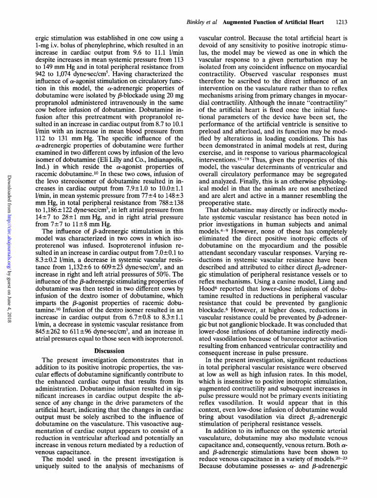
Right and Left Ventricular Filling PressuresBoth left and right atrial pressures increased with
dobutamine infusion (Figure 3). A progressive andsignificant (p=0.0004) increase in left atrial pressurewas noted with the baseline value of 13±5 mm Hgincreasing to 18+7 mm Hg with peak infusion andwith left atrial pressure significantly greater thanbaseline with each infusion rate. Similarly, right atrial
baseline 6 1 2 2 4
INFUSION RATE--MCGIKG/MIN
FIGURE 2. Plots ofchanges in pulmonary vascular hemody-namics with dobutamine administration. Panel A: Significantdecrease in pulmonary vascular resistance (PVR) noted withpeak dobutamine infusion rate only. Panel B: No change inmean pulmonary artery (PA) pressure observed with dobu-tamine administration.
pressure exhibited a moderate but statistically signif-icant (p =0.004) increase from 9±4 to 13±3 mm Hg.To test the specific role of the a- and fl-agonist
properties of dobutamine in mediating the observedcirculatory response, selective pharmacological stimu-lation of the vasculature was performed in a subset ofthe experimental animals. The influence of a-adren-
* =ANOVA Pc.005
I
E 17
0
13
CLi 13 -
11Xm 9 .
baseline 6 1 2 2 4
INFUSION RATE--MCG/KGIMINFIGURE 3. Plots of changes in left (panel A) and right(panel B) atrial pressures with dobutamine. A significantincrease in right atrialpressure is noted with peak infusion andin left atrial pressure at all infusion rates.
9.5-
aI-
CL
0
Ucr4cc
4cc
usE
CD
srCL
* = ANOVA pc.05660
610
560
510-460410-
360 -
310 -
260-
m
EE0z0ehpCL
0
z41
by guest on June 4, 2018http://circ.ahajournals.org/
Dow
nloaded from
Binkley et al Augmented Function of Artificial Heart 1213
ergic stimulation was established in one cow using a1-mg i.v. bolus of phenylephrine, which resulted in anincrease in cardiac output from 9.6 to 11.1 1/mindespite increases in mean systemic pressure from 113to 149 mm Hg and in total peripheral resistance from942 to 1,074 dyne.sec/cm5. Having characterized theinfluence of a-agonist stimulation on circulatory func-tion in this model, the a-adrenergic properties ofdobutamine were isolated by ,B-blockade using 20 mgpropranolol administered intravenously in the samecow before infusion of dobutamine. Dobutamine in-fusion after this pretreatment with propranolol re-sulted in an increase in cardiac output from 8.7 to 10.11/min with an increase in mean blood pressure from112 to 131 mm Hg. The specific influence of thea-adrenergic properties of dobutamine were furtherexamined in two different cows by infusion of the levoisomer of dobutamine (Eli Lilly and Co., Indianapolis,Ind.) in which reside the a-agonist properties ofracemic dobutamine.10 In these two cows, infusion ofthe levo stereoisomer of dobutamine resulted in in-creases in cardiac output from 7.9±1.0 to 10.0±1.11/min, in mean systemic pressure from 77±4 to 148±3mm Hg, in total peripheral resistance from 788±138to 1,186±122 dyne.sec/cm5, in left atrial pressure from14±7 to 28±1 mm Hg, and in right atrial pressurefrom 7±7 to 11±8 mm Hg.The influence of p-adrenergic stimulation in this
model was characterized in two cows in which iso-proterenol was infused. Isoproterenol infusion re-sulted in an increase in cardiac output from 7.0±0.1 to8.3±0.2 1/min, a decrease in systemic vascular resis-tance from 1.132+6 to 609±23 dyne.sec/cm5, and anincrease in right and left atrial pressures of 50%. Theinfluence of the p-adrenergic stimulating properties ofdobutamine was then tested in two different cows byinfusion of the dextro isomer of dobutamine, whichimparts the p-agonist properties of racemic dobu-tamine.'0 Infusion of the dextro isomer resulted in anincrease in cardiac output from 6.7±0.8 to 8.3±1.11/min, a decrease in systemic vascular resistance from845±262 to 611±96 dyne.sec/cm5, and an increase inatrial pressures equal to those seen with isoproterenol.
DiscussionThe present investigation demonstrates that in
addition to its positive inotropic properties, the vas-cular effects of dobutamine significantly contribute tothe enhanced cardiac output that results from itsadministration. Dobutamine infusion resulted in sig-nificant increases in cardiac output despite the ab-sence of any change in the drive parameters of theartificial heart, indicating that the changes in cardiacoutput must be solely ascribed to the influence ofdobutamine on the vasculature. This vasoactive aug-mentation of cardiac output appears to consist of areduction in ventricular afterload and potentially anincrease in venous return mediated by a reduction ofvenous capacitance.The model used in the present investigation is
uniquely suited to the analysis of mechanisms of
vascular control. Because the total artificial heart isdevoid of any sensitivity to positive inotropic stimu-lus, the model may be viewed as one in which thevascular response to a given perturbation may beisolated from any coincident influence on myocardialcontractility. Observed vascular responses musttherefore be ascribed to the direct influence of anintervention on the vasculature rather than to reflexmechanisms arising from primary changes in myocar-dial contractility. Although the innate "contractility"of the artificial heart is fixed once the initial func-tional parameters of the device have been set, theperformance of the artificial ventricle is sensitive topreload and afterload, and its function may be mod-ified by alterations in loading conditions. This hasbeen demonstrated in animal models at rest, duringexercise, and in response to various pharmacologicalinterventions.15-19 Thus, given the properties of thismodel, the vascular determinants of ventricular andoverall circulatory performance may be segregatedand analyzed. Finally, this is an otherwise physiolog-ical model in that the animals are not anesthetizedand are alert and active in a manner resembling thepreoperative state.That dobutamine may directly or indirectly modu-
late systemic vascular resistance has been noted inprior investigations in human subjects and animalmodels.4-9 However, none of these has completelyeliminated the direct positive inotropic effects ofdobutamine on the myocardium and the possibleattendant secondary vascular responses. Varying re-ductions in systemic vascular resistance have beendescribed and attributed to either direct p2-adrener-gic stimulation of peripheral resistance vessels or toreflex mechanisms. Using a canine model, Liang andHood6 reported that lower-dose infusions of dobu-tamine resulted in reductions in peripheral vascularresistance that could be prevented by ganglionicblockade.6 However, at higher doses, reductions invascular resistance could be prevented by p-adrener-gic but not ganglionic blockade. It was concluded thatlower-dose infusions of dobutamine indirectly medi-ated vasodilation because of baroreceptor activationresulting from enhanced ventricular contractility andconsequent increase in pulse pressure.
In the present investigation, significant reductionsin total peripheral vascular resistance were observedat low as well as high infusion rates. In this model,which is insensitive to positive inotropic stimulation,augmented contractility and subsequent increases inpulse pressure would not be primary events initiatingreflex vasodilation. It would appear that in thiscontext, even low-dose infusion of dobutamine wouldbring about vasodilation via direct p2-adrenergicstimulation of peripheral resistance vessels.
In addition to its influence on the systemic arterialvasculature, dobutamine may also modulate venouscapacitance and, consequently, venous return. Both a-and p-adrenergic stimulations have been shown toreduce venous capacitance in a variety of models.20-23Because dobutamine possesses a- and p-adrenergic
by guest on June 4, 2018http://circ.ahajournals.org/
Dow
nloaded from
1214 Circulation Vol 84, No 3 September 1991
agonist activities, it may augment venous return bystimulation of both of these receptor systems. Consis-tent with this hypothesis, Fuchs et al23 demonstrated adecrease in venous capacitance accompanying dobu-tamine infusion as measured by splanchnic volumeand found that this decrease could be prevented bya-blockade. Thus, both afterload reduction (via /2-agonist effects) and augmentation of venous return(resulting from a- and /3-agonist activities) may con-tribute to the increase in cardiac output mediated bydobutamine's influence on the vasculature.The subset of cows in which the responses to
specific a- and ,B-adrenergic stimulations were exam-ined further substantiates these mechanisms. a-Ad-renergic stimulation with phenylephrine resulted in anincrease in cardiac output similar in magnitude to thatobserved with dobutamine infusion. This occurreddespite marked increases in systemic arterial pressureand systemic vascular resistance. Because the pneu-matically driven artificial heart operates at a drivingpressure greater than even the peak systemic pressureachieved with phenylephrine, it will continue to ejectthe volume of blood returned by the venous circula-tion despite such elevations in afterload. As such, theartificial ventricle resembles the normally functioninghuman heart in its capacity to maintain stroke volumedespite elevations in afterload.24 Therefore, althoughthe artificial ventricle has a demonstrated sensitivity toafterload,16-19 in the setting of augmented venousreturn, a net increase in stroke volume may resultdespite an increase in afterload, especially if themagnitude of increased venous return is relativelygreater than the increase in afterload. Thus, the onlymechanism by which a-adrenergic stimulation couldincrease cardiac output in this model is throughaugmentation of venous return. Similarly, this wouldaccount for the observed response to dobutamineinfusion after /3-blockade with propranolol, in whichthe a-adrenergic activity of dobutamine would beunmasked, and with infusion of the levo isomer ofdobutamine, which possesses only a-agonist activity.As with phenylephrine, an increase in cardiac outputwas observed with infusion of these agents despitesubstantial increases in systemic pressure. The in-crease in atrial pressures with the levo isomer infusionfurther supports this mechanism.That the ,/-agonist properties of dobutamine contrib-
ute to the increase in cardiac output in this model isdemonstrated by the responses to the dextro isomer ofdobutamine, which possesses only /3-adrenergic stimu-lating properties, and to the infusion of isoproterenol.Infusion of the dextro isomer in two of the cowsresulted in an increase in cardiac output accompaniedby a reduction of systemic vascular resistance. Bothright and left atrial pressures increased with infusion ofthis isomer. Similarly, isoproterenol infusion resulted inan increase in cardiac output at all infusion rates andwas accompanied by a decrease in systemic vascularresistance and an increase in atrial pressures. Whetherthe increase in cardiac output associated with theseagents derives from the influence of /3-adrenergic stim-
ulation on afterload, from /-adrenergic-mediated re-duction of venous capacitance and augmented venousreturn, or from a combination of these mechanismscannot be completely resolved based on the currentdata. However, an increase in atrial pressure similar tothat seen after the levo isomer suggests that a primaryinfluence on venous return is an important componentof this response.The influence of dobutamine on the pulmonary
vasculature appears to be less marked than that notedfor the systemic vasculature. Although a decrease inpulmonary vascular resistance was noted, this occurredonly at the maximal infusion rate. This contrasts withthe marked sensitivity of the systemic vasculature inwhich significant decreases in resistance were noted ateven the lowest infusion rate. These differences mayarise because of variations in pulmonary and systemicvascular adrenergic receptor populations and affinitiesfor dobutamine. Significant decreases in pulmonaryvascular resistance accompanying dobutamine admin-istration have been reported in humans with congestiveheart failure.23,7 The present data suggest that such adecline in pulmonary vascular resistance may resultfrom indirect influences such as reductions in ventric-ular filling pressures or reflex responses to increasedcardiac output rather than from a direct effect on thepulmonary vasculature.
Unlike previous investigations in humans with con-gestive heart failure, dobutamine administration inthis model is associated with significant increases inright and left atrial pressures.2,37 This is consistentwith the increase in forward flow and venous returnobserved in this setting. Unlike the native ventricle,which can shift both to new pressure-volume rela-tions and to different points within a given relation inresponse to positive inotropic influence, the artificialventricle will operate along a relatively fixed pres-sure-volume curve; this will tend to increase fillingpressures in the setting of augmented venous return.These changes in atrial pressures and volumes may
themselves contribute to the increase in cardiacoutput through potentiation of atrial contractility byan effect similar to the Starling mechanism of ven-tricular muscle contraction. Alternatively, dobu-tamine may directly influence contractility of theatria and in this manner augment ventricular filling.However, the importance of atrial systole is uncertainin this model in which atrioventricular synchrony isnot preserved and, consequently, atrial filling occursat random intervals relative to ventricular filling.The observed increase in cardiac output in this
model was noted to occur with the first infusion rateand increase less markedly with subsequent infusions(Figure 1). This phenomenon is most likely a result ofthe fact that the maximal filling volume of theartificial ventricle was approached with the increasein cardiac output resulting from the first infusion,leaving little margin for further increase with subse-quent infusions. It is conceivable that if a greatermargin for increased filling were allowed, the cardiacoutput would continue to increase with further infu-
by guest on June 4, 2018http://circ.ahajournals.org/
Dow
nloaded from
Binkley et al Augmented Function of Artificial Heart 1215
sions rather than achieving the observed plateau.However, operating the artificial ventricle at fillingvolumes below those used in the present study wouldresult in inappropriately low cardiac outputs thatwould not be tolerated by the animal.The relevance of these observations to the clini-
cally observed hemodynamic response to dobutamineinfusion in patients with congestive heart failureprovides a focus for future investigation. Althoughdirect comparisons of the afterload sensitivity of thenormal or failing ventricle with that of the artificialheart have not been made, extrapolation from priorstudies in humans suggests that the artificial heartresembles the normal ventricle in terms of its sensi-tivity to afterload.24 In a report by Ross and Braun-wald,24 decreases in stroke volume were not seen inventricles with normal contractility until levels ofafterload approaching the drive pressure of the arti-ficial ventricle were achieved. The failing ventricle isknown to have a much greater sensitivity to afterload;thus, it may be speculated that in the setting ofventricular failure, the vasoactive properties of dobu-tamine may have an even more profound effect oncirculatory performance because of their modulationof loading conditions. This principle must be furthertested in humans with congestive heart failure and inanimal models of circulatory failure such as thosethat may be adapted from the present model.
ConclusionsThe present data indicate that the vasoactive prop-
erties of dobutamine contribute significantly to theimproved ventricular performance and augmentationof cardiac output associated with its administration.The model used in the present study effectivelyeliminates the inotropic influence of this agent andthus allows an analysis of the direct vascular effects ofdobutamine distinct from its influence on myocardialcontractility and attendant reflex vascular changes.Further elucidation of the mechanisms of vascularresponse to dobutamine and related positive inotro-pic agents may be provided through this model bycomparing and contrasting the hemodynamic re-sponse to agents that vary in their adrenergic recep-tor affinity and activity.
AcknowledgmentsThe authors wish to thank Anne Brandt and
Trichia Zavilla for their invaluable assistance in thepreparation of this manuscript.
References1. Tuttle RR, Mills J: Dobutamine: Development of a new
catecholamine to selectively increase cardiac contractility. CircRes 1975;36:185-196
2. Leier CV, Webel J, Bush CA: The cardiovascular effects of thecontinuous infusion of dobutamine in patients with severecardiac failure. Circulation 1977;56:468-472
3. Leier CV, Unverferth DV: Drugs five years later: Dobu-tamine. Ann Intem Med 1983;99:490-496
4. Vatner SF, McRitchie RJ, Braunwald E: Effects of dobu-tamine on left ventricular performance, coronary dynamics,
and distribution of cardiac output in conscious dogs. J ClinInvest 1974;53:1265-1273
5. Robie NW, Glodberg LI: Comparative systemic and regionalhemodynamic effects of dopamine and dobutamine. Am HeartJ 1975;90:340-345
6. Liang CS, Hood WB Jr: Dobutamine infusion in consciousdogs with and without autonomic nervous system inhibition:Effects on systemic hemodynamics, regional flows and cardiacmetabolism. J Pharmacol Exp Ther 1979;211:698-705
7. Leier CV, Heban P, Huss P, Bush CA, Lewis RP: Comparativesystemic and regional hemodynamic effects of dopamine anddobutamine in patients with cardiomyopathic heart failure.Circulation 1977;58:466-475
8. Lewis, GRJ, Poole Wison PA, Angerpointer TA, FarnsworthAE, Williams BT, Coltart DJ: Measurement of the circulatoryeffects of dobutamine, a new inotropic agent, in patientsfollowing cardiac surgery. Am Heart J 1978;95:301-307
9. Raifer SI, Borow DM, Lang RM, Neuman A, Carroll JD:Effects of dopamine on left ventricular afterload and contrac-tile state in heart failure: Relation to the activation of beta1-adrenoceptors and dopamine receptors. JAm Coll Cardiol1988;12:498-506
10. Ruffolo RR Jr, Spradlin TA, Pollock GD, Waddell JE, Mur-phy PJ: Alpha and beta adrenergic effects of the stereoisomersof dobutamine. J Pharmacol Exp Ther 1981;219:447-452
11. Olsen DB, Taenaka Y: State of the art and clinically appliedpneumatic artificial hearts. Crit Care Clin 1986;2:195-207
12. Frazier OH, Col'n R, Taenaka Y: Surgical technique andhemodynamic characteristics of partial cardiac replacementwith an artificial left ventricle. Tex Heart Inst J 1986;13:345-351
13. Anderson FL, DeVries WC, Anderson JL, Joyce LD: Evalu-ation of total artificial heart performance in man.Am J Cardiol1984;54:394-398
14. Willshaw P, Nielsen SD, Nanas J, Pichel RH, Olsen DB: Acardiac output monitor and diagnostic unit for pneumaticallydriven artificial hearts. Artif Organs 1984;8:215-219
15. Olsen DB, Grosse Siestrup CH, Unger R, Kless H, Kolff WJ,Bucherl ES: The non-cardiac intrinsic autoregulation of tissueperfusion in calves with total artificial hearts. Proc Eur SocArtif Organs 1977;4:263-270
16. Deleuze PH, Riebman JB, Paulis RDE, Olsen DB, LoisanceDY: Regulation of pneumatic total artificial heart function: Areview. Int JArtif Organs 1988;11:147-152
17. Uchida N, Ishikawa M, Watanabe T, Jacobs G, Oku T, Nasu M,Emoto H, Smith WA, Harasaki H, Kiraly R, Nose Y: Hemo-dynamic adaptation to exercise after total artificial heartimplantation. TransAm SocArtifIntem Organs 1987;33:240-244
18. Hennig E, Grosse Siestrup C, Krautzberger W, Kless H,Bucherl ES: The relationship of cardiac output and venouspressure in long surviving calves with total artificial heart.TransAm Soc Artif Intem Organs 1978;24:616-624
19. Goldenber I, Walker M, Roeller SK, Gobel FL, Joyce L,Jorgensen R, Madison JD, Pedersen WR: The Jarvik-7 totalartificial heart is preload and afterload dependent (abstract).Circulation 1987;6(suppl IV):IV-404
20. Kaiser GA, Ross J, Braunwald E: Alpha and beta adrenergicreceptor mechanisms in the systemic venous bed. J PharmacolExp Ther 1964;144:156-162
21. Van Maanen EF, Banning JW, Roebel LE, Morgan JP:Dopamine and norepinephrine increase venous return bystimulating alpha and beta adrenoceptors in the dog. J Car-diovasc Pharmacol 1988;11:627-634
22. Lee RW, Raya TE, Gay RG, Marcey 0, Goldman S: Beta-2adrenoceptor control of the venous circulation in intact dogs.JPharmacol Exp Ther 1987;242:1138-1143
23. Fuchs RM, Rutlen DL, Powell WJ Jr: Effect of dobutamine onsystemic capacity in the dog. Circ Res 1980;46:133-138
24. Ross J Jr, Braunwald E: The study of left ventricular functionin man by increasing resistance to ventricular ejection withangiotensin. Circulation 1964;29:739-749
KEY WORDS . positive inotropic agents * vascular mechanisms* congestive heart failure
by guest on June 4, 2018http://circ.ahajournals.org/
Dow
nloaded from

P F Binkley, K D Murray, K M Watson, P D Myerowitz and C V Leiervascular contribution of inotropic agents to augmented ventricular function.
Dobutamine increases cardiac output of the total artificial heart. Implications for
Print ISSN: 0009-7322. Online ISSN: 1524-4539 Copyright © 1991 American Heart Association, Inc. All rights reserved.
is published by the American Heart Association, 7272 Greenville Avenue, Dallas, TX 75231Circulation doi: 10.1161/01.CIR.84.3.1210
1991;84:1210-1215Circulation.
http://circ.ahajournals.org/content/84/3/1210the World Wide Web at:
The online version of this article, along with updated information and services, is located on
http://circ.ahajournals.org//subscriptions/
is online at: Circulation Information about subscribing to Subscriptions:
http://www.lww.com/reprints Information about reprints can be found online at: Reprints:
document. Permissions and Rights Question and Answer information about this process is available in the
located, click Request Permissions in the middle column of the Web page under Services. FurtherEditorial Office. Once the online version of the published article for which permission is being requested is
can be obtained via RightsLink, a service of the Copyright Clearance Center, not theCirculationpublished in Requests for permissions to reproduce figures, tables, or portions of articles originallyPermissions:
by guest on June 4, 2018http://circ.ahajournals.org/
Dow
nloaded from