do probiotics protect against the deleterious effects of a ... · do probiotics protect against the...
TRANSCRIPT

Do Probiotics Protect Against the Deleterious Effects of a High-Fat Diet?
Dissertation
Submitted to the faculty of the Virginia Polytechnic Institute and State University in partial fulfillment of the requirements for the degree of Doctor of Philosophy
In
Human Nutrition, Foods and Exercise
by
Gabrielle Francesca Fundaro
Chair: Matthew W. Hulver Madlyn I. Frisard
Kevin P. Davy Eva M. Schmelz
Andrew P. Neilson
May 15, 2014 Blacksburg, VA
Keywords: Probiotics, high-fat diet, metabolic endotoxemia, skeletal muscle
Do Probiotics Protect Against the Deleterious Effects of a High-Fat Diet?
Gabrielle Francesca Fundaro
ABSTRACT
High-fat diets and obesity have been linked to unfavorable changes in gut bacteria and
increased leakage of bacterially-derived lipopolysaccharide (endotoxin) from the
intestinal tract into circulation, which is associated with low-grade inflammation,
metabolic dysregulation and degradation of tight-junction proteins between intestinal
cells. Probiotic supplementation is the practice of ingesting live strains of bacteria that are
proposed to have a beneficial effect on the host by enriching the intestine with healthy
bacteria. The purpose of this project was to determine if probiotic supplementation would
prevent increased inflammatory tone, decreased oxidative capacity, and decreased tight-
junction protein expression associated with high-fat feeding and elevated endogenous
endotoxin. Male C57BL/6J mice were fed either a control (CD, 10% fat) or high-fat
(HFD, 60% fat) diet for 4 weeks while receiving a daily oral gavage of water (C-VSL#3,
HF-VSL#3) or probiotics (C+VSL#3, HF+VSL#3) equivalent to 1.2 billion live cultures.
Changes in body weight, body composition, respiratory exchange ratio, energy
expenditure, and glucose and insulin tolerance were measured in live mice. Markers of
metabolic function were measured in whole muscle homgenates and mitochondria
isolated from red and white skeletal muscle. Plasma endotoxin was measured in blood
collected from fasted mice at the time of euthanization. The large and small intestines
were collected and mRNA levels of tight-junction proteins and markers of nutrient
sensing were measured. To determine a possible protective effect against endogenous
LPS, a second cohort of mice were given an intraperitoneal injection of 0.1µg/kg LPS or
iii
saline to induce endotoxemia after four weeks of the aforementioned feeding protocol.
Markers of metabolic function and inflammation were measured in mitochondria, skeletal
muscle and liver. VSL#3 supplementation improved glucose homeostasis and markers of
inflammation while enhancing nutrient sensing in the gut.
iv
Table of Contents
CHAPTER I: INTRODUCTION 1 CHAPTER II: REVIEW OF LITERATURE 3 2. Introduction 3 3. The Gut Microbiome and Gut Colonization 5 3.1 The Microbiome 5 3.2 Colonization 6 4. Mechanisms of Metabolic Influence 7
4.1 Energy Harvesting 7 4.2 Short Chain Fatty Acids & G-protein Coupled Receptor Signaling 8
4.3 Fatty Acid Oxidation & de novo Fatty Acid Synthesis 9 4.4 Fasting-Induced Adipose Factor & Fatty Acid Uptake 10
4.5 Endocannabinoid System 10 4.6 Metabolic Endotoxemia 13 5. Toll-Like Receptor 4 Activation and Metabolic Dysregulation 14
5.1 TLR-4 Signaling in Skeletal Muscle 14 5.2 TLR-4 Signaling in the Intestine 15
5.3 Emerging Links between Skeletal Muscle and the Microbiome 16 6. Probiotics 17
6.1 Benefits of Probiotic Supplementation 17 6.2 Probiotic Therapy for Inflammatory Bowel Diseases 18 6.3 Probiotics and Gut Permeability 19 6.4 Probiotics and Metabolic Syndrome 21 6.5 Possible Role of Probiotics in Lipopolysaccharide Clearance 22
7. Limitations and Unanswered Questions 23 8. Conclusion 25 CHAPTER III: SPECIFIC AIMS AND HYPOTHESES 27 CHAPTER IV: THE EFFECTS OF VSL#3 ON GLUCOSE HOMEOSTASIS AND INTESTINAL MARKERS OF NUTRIENT SENSING DURING HIGH-FAT FEEDING 4.1 Abstract 30 4.2 Introduction 31 4.3 Methods 33 4.4 Results 37 4.5 Discussion 44 CHAPTER V: VSL#3 SUPPLEMENTATION REDUCES TLR-2, TLR-4 AND TNFα EXPRESSION IN SKELETAL MUSCLE
5.1 Abstract 53 5.2 Introduction 54 5.3 Methods 55 5.4 Results 57
v
5.5 Discussion 63 CHAPTER VI: IMPLICATIONS & FUTURE DIRECTIONS 67 LITERATURE CITED 70
vi
DEDICATION
I dedicate this dissertation to my grandfather, Richard Hagerty, who taught me the value
of lifelong learning and discovery. Instead of simply telling me the answer to one of my
many questions, he would guide me through the process of learning the answer myself.
He embodied the spirit of research and imbued in me an appreciation for the search of
new knowledge. That passion has motivated me throughout my doctoral career and will
motivate my future students in the same way.

vii
ACKNOWLEDGEMENTS
I would like to thank Dr. Matthew Hulver for his ceaseless confidence in my abilities to
perform research and teach. He has always supported my teaching efforts, and without
his thoughtful nomination I would not be one of the inaugural members of the Graduate
Teaching Scholars. At times, Dr. Hulver had more confidence in my abilities than I did,
and his calm encouragement was a great source of motivation. I would also like to thank
Dr. Donna Westfall-Rudd who has advised me as a Graduate Teaching Scholar for the
latter half of my doctorate. Dr. Westfall-Rudd has been a well of advice and guidance,
and from her I have learned what it really takes to be a dynamic teacher. To the rest of
my committee members—Dr. Madlyn Frisard, Dr. Kevin Davy, Dr. Eva Schmelz, and
Dr. Andrew Neilson—I give my gratitude for your thoughtful advice and insight.
I would like to thank my parents, Micheal and Silke Fundaro, for all their love, support
and advice on this journey. I am incredibly thankful to my wonderful fiance’, Jeffery
Willis, who has been so understanding, patient, and loving, especially when stress has
gotten the best of me. Additionally, I wish to thank my amazing and hilarious friends and
colleagues, whose humor and commiseration has helped me take myself less seriously.
Finally, I thank my grandmothers, Penny Hagerty and Evelyn Fundaro, for their love and
support.

viii
LIST OF FIGURES
Figure One. Changes in body mass (A), total weight gain (B) and changes in % body fat
(C) in mice fed a control or high-fat diet with or without supplementation of VSL#3
probiotics at 1.2 billion cultures per day (n=10-15). 38
Figure Two. Total fat mass in grams (A) and total body weight (B) in the fourth week of
feeding (n=10-15). 39
Figure Three. Levels of 12-hour fasted glucose in all mice (n=14-15) (A). 41
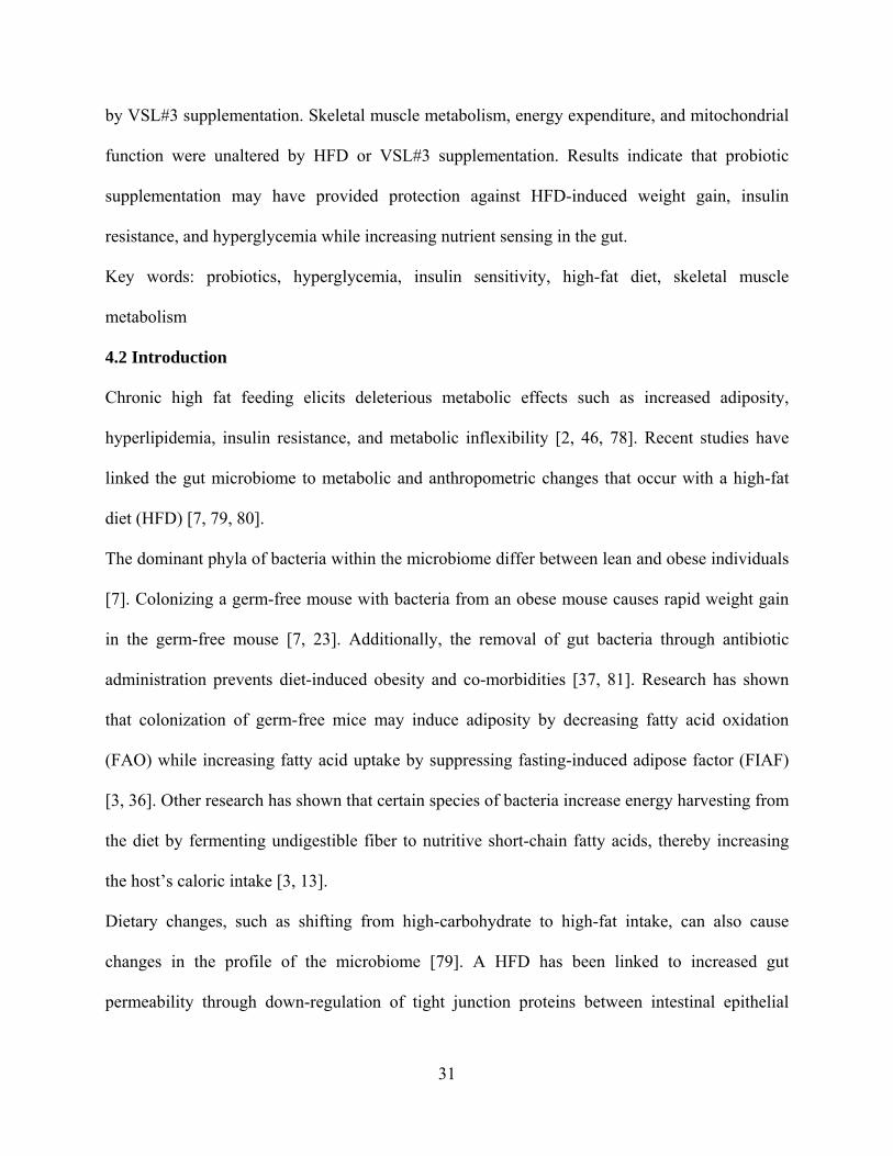
Figure Four. LPS bioactivity in plasma following a 12-hour fast (n=5-8) (A). 43
Figure Five. mRNA expression levels of glucagon-like peptide 2 receptor (n=5-10) (A)
and fasting-induced adipose factor in proximal sections of small intestine (B) (n=4-7). 45
Figure Six. mRNA expression levels of TLR-2 (A), TLR-4 (B), and TNFa (C) in white
skeletal muscle of mice injected with saline or low-dose LPS (0.1 µg/kg BW) after 4
weeks with or without VSL#3 supplementation (n=8-9). 60
Figure Seven. mRNA expression levels of TLR-2 (A), TLR-4 (B), and TNFa (C) in red
skeletal muscle of mice injected with saline or low-dose LPS (0.1 µg/kg BW) after 4
weeks with or without VSL#3 supplementation (n=8-10). 62
Figure Eight. mRNA expression levels of TNFa (A), IL-10 (B) and SREBP-1 (C) in
liver of mice injected with low-dose LPS (0.1 mg/kg BW) after 4 weeks with or without
VSL#3 supplementation (n=7-10). 63
ix
LIST OF TABLES
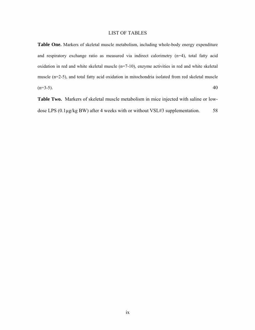
Table One. Markers of skeletal muscle metabolism, including whole-body energy expenditure
and respiratory exchange ratio as measured via indirect calorimetry (n=4), total fatty acid
oxidation in red and white skeletal muscle (n=7-10), enzyme activities in red and white skeletal
muscle (n=2-5), and total fatty acid oxidation in mitochondria isolated from red skeletal muscle
(n=3-5). 40
Table Two. Markers of skeletal muscle metabolism in mice injected with saline or low-
dose LPS (0.1µg/kg BW) after 4 weeks with or without VSL#3 supplementation. 58
x
LIST OF ABBREVIATIONS
Lipopolysaccharide (LPS)
Toll-Like Receptor 4 (TLR-4)
Fatty Acid Oxidation (FAO)
Fatty Acid Synthase (FAS)
Carbohydrate Responsive Element Binding Protein (ChREBP)
Sterol Responsive Element Binding Protein (SREBP-1)
5’ Adenosine Monophosphate-Activated Protein Kinase (AMPK)
Short-Chain Fatty Acids (SCFA)
G-Protein Coupled Receptors (GPCR)
Acetyl CoA Carboxylase (ACC)
Carnitine:Palmitoyl Transferase-1 (CPT1)
Fasting-Induced Adipose Factor (FIAF)
Lipoprotein Lipase (LPL)
Cannabinoid Receptor 1 and 2 (CB1 and CB2)
Mitogen-Activated Protein Kinase (MAPK)
Extracellular Signal-Related Kinase (ERK)
Nuclear Factor kappa B (NFκB)
Fatty Acid Amide Hydrolase (FAAH)
Monoacylglycerol Hydrase (MGL)
Tumor Necrosis Factor α (TNFα)
Monocyte Chemoattractant Protein-1 (MCP-1)
Crohn’s Disease (CD)

xi
Ulcerative Colitis (UC)
Irritable Bowel Disease (IBD)
Colony Forming Units (CFU)
Zonula Occludins 1, 2 and 3 (ZO-1, ZO-2, ZO-3)
Glucagon-Like Peptide 2 (GLP-2)
Interleukin 10 (IL-10)
Monoacylglycerols (MAG)
Diacylglycerols (DAG)
Very Low Density Lipoproteins (VLDL)
Low Density Lipoproteins (LDL)
1
CHAPTER I: INTRODUCTION
Rates of both child and adult obesity in the United States have increased drastically in the past 30
years [1]. Obesity is closely associated with metabolic dysregulation including dyslipidemia,
insulin resistance, and type II diabetes [2]. High-fat diets, obesity, and type II diabetes have been
linked to unfavorable changes in gut bacteria and increased leakage of bacterially-derived
lipopolysaccharide (endotoxin) from the intestinal tract into circulation[3]. Lipopolysaccharide,
or LPS, may migrate from the intestine into circulation via compromised tight junction proteins,
resulting in a condition known as ‘metabolic endotoxemia’[4]. When LPS binds its ligand, Toll-
like receptor 4 (TLR-4), an inflammatory cascade is initiated and inflammatory cytokines are
produced [5]. Persistent elevation of plasma endotoxin can induce chronic low-grade
inflammation which has been implicated in the development of metabolic dysregulation and gut
permeability [6].
Recent research has shown that the profiles of intestinal bacteria differ between lean and obese
individuals, and obese individuals are likely to present with ‘dysbiosis’ or an over-abundance of
non-beneficial gut bacteria [7]. Certain bacterial profiles favor increased energy harvesting from
the host diet, increased intestinal permeability, and higher levels of gram-negative bacteria which
contain LPS. In contrast, some strains of bacteria are commonly found in lean hosts and are
associated with greater insulin sensitivity and integrity of intestinal tight junctions as compared
to obese hosts [8]. Additionally, new links have been discovered between LPS and the
endocannabinoid system which can regulate feeding behaviors, lipid uptake, and adipose cell
differentiation [9-11].
Probiotic supplementation is the practice of ingesting live, non-pathogenic bacteria that can
enrich the bacterial colony of the host and promote gut health [12]. Beneficial bacteria provided
2
through probiotic supplementation have been shown to decrease gut permeability, decrease
inflammatory markers, improve diet-induced insulin resistance, and increase the health of
individual enterocytes [13]. However, most of these findings have occurred in murine models,
and human studies have not consistently replicated these results [14, 15]. Probiotic
supplementation has been utilized primarily as a therapy for inflammatory bowel diseases,
including colitis and irritable bowel syndrome, to correct the overgrowth of harmful bacteria and
improve mucous production to protect the intestinal lining [16].
Although there are clear links between the profile of gut bacteria and the phenotype of the host,
few studies have examined the effect that enrichment with probiotics may have on preventing or
treating obesity [3]. In most cases, studies illustrate correlations between host phenotype and pre-
existing colonies, and the role of bacteria in actually inducing obesity or endotoxemia remains to
be seen. While several studies have examined the protective effects of single-strain probiotics
against diet-induced obesity and metabolic regulation, no other study has utilized a multi-strain
probiotic as an intervention to determine effects on skeletal muscle metabolism. Therefore, it is
the purpose of this study to determine the role of multi-strain probiotic supplementation as a non-
invasive dietary method for protection against high-fat diet-induced metabolic dysregulation.
3
CHAPTER II: REVIEW OF LITERATURE
2. Introduction
In the past 30 years, obesity rates have doubled in adults and tripled in children. Today, a
staggering 17% of children and over 30% of adults are classified as obese [1]. It is well
documented that obesity is accompanied by insulin resistance, dyslipidemia, and type II diabetes,
the latter of which affects 220 million people globally [2]. Obesity is now considered a low-
grade inflammatory condition, causing immune responses that dysregulate metabolism and lead
to the aforementioned co-morbidities [17]. Both obesity and type II diabetes have been linked to
metabolic endotoxemia, a modest increase in circulating levels of lipopolysaccharide (LPS)
which binds to Toll-like receptor 4 (TLR-4) and activates the innate immune system, resulting in
low-grade inflammation [5, 18]. In response to pro-inflammatory signaling, skeletal muscle
becomes insulin-resistant and loses oxidative capacity, thereby resulting in the storage of fats
rather than oxidation, a state known as metabolic dysregulation or metabolic inflexibility. [19].
In recent years, researchers have also discovered links between obesity and the gut microbiome,
which refers to the bacteria that colonize the length of the intestinal tract. Obese and diabetic
individuals’ bacterial profiles and gut permeability differ significantly from those of lean
individuals [2, 9, 20]. These differences correlate with changes in insulin sensitivity, propensity
for diet-induced obesity, endotoxemia, circulating cytokine concentrations, and gut hormones [2,
21, 22]. Colonizing germ-free mice (lacking gut bacteria) with gut bacteria from normal, diet-
induced obese mice causes dramatic weight gain independent of food intake [7, 23]. When ob/ob
mice, which lack the appetite-regulating hormone leptin, are used for colonization, this weight
gain effect is exacerbated. Cani, et al. has coined the terms ‘dysbiosis’ and ‘microbesity’ to
describe a gut microbiome which favors obesity and leads to increased gut permeability and

4
plasma LPS due to its overgrowth of pathogenic bacteria [7, 13, 24]. While the mechanisms are
not clear, it is becoming more obvious that the diet readily influences the gut microbiome, and in
turn the bacteria influence the metabolic health of their host.
The human intestinal tract is colonized by roughly 10 trillion bacteria comprised of hundreds of
species [7]. Gut bacteria were initially perceived as benign organisms with no influence on
human health unless they entered the blood stream and induced sepsis, but in the past 20 years
they have been recognized for their potential to influence intestinal cell health and whole-body
metabolism [13]. Now, the gut microbiome is being considered as a potential therapeutic target
to combat obesity and its related complications, including low-grade inflammation and metabolic
dysregulation. Recent research has begun to elucidate a “brain-gut-microbiota axis,” wherein the
gut bacteria influence energy balance and obesity via regulation of gut hormones and activation
of the endocannabinoid receptors [11, 17]. Research has thus far focused on the use of
supplements, including prebiotics and probiotics, to influence the activity and bacterial variety of
the gut microbiome.
The conceptualization and commercialization of probiotics—bacteria that positively affect a
host’s health—have led to new research pursuits to better understand the role of the gut
microbiome and methods by which to improve its beneficial properties [12]. It is hypothesized
that probiotics can effectively enrich the gut to shift a potentially harmful microbiome phenotype
to one that benefits and protects the host. In humans, probiotic supplementation has correlated
with decreased plasma cholesterol levels, decreased bowel-disease-related intestinal
inflammation, decreased diarrhea and decreased visceral fat mass [16, 25, 26]. In mice, they
have been shown to increase intestinal cell and mucous proliferation, thereby decreasing gut
permeability and possibly indirectly reducing the harmful effects of a high-fat diet in a

5
microbiome-dependent manner [12, 27]. Prebiotics—fibers that are indigestible by humans but
fermentable by bacteria—have been used as successful therapies to improve the health of
intestinal cells and reduce high-fat diet-induced metabolic dysregulation by increasing certain
strains of gut bacteria [28]. The bacteria ferment these fibers to short-chain fatty acids, which can
then be utilized for energy by the bacteria and intestinal cells, promoting proliferation and the
production of proteins that increase gut integrity [29]. The possibility to reduce and even prevent
diet-induced obesity and metabolic dysregulation with non-invasive dietary supplements shows
promise in combating the current epidemic of obesity and its co-morbidities [21]. This review
discusses recent research illustrating the mechanisms by which gut bacteria and probiotics can
influence skeletal muscle metabolism, intestinal function and integrity, and obesity-related
endotoxemia. In addition, it proposes possible avenues for further research into proposed
processes by which probiotic supplementation may ameliorate the deleterious inflammatory
effects of chronic endotoxemia.
3. The Gut Microbiome and Gut Colonization
3.1 The Microbiome
The gut microbiome is essentially a microscopic ecosystem composed of trillions of bacteria
living off the intestinal contents of the host and producing both waste products and energy [7].
The propensity of the bacteria to convert fibrous material to energy usable by the host has been
shown to greatly influence the metabolic health of the host [21]. Similarly, by-products of the
death of certain bacteria have been shown to have deleterious effects on the metabolic health of
the host. ‘Good’ bacteria are non-pathogenic and bind to the epithelial wall of the lumen,
promoting cell and mucous proliferation as well as the formation of the antibody IgA to fight
infectious bacteria [30]. The ‘bad’ bacteria release toxins in the form of lipopolysaccharide,

6
which can induce inflammation and metabolic dysregulation at the level of the intestine as well
as skeletal muscle [6, 31]. Certain bacteria have been linked to obesity and overweight in mice
and humans, while others appear to prevent diet-induced obesity and insulin resistance [3, 13].
Bacterial strains compete for the available nutrients, using bacteriocins and controlling luminal
pH to kill their competitors and prevent their proliferation [12]. Controlling the dominating
species of bacteria through the use of probiotics has been shown to benefit the health of the host,
and as researchers classify the expansive number of bacterial species, correlations can be found
between the genotype of the gut microbiome and the phenotype of its host [2, 3, 7, 21, 22, 32].
3.2 Colonization
In utero, infants’ intestinal tracts are completely free of bacteria. Even before birth, the mother’s
gut microbiome can influence the infant’s birth weight, and from the beginning of life, the
profile of the inherited gut microbiota correlates with weight gain through adolescence [33].
Colonization of the gut begins at birth as the infant is exposed to the vaginal canal, and continues
throughout infancy while the infant nurses and is exposed to a bacteria-rich environment up to
age two, at which point the gut microbiome stabilizes [32, 34]. Tsukumo, et al. reported that
cesarean sections result in a delay of infant gut colonization by up to a month compared to babies
birthed vaginally [27]. In addition, breastfeeding was shown to colonize the infant gut primarily
with the beneficial Bifidobacterium, which produced high amounts of acetate and lactate. These
acidic compounds reduced the pH of the intestine making it difficult for pathogenic bacteria to
flourish. Many studies have illustrated links between the type of bacteria colonizing the gut and
the degree of obesity and insulin resistance in the host. In humans, the most prevalent phyla of
bacteria are the Firmicutes, Actinobacteria and Bacteroidetes, which make up roughly 80% of
the total bacterial population [21]. The dominant phyla in an individual’s gut can determine its

7
“enterotype”, or co-occurring genre and species related to a specific phyla, and in turn, the
enterotype influences the phenotype of the host [3]. High levels of Firmicutes have been linked
with obesity and high amounts of Bacteriodetes with a propensity toward leanness [20, 21].
Proteobacteria, a Gram-negative phylum, have been measured at unusually high levels in
insulin-resistant humans [3]. Dietary factors can also influence the enterotype of the gut
according to studies that have shown increased Bacteroides levels during a diet high in protein
and saturated fat, and increased levels of Prevotella (Firmicutes) during a diet high in simple
sugars and carbohydrates [3].
Obesity has clear links to the composition and metabolic activity of gut bacteria [2, 3, 6, 9]. The
microbiome of obese individuals is less diverse and has a high ratio of Firmicutes to Bacteroides
[3]. Obese individuals also contain greater numbers of methanogens, or archaea that produce
methane, which makes fiber fermentation more efficient and allows for greater energy harvesting
from the diet [3, 9]. It appears that obese individuals contain bacteria that are adept at efficiently
extracting energy from the host diet, which, in cases of obesity, is often Westernized and high in
fat and sugar [3]. LPS-producing, pathogenic bacteria are also higher in both obese individuals
and those with Type II diabetes while some beneficial strains are reduced; this phenomenon is
termed “gut dysbiosis [7, 13, 21].” Fortunately, these effects can be ameliorated when
individuals lose weight as a result of a calorie-restricted diet [3]. While the mechanism has not
been fully elucidated, there are several theories that attempt to explain this phenomenon.
4. Mechanisms of Metabolic Influence
4.1 Energy Harvesting
One possible cause of a propensity toward obesity with certain gut colonization points to
products of fiber fermentation by gut bacteria. Gut bacteria are capable of fermenting fibers

8
undigestible by humans, but the products differ [21]. While Bacteriodetes produce hydrogen,
Firmicutes are capable of producing short-chain fatty acids (SCFA) which can be used for
energy by the intestinal cells and as precursors to fatty acids and cholesterol [13]. Although the
ingested fiber isn’t bioavailable to a human gut, the SCFA’s produced from it can account for 6-
10% of basal energy requirements for people in developed countries [3]. This is known as
‘energy harvesting’, and it is one possible explanation for increased obesity in individuals
colonized with high levels of this bacteria.
4.2 Short Chain Fatty Acids & G-protein Coupled Receptor Signaling
Recent research has shown these SCFA’s can act as ligands to enteroendocrine cell-bound G-
protein coupled receptors (GPR) in the gut which regulate energy homeostasis [3, 9, 13, 21].
GPR43 expression is increased in subcutaneous fat of mice fed a high-fat diet. Binding of GPR
43 by the SCFA’s acetate and propioniate has been linked to increased adipogenesis, inhibited
lipolysis, and decreased whole-body energy expenditure [3, 9, 13, 21]. When GPR41 was bound
by the SCFA butyrate, SCFA uptake increased. Meanwhile, GPR 41 -/- mice were resistant to
diet-induced obesity even after conventionalization, indicating that this receptor may be required
for the gut microbiome to promote diet-induced obesity [3, 9, 13, 21].
However, it should be noted that in some cases supplementation of certain fibers known as
preobiotics led to increased resting energy expenditure and insulin sensitivity and a decrease in
de novo fatty acid synthesis in mice which contradicts the idea that fiber is a singular cause of
increase adipogenesis [22, 29]. Gao, et. al showed that butyrate, a SCFA product of fiber
fermentation, increased insulin sensitivity and energy expenditure in mice by increasing PGC-1a
activity in skeletal muscle [29]. In addition, the enterocytes of the intestine can utilize the
SCFA’s for energy, which generally results in greater proliferation and tight-junction integrity,
9
thereby reducing the negative effects of high-fat feeding on the gut. Due to these conflicting
results, the ‘energy harvesting’ theory has lost some support in recent years.
4.3 Fatty Acid Oxidation & de novo Fatty Acid Synthesis
Another possible explanation for decreased fatty acid oxidation in animals colonized with gut
bacteria is the suppression of AMP-activated protein kinase (AMPK), which acts as an energy
gauge and deactivates acetyl-CoA carboxylase (ACC) to increase mitochondrial fatty-acid
oxidation . AMPK becomes active when levels of ATP decrease due to its dephosphorylation
during times of high energy demand [35]. Active AMPK phosphorylates and suppresses the
activity of ACC. ACC inactivation prevents the conversion of acetyl CoA to malonyl CoA.
When malonyl CoA levels are low, fatty acids can enter the mitochondria via carnitine:palmitoyl
transferase-1 (CPT1) where they can be oxidized to produce energy. When AMPK is suppressed,
malonyl CoA levels remain elevated, preventing fatty acids from entering the mitochondria
where they would be oxidized. Multiple studies and reviews reported germ-free mice
experienced increased AMPK activity resulting in elevated PGC-1a levels, which increases
mitochondrial biogenesis and oxidative capacity [7, 9, 36]. In a murine model of high-fat diet-
induced obesity, mice lacking gut bacteria experienced obesity resistance, 40% more
phosphorylated AMPK and ACC but a 15% decrease in CPT1, indicating lower levels of both
fatty acid synthesis and oxidation, respectively [3]. Introduction of gut bacteria into germ-free
mice resulted in suppression of AMPK and a subsequent decrease in fat oxidation. ACC and
fatty acid synthase (FAS) can also be controlled by Carbohydrate Responsive Element Binding
Protein (ChREBP) and Sterol Responsive Element Binding Protein (SREBP-1), both of which
increase in hepatic cells after gut colonization [3, 7]. This leads to increased de novo fatty acid

10
synthesis and higher monosaccharide uptake from the intestine in colonized mice as compared to
germ-free mice.
4.4 Fasting-induced Adipose Factor & Fatty Acid Uptake
Gut colonization, or the addition of gut bacteria to a previously bacteria-free mouse, has also
been shown to suppress fasting-induced adipose factor (FIAF), a lipoprotein lipase (LPL)
inhibitor [3, 13]. The suppression of FIAF led to greater LPL activity and therefore greater
triglyceride uptake and possible storage in adipocytes. One study showed a decrease in ileal
FIAF expression and a 122% increase in LPL activity in epididymal adipose tissue along with
increased adiposity after conventionalization of germ-free mice [3]. Conventionalized mice—
those who have undergone manual colonization—experience greater uptake and clearance of
serum lipids compared to germ-free mice as well. In contrast, during high fat feeding, treatment
with antibiotics intended to kill bacteria ameliorated diet-induced weight gain [37]. Germ-free
mice have been shown to have reduced intestinal vascularity and digestive capabilities, so it is
possible that some protection against diet-induced obesity is due to inefficient nutrient digestion
and absorption [32].
4.5 Endocannabinoid System
Known as the ‘brain-gut’ axis, the endocannabinoid system connects the brain, gut, and liver
through the vagus nerve, hypothalamus, and gastric hormone production to regulate certain
digestive actions and signal energy requirements [3, 7, 11, 17]. Endocannabinoids are
endogenous ligands, including anandamide and AG-2, which bind to cannabinoid receptors that
are expressed in skeletal muscle, adipose tissue, liver, pancreas, bone, immune cells, and the
nervous system [11, 17]. Cannabinoid receptors (CB1 and CB2) are G-coupled protein receptors
that inhibit adenylate cyclase and cAMP production when bound and modify gene transcription
11
through a mitogen-activated protein kinase (MAPK), extracellular signal-related kinase (ERK)
and nuclear factor kappa B (NFkB) pathway [17]. Endocannabinoids are produced only when
needed and once synthesized, they are rapidly degraded by fatty acid amide hydrolase (FAAH)
and monoacylglycerol hydrase (MGL). It has been shown that high-fat feeding, obesity, gut
microbiota, and LPS play influential roles in endocannabinoid system activity [11, 17]. Obese
individuals experience greater endocannabinoid eCB) levels in plasma and adipose tissue as well
as increased CB1 expression compared to lean individuals [11, 17]. Muccioli, et. al showed that
acute high-fat feeding increased intestinal eCB production and suppressed FAAH and MGL, but
Cluny, et. al reported that chronic high-fat feeding resulted in normal eCB system function,
which may indicate that obesity rather than high-fat feeding may cause chronic dysregulation of
the eCB system [11, 17]. LPS alone has been shown to increase the synthesis of
endocannabinoids and CB1 receptor expression in cell culture and mice, mimicking the effect of
diet-induced obesity [7]. In addition, CB1 receptor activation has been linked to higher LPS
levels in obese mice, indicating a possible self-promoting cycle [11, 17]. The eCB system has
been shown to control gut permeability by changing the distribution of tight junction proteins,
which exist between enterocytes to regulate influx and efflux of various substances in the
intestine, including LPS [17]. Muccioli, et. al blocked the CB1 receptor using a receptor
antagonist which reduced plasma LPS levels and decreased gut permeability [11]. In contrast, the
application of LPS and a CB1 agonist to intestinal cells resulted in decreased tight junction
protein expression. Evidence supports the theory that increases in LPS and CB1 receptor
activation due to obesity correlates with increased gut permeability, allowing LPS to enter
circulation and cause an inflammatory response. Fortunately, gut bacteria were shown to have
exerted a great deal of influence over the eCB system after treatment with prebiotics (fibers)

12
[11]. Presumably, this was due to an increase in non-pathogenic bacteria and/or an increase in
fermentation of fiber to SCFA’s that the bacteria could use for energy as well as a pH buffer to
decrease the growth of pathogenic strains. Obese mice fed a prebiotic fiber for 5 weeks
experienced decreased intestinal CB1 expression, plasma LPS, and anandamide production.
Similarly, obese mice treated with a CB1 receptor antagonist for 12 days showed reduced plasma
LPS and increased gut barrier proteins, indicating decreased gut permeability [11]. In another
study, obese mice fed prebiotic fiber for two weeks experienced a normalization of eCB tone in
the gut, decreased gut permeability and plasma LPS, and a stunted fat mass growth [28].
Interestingly, these reductions were related to increases in markers of adipocyte differentiation
and lipogenesis, including ACC, FAS, and SREBP-1 [11]. This could be indicative of the
production of new adipocytes to prevent hypertrophy of other adipose cells, which leads to
inflammation. As adipocytes undergo hypertrophy, they can become hypoxic and starved of
nutrients due to the distance between the cell wall and the nucleus. In response, they secrete low
levels of cytokines such as tumor necrosis factor α (TNFα) and monocyte chemoattractant
protein-1 (MCP-1) [17]. LPS was shown to inhibit this adipogenic effect [11]. A logical
explanation for these phenomena would be as follows: obesity-induced CB1 expression led to
increased gut permeability and LPS levels; the LPS was free to circulate and increase
inflammation while simultaneously preventing adipogenesis, thereby increasing adipose cell size
and furthering low-grade inflammation.
There are many possibilities and conflicting views surrounding the type and mode of bacterial
influence on the host’s metabolic health. Currently it is only clear that these findings are reliant
on the specific species of bacteria being introduced into the gut and the pre-existing gut
microbiome of the host. These findings suggest a high-fat diet and/or obesity in conjunction with
13
a Firmicute- and Gram-negative-rich bacterial colony resulted in greater energy harvesting,
increased circulating LPS, increased fatty acid synthesis, dysregulated hormone response to
feeding, and increased eCB tone leading to gut leakiness [20, 28].
4.6 Metabolic Endotoxemia
When considering obesity as an inflammatory state, another possibility arises linking gut
microbiota to inflammation via lipopolysaccharide (LPS), or endotoxin. The cell walls of Gram-
negative bacteria in the intestine contain the LPS which acts as a ligand to Toll-like receptor 4
(TLR-4) of the immune system [22]. Plasma LPS levels in normal, healthy individuals generally
remain under 0.2 ng/mL, but a high-fat diet can increase this to 2 ng/mL and individuals with
increased gut permeability can show levels up to 10 ng/ml [6]. Studies have illustrated that LPS
can bind TLR-4 and cause metabolic endotoxemia, a chronic, low-grade inflammation due to
consistently, moderately elevated levels of cytokines [5].
When TLR-4 is activated, it causes the production of inflammatory cytokines, which have been
shown to play a role in insulin resistance. TNFa, for instance, phosphorylates serine residues on
insulin receptors, rendering them inactive [36]. In mice, low-dose LPS injections were shown to
increase expression of TNFa and multiple inflammatory interleukins [38]. In addition, insulin-
stimulated muscle glucose uptake was inhibited and insulin clearance was reduced, indicating
some level of insulin-resistance at the level of muscle tissue. High-fat feeding has been linked to
a greater level of Gram-negative bacteria in the gut and obesity has been linked to greater TLR-4
expression, and both have been linked to increased plasma LPS levels [3, 21, 24].
The tight junctions that separate enterocytes become compromised due to down-regulation of
certain hormones during high-fat feeding, obesity and diabetes, and have also been associated
with a loss of Bifidobacterium (Actinobacteria) [3]. Impaired tight junctions result in increased
14
gut permeability [39]. Both in vitro and in vivo models have illustrated that physiologically-
relevant levels of LPS ranging from .1-10 ng/mL can cause increased gut permability and TLR-4
expression in gut epithelial cells [6]. LPS has been shown to associate with chylomicrons, which
transport fatty acids to peripheral tissues, and plasma LPS levels are elevated after high-fat
feeding and in obese individuals [40-42]. Additionally, obesity has been associated with
increased bacterial translocation of Gram-negative bacteria from the lumen and mucosa to the
blood and mesenteric adipose tissue in mice after one week of high-fat feeding compared with
chow-fed mice [3]. While the mechanisms are still unclear, these findings illustrate that LPS may
translocate through the enterocyte, be carried out on a chylomicron, or leak through the
paracellular junction. Once in circulation, free LPS can bind TLR-4 which may lead to the
condition referred to as endotoxemia [5].
5. Toll-Like Receptor 4 Activation and Metabolic Dysregulation
5.1 TLR-4 Signaling in Skeletal Muscle
Skeletal muscle is a metabolically-active organ that makes up a great deal of total body mass and
is active in the endocrine system, making it a main site of substrate disposal as well as
cytokine—or myokine—production [5, 43]. Healthy skeletal muscle responds to an increase in
glucose or fatty acid availability by increasing oxidation of the dominant substrate [44]. This
cycle, named for its discoverer Sir Philip Randle, has been considered as an originating site of
metabolic dysregulation since it regulates both fatty acid and glucose oxidation [45]. Chronic
high-fat feeding has been shown to induce insulin resistance via the Randle cycle by inhibiting
the activity of pyruvate dehydrogenase and phosphofructokinase, thereby preventing glucose
oxidation through glycolysis. The inability of muscle to adapt to increased substrate availability
has been termed ‘metabolic inflexibility’. It is associated with inflammatory diseases such as

15
obesity and diabetes as well as high-fat feeding and excess caloric intake resulting in adipocyte
hypertrophy [44, 46].
Skeletal muscle cells express TLR-4 at the cell surface where it can bind LPS that is carried
through the plasma by LPS binding protein. Once bound, TLR-4 induces an inflammatory
cascade resulting in the release of cytokines linked to skeletal muscle insulin resistance and
metabolic inflexibility [47, 48]. In diabetes, obesity, or during high-fat feeding when circulating
LPS levels are high, skeletal muscle metabolism becomes abnormal, resulting in ‘metabolic
inflexibility’ [44, 46, 49]. This results in a propensity toward glucose utilization and increased
adipose and intramuscular fat deposition [50]. The cycle of inflammation and dysregulation is
furthered as fat cell size increases due to greater fat deposition, causing inflammatory cytokines
to be released from adipose tissue as well [23]. TLR-4 knockout mice are resistant to the
obesogenic, metabolic, and inflammatory effects of either a high-fat diet or LPS infusion,
indicating that this is a key regulator in the development of obesity and metabolic inflexibility
[3].
5.2 TL-4 Signaling in the Intestine
In a healthy gut, TLR-4 is expressed at very low levels and there is little inflammatory response
to the constant presence of LPS [51, 52]. However, obesity and high doses of LPS increase the
expression of TLR-4 and CD-14, respectively. CD14 associates LPS with the TLR-4 receptor.
These have been found in the Golgi apparatus of mucosal cells in the intestinal crypts. There is
little expression of TLR-4 on the apical membrane enterocytes, most likely due to the constant
presence of LPS in the lumen which would induce high levels of inflammation [53]. Most
luminal LPS is bound to micelles and packaged in chylomicrons before eventual deactivation in
the liver. However, free LPS can bind to CD14 and can then be internalized to facilitate the
16
binding of LPS to TLR-4 which is co-localized with the Golgi apparatus. At that point the
immune response begins, leading to inflammation in the gut. TLR’s have been shown to regulate
the gut immune response via influence on tight junction proteins and the production of
inflammatory cytokines which are essential to fending off pathogens, maintaining homeostasis
[54]. Mice lacking either TLR-4, MyD88 (a TLR pathway intermediate), or gut bacteria
experience increased disease states and higher morbidity in models of intestinal inflammation,
illustrating the essentiality of both the microbiome and TLR signaling in maintaining gut health.
Pathologies arise when this signaling pathway is dysregulated, resulting in chronic inflammation.
In the intestine, this manifests as inflammatory bowel disease; in skeletal muscle, this manifests
as metabolic inflexibility.
5.3 Emerging Links between Skeletal Muscle and the Microbiome
Few studies have examined the link between skeletal muscle metabolism and the gut
microbiome, but they appear to be linked through circulating LPS, which is increased in obesity,
type II diabetes, and high-fat feeding [6, 31, 49]. Obese and diabetic individuals have higher
circulating LPS levels, greater numbers of Gram-negative bacteria, and greater TLR-4
expression than lean individuals [3, 5]. Visceral adiposity—high levels of adipose tissue around
the organs—has also been linked to increased gut permeability [55]. A high-fat diet increases
both intestinal permeability and numbers of Gram-negative bacteria which release LPS [2]. LPS
can bind to TLR-4 on skeletal muscle and enterocytes resulting in an inflammatory cascade.
TLR-4 is abundant in skeletal muscle and has been shown to play a role in FA-induced insulin
resistance and obesity-related LPS-induced inflammation [5]. TLR-4 binding results in the
release of inflammatory cytokines such as tumor necrosis factor a (TNFa), which has been
shown to induce insulin resistance in skeletal muscle, and the transcription factor NF-kB which
17
may lead to increased gut permeability by decreasing tight-junction proteins [6, 31, 38]. This
heightened expression of TLR-4 in concert with elevated intestinal permeability and Gram-
negative bacteria may result in a self-promoting cycle of endotoxemia and metabolic
dysregulation in obese or diabetic individuals and those eating a chronic high-fat diet. The end
result is metabolic inflexibility in skeletal muscle, one of the largest metabolically active tissues
in the body.
6. Probiotics
6.1 Benefits of Probiotic Supplementation
Probiotic supplementation is the practice of ingesting live, non-pathogenic bacteria that can
enrich the bacterial colony of the host and promote gut health [12]. Enrichment refers to the
addition of healthy bacteria and should not be confused with colonization, which refers to adding
an entirely new population of bacteria to a previously uncolonized gut. The host and bacteria
share a symbiotic or mutualistic relationship; the bacteria are able to produce energy from non-
digestible fibers and other nutrients provided by the host, and the health of the host is greatly
influenced by the bacteria [56]. Beneficial bacteria provided through probiotic supplementation
have been shown to decrease gut permeability, decrease inflammatory markers, improve diet-
induced insulin resistance, and increase the health of individual enterocytes [13]. Probiotic
supplementation has been utilized primarily as a therapy for inflammatory bowel diseases,
including colitis and irritable bowel syndrome, to correct the overgrowth of harmful bacteria and
improve mucous production to protect the intestinal lining [16]. Recently probiotics have
become more widely available for regular use by individuals without bowel illness.
Probiotics are generally available as over-the-counter nutritional supplements containing
millions to billions of bacteria in a capsule that protects them from the highly acidic contents of
18
the stomach and duodenum, but breaks down during digestion so the bacteria can enrich
primarily the large intestine. Because probiotics are considered a dietary supplement and not a
drug, the Food and Drug Administration only regulates their safety, efficacy, and accuracy of
labeling [57]. Currently little is known about appropriate dosing and effectiveness. Generally the
host must ingest enough capsules to release multiple billions of bacteria into their intestines each
day for effective enrichment, and some companies recommend doses into the hundreds of
billions for individuals with inflammatory bowel diseases. In addition, the probiotic bacteria
must be capable of surviving transit through the highly acidic stomach and then adhere to the
mucous layer of the enterocytes upon entering the intestines [58]. While the mechanisms of
probiotics in improving host health are not entirely clear, it is theorized that they function to
block the growth and adhesion of pathogenic bacteria to the intestinal mucous, enhance the
innate immune response via Toll-like receptors located in the enterocytes, and provide substrates
for enterocyte signaling and energy production [3, 12, 13]. Probiotics have also been shown to
protect gut health by changing the pH of the gut, outcompeting pathogenic bacteria, and
increasing mucous production [12, 58]. In addition, some probiotic bacteria produced
bacteriocins, which kill competing bacteria. Certain probiotic bacteria also increase the
expression of MUC genes which enhances the production of mucous covering the enterocytes [3,
12]. This protects them from the highly acidic environment and also prevents pathogenic bacteria
from coming into contact with the enterocytes. More research is needed to further elucidate the
extent to which these proposed mechanisms occur and how effectively the probiotic bacteria
enrich the pre-existing colonies.
6.2 Probiotic Therapy for Inflammatory Bowel Diseases
19
Probiotic supplementation in humans has been shown to reduce the severity of diarrhea and gut
permeability in Crohn’s disease (CD), ulcerative colitis (UC), and irritable bowel disease (IBD)
[16, 27, 58, 59]. In doses of 10-20 billion colony-forming units (CFUs), probiotics reduced
infectious and antibiotic-associated diarrhea duration and occurrence in children and adults.
Remission in CD and UC was increased significantly in patients who supplemented probiotics
for 6 months and 6 weeks, respectively [59] . Adults and children with IBD experienced
significantly less bloating and fewer relapses while taking probiotics compared to placebo [16].
A murine model of UC showed complete prevention of colitis-related gut permeability and
enterocyte apoptosis during probiotic supplementation, suggesting that probiotics may increase
proteins associated with maintaining cellular tight junctions [27].
6.3 Probiotics and Gut Permeability
Gut permeability refers to the ability of substances to leak between enterocytes. High gut
permeability is linked to increased plasma LPS, most likely because LPS from Gram-negative
bacteria is a small molecule that can diffuse between the intestinal cells [22]. Possible causes of
increased gut permeability include high-fat feeding, visceral adiposity, type II diabetes, and
obesity [2, 55, 60]. However, even in these states, probiotics have been shown to decrease gut
permeability in mice through a variety of mechanisms, including the promotion of hormone
production and the fermentation of fibers into SCFA’s the enterocytes can utilize for energy [58].
Due to the variability in pre-existing gut colonies, many of the mechanisms are unclear and the
findings are not always consistent. However, some studies do illustrate the beneficial effects and
provide compelling explanations [12, 27, 58].
Tight junctions occur at the apical membrane between two epithelial cells [30]. At the site of the
junction (the ‘kissing point’) are the transmembrane receptors occludin and claudin, which
20
associate in the intermembrane space. They are held in place by zonula-occludins-1,2 and 3 (ZO-
1,2,3) that associate with actin which can manipulate the size of the tight junction through
relaxation or contraction. Multiple studies have shown that endotoxemia, high-fat feeding, and
obesity are associated with intestinal permeability, or leakiness [20, 21, 41]. Impaired tight-
junctions can lead to bacterial translocation of intestinal bacteria into the circulatory and lymph
system, causing inflammation.
In contrast, probiotic supplementation correlated with the increased tight junction proteins
including zonula-occludins 1 (ZO-1) and occludin [12, 27, 58]. The expression of these proteins
may, in part, be regulated by glucagon-like peptide 2 (GLP-2), an intestinal hormone secreted to
facilitate nutrient absorption in response to nutrient intake. It is also involved in intestinal cell
growth, maintenance of tight junctions, inflammation reduction, and the formation of
chylomicrons [39]. Cani, et. al showed that prebiotic (fiber) supplementation caused a selective
increase in Bifidobacterium, a bacterial strain commonly found in probiotic supplements [37].
The increase of Bifidobacterium correlated with increased GLP-2 production, which is associated
with increased tight junction proteins. While it is not yet clear how GLP-2 promotes cell growth
and junction integrity, some studies have shown that it acts on insulin-like growth factor 1 (IGF-
1) which is a peptide that promotes cell proliferation, survival, and differentiation [39, 61]. It
may also prevent apoptosis in a PI3Kase-dependent manner. GLP-2 has also been shown to
increase the anti-inflammatory cytokine interleukin-10 (IL-10) and decrease levels of TNFa and
other inflammatory cytokines. In reducing levels of these cytokines, it reduces potential down-
regulation of the tight junction proteins. While studies have yet to link probiotic supplementation
with an increase in GLP-2 production, it is often referred to as a mechanism by which changing
the gut microbiome through probiotic supplementation can positively influence host health [3, 7,
21
13, 58]. Metabolic endotoxemia is generally related to a decrease in Bifidobacterium and a
subsequent increase in gut permeability; enriching the gut with this bacteria through probiotic
supplementation could repair gut permeability in a GLP-2 dependent manner [3].
Probiotic supplementation has also been shown to prevent the inflammatory-bowel-disease-
induced enterocyte apoptosis, as well as losses of the tight-junction proteins ZO-1, occludin, and
the claudins at the apical side of enterocytes [27]. The induction of colitis in murine models
caused increased gut permeability by reducing both protein levels and membrane localization of
these proteins, compromising the tight-junction complexes. In addition, enterocyte apoptosis
increased and the diversity of the gut bacteria decreased. Probiotic supplementation completely
ameliorated these effects, maintaining normal tight-junction function and the profile of the gut
bacteria.
6.4 Probiotics and Metabolic Syndrome
Recently, researchers have begun to examine the effects of probiotic supplementation on obesity
as an inflammatory disease and its related metabolic dysfunctions, such as insulin resistance,
which are commonly grouped under the title of metabolic syndrome [14, 26]. Although there is
conflict regarding the specific strains related to an obese or lean phenotype, there is consistent
evidence that both the microbiome and gut integrity is less robust in obese humans and mice
versus their lean counterparts [3]. This has led researchers to postulate that enriching the gut with
beneficial bacteria may lead to improvements in anthropometric measurements. However, only a
limited number of studies have shown minor improvement in biomarkers of metabolic health in
obese humans, including weight loss, improved waist-hip ratio, decreased body mass index, and
decreased blood pressure [26, 62]. Human studies utilizing probiotics as an intervention during
metabolic syndrome or obesity showed no effect on gut permeability or inflammatory markers,
22
though insulin sensitivity improved in some cases [14, 15, 63, 64]. Effects in murine models of
obesity are striking in comparison, as mice fed probiotics consistently exhibit increased insulin
sensitivity and fatty acid oxidation in addition to decreased adiposity, endotoxemia, and markers
of inflammation [65-68].
6.5 Possible Role of Probiotics in Lipopolysaccharide Clearance
LPS in the gut can be rendered temporarily inactive during fat absorption before its eventual
deacetylation and inactivation in Kupffer cells, a process which is dependent upon the
concentration of lipopolysaccharide binding protein and the number of chylomicrons produced
[40, 69]. LPS associates with the micelle, a globule of digested fatty acids which is taken up by
the enterocyte and disassembled. The contents of the micelle are repackaged as a nascent
chylomicron, which binds the lipid A portion of the LPS molecule, rendering the LPS incapable
of binding TLR-4. The chylomicron then enters circulation via the lymph system and can
scavenge excess LPS circulating in the plasma [70]. Once it reaches the liver the LPS
disassociates before being deacetylated and filtered from the body. This process prevents
uncontrolled LPS binding and excess inflammation in response to the LPS, which is constantly
being released from the intestine as Gram-negative bacteria die.
CD36, a fatty acid uptake receptor, binds monoacylglycerols (MAGs) and diacylglycerols
(DAGs) as they approach the brush border of the enterocytes, and then transports them to the
endoplasmic reticulum where they are packaged into chylomicrons. LPS bound to micelles
which carry the MAGs and DAGs is also bound in the chylomicron because LPS associates with
ApoB48 , the apolipoprotein attached to chylomicrons to identify them to pathogen-sensing cells.
Increased LPS association with chylomicrons reduced levels of free LPS available to bind TLR-4
and cause inflammation [40, 71]. GLP-2 has been shown to increase chylomicron formation in a

23
CD-36 dependent manner [71]. A selective increase in Bifidobacteirum, a strain used in nearly
all multi-strain probiotic supplementation, has been found to increase GLP-2 expression [37].
This is one proposed mechanism by which GLP-2 increases LPS clearance, and how probiotics
might indirectly promote clearance through increasing GLP-2 production.
As chylomicrons distribute triglycerides to the periphery, their density changes as cholesterol
levels remain generally stable. Once devoid of triglycerides, chylomicron remnants return to the
liver where they can be re-used in the synthesis of very-low density lipoproteins (VLDL). VLDL
cholesterol is distributed to the periphery and eventually the lipoproteins become low-density
lipoproteins (LDL). This LDL cholesterol can be oxidized, which has been regarded as
deleterious to health. Normally, oxidized cholesterol contributes to atherosclerotic plaque.
However, it has been shown that oxidized LDL cholesterol can inhibit formation of the complex
of LPS, CD-14, and TLR-4 required for TLR-4 activation [72]. Schlieffen, et. al showed that
oxidized phospholipids competed with LPS for TLR-4 binding, and when bound to TLR-4 did
not produce levels of inflammation equal to those induced by LPS [73]. This mechanism is
novel, however, and requires more research to determine whether the benefits outweigh the
increased disease risk. This pathway may serve as a mechanism to control metabolic
dysregulation induced by a high-fat diet by utilizing otherwise harmful oxidized LDL cholesterol
in a protective manner.
7. Limitations and Unanswered Questions
Currently there is little conclusive evidence supporting the hypothesis that probiotic
supplementation is effective in improving human health. While murine models have successfully
and repeatedly illustrated benefits, there are intrinsic limitations to testing these hypotheses in
humans.
24
Compared to laboratory mice, there is great variability between individual human lifestyles,
diets, and genetic profiles. As a result, there is variability between individual microbiomes which
are influenced by all of these factors [74]. Mice engage in coprophagia, or the ingestion of their
own fecal matter, which has been shown to influence markers in serum and could affect the
microbiome [75]. It is also important to note that probiotic strains are native to the human gut,
but not all of them are commonly found in the murine gut. Rawls, et al. showed that the profile
of transplanted gut bacteria will change to match that of the native microbiome, illustrating that
the microbiomes of different species are adaptable but certainly variable [76]. It is possible that
coprophagia and enrichment with foreign strains of bacteria could confound the results seen in
murine models of probiotic supplementation.
While a trend exists in the predominating bacterial phyla between lean and obese humans,
variability and conflicting information confound correlational evidence that certain strains of
bacteria are specifically beneficial or pathogenic [3]. While laboratory mice can be reared
without gut bacteria and are so genetically similar that there is little difference in their gut
microbiome at baseline, humans are extremely variable and there is no feasible or ethical way to
induce total annihilation of their gut bacteria. Therefore, it is nearly impossible to control this
variable in human subject testing. Additionally, while it is possible to test the expression of tight-
junction protein-related genes directly from mouse intestinal tissue, it is not feasible to remove
sections of human intestine for this purpose. Though it is possible to test gut permeability
through the ingestion of sugars that cross the intestinal barrier through paracellular junctions,
thus far this method has provided little evidence that probiotics improve obesity-related gut
permeability in human models although probiotic supplementation has been effective in murine
models of gut permeability [14, 27].

25
Most studies examining the effects of probiotics or gut bacteria in humans rely on
anthropomorphic measurements, inflammatory markers in plasma, gut permeability markers in
urine, and bacterial DNA analysis from fecal samples [26]. However, this provides little
evidence that probiotic supplementation changes the profiles of the living microbiome within a
human intestine. While it is possible that fecal samples are representative of the actual profile of
the microbiome, there is also a chance that it is only representative of the bacteria that are failing
to compete for nutrients and adhesion to intestinal mucuous [77]. In this case, the fecal sample
would not be an effective measurement of active bacteria capable of influencing host
metabolism.
In addition to the limitations inherent to human research, this area of study is also full of
opportunities for future research. While links are emerging between gut permeability, plasma
LPS, metabolic disease, and the gut microbiome, there are very few clear connections due to a
lack of mechanistic studies and many questions remain. What are the mechanisms by which gut
bacteria regulate gut permeability? What are the roles of gut peptides and the endocrine system
in the cross-talk between gut bacteria and peripheral metabolism? What are the deciding factors
in forming an individual gut microbiome, and could researchers one day synthesize gut
microbiomes for implantation to ‘cure’ obesity?
8. Conclusion
The gut microbiome is capable of influencing the metabolic function of its host through a variety
of mechanisms that have yet to be fully elucidated. Studies have shown that the production of
SCFA’s, increases in tight junction proteins and GLP-2, and improved LPS clearance are all
ways the gut bacteria promote health. In contrast, LPS release by some bacteria can be
deleterious to the metabolic regulation of the host. High-fat feeding, obesity, and diabetes have

26
all been linked to increased gut permeability which allows LPS to leak between the enterocytes
of the intestine and easily reach the blood stream. Probiotic supplementation has correlated with
positive changes in gut permeability, visceral adiposity, and markers of endotoxemia and
inflammation. The gut appears to be connected to skeletal muscle metabolism through the release
of LPS. It is hypothesized that LPS may travel through paracellular junctions between intestinal
cells whose permeability has increased as a result of a high-fat diet and/or obesity. Once the LPS
enters circulation, it can bind TLR-4 on skeletal muscle and cause an inflammatory response
which, over time, causes the skeletal muscle to become insulin resistant and metabolically
inflexible. The impairment of skeletal muscle metabolism can lead to type II diabetes and
obesity. It may be possible to prevent or treat certain aspects of these metabolic diseases through
the use of probiotics to enrich the gut with beneficial bacteria. However, intrinsic limitations in
human research must be overcome in order to fully explore and elucidate possible mechanisms
by which probiotics can impact the native gut microbiome and cause changes in host health.
27
CHAPTER III: SPECIFIC AIMS AND HYPOTHESES
Despite a plethora of research and attention aimed at improving the obesity epidemic, obesity is
still a prevalent disease in the United States and is becoming a serious concern all over the world.
Diet-induced obesity has been linked to insulin resistance, type II diabetes, dyslipidemia, and
overall metabolic dysregulation characterized by metabolic inflexiblity in skeletal muscle.
Additionally, obesity is associated with dysbiosis of the intestinal tract leading to the loss of
intestinal tight junction proteins and possibly metabolic endotoxemia.
Metabolic endotoxemia can induce low-grade inflammation which appears to play a major role
in the development of reduced oxidative capacity related to metabolic inflexibility. The LPS, or
endotoxin, must leave the intestinal tract and enter circulation in order to induce metabolic
endotoxemia. The gut bacteria of obese individuals is characterized by increased potential for
energy harvesting, and high-fat feeding and obesity correlate with increased plasma endotoxin
levels. Our lab and others have shown increased plasma endotoxin levels following a high-fat
meal in both humans and murine models. Additionally, both high-fat feeding and LPS
stimulation reduced insulin sensitivity and glucose uptake, both of which are indicative of
metabolic inflexibility. Research has shown that probiotic supplementation can increase the
expression of intestinal tight junction proteins in models of inflammatory bowel diseases.
Additionally, specific strains of probiotic bacteria improved insulin sensitivity in mouse models.
Our working hypothesis is that probiotic supplementation will prevent the deleterious of a high-
fat diet, which include adiposity, metabolic endotoxemia, low-grade inflammation, disrupted
glucose homeostasis and reduced oxidative capacity by reducing fatty acid uptake, maintaining
the expression of tight junction proteins, maintaining insulin and glucose sensitivity, and
maintaining normal regulation of the immune response. The specific aims of this project include:

28
Specific Aim 1: To determine the effects of 4 weeks of a high fat diet with or without VSL#3
probiotic supplementation on weight gain and body composition, respiratory exchange
ratio and energy expenditure, glucose and insulin tolerance, mitochondrial biogenesis, and
skeletal muscle substrate handling in C57BL/6 mice.
Hypothesis: A high fat diet will induce weight gain and increased fat mass, reduced oxidative
capacity, reduced energy expenditure, reduced glucose and insulin tolerance, and reduced
mitochondrial function. VSL#3 will prevent this effect.
Objective 1: Feed C57BL6 mice a high fat or control diet with or without VSL#3 probiotic
supplementation. Measure changes in bodyweight and body composition during 4 weeks of
feeding.
Objective 2: Feed C57BL6 mice a high fat or control diet with or without VSL#3 probiotic
supplementation. Measure respiratory exchange ratio and energy expenditure in vivo. Determine
insulin and glucose tolerance in vivo.
Objective 3: Feed C57BL6 mice a high fat or control diet with or without VSL#3 probiotic
supplementation. Measure skeletal muscle substrate handling and mitochondrial function ex vivo
after feeding a high-fat diet with or without probiotic supplementation.
Specific Aim 2: To study the effects of 4 weeks of a high fat diet +/- VSL#3 on changes in
plasma endotoxin, tight junction protein expression, and markers of lipid handling in the
small intestine.
Hypothesis: A high fat diet will reduce expression of tight-junction proteins and increase plasma
endotoxin levels. VSL#3 supplementation will maintain normal expression of tight-junction
proteins and prevent metabolic endotoxemia. VSL#3 supplementation will decrease genes
associated with fatty acid uptake in small intestine epithelial cells.

29
Objective 1: Feed mice a high fat diet with or without VSL#3 supplementation for 4 weeks.
Measure plasma lipopolysaccharide, tight junction protein mRNA expression, and expression of
genes associated with fatty acid handling in intestinal tissue.
Specific Aim 3: To study the effects of 4 weeks of a high fat diet +/- VSL#3 and LPS
injection on LPS-mediated alterations in markers of inflammation in skeletal muscle and
liver as well as mitochondrial function in skeletal muscle.
Hypothesis: A high fat diet will induce inflammation in skeletal muscle and liver while
decreasing mitochondrial function in skeletal muscle. This will be exacerbated by LPS injection
but reduced by VSL#3 supplementation.
Objective 1: Feed mice a high fat diet with or without VSL#3 supplementation. Inject mice with
a low dose of LPS or saline after 4 weeks of feeding. Measure skeletal muscle inflammation,
substrate handling and mitochondrial function in skeletal muscle tissue.
Objective 2: Feed mice a high fat diet with or without VSL#3 supplementation. Inject mice with
a low dose of LPS or saline after 4 weeks of feeding. Measure markers of inflammation in liver
tissue.
30
CHAPTER IV: THE EFFECTS OF VSL#3 ON GLUCOSE HOMEOSTASIS AND
INTESTINAL MARKERS OF NUTRIENT SENSING DURING HIGH-FAT FEEDING
4.1 Abstract
High-fat diets and obesity have been linked to unfavorable changes in gut bacteria and tight
junction proteins leading to increased leakage of bacterially-derived lipopolysaccharide
(endotoxin) from the intestinal tract into circulation. Metabolic endotoxemia, or chronically
elevated circulating endotoxin, is associated with low-grade inflammation and metabolic
dysregulation. Probiotic supplementation is the practice of ingesting live strains of bacteria that
are proposed to have a beneficial effect on the host by enriching the intestine with healthy
bacteria. The purpose of this project was to determine if probiotic supplementation would
prevent the dysregulation of metabolism and tight-junction proteins associated with high fat
feeding in mice. Male C57BL/6J mice were fed either a control (CD) (10% fat) or high-fat
(HFD) (60% fat) diet for 4 weeks while receiving a daily oral gavage of water (C-VSL#3, HF-
VSL#3) or probiotics (CD+VSL#3, HF+VSL#3) equivalent to 1.2 billion live cultures. Changes
in body weight, body composition, respiratory exchange ratio,energy expenditure, and glucose
and insulin tolerance were measured in live mice. Markers of metabolic function were measured
in mitochondria and red and white skeletal muscle. Plasma endotoxin was measured in blood
collected from fasted mice. The large and small intestines were collected and mRNA levels of
tight-junction proteins and markers of nutrient sensing were measured. Fasting glucose and
glucose AUC were significantly reduced during glucose and insulin tolerance tests with VSL#3
supplementation. VSL#3 supplementation significantly reduced expression levels of fasting-
induced adipose factor while increasing levels of GLP-2 receptor expression in the small
intestine. Body composition, tight junction proteins and fasting plasma endotoxin were unaltered
31
by VSL#3 supplementation. Skeletal muscle metabolism, energy expenditure, and mitochondrial
function were unaltered by HFD or VSL#3 supplementation. Results indicate that probiotic
supplementation may have provided protection against HFD-induced weight gain, insulin
resistance, and hyperglycemia while increasing nutrient sensing in the gut.
Key words: probiotics, hyperglycemia, insulin sensitivity, high-fat diet, skeletal muscle
metabolism
4.2 Introduction
Chronic high fat feeding elicits deleterious metabolic effects such as increased adiposity,
hyperlipidemia, insulin resistance, and metabolic inflexibility [2, 46, 78]. Recent studies have
linked the gut microbiome to metabolic and anthropometric changes that occur with a high-fat
diet (HFD) [7, 79, 80].
The dominant phyla of bacteria within the microbiome differ between lean and obese individuals
[7]. Colonizing a germ-free mouse with bacteria from an obese mouse causes rapid weight gain
in the germ-free mouse [7, 23]. Additionally, the removal of gut bacteria through antibiotic
administration prevents diet-induced obesity and co-morbidities [37, 81]. Research has shown
that colonization of germ-free mice may induce adiposity by decreasing fatty acid oxidation
(FAO) while increasing fatty acid uptake by suppressing fasting-induced adipose factor (FIAF)
[3, 36]. Other research has shown that certain species of bacteria increase energy harvesting from
the diet by fermenting undigestible fiber to nutritive short-chain fatty acids, thereby increasing
the host’s caloric intake [3, 13].
Dietary changes, such as shifting from high-carbohydrate to high-fat intake, can also cause
changes in the profile of the microbiome [79]. A HFD has been linked to increased gut
permeability through down-regulation of tight junction proteins between intestinal epithelial

32
cells, leading to an increase in plasma lipopolysaccharide (LPS), also known as endotoxin [2, 24,
42]. Chronically-elevated LPS (metabolic endotoxemia) has been implicated as a possible cause
of metabolic inflexibility and insulin resistance due to its pro-inflammatory properties [24, 49,
82].
Probiotics are live, non-pathogenic bacteria that can be added to the diet through supplements or
fermented products in order to enrich the gut microbiome [12]. Recently, probiotic
supplementation has become a proposed therapy to prevent the deleterious effects of chronic
high-fat feeding, such as excess adiposity, insulin resistance, gut permeability and metabolic
endotoxemia [7]. Human trials have illustrated that certain probiotic strains can prevent visceral
adiposity and dyslipidemia, but findings concerning insulin resistance are inconsistent and there
is no evidence of protection against HFD-induced gut permeability [3, 14, 15, 25, 62, 83, 84].
Animal studies have illustrated improved insulin sensitivity, reduced weight gain, reduced
plasma endotoxin, and increased markers of FAO [8, 65-67, 85-87]. In addition, animal studies
of inflammatory bowel diseases showed that probiotics maintained tight junction proteins
including ZO-1 and occludin, and a prebiotic-associated increase in specific bacteria led to an
increase in glucagon-like peptide 2 (GLP-2), which may prevent the HFD-induced loss of tight
junction proteins [27, 39, 60, 88].
The majority of the aforementioned studies examining the effect of probiotic supplementation
during HFD utilized single-strain probiotics, primarily within the Lactobacillus and
Bifidobacterium genera. VSL#3 is a probiotic medical food containing eight strains of bacteria
from the genera Lactobacillus, Bifidobacterium, and Streptococcus. It has classically been
studied as an effective therapy for individuals with inflammatory bowel disease, especially
ulcerative colitis [89, 90]. Inflammatory bowel diseases often lead to increased gut permeability
33
through the loss of tight junction proteins, and VSL#3 has been shown to maintain normal levels
of these proteins [27]. A limited number of studies have illustrated its anti-inflammatory and
anti-steatotic effects in the liver during HFD [68, 91-93]. Additionally, Ma, et al. showed that
VSL#3 supplementation protected against HFD-induced glucose intolerance following a glucose
tolerance test in mice [68]. Thus far, evidence supports VSL#3 as a promising intervention to
protect against the deleterious metabolic effects of HFD. However, to date the researchers are
unaware of any study that has examined this multi-strain probiotic as it pertains to the prevention
of HFD-induced adiposity, whole-body insulin resistance, metabolic endotoxemia or changes in
fatty acid handling. The purpose of this study is to determine the potential protective effect of
VSL#3 against the aforementioned deleterious effects of chronic high-fat feeding.
4.3 Methods
Animal studies. Male C57Bl/6J mice were ordered from Jackson Laboratories (Bar Harbor,
Maine, USA) at 5 weeks of age and single-housed. They were acclimated to standard show for
one week and scruffed daily for acclimation to being handled. The mice were divided into 4
groups (n=20): Control (10% fat) –VSL#3, Control +VSL#3, High Fat (60% fat) –VSL#3, and
High Fat +VSL#3. Probiotic supplementation was administered by oral gavage with 150uL
Omnipur water with or without 45mg of VSL#3 powder (1.2 billion cells) (Sigma-Tau
Laboratories, Gaithersburg, MD, USA). The feeding lasted 4 weeks, at which point mice were
euthanized by CO2 asphyxiation followed by cervical dislocation. All studies were conducted
under an approved protocol by the Institutional Animal Care and Use committee at Virginia
Tech, Blacksburg, VA.
Anthropometric changes. Body mass was recorded weekly. Body composition was measured at
week 0 and week 4 using the Bruker MiniSpec magnetic nMR machine (Bruker Biospin Corp.,

34
Billerica, MA, USA).
Whole-body glucose and insulin sensitivity. Whole-body glucose tolerance and insulin tolerance
were measured using an intraperitoneal glucose tolerance test or insulin tolerance test following
12 or 4 hours of fasting, respectively, as previously described [94].
Mouse energy metabolism. A Labmaster system (TSE Systems, Chesterfield, MO, USA) was
used to measure indirect calorimetry and locomotor activity in individual mice. Metabolic
chambers were used to measure VO2 consumption and VCO2 production in individual mice. Air
with a concentration of 20.9% oxygen and 0.05% CO2 (Airgas Company, Radnor Twp.,
Pennsylvania, USA) flowed into the TSE system at a rate of 0.4 L/min. Mouse ambulatory
movements were recorded via a photobeam-based activity monitoring system. Data on the
aforementioned parameters was collected every 15 minutes. The results were used to calculate
the respiratory exchange ratio (RER) and total energy expenditure/gram lean mass (EE). The
formula VO2 × [3.815 + (1.232 × RER)] × 4.1868 was used to calculate energy expenditure
(kJ/h), which was then normalized to the lean mass determined by NMR [95]. Measurements
were made continuously and simultaneously for 48 hours after approximately 20 hours of
adaptation for singly housed mice. The average values for the last 24 hours were used for
analysis.
Assay of fatty acid oxidation. Quadriceps and gastrocnemius muscles were fractioned into red
and white portions and collected for analysis. Fatty acid oxidation was assessed by measuring
and summing 14CO2 production and 14C-labeled acid-soluble metabolites from the oxidation of
[1-14C]-palmitic acid (Perkin-Elmer, Waltham, MA), as previously described [5]. Citrate
synthase, phosphofructokinase, and malate dehydrogenase maximal activities were measured in
skeletal muscle as previously described [5].

35
Mitochondrial isolation and respiration analysis. Mitochondria were isolated from red
gastrocnemius muscle as previously described with modifications [96]. Tissue samples were
collected in buffer containing 67mM sucrose, 50mM Tris/HCl, 50mM KCl, 10mM EDTA/ Tris,
and 10% bovine serum albumin (all from Sigma-Aldrich, St. Louis, MO). Samples were minced
and digested in 0.05% trypsin (Invitrogen, Carlsbad, CA) for 30 minutes. Samples were
homogenized and mitochondria were isolated by differential centrifugation. The experiments
consisted of 3-min mixing, 2-min wait, and 3-min measurement cycle. Oxygen consumption was
measured under basal conditions in the presence of the mitochondrial inhibitors 0.5 μmol/L
oligomycin (Calbiochem) or 0.25 μmol/L rotenone (Sigma), or in the presence of the
mitochondrial uncoupler, 0.3 μmol/L carbonylcyanide-p-trifluoromethoxyphenylhydrazone
FCCP (Sigma) to assess maximal oxidative capacity. All experiments were performed at 37 °C.
Oxygen consumption rate (OCR) was calculated by the oligomycin or FCCP-induced rates
expressed as pmols/minute. Respiratory control ratio (RCRo) was calculated as the ratio between
ADP-stimulated respiration and respiration in the presence of oligomycin, an ATP synthase
inhibitor. Data are presented as the mean of three independent experiments performed in
replicates of five and corrected for protein.
Plasma endotoxin. Fasting plasma endotoxin was measured in blood collected from cardiac
punch after mice were sacrificed at the end of 4 weeks. Plasma endotoxin concentrations were
determined using PyroGene Recombinant Factor C Endotoxin Detection fluorescence assay kits
(Lonza Walkersville, Inc.,Walkersville, MD, USA). Limulus Amebocyte Lysate assay plates,
Limulus Amebocyte Lysate reagent water, and pyrogen-free tubes (Lonza) were used to
minimize exogenous endotoxin contamination. Assays were performed in duplicate on 25µL
diluted plasma diluted in 75µL LAL water. Fluorescence was quantified using a BioTek

36
Synergy2 plate reader (Winooski, VT, USA), and data were analyzed using Gen 5 software
(v1.08; BioTek).
RNA extraction and qRT-PCR. Intestine was collected from mice after sacrifice. The small
intestine was separated into three equal sections (proximal, medial and distal) and the large
intestine was separated from the distal section of small intestine. Intestinal contents were
extruded and sections were minced before being placed in TRIzol (Life Technologies, Grand
Island, NY, USA) and flash-frozen in liquid nitrogen. RNA was extracted using an RNeasy Mini
Kit (Qiagen, Valencia, CA) and DNase I treatment (Qiagen, Valencia, CA), according to the
manufacturer's instructions. qRT-PCR was performed using a Life Technologies ViiA 7 rtPCR
(Life Technologies, Grand Island, NY) instrument and TaqMan Universal PCR Master Mix
(Applied Biosystems, Foster City, CA) was used according to the manufacturer's specifications.
Target gene expression in rodent intestine was normalized to β-actin RNA levels. Primers for
ZO-1, ZO-2, Occludin, FIAF and GLP-2 were purchased as pre-validated assays from Life
Technologies (Grand Island, NY). Relative quantification of target genes were calculated using
the 2−ΔCT method which has been described in Applied Biosystems User Bulletin no. 2 (P/N
4303859).
Statistical analysis. All data are represented as mean ± SEM. A 2-way ANOVA with multiple
comparisons was performed to determine an interaction between diet x probiotic
supplementation as well as main effects. If a significant interaction or main effect was found, an
unprotected Fisher’s LSD was performed post-hoc to determine differences between groups.
Differences were only reported within the statistically significant main effect. Analysis was
completed using GraphPad Prism 6 (La Jolla, CA, USA). Data was considered significant at
p<.05.

37
4.4 Results
Body mass, weight gain and body composition. Mouse body weights were not stratified until
week four, indicating a lack of HFD-induced obesity for the first three weeks of feeding (Figure
1A). After 4 weeks of feeding, HFD mice had gained significantly more weight (Figure 1B) and
exhibited a significantly greater increase in %BF (Figure 1C) than control fed mice regardless of
VSL#3 supplementation. HF-VSL#3 mice exhibited significantly greater fat mass than C-VSL#3
mice (Figure 2A). However, the body weights (Figure 2B) and total fat mass of C+VSL#3 and
HFD+VSL#3 (Figure 2A) were not significantly different at week four. There were no
differences in lean mass (data not shown).

38

39
Respiratory exchange ratio and energy expenditure. RER and EE were measured to determine
whether VSL#3 supplementation would influence substrate preference or metabolic rate.
Regardless of VSL#3 probiotic supplementation, HFD resulted in a significantly lower
respiratory exchange ratio (RER) in mice compared to control diet (Table 1). These results are
indicative of the switch from primarily glucose oxidation to fatty acid oxidation during high fat
feeding. There were no changes in energy expenditure (Table 1).

40
Markers of Skeletal Muscle Metabolism
-VSL#3 +VSL#3
Control High Fat Control High Fat
Energy Expenditure (KJ/kg FFM/hr) 104.77±2.63 107.38±1.76 106.98±3.03 104.24±2.28
Respiratory Exchange Ratio (vCO2/vO2)
0.85±.03 0.75±.01 0.86±.03 0.75±.01
White Skeletal Muscle
Total Fatty Acid Oxidation 16.59±2.64 18.40±2.54 15.64±2.22 19.94±2.56
Citrate Synthase 373.10±38.21 451.47±53.31 473.42±71.69 505.24±81.37
Phosphofructokinase 34.23±11.57 62.50±9.10 50.65±9.52 46.67±4.15
Malate Dehydrogenase 590.33±185.27 408.66±39.43 412.72±42.24 447.58±33.96
Red Skeletal Muscle
Total Fatty Acid Oxidation 76.93±9.13 78.93±9.57 68.50±10.15 78.81±11.00
Mitochondrial Fatty Acid Oxidation 111.07±6.19 109.46±10.37 98.44±10.36 98.71±13.93
Citrate Synthase 746.86±54.4 814.54±90.53 720.63±62.40 794.02±62.07
Phosphofructokinase 22.66±7.78 37.50±4.84 40.5±7.72 36.44±4.30
Malate Dehydrogenase 883.70±99.86* 517.97±79.69* 625.77±49.19 485.70±178.78
Table 1. Markers of skeletal muscle metabolism, including whole-body energy expenditure and respiratory exchange ratio as measured via indirect calorimetry (n=4), total fatty acid oxidation in red and white skeletal muscle (n=7-10), enzyme activities in red and white skeletal muscle (n=2-5), and total fatty acid oxidation in mitochondria isolated from red skeletal muscle (n=3-5). Fatty acid oxidation is expressed as µmol/mg protein/hr. Enzyme activity is expressed as nmol/mg protein/min. *MDH enzyme activity in red muscle was significantly different in C-VSL#3 versus HF-VSL#3 mice (p<.05). All data are presented as mean±SEM.
Fasting glucose, insulin tolerance, and glucose tolerance. Following a 12-hour fast, blood
glucose was measured to determine the presence of HFD-induced hyperglycemia. Fasting blood
glucose was significantly lower in HF+VSL#3 mice compared to HF-VSL#3 mice (Figure 3A).
Interestingly, glucose levels in C+VSL#3 and HF+VSL#3 mice were not significantly different.
Fasting blood glucose of C-VSL#3 mice was not significantly different from any treatment
group. An i.p. glucose tolerance test (GTT) was performed to determine whole-body glucose
sensitivity. There were no significant differences in blood glucose at any time point during the

41
GTT (Figure 3B). However, VSL#3 supplementation resulted in a significantly reduced glucose
AUC regardless of diet, and C+VSL#3 mice trended toward a decrease in glucose AUC
compared to C-VSL#3 mice (p=.08) (Figure 3C). An i.p. insulin tolerance test (ITT) was
performed to determine whole-body insulin sensitivity as well. Following a 4-hour fast, blood
glucose levels in HFD mice were significantly higher than those of CD mice regardless of
VSL#3 supplementation, but there were no significant differences at any other time point during
the ITT (Figure 3D). HF-VSL#3 mice exhibited significantly higher glucose AUC values than C-
VSL#3 mice (Figure 3E). However, similar to the fasting glucose results, C+VSL#3 and
HF+VSL#3 values were not significantly different. These results demonstrate that VSL#3
supplementation leads to reduced blood glucose whether in a metabolically fed or fasted state.

42
Skeletal muscle fatty acid oxidation and enzyme activity. Total fatty acid oxidation, CO2
production, and acid-soluble metabolite (ASM) production was measured in red and white
skeletal muscle homogenate to determine whether VSL#3 supplementation would protect against
high-fat feeding-induced loss of fatty acid oxidative capacity indicated by reduced CO2
production and increased ASM’s. Neither VSL#3 supplementation nor the HFD elicited changes
in total fatty acid oxidation in white or red skeletal muscle (Table 1). Similarly, CO2 production
and acid-soluble metabolite production were unchanged (data not shown). Malate dehydrogenase
activity was significantly decreased in the red muscle of HF-VSL#3 mice compared to C-VSL#3
mice, but there was no significant difference between C+VSL#3 and HF+VSL#3 mice (Table 1).
These results indicate that fatty acid oxidative capacity was not reduced during high fat feeding.
Mitochondrial fatty acid oxidation and biogenesis. Mitochondrial measures were taken to
determine whether VSL#3 would affect the oxidative capacity and functionality of mitochondria.
There were no differences in mitochondrial biogenesis (data not shown) or fatty acid oxidation
between any groups (Table 1).
Fasting plasma LPS. Plasma LPS was measured in blood collected from cardiac punches
following animal sacrifice to determine whether VSL#3 supplementation could reduce HFD-
induced metabolic endotoxemia. Regardless of VSL#3 supplementation, plasma LPS was
significantly and unexpectedly reduced in HFD mice compared to control-fed animals (Figure
4A). These are, perhaps, indicative of a significant difference in the clearance or bioactivity of
LPS in HFD versus control-fed animals during the fasted state.
43
Gene expression of tight junction proteins. mRNA expression of the tight junction proteins ZO-
1, ZO-2, and occludin were measured to determine whether VSL#3 supplementation would
protect against the high-fat feeding-induced reduction in expression levels. Interestingly, ZO-2
expression in the medial section of small intestine was significantly increased in HFD mice
compared to control fed mice (Figure 4B). VSL#3 supplementation trended toward an increase in
ZO-2 expression (p=.08). There were no changes in any other measures (data not shown).
Expression of genes associated with fatty acid digestion and absorption. Expression levels of
GLP-2 receptor and FIAF were measured in the small intestine to determine whether VSL#3
supplementation would influence HFD-induced changes in these markers of fatty acid handling.
In the proximal section, expression of GLP-2 receptor was significantly lower in HF+VSL#3
mice compared to C+VSL#3 mice, while the levels in mice that did not receive VSL#3
supplementation were not significantly different (Figure 5A). There were no changes in FIAF
(Figure 5B). Interestingly, in the medial section of the small intestine, there were no changes in
GLP-2 receptor (Figure 5C) but changes occurred in FIAF. Expression of FIAF was significantly

44
higher in HF+VSL#3 mice compared to C+VSL#3 mice, but the differences between HF-VSL#3
and C-VSL#3 mice were not significant (Figure 5D). There were no differences in any measures
in the distal section of the small intestine (data not shown). These results indicate that VSL#3
supplementation exerts effects in the proximal and medial sections of small intestine which
influences genes associated with fatty acid digestion and absorption.
4.5 Discussion
Our objective was to determine whether supplementation with the multi-strain probiotic VSL#3
would protect against HFD-induced weight gain, metabolic dysregulation, endotoxemia, and loss
of tight-junction proteins. Our results show that VSL#3 probiotic supplementation provides
45
modest protection against diet-induced weight gain and insulin resistance during HFD.
Additionally, VSL#3 supplementation provided significant protection against HFD-induced
fasting hyperglycemia. Interestingly, compared to mice that did not receive probiotics, mice
given VSL#3 supplementation experienced greater changes in FIAF and GLP-2 receptor
expression when comparing control diet to HFD. While this is the first study that has utilized
VSL#3 in such a way, it is in agreement with previous studies that have used single-strain
probiotics as interventions during HFD.
Multiple studies have illustrated that single-strain probiotics in the Lactobacillus and
Bifidobacterium genera are protective against hyperglycemia and insulin resistance during HFD
in rodent models [65, 67, 86, 97]. The current study is in agreement with these studies as well as
a previous study which illustrated that VSL#3 supplementation protected against glucose
intolerance after 8 weeks of HFD in mice [68]. Our study showed that fasting glucose was
significantly lower in HF+VSL#3 mice compared to HF-VSL#3 mice. Kim, et al. found that
supplementation with a single-strain probiotic during HFD reduced the expression of the
gluconeogenic genes PEPCK and glucose-6-phosphotase [65]. It is possible that VSL#3
supplementation elicited the same effect, resulting in lower levels of fasting blood glucose.
Interestingly, this effect was not seen in control-fed mice; this is similar to Kim, et al. who found
a significant improvement in glucose tolerance in HFD but not control-fed mice [65]. The results
of the insulin tolerance test indicate that VSL#3 supplementation may have elicited intermediate
effects in preventing insulin resistance based on similarities of glucose AUC in control and HFD
mice fed VSL#3 probiotics. Similar results were seen during the glucose tolerance test, during
which +VSL#3 mice trended toward decreased blood glucose levels compared to –VSL#3 mice.
Although a number of studies have shown similar results, few have examined a possible

46
mechanism. Kim, et al. found increased expression levels of GLUT4 in the skeletal muscle of
HFD mice fed a single-strain probiotic, which could indicate increased insulin-dependent
glucose uptake [65]. In a study utilizing a combination of fiber and single-strain probiotic during
HFD, hepatic insulin-receptor substrate-1 (IRS-1) serine phosphorylation was decreased by
supplementation, indicating the protection of insulin signalling [86]. Possibly due to the short
duration of the study, the results of our insulin and glucose tolerance tests indicate that the HFD
mice were still sensitive to insulin and glucose tolerant. In this case, it could be extrapolated that
VSL#3 supplementation may play a key role in preventing insulin resistance by maintaining
euglycemia during the early stages of HFD-induced metabolic changes.
In –VSL#3 mice, the high fat diet significantly increased fat mass, percent body fat, total weight
gain, and body weight at the end of 4 weeks of HFD compared to control-fed mice. While some
of these findings also occurred in +VSL#3 mice, total fat mass and week 4 body weight was not
significantly different between control+VSL#3 and HF+VSL#3 mice, indicating intermediate
protective effects against diet-induced weight gain and adiposity. These results are in agreement
with other studies that have shown protection against HFD-induced weight gain [23, 65, 87].
Other studies utilized longer feeding protocols of 10-12 weeks, which may explain the greater
effects exhibited by their probiotic interventions. Similar to the previous glucose tolerance and
insulin resistance data, because of the short length of our feeding protocol, it is likely that these
data represent prevention of excess fat mass and weight gain considering the differences in body
weight which emerged at four weeks of feeding. It could be extrapolated that a longer feeding
protocol would result in a greater stratification of weight gain and body fat between groups, and
indeed the time-courses of weight gain in previous studies illustrated that significant differences
did not occur until after 3 to 4 weeks of HFD [8, 78].
47
Ours is the first study that has examined the effects of a multi-strain probiotic on the expression
of fasting-induced adipose factor (FIAF or ANGPTL4), lipoprotein lipase inhibitor that likely
acts on pancreatic lipase in the small intestine [98]. A previous study has shown that a
Lactobacillus strain decreased fat storage through an increase in the expression FIAF [87]. In
contrast, FIAF was suppressed in germ-free mice after colonization with bacteria from obese
mice, leading to increased fatty acid uptake [99]. These conflicting results were likely due to the
fact that the microbiome of obese mice exhibits increased capacity for energy harvesting from
the diet while specific bacterial species within a single genus can have drastically different
metabolic effects on the host [36, 99]. Additionally, the short-chain fatty acid butyrate, a product
of fermentation by certain bacterial strains, has been shown to drastically increase FIAF
expression [98]. Given that FIAF expression has been shown to increase with probiotic
supplementation and high-fat feeding, we expected that its expression would increase in mice fed
HFD as well as those given VSL#3. In the medial small intestine section of –VSL#3 mice, levels
in control-fed and HFD mice were not significantly different. However, VSL#3 supplementation
resulted in significantly different levels between control-fed and HFD mice, with greater
expression in the HFD mice. While the current study is in agreement with Aronsson, et al.
regarding an increase in FIAF with probiotic supplementation, their study did not include a HFD
treatment, so these findings are novel. Increased expression of FIAF indicates reduced fatty acid
(FA) uptake at the level of the intestine, which could explain the protection against weight gain
and insulin resistance since elevated free fatty acids can cause inflammation-induced insulin
resistance in peripheral tissues [100].
Studies have linked increases in Bifidobacterium to increases in circulating GLP-2, which is
secreted from enteroendocrine cells following food intake and is involved in chylomicron
48
formation [39, 60, 88]. It is also suspected to play a role in maintaining tight junction proteins
and regulating intestinal cell proliferation [71]. Due to the short half-life of this peptide and the
overnight fast of our mice, it was prudent in our case to instead measure the expression of the
GLP-2 receptor at the level of the intestine, where it is negatively regulated by circulating GLP-2
[61]. Based on the proposed regulation of GLP-2 receptor by its ligand, we expected to see a
decrease in receptor expression during HFD compared to control-fed mice as GLP-2 levels
would be highest due to increased nutrient intake and need for chylomicron formation. This was
supported by our data which showed a significant decrease in GLP-2 receptor in HF+VSL#3
mice compared to C+VSL#3 mice in proximal small intestine. In contrast, the difference
between C-VSL#3 and HF-VSL#3 mice was not significant. These results may indicate that
VSL#3 supplementation improved nutrient sensing by enteroendocrine cells, thereby increasing
GLP-2 to a greater extent during HFD. Despite the possible increase in the GLP-2 peptide based
on these assumptions, there were no changes in tight junction proteins with the exception of ZO-
2 in the medial intestine. Unexpectedly, ZO-2 expression increased modestly during high-fat
feeding regardless of probiotic supplementation. Other studies have shown that a HFD decreases
the expression of other tight junction proteins such as occludin and ZO-1, while probiotic
supplementation can increase their expression [27, 80, 88]. One study has shown enhanced
expression of ZO-2 expression in mice fed a single-strain probiotic, but none have shown an
increase during HFD [101]. This could be indicative of a compensatory mechanism, but due to
the lack of changes in any other tight junction proteins, this finding must be interpreted
conservatively.
While several studies have shown that a high-fat meal acutely increases post-prandial plasma
endotoxin levels in mice and humans, fewer studies have consistently replicated this finding in a

49
fasted state [24, 42, 94, 102]. The endotoxin concentrations of the mice in this study are similar
to those seen in other studies which have shown post-prandial or fasting levels of LPS that
reached roughly 5-8EU/mL, indicating that our mice may have exhibited metabolic endotoxemia
[24, 83]. Cani, et al. illustrated that chronic HFD resulted in metabolic endotoxemia, or
chronically elevated levels of plasma LPS that could induce low-grade inflammation [24].
However, our results show higher levels of plasma endotoxin in fasted mice fed the control diet.
Interestingly, the time course of Cani, et al. illustrated a short period of time during which
plasma LPS was higher in control than HFD mice, and it is within this window of time that our
mice were sacrificed and blood was collected to measure plasma endotoxin, possibly indicating
normal circadian fluctuations in endotoxin. Lipopolysaccharide binding protein (LBP) is released
primarily from the liver in response to the presence of inflammatory cytokines such as
interleukin-6, and binds the lipid A portion of LPS [103]. Once bound, the LPS/LBP complex
can associate with immune receptors or circulating lipoproteins such as chylomicrons. While
either interaction can result in an inflammatory response, the latter results in hepatic LPS
deacetylation and clearance as well [69, 104]. Chylomicrons reduce LPS toxicity by binding it
via LBP and transporting it to the liver where the inflammatory lipid A portion is removed [40,
70]. In a fasted state, the chylomicrons would have already been taken up by hepatocytes,
resulting in low levels of plasma LPS. It is likely that the LPS had been cleared from the blood
more rapidly under high-fat feeding conditions, perhaps as a result of post-prandial metabolic
endotoxemia.
There were no changes in skeletal muscle fatty acid oxidation, and the significant differences in
respiratory exchange ratio were appropriate given the high fat content of the HFD compared to
the control diet. Malate dehydrogenase (MDH) was significantly reduced during high-fat feeding

50
without VSL#3 supplementation, but the mechanism and effect of this observation is unclear.
Malate dehydrogenase is allosterically regulated by citrate, but no changes in citrate synthase
were observed, making citric acid cycle flux unlikely [105]. A previous study showed that MDH
expression was increased after high-fat feeding, which contradicts our findings [106]. These
results indicate a limitation in the duration of the feeding protocol. The mice did not experience
HFD-induced metabolic dysregulation, likely due to the short duration of the study resulting in
the lack of excessive adiposity in HFD mice. This may have been due to the stress or discomfort
of being handled and gavaged daily eliciting a stress response that would increase energy-
producing pathways and blunt weight gain. The lack of HFD-induced metabolic dysregulation
does limit the scope of our results because the mice did not serve as models of HFD-induced
obesity or late-stage metabolic inflexibility. However, Hildebrandt, et al. showed that HFD-
induced changes at the level of the gut microbiome occurred without the presence of obesity, and
these changes resulted in modified fatty acid handling and weight gain [107]. Despite the fact
that there was no metabolic inflexibility at the level of skeletal muscle, our results still illustrated
HFD-induced changes in metabolism and therefore opportunities for VSL#3 supplementation to
maintain normal metabolic function.
In addition to the short duration of the feeding protocol, a second limitation of this study is the
lack of information about whether, or how, VSL#3 or HFD may have influenced the
microbiome. Other studies have shown that diet rapidly changes the profile of gut microbiota,
and there is evidence that HFD may lead to dysbiosis, or an overabundance of non-beneficial
bacteria [20, 80, 107]. Studies have also shown that VSL#3 supplementation led to
Bifidobacterium and Lactobacillus enrichment as well as an overall increase in species diversity
[108, 109]. Based on previous data, it is reasonable to surmise that changes at the level of the
51
microbiome may have occurred, and that these changes influenced the current results.
Additionally, this may explain why intestinal changes were localised in the proximal and medial
sections of small intestine despite the fact that the large intestine is the most densely populated
with bacteria [110]. The phyla Actinobacteria, which contains the genus Bifidobacterium,
colonizes the length of the small intestine, while Lactobacillus species are hardy and can
colonize the stomach and duodenum where acidity is too high for dense colonization. VSL#3
consists of multiple species of both the aforementioned genera, so it is possible that the greatest
enrichment occurred in the proximal and medial sections of intestine.
This study was the first to examine the effects of VSL#3, a multi-strain probiotic, on HFD-
induced metabolic derangements in skeletal muscle. Interestingly, our results occurred
independently from metabolic changes at the level of skeletal muscle or mitochondria, which
may point to mechanisms occurring at the level of intestine or liver. Our results show that
VSL#3 significantly reduced fasting glucose and provided modest protection against the
development of insulin resistance during high-fat feeding. While the mechanisms are unknown,
based on other studies this could be due to a decrease in gluconeogenesis in the liver or increased
insulin signaling at the level of skeletal muscle. In addition, our results include novel findings
that VSL#3 supplementation increased FIAF expression and reduced GLP-2 receptor expression
during HFD, which may indicate reduced fatty acid uptake and increased nutrient sensing by
enteroendocrine cells. The increase in FIAF without a change in FAO indicates that the
protection against weight gain by week four may have been due to decreased triglyceride
digestion in the gut. The decrease in GLP-2 receptor may reflect an increase in the GLP-2
peptide which has been shown to protect against HFD induced gut permeability. In our case this
may have been a mechanism by which the HFD-fed mice exhibited significantly reduced fasting

52
plasma LPS. Based on these results, VSL#3 is a novel potential therapy in preventing
deleterious effects of HFD, and given its previous safe use in humans, should be utilized further
in clinical trials.
53
CHAPTER V: VSL#3 SUPPLEMENTATION REDUCES TLR-2, TLR-4 AND TNFα
EXPRESSION IN SKELETAL MUSCLE
6.1 Abstract
Both high-fat diets and obesity have been linked to changes in gut bacteria which may lead
increased leakage of bacterially-derived lipopolysaccharide (endotoxin) from the intestinal tract
into circulation where it can induce low-grade inflammation. Chronically-elevated levels of
plasma endotoxin can induce persistent low-grade inflammation resulting in a condition known
as ‘metabolic endotoxemia’ which has been implicated in the development of impaired fatty acid
oxidation. Probiotic supplementation is the practice of ingesting live strains of bacteria that are
proposed to have a beneficial effect on the host by enriching the intestine with healthy bacteria.
The purpose of this project was to determine if probiotic supplementation would prevent the
inflammatory and metabolic stress induced in skeletal muscle by a model of metabolic
endotoxemia. Male C57BL/6J mice (n=5 per group) were fed either a control (CD) (10% fat) or
high-fat (HFD) (60% fat) diet for 4 weeks while receiving a daily oral gavage of water or
probiotics equivalent to 1.2 billion live cultures. Mice received an injection of saline or
lipopolysaccharide (0.1µg/kg bodyweight) 4 hours before being sacrificed for tissue collection.
Markers of metabolic function and inflammation were measured in mitochondria, skeletal
muscle and liver. Due to a lack of high-fat feeding effect, groups were collapsed across diet
treatments and the interaction between low-dose LPS injection and probiotic supplementation
was measured. VSL#3 supplementation was associated with decreased mRNA expression levels
of TLR-4, TLR-2 and TNFa in skeletal muscle following saline injection. While it did not
protect against LPS-induced changes in TNFa or IL-10 in the liver, VSL#3 supplementation
amplified both the inflammatory and anti-inflammatory responses to low-dose LPS injection.
54
Results indicate that probiotic supplementation modifies TLR-2, TLR-4 and TNFa mRNA
expression in skeletal muscle and TNFa and IL-10 expression in the liver.
Key words: probiotics, inflammation, lipopolysaccharide
6.2 Introduction
High-fat feeding has been closely associated with increased leakage of bacterially-derived
lipopolysaccharide (LPS), or endotoxin, from the intestinal tract into circulation [3, 42, 49, 80].
Chronically-elevated levels of plasma endotoxin, or ‘metabolic endotoxemia, can induce
persistent low-grade inflammation [4, 7]. This low-grade inflammation has been implicated in
the development of metabolic dysregulation [24, 38, 44, 111, 112]. Elevated levels of LPS have
been found in obese and type II diabetic individuals, indicating a link to metabolic dysregulation
[113, 114].Toll-like receptor 4 (TLR-4), an integral part of the innate immune system, is a
pathogen-associated molecular pattern (PAMP) receptor that is abundantly expressed in skeletal
muscle, intestinal epithelial cells, and to a lesser extent in various liver cells [5, 6, 115]. LPS
binds TLR-4, resulting in the release of inflammatory cytokines such as tumor necrosis factor α
(TNFα), which may induce insulin resistance and the development of liver steatosis [31, 38, 92].
Metabolic endotoxemia, or chronically-elevated levels of LPS, could induce chronic
inflammation and metabolic dysregulation through chronic activation of the TLR-4 pathway
[24]. Similarly, TLR-2 has been shown to initiate the same inflammatory cascade in high-
glucose environments and in conjunction with TLR-4 activation [100].
Probiotic supplementation is the practice of ingesting live, non-pathogenic bacteria that may
enrich the bacterial colony of the host and promote gut health [12]. Although there are clear links
between diet, gut bacteria, and metabolic function, few studies have examined the effect that
probiotic supplementation may have on preventing or treating inflammation and metabolic

55
dysregulation induced by metabolic endotoxemia in skeletal muscle [3, 65]. Beneficial bacteria
provided through probiotic supplementation have been shown to normalize inflammatory tone in
plasma, intestine and liver in human and animal models [8, 90, 116, 117]. Common findings
include a decrease in pro-inflammatory TNFα and an increase in the anti-inflammatory cytokine
IL-10 [91, 93, 118].
Previous studies have primarily utilized single-strain probiotics, commonly from the
Lactobacillus and Bifidobacterium genera. VSL#3 is a multi-strain probiotic, containing strains
from the aforementioned genera as well as Streptococcus [119]. VSL#3 is a highly effective
therapy in treating inflammatory bowel diseases (IBD), likely due to its high concentration and
multiple strains of bacteria [89, 119]. Despite its effectiveness in treating IBD, it has not yet been
studied as a therapy against inflammation in skeletal muscle. Indeed, the researchers are not
aware of any studies that have examined the effects of a multi-strain probiotic on skeletal muscle
metabolism or inflammation in a model of metabolic endotoxemia. Therefore, it was the purpose
of this study to determine a possible role of probiotic supplementation as a non-invasive dietary
method for protection against metabolic endotoxemia. Researchers hypothesized that probiotic
supplementation would abrogate the inflammatory effects of a low-dose LPS injection modeling
metabolic endotoxemia, thereby preventing subsequent metabolic dysregulation.
6.3 Methods
Animal Studies. Male C57Bl/6J mice were ordered from Jackson Laboratories (Bar Harbor,
Maine, USA) at 5 weeks of age and single-housed. They were acclimated to standard show for
one week and scruffed daily for acclimation to being handled. The mice were divided into 4
groups (n=5): Control (10% fat) –VSL#3 (C-VSL#3), Control +VSL#3 (C+VSL#3), High Fat
(60% fat) –VSL#3 (HF-VSL#3), and High Fat+VSL#3 (HF+VSL#3). Probiotic supplementation

56
was administered by oral gavage with 150uL Omnipur water with or without 43mg of VSL#3
powder (1.2 billion cells) (Sigma-Tau Laboratories, Gaithersburg, MD, USA). The feeding lasted
4 weeks, at which point mice received an intraperitoneal injection of saline or lipopolysaccharide
(LPS) (0.1µg/kg bodyweight) 4 hours before being sacrificed via inhalation of CO2. All studies
were conducted under an approved protocol by the Institutional Animal Care and Use committee
at Virginia Tech, Blacksburg, VA.
Assay of Fatty Acid Oxidation. Quadriceps and gastrocnemius muscles were fractioned into red
and white portions and collected for analysis. Fatty acid oxidation was assessed by measuring
and summing 14CO2 production and 14C-labeled acid-soluble metabolites from the oxidation of
[1-14C]-palmitic acid (Perkin-Elmer, Waltham, MA), as previously described [5]. Citrate
synthase, phosphofructokinase, and malate dehydrogenase maximal activities were measured in
skeletal muscle as previously described [5].
Mitochondrial isolation and respiration analysis. Mitochondria were isolated from red
gastrocnemius muscle as previously described with modifications [96]. Tissue samples were
collected in buffer containing 67mM sucrose, 50mM Tris/HCl, 50mM KCl, 10mM EDTA/ Tris,
and 10% bovine serum albumin (all from Sigma-Aldrich, St. Louis, MO). Samples were minced
and digested in 0.05% trypsin (Invitrogen, Carlsbad, CA) for 30 minutes. Samples were
homogenized and mitochondria were isolated by differential centrifugation. The experiments
consisted of 3-min mixing, 2-min wait, and 3-min measurement cycle. Oxygen consumption was
measured under basal conditions in the presence of the mitochondrial inhibitors 0.5 μmol/L
oligomycin (Calbiochem) or 0.25 μmol/L rotenone (Sigma), or in the presence of the
mitochondrial uncoupler, 0.3 μmol/L carbonylcyanide-p-trifluoromethoxyphenylhydrazone
FCCP (Sigma) to assess maximal oxidative capacity. All experiments were performed at 37 °C.

57
Oxygen consumption rate (OCR) was calculated by the oligomycin or FCCP-induced rates
expressed as pmols/minute. Respiratory control ratio (RCRo) was calculated as the ratio between
ADP-stimulated respiration and respiration in the presence of oligomycin, an ATP synthase
inhibitor. Data are presented as the mean of three independent experiments performed in
replicates of five and corrected for protein.
RNA extraction and qRT-PCR. RNA was extracted using an RNeasy Mini Kit (Qiagen) and
DNase I treatment (Qiagen, Valencia, CA), according to the manufacturer's instructions. qRT-
PCR was performed using a Life Technologies ViiA 7 rtPCR (Life Technologies, Grand Island,
NY) instrument and TaqMan Universal PCR Master Mix (Applied Biosystems, Foster City, CA)
was used according to the manufacturer's specifications. Target gene expression in rodent
intestine was normalized to β-actin RNA levels. Primers for TLR-2, TLR-4, TNFα, IL-10 and β-
actin were purchased as prevalidated assays from Life Technologies (Grand Island, NY).
Relative quantification of target genes were calculated using the 2−ΔCT method which has been
described in Applied Biosystems User Bulletin no. 2 (P/N 4303859).
Statistical Analysis. Due to a lack of diet effect measured using 3-way ANOVA F-test values, the
model was simplified by collapsing across diets. Data were then analyzed using 2-way ANOVA
with an unprotected Fisher’s LSD post-hoc when significant interaction or main effects were
found. Analysis was completed using GraphPad Prism 6 (La Jolla, CA, USA). All data are
represented as mean ± SEM and significance is set at p<.05.
6.4 Results
Markers of Fatty Acid Oxidation in Skeletal Muscle and Mitochondria. Neither LPS injection nor
supplementation with VSL-3 probiotics had an effect on total, complete or incomplete fatty acid
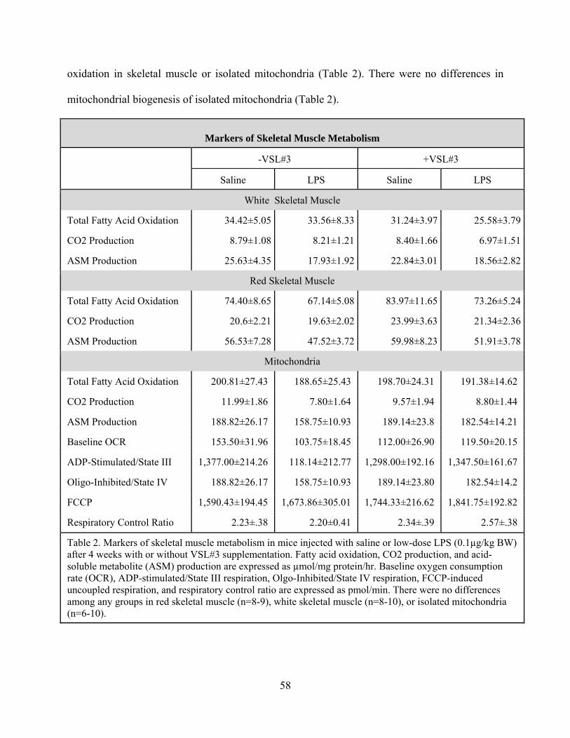
58
oxidation in skeletal muscle or isolated mitochondria (Table 2). There were no differences in
mitochondrial biogenesis of isolated mitochondria (Table 2).
Markers of Skeletal Muscle Metabolism
-VSL#3 +VSL#3
Saline LPS Saline LPS
White Skeletal Muscle
Total Fatty Acid Oxidation 34.42±5.05 33.56±8.33 31.24±3.97 25.58±3.79
CO2 Production 8.79±1.08 8.21±1.21 8.40±1.66 6.97±1.51
ASM Production 25.63±4.35 17.93±1.92 22.84±3.01 18.56±2.82
Red Skeletal Muscle
Total Fatty Acid Oxidation 74.40±8.65 67.14±5.08 83.97±11.65 73.26±5.24
CO2 Production 20.6±2.21 19.63±2.02 23.99±3.63 21.34±2.36
ASM Production 56.53±7.28 47.52±3.72 59.98±8.23 51.91±3.78
Mitochondria
Total Fatty Acid Oxidation 200.81±27.43 188.65±25.43 198.70±24.31 191.38±14.62
CO2 Production 11.99±1.86 7.80±1.64 9.57±1.94 8.80±1.44
ASM Production 188.82±26.17 158.75±10.93 189.14±23.8 182.54±14.21
Baseline OCR 153.50±31.96 103.75±18.45 112.00±26.90 119.50±20.15
ADP-Stimulated/State III 1,377.00±214.26 118.14±212.77 1,298.00±192.16 1,347.50±161.67
Oligo-Inhibited/State IV 188.82±26.17 158.75±10.93 189.14±23.80 182.54±14.2
FCCP 1,590.43±194.45 1,673.86±305.01 1,744.33±216.62 1,841.75±192.82
Respiratory Control Ratio 2.23±.38 2.20±0.41 2.34±.39 2.57±.38
Table 2. Markers of skeletal muscle metabolism in mice injected with saline or low-dose LPS (0.1µg/kg BW) after 4 weeks with or without VSL#3 supplementation. Fatty acid oxidation, CO2 production, and acid-soluble metabolite (ASM) production are expressed as µmol/mg protein/hr. Baseline oxygen consumption rate (OCR), ADP-stimulated/State III respiration, Olgo-Inhibited/State IV respiration, FCCP-induced uncoupled respiration, and respiratory control ratio are expressed as pmol/min. There were no differences among any groups in red skeletal muscle (n=8-9), white skeletal muscle (n=8-10), or isolated mitochondria (n=6-10).
59
Markers of Inflammation in White Skeletal Muscle. Expression levels of TNFα were significantly
reduced in saline+VSL#3 mice and LPS-VSL#3 mice compared to saline-VSL#3 mice; levels in
LPS+VSL#3 mice were not significantly different from any group (Figure 6C). There were no
differences in TLR-2 (Figure 6A) or TLR-4 (Figure 6B) mRNA expression levels.

60
61
Markers of Inflammation in Red Skeletal Muscle. Expression of TLR-2 was significantly
different in saline-VSL#3 mice compared to saline+VSL#3, but in mice that received an LPS
injection these levels did not differ from any group (Figure 7A). TLR-4 expression levels were
significantly lower in saline+VSL#3 mice and LPS-VSL#3 mice compared to saline-VSL#3
mice (Figure 7B). Expression levels of TLR-4 in mice that received both LPS and VSL#3
supplementation were not significantly different from any group. There were no changes in
TNFα expression (Figure 7C).

62
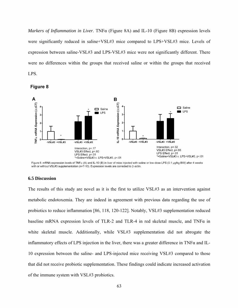
63
Markers of Inflammation in Liver. TNFα (Figure 8A) and IL-10 (Figure 8B) expression levels
were significantly reduced in saline+VSL#3 mice compared to LPS+VSL#3 mice. Levels of
expression between saline-VSL#3 and LPS-VSL#3 mice were not significantly different. There
were no differences within the groups that received saline or within the groups that received
LPS.
6.5 Discussion
The results of this study are novel as it is the first to utilize VSL#3 as an intervention against
metabolic endotoxemia. They are indeed in agreement with previous data regarding the use of
probiotics to reduce inflammation [86, 118, 120-122]. Notably, VSL#3 supplementation reduced
baseline mRNA expression levels of TLR-2 and TLR-4 in red skeletal muscle, and TNFα in
white skeletal muscle. Additionally, while VSL#3 supplementation did not abrogate the
inflammatory effects of LPS injection in the liver, there was a greater difference in TNFα and IL-
10 expression between the saline- and LPS-injected mice receiving VSL#3 compared to those
that did not receive probiotic supplementation. These findings could indicate increased activation
of the immune system with VSL#3 probiotics.
64
VSL#3 supplementation resulted in significantly reduced mRNA expression levels of TLR-2 and
TLR-4 in red muscle and TNFα in the white muscle of saline-injected mice. Interestingly, in
mice not receiving VSL#3 supplementation, a low-dose LPS injection did not increase mRNA
expression of TLR-4 or TNFα compared to saline-injected mice given VSL#3 supplementation.
Given our low dosage and time course, it is difficult to ascertain whether this could be due LPS-
induced downregulation of these receptors or a latent decrease in mRNA expression in response
to elevated protein levels. Other studies have shown that various forms of LPS administration
can reduce TLR-4 protein levels in adipose tissue while eliciting only minor changes in TLR-2
or TLR-4 in skeletal muscle [47, 123]. Regardless, it can be extrapolated that VSL#3 probiotic
supplementation may have similar effects on the immune system as an acute low-dose LPS
challenge, which may be beneficial. Studies have shown that cells treated with low doses of LPS
(1µg/mL) can become endotoxin resistant, leading to reduced inflammatory responses to
subsequent LPS challenges [124]. Alternatively, the lipoteichoic acid or peptidoglycan found on
the gram-positive bacteria in VSL#3 could have entered circulation and activated TLR-2,
causing release of TNFα and NFκB in a similar fashion to TLR-4 after LPS treatment [125]. By
activating the immune system, VSL#3 supplementation could reduce the inflammatory response
to metabolic endotoxemia.
The liver is a main site of LPS clearance [126]. There, it is taken up primarily by Kupffer cells
which release inflammatory cytokines such as TNFα and interleukins. Our results illustrate that
the low-dose LPS injection resulted in a significant increase in TNFα as well as the anti-
inflammatory cytokine IL-10 which is activated in Kupffer cells to abrogate inflammatory
responses [127]. However, in contrast to previous studies, VSL#3 supplementation did not
reduce LPS-induced liver inflammation. Interestingly, mice who received VSL#3

65
supplementation exhibited a greater difference in TNFα and IL-10 mRNA expression between
saline and LPS conditions compared to mice who did not receive the probiotic. This supports our
theory that basal levels of inflammation may have been lower with VSL#3 supplementation. The
elevated inflammatory response in the liver also indicates that LPS may have been cleared by the
four-hour time point at which the tissues were collected, which may explain the lack of LPS
effect in skeletal muscle metabolism and mitochondrial biogenesis.
The 4-hour low-dose LPS treatment elicited no changes in skeletal muscle metabolism or
mitochondrial biogenesis. Previous studies have shown that a low-dose LPS challenge reduced
fatty acid uptake and oxidation in both cell culture and animal models [5, 128]. This is a
limitation of the current study because it indicates that the LPS dose may not have been
sufficient to induce metabolic dysregulation, therefore we could not observe VSL#3 as a
potential protective intervention. Similarly, previous studies have shown increased mitochondrial
biogenesis after a high dose of LPS, but we did not observe this, perhaps due to the low dosage
administered [129, 130]. The lack of metabolic effects indicates that the model may have better
represented the acute endotoxemia that may follow a high-fat meal rather than the chronic
metabolic endotoxemia linked to insulin resistance and obesity [24, 41].
The original purpose of this study was to determine whether VSL#3 supplementation could
protect against the deleterious effects of an LPS challenge modelling metabolic endotoxemia
during high-fat feeding. However, due to the lack of diet effect in any parameter, the diet factor
was removed in order to simplify the model. Therefore, the study examined instead the potential
protective effects of VSL#3 against an acute, low-dose LPS challenge modelling acute metabolic
endotoxemia. Our results showed that VSL#3 supplementation may reduce basal inflammatory
tone in skeletal muscle, which could protect against metabolic dysregulation associated with

66
chronic inflammation caused by metabolic endotoxemia.
67
CHAPTER VI: IMPLICATIONS & FUTURE DIRECTIONS
High-fat diets and obesity have been linked to metabolic endotoxemia and metabolic
dysregulation characterized by the inability of skeletal muscle to utilize appropriate substrate [5,
24]. Rates of obesity have reached epidemic proportions while obesity-related medical costs are
estimated to reach over $209 billion annually [131]. The gut microbiome has been shown to
influence fatty acid uptake, oxidation, and synthesis, as well as adipogenesis and lipogenesis [11,
65, 87, 99]. In both humans and mice, an obesogenic bacterial profile has been identified, and the
term ‘dysbiosis’ now refers to the overabundance of non-beneficial bacteria in conjunction with
a loss of bacterial variety and a predisposition to increased energy harvesting from the diet [4].
Recent discoveries showing the link between high-fat diets, obesity and the gut microbiome have
led to new studies examining the potential role of probiotics in preventing or treating diet-
induced obesity. Probiotic supplementation is a non-invasive, dietary intervention that has been
safely used in children and adults with some promising results.
The current study tested whether supplementation with VSL#3, a multi-strain probiotic, would
prevent inflammation, metabolic inflexibility, and loss of intestinal tight junction proteins during
high-fat feeding or metabolic endotoxemia. This was the first study to utilize VSL#3 as a dietary
intervention during high-fat feeding or metabolic endotoxemia, so the results are novel, but they
are largely in agreement with previous studies utilizing single or double probiotic strains during
high-fat feeding. Overall, these results demonstrate that VSL#3 supplementation 1) significantly
lowered fasting blood glucose, 2) increased FIAF and decreased GLP-2 expression during high-
fat feeding, and 3) reduced basal expression levels of TLR-2, TLR-4 and TNFα in skeletal
muscle. VSL#3 supplementation was not shown to protect against an increase in percent body fat
during high-fat feeding or LPS-induced inflammation in skeletal muscle or liver. In this model, 4
68
weeks of high-fat feeding did not induce metabolic inflexibility in skeletal muscle, so potential
protection through VSL#3 supplementation could not be observed.
Because of the intervention design of this study, mechanisms of influence were examined in
breadth but not depth, opening a great number of avenues for future directions. The major
limitation of this study was due to the short feeding protocol, so future studies aiming to
intervene in a model of high-fat diet-induced metabolic dysregulation should extend their
protocol until metabolic dysregulation is observed through glucose or insulin tolerance tests. In
addition, future studies aiming to utilize VSL#3 in models of metabolic endotoxemia should
measure plasma endotoxin and markers of inflammation in living mice at regular intervals post-
injection to determine peak inflammatory response and LPS clearance rates. This would ensure a
model of metabolic endotoxemia rather than a latent inflammatory response.
Additionally, mechanistic studies must be performed to determine which pathways are being
affected by VSL#3 supplementation. A decrease in 12-hour fasting blood glucose indicates that
supplementation has chronic effects, perhaps at the level of the liver where glycogenolysis and
gluconeogenesis take place during a fasted state to maintain euglycemia. Recently, Kim, et al.
illustrated decreased expression of genes that regulate gluconeogenesis in addition to increased
GLUT4 expression after supplementation with a single-strain probiotic [65]. Future studies
should measure changes in these regulatory genes after supplementation with VSL#3 to
determine mechanistic action. In the current study, VSL#3 supplementation increased FIAF
mRNA expression and decreased GLP-2 receptor mRNA expression, findings that are
complementary to other studies that have shown an increase in circulating FIAF and GLP-2 [60,
87]. Future studies using VSL#3 supplementation should quantify the protein levels of these
genes to determine their activity. Similarly, mRNA expression levels of TLR-2, TLR-4 and

69
TNFa were reduced, but this may or may not translate to reductions in protein levels, and that in
turn does not necessarily translate to the inflammatory state. A previous study by Raso, et al.
illustrated a simultaneous decrease in mRNA expression and protein levels of TLR-4 after
intervention with a single-strain probiotic and prebiotic fibers [86]. In order to better determine
the inflammatory state, future studies to measure levels of membrane-bound TLR-2 and TLR-4
and nuclear TNFα.
VSL#3 has been safely used in many clinical trials to treat inflammation during inflammatory
bowel diseases and steatotic liver diseases. Here, we have shown that VSL#3 supplementation
can also reduce fasting hyperglycemia and insulin resistance during high-fat feeding while
enhancing nutrient sensing in the gut in a murine model. Given the limited but compelling data
showing the anti-inflammatory and protective mechanisms of VSL#3 during high-fat feeding,
future studies should explore its mechanisms and potential influence in preventing diet-induced
obesity and its co-morbidities. VSL#3 supplementation could serve as a non-invasive method for
combating the obesity epidemic.
70
LITERATURE CITED 1. Centers for Disease Control and Prevention DoDT: Obesity and Diabetes. 2009. 2. de Kort S, Keszthelyi D, Masclee AA: Leaky gut and diabetes mellitus: what is the
link? Obes Rev 2011, 12:449-458. 3. Shen J, Obin MS, Zhao L: The gut microbiota, obesity and insulin resistance. Mol
Aspects Med 2013, 34:39-58. 4. Cani PD, Delzenne NM: Interplay between obesity and associated metabolic
disorders: new insights into the gut microbiota. Curr Opin Pharmacol 2009, 9:737-743.
5. Frisard MI, McMillan RP, Marchand J, Wahlberg KA, Wu Y, Voelker KA, Heilbronn L, Haynie K, Muoio B, Li L, Hulver MW: Toll-like receptor 4 modulates skeletal muscle substrate metabolism. Am J Physiol Endocrinol Metab 2010, 298:E988-998.
6. Guo S, Al-Sadi R, Said HM, Ma TY: Lipopolysaccharide Causes an Increase in Intestinal Tight Junction Permeability in Vitro and in Vivo by Inducing Enterocyte Membrane Expression and Localization of TLR-4 and CD14. Am J Pathol 2013, 182:375-387.
7. Cani PD, Delzenne NM: The gut microbiome as therapeutic target. Pharmacol Ther 2011, 130:202-212.
8. Cani PD, Neyrinck AM, Fava F, Knauf C, Burcelin RG, Tuohy KM, Gibson GR, Delzenne NM: Selective increases of bifidobacteria in gut microflora improve high-fat-diet-induced diabetes in mice through a mechanism associated with endotoxaemia. Diabetologia 2007, 50:2374-2383.
9. Das UN: Obesity: genes, brain, gut, and environment. Nutrition 2010, 26:459-473. 10. Matias I, Di Marzo V: Endocannabinoids and the control of energy balance. Trends
Endocrinol Metab 2007, 18:27-37. 11. Muccioli GG, Naslain D, Backhed F, Reigstad CS, Lambert DM, Delzenne NM, Cani
PD: The endocannabinoid system links gut microbiota to adipogenesis. Mol Syst Biol 2010, 6:392.
12. Sherman PM, Ossa JC, Johnson-Henry K: Unraveling mechanisms of action of probiotics. Nutr Clin Pract 2009, 24:10-14.
13. Delzenne NM, Cani PD: Interaction between obesity and the gut microbiota: relevance in nutrition. Annu Rev Nutr 2011, 31:15-31.
14. Leber B, Tripolt NJ, Blattl D, Eder M, Wascher TC, Pieber TR, Stauber R, Sourij H, Oettl K, Stadlbauer V: The influence of probiotic supplementation on gut permeability in patients with metabolic syndrome: an open label, randomized pilot study. Eur J Clin Nutr 2012, 66:1110-1115.
15. Tripolt NJ, Leber B, Blattl D, Eder M, Wonisch W, Scharnagl H, Stojakovic T, Obermayer-Pietsch B, Wascher TC, Pieber TR, et al: Short communication: Effect of supplementation with Lactobacillus casei Shirota on insulin sensitivity, beta-cell function, and markers of endothelial function and inflammation in subjects with metabolic syndrome--a pilot study. J Dairy Sci 2013, 96:89-95.
16. Pham M, Lemberg DA, Day AS: Probiotics: sorting the evidence from the myths. Med J Aust 2008, 188:304-308.
17. Cluny NL, Reimer RA, Sharkey KA: Cannabinoid signalling regulates inflammation and energy balance: The importance of the brain-gut axis. Brain Behav Immun 2012.

71
18. Cani PD, Delzenne NM, Amar J, Burcelin R: Role of gut microflora in the development of obesity and insulin resistance following high-fat diet feeding. Pathol Biol (Paris) 2008, 56:305-309.
19. Stump CS, Henriksen EJ, Wei Y, Sowers JR: The metabolic syndrome: role of skeletal muscle metabolism. Ann Med 2006, 38:389-402.
20. Serino M, Luche E, Gres S, Baylac A, Berge M, Cenac C, Waget A, Klopp P, Iacovoni J, Klopp C, et al: Metabolic adaptation to a high-fat diet is associated with a change in the gut microbiota. Gut 2012, 61:543-553.
21. Diamant M, Blaak EE, de Vos WM: Do nutrient-gut-microbiota interactions play a role in human obesity, insulin resistance and type 2 diabetes? Obes Rev 2011, 12:272-281.
22. Delzenne NM, Cani PD: Gut microbiota and the pathogenesis of insulin resistance. Curr Diab Rep 2011, 11:154-159.
23. Takemura N, Okubo T, Sonoyama K: Lactobacillus plantarum strain No. 14 reduces adipocyte size in mice fed high-fat diet. Exp Biol Med (Maywood) 2010, 235:849-856.
24. Cani PD, Amar J, Iglesias MA, Poggi M, Knauf C, Bastelica D, Neyrinck AM, Fava F, Tuohy KM, Chabo C, et al: Metabolic endotoxemia initiates obesity and insulin resistance. Diabetes 2007, 56:1761-1772.
25. Guo Z, Liu XM, Zhang QX, Shen Z, Tian FW, Zhang H, Sun ZH, Zhang HP, Chen W: Influence of consumption of probiotics on the plasma lipid profile: a meta-analysis of randomised controlled trials. Nutr Metab Cardiovasc Dis 2011, 21:844-850.
26. Sanz Y, Rastmanesh R, Agostonic C: Understanding the role of gut microbes and probiotics in obesity: How far are we? Pharmacol Res 2013, 69:144-155.
27. Mennigen R, Nolte K, Rijcken E, Utech M, Loeffler B, Senninger N, Bruewer M: Probiotic mixture VSL#3 protects the epithelial barrier by maintaining tight junction protein expression and preventing apoptosis in a murine model of colitis. Am J Physiol Gastrointest Liver Physiol 2009, 296:G1140-1149.
28. Delzenne NM, Neyrinck AM, Cani PD: Modulation of the gut microbiota by nutrients with prebiotic properties: consequences for host health in the context of obesity and metabolic syndrome. Microb Cell Fact 2011, 10 Suppl 1:S10.
29. Gao Z, Yin J, Zhang J, Ward RE, Martin RJ, Lefevre M, Cefalu WT, Ye J: Butyrate improves insulin sensitivity and increases energy expenditure in mice. Diabetes 2009, 58:1509-1517.
30. Yu LC, Wang JT, Wei SC, Ni YH: Host-microbial interactions and regulation of intestinal epithelial barrier function: From physiology to pathology. World J Gastrointest Pathophysiol 2012, 3:27-43.
31. Andreasen AS, Kelly M, Berg RM, Moller K, Pedersen BK: Type 2 diabetes is associated with altered NF-kappaB DNA binding activity, JNK phosphorylation, and AMPK phosphorylation in skeletal muscle after LPS. PLoS ONE 2011, 6:e23999.
32. Tsukumo DM, Carvalho BM, Carvalho-Filho MA, Saad MJ: Translational research into gut microbiota: new horizons in obesity treatment. Arq Bras Endocrinol Metabol 2009, 53:139-144.
33. Sanz Y: Gut microbiota and probiotics in maternal and infant health. Am J Clin Nutr 2011, 94:2000S-2005S.
72
34. Salminen S, Isolauri E: Opportunities for improving the health and nutrition of the human infant by probiotics. Nestle Nutr Workshop Ser Pediatr Program 2008, 62:223-233; discussion 233-227.
35. Backhed F, Manchester JK, Semenkovich CF, Gordon JI: Mechanisms underlying the resistance to diet-induced obesity in germ-free mice. Proc Natl Acad Sci U S A 2007, 104:979-984.
36. Cani PD, Delzenne NM: The role of the gut microbiota in energy metabolism and metabolic disease. Curr Pharm Des 2009, 15:1546-1558.
37. Cani PD, Bibiloni R, Knauf C, Waget A, Neyrinck AM, Delzenne NM, Burcelin R: Changes in gut microbiota control metabolic endotoxemia-induced inflammation in high-fat diet-induced obesity and diabetes in mice. Diabetes 2008, 57:1470-1481.
38. Mulligan KX, Morris RT, Otero YF, Wasserman DH, McGuinness OP: Disassociation of muscle insulin signaling and insulin-stimulated glucose uptake during endotoxemia. PLoS ONE 2012, 7:e30160.
39. Rowland KJ, Brubaker PL: The "cryptic" mechanism of action of glucagon-like peptide-2. Am J Physiol Gastrointest Liver Physiol 2011, 301:G1-8.
40. Vreugdenhil AC, Rousseau CH, Hartung T, Greve JW, van 't Veer C, Buurman WA: Lipopolysaccharide (LPS)-binding protein mediates LPS detoxification by chylomicrons. J Immunol 2003, 170:1399-1405.
41. Laugerette F, Vors C, Geloen A, Chauvin MA, Soulage C, Lambert-Porcheron S, Peretti N, Alligier M, Burcelin R, Laville M, et al: Emulsified lipids increase endotoxemia: possible role in early postprandial low-grade inflammation. J Nutr Biochem 2011, 22:53-59.
42. Pendyala S, Walker JM, Holt PR: A high-fat diet is associated with endotoxemia that originates from the gut. Gastroenterology 2012, 142:1100-1101 e1102.
43. Pedersen BK: Muscles and their myokines. J Exp Biol 2011, 214:337-346. 44. Corpeleijn E, Saris WH, Blaak EE: Metabolic flexibility in the development of insulin
resistance and type 2 diabetes: effects of lifestyle. Obes Rev 2009, 10:178-193. 45. Kelley DE, He J, Menshikova EV, Ritov VB: Dysfunction of mitochondria in human
skeletal muscle in type 2 diabetes. Diabetes 2002, 51:2944-2950. 46. Jans A, Sparks LM, van Hees AM, Gjelstad IM, Tierney AC, Riserus U, Drevon CA,
Roche HM, Schrauwen P, Blaak EE: Transcriptional metabolic inflexibility in skeletal muscle among individuals with increasing insulin resistance. Obesity (Silver Spring) 2011, 19:2158-2166.
47. Lang CH, Silvis C, Deshpande N, Nystrom G, Frost RA: Endotoxin stimulates in vivo expression of inflammatory cytokines tumor necrosis factor alpha, interleukin-1beta, -6, and high-mobility-group protein-1 in skeletal muscle. Shock 2003, 19:538-546.
48. McNicol FJ, Hoyland JA, Cooper RG, Carlson GL: Skeletal muscle contractile properties and proinflammatory cytokine gene expression in human endotoxaemia. Br J Surg 2010, 97:434-442.
49. Kelly CJ, Colgan SP, Frank DN: Of microbes and meals: the health consequences of dietary endotoxemia. Nutr Clin Pract 2012, 27:215-225.
50. Bruce CR, Dyck DJ: Cytokine regulation of skeletal muscle fatty acid metabolism: effect of interleukin-6 and tumor necrosis factor-alpha. Am J Physiol Endocrinol Metab 2004, 287:E616-621.
73
51. Hornef MW, Frisan T, Vandewalle A, Normark S, Richter-Dahlfors A: Toll-like receptor 4 resides in the Golgi apparatus and colocalizes with internalized lipopolysaccharide in intestinal epithelial cells. J Exp Med 2002, 195:559-570.
52. Hornef MW, Normark BH, Vandewalle A, Normark S: Intracellular recognition of lipopolysaccharide by toll-like receptor 4 in intestinal epithelial cells. J Exp Med 2003, 198:1225-1235.
53. Abreu MT: Toll-like receptor signalling in the intestinal epithelium: how bacterial recognition shapes intestinal function. Nat Rev Immunol 2010, 10:131-144.
54. Rakoff-Nahoum S, Paglino J, Eslami-Varzaneh F, Edberg S, Medzhitov R: Recognition of commensal microflora by toll-like receptors is required for intestinal homeostasis. Cell 2004, 118:229-241.
55. Gummesson A, Carlsson LM, Storlien LH, Backhed F, Lundin P, Lofgren L, Stenlof K, Lam YY, Fagerberg B, Carlsson B: Intestinal permeability is associated with visceral adiposity in healthy women. Obesity (Silver Spring) 2011, 19:2280-2282.
56. Backhed F, Ley RE, Sonnenburg JL, Peterson DA, Gordon JI: Host-bacterial mutualism in the human intestine. Science 2005, 307:1915-1920.
57. Regulatory oversight and safety of probiotic use. Emerg Infect Dis [serial on the Internet]. [ http://wwwnc.cdc.gov/eid/article/16/11/10-0574.htm]
58. Rupa P, Mine Y: Recent Advances in the Role of Probiotics in Human Inflammation and Gut Health. J Agric Food Chem 2012.
59. Mack DR: Probiotics in inflammatory bowel diseases and associated conditions. Nutrients 2011, 3:245-264.
60. Cani PD, Possemiers S, Van de Wiele T, Guiot Y, Everard A, Rottier O, Geurts L, Naslain D, Neyrinck A, Lambert DM, et al: Changes in gut microbiota control inflammation in obese mice through a mechanism involving GLP-2-driven improvement of gut permeability. Gut 2009, 58:1091-1103.
61. Estall JL, Yusta B, Drucker DJ: Lipid raft-dependent glucagon-like peptide-2 receptor trafficking occurs independently of agonist-induced desensitization. Mol Biol Cell 2004, 15:3673-3687.
62. Kadooka Y, Sato M, Imaizumi K, Ogawa A, Ikuyama K, Akai Y, Okano M, Kagoshima M, Tsuchida T: Regulation of abdominal adiposity by probiotics (Lactobacillus gasseri SBT2055) in adults with obese tendencies in a randomized controlled trial. Eur J Clin Nutr 2010, 64:636-643.
63. Gobel RJ, Larsen N, Jakobsen M, Molgaard C, Michaelsen KF: Probiotics to adolescents with obesity: effects on inflammation and metabolic syndrome. J Pediatr Gastroenterol Nutr 2012, 55:673-678.
64. Andreasen AS, Larsen N, Pedersen-Skovsgaard T, Berg RM, Moller K, Svendsen KD, Jakobsen M, Pedersen BK: Effects of Lactobacillus acidophilus NCFM on insulin sensitivity and the systemic inflammatory response in human subjects. Br J Nutr 2010, 104:1831-1838.
65. Kim SW, Park KY, Kim B, Kim E, Hyun CK: Lactobacillus rhamnosus GG improves insulin sensitivity and reduces adiposity in high-fat diet-fed mice through enhancement of adiponectin production. Biochem Biophys Res Commun 2013, 431:258-263.
74
66. Chen JJ, Wang R, Li XF, Wang RL: Bifidobacterium longum supplementation improved high-fat-fed-induced metabolic syndrome and promoted intestinal Reg I gene expression. Exp Biol Med (Maywood) 2011, 236:823-831.
67. Naito E, Yoshida Y, Makino K, Kounoshi Y, Kunihiro S, Takahashi R, Matsuzaki T, Miyazaki K, Ishikawa F: Beneficial effect of oral administration of Lactobacillus casei strain Shirota on insulin resistance in diet-induced obesity mice. J Appl Microbiol 2011, 110:650-657.
68. Ma X, Hua J, Li Z: Probiotics improve high fat diet-induced hepatic steatosis and insulin resistance by increasing hepatic NKT cells. J Hepatol 2008, 49:821-830.
69. Shao B, Munford RS, Kitchens R, Varley AW: Hepatic uptake and deacylation of the LPS in bloodborne LPS-lipoprotein complexes. Innate Immun 2012, 18:825-833.
70. Spitzer AL, Chuang KI, Victorino GP, Kasravi B, Curran B, Lee D, Harris HW: Chylomicrons combined with endotoxin moderate microvascular permeability. Innate Immun 2011, 17:283-292.
71. Newberry EP, Davidson NO: Intestinal lipid absorption, GLP-2, and CD36: still more mysteries to moving fat. Gastroenterology 2009, 137:775-778.
72. Erridge C: Oxidized phospholipid inhibition of LPS-signaling: a good side to the bad guys? Arterioscler Thromb Vasc Biol 2009, 29:337-338.
73. von Schlieffen E, Oskolkova OV, Schabbauer G, Gruber F, Bluml S, Genest M, Kadl A, Marsik C, Knapp S, Chow J, et al: Multi-hit inhibition of circulating and cell-associated components of the toll-like receptor 4 pathway by oxidized phospholipids. Arterioscler Thromb Vasc Biol 2009, 29:356-362.
74. Lozupone C, Faust K, Raes J, Faith JJ, Frank DN, Zaneveld J, Gordon JI, Knight R: Identifying genomic and metabolic features that can underlie early successional and opportunistic lifestyles of human gut symbionts. Genome Res 2012, 22:1974-1984.
75. Groen A, Kunne C, Oude Elferink RP: Increased serum concentrations of secondary bile salts during cholate feeding are due to coprophagy. A study with wild-type and Atp8b1-deficient mice. Mol Pharm 2006, 3:756-761.
76. McFall-Ngai M: Love the one you're with: vertebrate guts shape their microbiota. Cell 2006, 127:247-249.
77. Schluter J, Foster KR: The evolution of mutualism in gut microbiota via host epithelial selection. PLoS Biol 2012, 10:e1001424.
78. Podrini C, Cambridge EL, Lelliott CJ, Carragher DM, Estabel J, Gerdin AK, Karp NA, Scudamore CL, Sanger Mouse Genetics P, Ramirez-Solis R, White JK: High-fat feeding rapidly induces obesity and lipid derangements in C57BL/6N mice. Mamm Genome 2013, 24:240-251.
79. Tagliabue A, Elli M: The role of gut microbiota in human obesity: recent findings and future perspectives. Nutr Metab Cardiovasc Dis 2013, 23:160-168.
80. Moreira AP, Texeira TF, Ferreira AB, Peluzio Mdo C, Alfenas Rde C: Influence of a high-fat diet on gut microbiota, intestinal permeability and metabolic endotoxaemia. Br J Nutr 2012, 108:801-809.
81. Carvalho BM, Guadagnini D, Tsukumo DM, Schenka AA, Latuf-Filho P, Vassallo J, Dias JC, Kubota LT, Carvalheira JB, Saad MJ: Modulation of gut microbiota by antibiotics improves insulin signalling in high-fat fed mice. Diabetologia 2012, 55:2823-2834.

75
82. Cani PD, Delzenne NM: Involvement of the gut microbiota in the development of low grade inflammation associated with obesity: focus on this neglected partner. Acta Gastroenterol Belg 2010, 73:267-269.
83. Lee SJ, Bose S, Seo JG, Chung WS, Lim CY, Kim H: The effects of co-administration of probiotics with herbal medicine on obesity, metabolic endotoxemia and dysbiosis: A randomized double-blind controlled clinical trial. Clin Nutr 2013.
84. Asemi Z, Samimi M, Tabassi Z, Naghibi Rad M, Rahimi Foroushani A, Khorammian H, Esmaillzadeh A: Effect of daily consumption of probiotic yoghurt on insulin resistance in pregnant women: a randomized controlled trial. Eur J Clin Nutr 2013, 67:71-74.
85. Shen J, Obin MS, Zhao L: The gut microbiota, obesity and insulin resistance. Mol Aspects Med 2012.
86. Raso GM, Simeoli R, Iacono A, Santoro A, Amero P, Paciello O, Russo R, D'Agostino G, Di Costanzo M, Canani RB, et al: Effects of a Lactobacillus paracasei B21060 based synbiotic on steatosis, insulin signaling and toll-like receptor expression in rats fed a high-fat diet. J Nutr Biochem 2014, 25:81-90.
87. Aronsson L, Huang Y, Parini P, Korach-Andre M, Hakansson J, Gustafsson JA, Pettersson S, Arulampalam V, Rafter J: Decreased fat storage by Lactobacillus paracasei is associated with increased levels of angiopoietin-like 4 protein (ANGPTL4). PLoS ONE 2010, 5.
88. Bomhof MR, Saha DC, Reid DT, Paul HA, Reimer RA: Combined effects of oligofructose and Bifidobacterium animalis on gut microbiota and glycemia in obese rats. Obesity (Silver Spring) 2014, 22:763-771.
89. Cary VA, Boullata J: What is the evidence for the use of probiotics in the treatment of inflammatory bowel disease? J Clin Nurs 2010, 19:904-916.
90. Camilleri M: Probiotics and irritable bowel syndrome: rationale, mechanisms, and efficacy. J Clin Gastroenterol 2008, 42 Suppl 3 Pt 1:S123-125.
91. Esposito E, Iacono A, Bianco G, Autore G, Cuzzocrea S, Vajro P, Canani RB, Calignano A, Raso GM, Meli R: Probiotics reduce the inflammatory response induced by a high-fat diet in the liver of young rats. J Nutr 2009, 139:905-911.
92. Li Z, Yang S, Lin H, Huang J, Watkins PA, Moser AB, Desimone C, Song XY, Diehl AM: Probiotics and antibodies to TNF inhibit inflammatory activity and improve nonalcoholic fatty liver disease. Hepatology 2003, 37:343-350.
93. Ma YY, Li L, Yu CH, Shen Z, Chen LH, Li YM: Effects of probiotics on nonalcoholic fatty liver disease: a meta-analysis. World J Gastroenterol 2013, 19:6911-6918.
94. Dorenkott MR, Griffin LE, Goodrich KM, Thompson-Witrick KA, Fundaro G, Ye L, Stevens JR, Ali M, O'Keefe SF, Hulver MW, Neilson AP: Oligomeric cocoa procyanidins possess enhanced bioactivity compared to monomeric and polymeric cocoa procyanidins for preventing the development of obesity, insulin resistance, and impaired glucose tolerance during high-fat feeding. J Agric Food Chem 2014, 62:2216-2227.
95. Jiao Y, George SK, Zhao Q, Hulver MW, Hutson SM, Bishop CE, Lu B: Mex3c mutation reduces adiposity and increases energy expenditure. Mol Cell Biol 2012, 32:4350-4362.
96. Frezza C, Cipolat S, Scorrano L: Organelle isolation: functional mitochondria from mouse liver, muscle and cultured fibroblasts. Nat Protoc 2007, 2:287-295.
76
97. Andersson U, Branning C, Ahrne S, Molin G, Alenfall J, Onning G, Nyman M, Holm C: Probiotics lower plasma glucose in the high-fat fed C57BL/6J mouse. Benef Microbes 2010, 1:189-196.
98. Dijk W, Kersten S: Regulation of lipoprotein lipase by Angptl4. Trends Endocrinol Metab 2014, 25:146-155.
99. Backhed F, Ding H, Wang T, Hooper LV, Koh GY, Nagy A, Semenkovich CF, Gordon JI: The gut microbiota as an environmental factor that regulates fat storage. Proc Natl Acad Sci U S A 2004, 101:15718-15723.
100. Yin J, Peng Y, Wu J, Wang Y, Yao L: Toll-like receptor 2/4 links to free fatty acid-induced inflammation and beta-cell dysfunction. J Leukoc Biol 2014, 95:47-52.
101. Ukena SN, Singh A, Dringenberg U, Engelhardt R, Seidler U, Hansen W, Bleich A, Bruder D, Franzke A, Rogler G, et al: Probiotic Escherichia coli Nissle 1917 inhibits leaky gut by enhancing mucosal integrity. PLoS ONE 2007, 2:e1308.
102. Erridge C, Attina T, Spickett CM, Webb DJ: A high-fat meal induces low-grade endotoxemia: evidence of a novel mechanism of postprandial inflammation. Am J Clin Nutr 2007, 86:1286-1292.
103. Schumann RR: Old and new findings on lipopolysaccharide-binding protein: a soluble pattern-recognition molecule. Biochem Soc Trans 2011, 39:989-993.
104. Gegner JA, Ulevitch RJ, Tobias PS: Lipopolysaccharide (LPS) signal transduction and clearance. Dual roles for LPS binding protein and membrane CD14. J Biol Chem 1995, 270:5320-5325.
105. Mullinax TR, Mock JN, McEvily AJ, Harrison JH: Regulation of mitochondrial malate dehydrogenase. Evidence for an allosteric citrate-binding site. J Biol Chem 1982, 257:13233-13239.
106. Nemeth PM, Rosser BW, Choksi RM, Norris BJ, Baker KM: Metabolic response to a high-fat diet in neonatal and adult rat muscle. Am J Physiol 1992, 262:C282-286.
107. Hildebrandt MA, Hoffmann C, Sherrill-Mix SA, Keilbaugh SA, Hamady M, Chen YY, Knight R, Ahima RS, Bushman F, Wu GD: High-fat diet determines the composition of the murine gut microbiome independently of obesity. Gastroenterology 2009, 137:1716-1724 e1711-1712.
108. Kuhbacher T, Ott SJ, Helwig U, Mimura T, Rizzello F, Kleessen B, Gionchetti P, Blaut M, Campieri M, Folsch UR, et al: Bacterial and fungal microbiota in relation to probiotic therapy (VSL#3) in pouchitis. Gut 2006, 55:833-841.
109. Appleyard CB, Cruz ML, Isidro AA, Arthur JC, Jobin C, De Simone C: Pretreatment with the probiotic VSL#3 delays transition from inflammation to dysplasia in a rat model of colitis-associated cancer. Am J Physiol Gastrointest Liver Physiol 2011, 301:G1004-1013.
110. Sekirov I, Russell SL, Antunes LC, Finlay BB: Gut microbiota in health and disease. Physiol Rev 2010, 90:859-904.
111. Remels AH, Gosker HR, Bakker J, Guttridge DC, Schols AM, Langen RC: Regulation of skeletal muscle oxidative phenotype by classical NF-kappaB signalling. Biochim Biophys Acta 2013, 1832:1313-1325.
112. Steinberg GR: Inflammation in obesity is the common link between defects in fatty acid metabolism and insulin resistance. Cell Cycle 2007, 6:888-894.
77
113. Liang H, Hussey SE, Sanchez-Avila A, Tantiwong P, Musi N: Effect of lipopolysaccharide on inflammation and insulin action in human muscle. PLoS ONE 2013, 8:e63983.
114. Lassenius MI, Pietilainen KH, Kaartinen K, Pussinen PJ, Syrjanen J, Forsblom C, Porsti I, Rissanen A, Kaprio J, Mustonen J, et al: Bacterial endotoxin activity in human serum is associated with dyslipidemia, insulin resistance, obesity, and chronic inflammation. Diabetes Care 2011, 34:1809-1815.
115. Soares JB, Pimentel-Nunes P, Roncon-Albuquerque R, Leite-Moreira A: The role of lipopolysaccharide/toll-like receptor 4 signaling in chronic liver diseases. Hepatol Int 2010, 4:659-672.
116. Asemi Z, Jazayeri S, Najafi M, Samimi M, Mofid V, Shidfar F, Foroushani AR, Shahaboddin ME: Effects of daily consumption of probiotic yoghurt on inflammatory factors in pregnant women: a randomized controlled trial. Pak J Biol Sci 2011, 14:476-482.
117. Chen CC, Kong MS, Lai MW, Chao HC, Chang KW, Chen SY, Huang YC, Chiu CH, Li WC, Lin PY, et al: Probiotics have clinical, microbiologic, and immunologic efficacy in acute infectious diarrhea. Pediatr Infect Dis J 2010, 29:135-138.
118. Calcinaro F, Dionisi S, Marinaro M, Candeloro P, Bonato V, Marzotti S, Corneli RB, Ferretti E, Gulino A, Grasso F, et al: Oral probiotic administration induces interleukin-10 production and prevents spontaneous autoimmune diabetes in the non-obese diabetic mouse. Diabetologia 2005, 48:1565-1575.
119. Camilleri M: Probiotics and irritable bowel syndrome: rationale, putative mechanisms, and evidence of clinical efficacy. J Clin Gastroenterol 2006, 40:264-269.
120. Rishi P, Bharrhan S, Singh G, Kaur IP: Effect of Lactobacillus plantarum and L-arginine against endotoxin-induced liver injury in a rat model. Life Sci 2011, 89:847-853.
121. Loguercio C, Federico A, Tuccillo C, Terracciano F, D'Auria MV, De Simone C, Del Vecchio Blanco C: Beneficial effects of a probiotic VSL#3 on parameters of liver dysfunction in chronic liver diseases. J Clin Gastroenterol 2005, 39:540-543.
122. Wang Y, Li Y, Xie J, Zhang Y, Wang J, Sun X, Zhang H: Protective effects of probiotic Lactobacillus casei Zhang against endotoxin- and d-galactosamine-induced liver injury in rats via anti-oxidative and anti-inflammatory capacities. Int Immunopharmacol 2013, 15:30-37.
123. Gabler NK, Spencer JD, Webel DM, Spurlock ME: n-3 PUFA attenuate lipopolysaccharide-induced down-regulation of toll-like receptor 4 expression in porcine adipose tissue but does not alter the expression of other immune modulators. J Nutr Biochem 2008, 19:8-15.
124. Morris M, Li L: Molecular mechanisms and pathological consequences of endotoxin tolerance and priming. Arch Immunol Ther Exp (Warsz) 2012, 60:13-18.
125. Schwandner R, Dziarski R, Wesche H, Rothe M, Kirschning CJ: Peptidoglycan- and lipoteichoic acid-induced cell activation is mediated by toll-like receptor 2. J Biol Chem 1999, 274:17406-17409.
126. van Oosten M, van de Bilt E, van Berkel TJ, Kuiper J: New scavenger receptor-like receptors for the binding of lipopolysaccharide to liver endothelial and Kupffer cells. Infect Immun 1998, 66:5107-5112.
78
127. Kawaratani H, Tsujimoto T, Douhara A, Takaya H, Moriya K, Namisaki T, Noguchi R, Yoshiji H, Fujimoto M, Fukui H: The effect of inflammatory cytokines in alcoholic liver disease. Mediators Inflamm 2013, 2013:495156.
128. Memon RA, Fuller J, Moser AH, Smith PJ, Feingold KR, Grunfeld C: In vivo regulation of acyl-CoA synthetase mRNA and activity by endotoxin and cytokines. Am J Physiol 1998, 275:E64-72.
129. Hickson-Bick DL, Jones C, Buja LM: Stimulation of mitochondrial biogenesis and autophagy by lipopolysaccharide in the neonatal rat cardiomyocyte protects against programmed cell death. J Mol Cell Cardiol 2008, 44:411-418.
130. Suliman HB, Welty-Wolf KE, Carraway M, Tatro L, Piantadosi CA: Lipopolysaccharide induces oxidative cardiac mitochondrial damage and biogenesis. Cardiovasc Res 2004, 64:279-288.
131. Apovian CM: The clinical and economic consequences of obesity. Am J Manag Care 2013, 19:s219-228.