dna supercoiling, a critical signal regulating the basal
TRANSCRIPT

DNA supercoiling, a critical signal regulating thebasal expression of the lac operon in EscherichiacoliGeraldine Fulcrand, Florida International UniversitySamantha Dages, Florida International UniversityXiaoduo Zhi, Florida International UniversityPrem Chapagain, Florida International UniversityBernard S. Gerstman, Florida International UniversityDavid Dunlap, Emory UniversityFenfei Leng, Florida International University
Journal Title: Scientific ReportsVolume: Volume 6Publisher: Nature Publishing Group | 2016-01-14, Pages 19243-19243Type of Work: Article | Final Publisher PDFPublisher DOI: 10.1038/srep19243Permanent URL: https://pid.emory.edu/ark:/25593/rjw8n
Final published version: http://dx.doi.org/10.1038/srep19243
Copyright information:This is an Open Access work distributed under the terms of the CreativeCommons Attribution 4.0 International License(http://creativecommons.org/licenses/by/4.0/).
Accessed October 22, 2021 8:26 PM EDT

1Scientific RepoRts | 6:19243 | DOI: 10.1038/srep19243
www.nature.com/scientificreports
DNA supercoiling, a critical signal regulating the basal expression of the lac operon in Escherichia coliGeraldine Fulcrand1,2, Samantha Dages1,2, Xiaoduo Zhi1,2, Prem Chapagain1,3, Bernard S. Gerstman1,3, David Dunlap4 & Fenfei Leng1,2
Escherichia coli lac repressor (LacI) is a paradigmatic transcriptional factor that controls the expression of lacZYA in the lac operon. This tetrameric protein specifically binds to the O1, O2 and O3 operators of the lac operon and forms a DNA loop to repress transcription from the adjacent lac promoter. In this article, we demonstrate that upon binding to the O1 and O2 operators at their native positions LacI constrains three (−) supercoils within the 401-bp DNA loop of the lac promoter and forms a topological barrier. The stability of LacI-mediated DNA topological barriers is directly proportional to its DNA binding affinity. However, we find that DNA supercoiling modulates the basal expression from the lac operon in E. coli. Our results are consistent with the hypothesis that LacI functions as a topological barrier to constrain free, unconstrained (−) supercoils within the 401-bp DNA loop of the lac promoter. These constrained (−) supercoils enhance LacI’s DNA-binding affinity and thereby the repression of the promoter. Thus, LacI binding is superhelically modulated to control the expression of lacZYA in the lac operon under varying growth conditions.
The Escherichia coli lac operon is a paradigm for transcriptional regulation in prokaryotes1,2. The primary reg-ulator is the lac repressor (LacI) that specifically binds to operators at the lac promoter region3 and inhibits transcription from the lac promoter in the absence of an inducer4. There are three operators: the primary O1 operator located 11 bp downstream of the transcription-starting site from the lac promoter1,4 and two auxiliary O2 and O3 operators located at 412 and − 82 bp, respectively, of the promoter region4. Since only about ten copies of LacI tetramer are simultaneously present in each E. coli cell5, the function of O2 and O3 is to increase the local concentration of LacI around the lac promoter6 and therefore increase the efficiency of repression4,7. The sec-ond regulator is the cAMP-CRP complex (CRP refers to cAMP receptor protein) that binds to the CRP binding site centered at position − 61.5 bp upstream from the transcription-starting site of the lac promoter8. Binding of cAMP-CRP complex to its binding site at the lac promoter strongly activates transcription initiation from the E. coli lac promoter9. The deletion of crp from the chromosome or the CRP-binding site from the lac promoter greatly reduced the promoter strength in the presence of an inducer10,11. This dual control of the lac promoter by LacI and CRP ensures the maximal repression in the absence of an inducer12. However, when glucose level is low and lactose level is high, the operon is fully activated13,14.
In Escherichia coli, DNA is typically (−) supercoiled15–17. In fact, DNA supercoiling plays critical roles in several essential DNA metabolic pathways, such as DNA replication, transcription, and recombination15–17. DNA supercoiling is also involved in the regulation of the lac operon. For instance, it has been demonstrated that negative supercoiling enhances the binding of LacI to lac operators18,19 and promotes LacI-mediated DNA loop-ing20–22. Additionally, LacI induces supercoiling within the LacI-lac O1 complexes and retains certain superhelical energy in the complexes as well18,23,24. Recently, we showed that binding of LacI tetramers to tandem copies of lac O1 operators in different locations of a DNA molecule separated a supercoiled DNA molecule into two topologi-cally independent, looped domains25. These supercoiled domains are highly stable and supercoils diffuse through the LacI loop closure with a half-life of 112 min25. A topological barrier model in which nucleoprotein complexes confine DNA supercoils to localized regions is consistent with these results25. These results also led us to pose the following questions: Is LacI able to form topological barriers and constrain supercoils in the lac promoter region
1Biomolecular Sciences Institute, Florida International University, Miami, FL 33199. 2Department of Chemistry & Biochemistry, Florida International University, Miami, FL 33199. 3Department of Physics, Florida International University, Miami, FL 33199. 4Department of Physics, Emory University, Atlanta, GA 30322. Correspondence and requests for materials should be addressed to F.L. (email: [email protected])
received: 12 October 2015
accepted: 10 December 2015
Published: 14 January 2016
OPEN

www.nature.com/scientificreports/
2Scientific RepoRts | 6:19243 | DOI: 10.1038/srep19243
upon binding to the O1, O2, and O3 operators at their native positions? If so, is such trapped superhelicity biolog-ically significant? Here utilizing combined approaches of biochemical assays, bacterial genetics, and atomic force microscopy, we show that upon simultaneously binding the O1 and O2 operators LacI forms a topological barrier that divides a supercoiled plasmid DNA molecule containing the lac promoter region into distinct topological domains. We also demonstrate that DNA supercoiling is an important modulator of the basal level of gene expres-sion from the lac operon and that the LacI-mediated topological barrier plays an essential role in this process.
ResultsThe stability of LacI-mediated DNA topological barriers is correlated with their DNA binding affinity. Our previous results showed that two LacI tetramers bound to the four-lac O1 operators of plasmid pCB115 and formed two highly stable LacI-lac O1 nucleoprotein complexes25. These established a topological barrier and divided the supercoiled DNA molecule into two topological domains, a nicked & relaxed domain and a supercoiled domain25. Using similar approaches (Supplementary Fig. S1), we determined the stability of LacI-mediated topological barriers for plasmids pCB116, pCB108, and pCB109 that contain, respectively, 8, 16, and 32 lac O1 operators in two different locations spaced ~1.2 and 2.9 kb apart (Supplementary Fig. S2). In the absence of IPTG, the multiple LacI-mediated topological barriers have similar t1/2 of ~120 min (Table 1 and Supplementary Fig. S3a–c). The only exception is the DNA topological barrier for plasmid pCB126 with a t1/2 of 0.87 ± 0.33 min that results from binding of one molecule of LacI to two lac O1 operators (Table 1). Intriguingly, even in the presence of IPTG, LacI was able to form multiple LacI-lac O1 complexes and block supercoil diffusion (Table 1 and Supplementary Fig. S3d–g). These results suggest that IPTG is not capable of dissociating LacI from the tandem copies of lac O1 operators, which is consistent with previously published results26. In order to detect and determine the stability of LacI-mediated topological barriers for plasmid pCB126 that has single copies of lac O1 operators (Fig. 1a), significantly more Nt.BbvCI was used, which was able to nick more than 90% of supercoiled pCB126 molecules within 5 seconds (Fig. 1b and Supplementary Fig. S4a,b). In this way, we determined the t1/2 of the topological barrier mediated by one LacI tetramer to be 52 ± 12 sec (0.87 ± 0.33 min) (Fig. 1c,d and Table 1). Although the t1/2 of the LacI-mediated topological barrier on pCB126 is significantly lower than that of the mul-tiple LacI-mediated topological barriers, one LacI tetramer significantly blocked supercoil diffusion, which oth-erwise would be on the order of milliseconds27,28.
Next, we constructed a series of plasmid DNA templates that contain different pairs of O1, O2, O3, and Os operators (Supplementary Fig. S2; Os represents the symmetric lac operator29). We then determined t1/2 val-ues of the LacI-mediated topological barriers for these DNA templates. Our results in Table 2 clearly demon-strat that the stability of the LacI-mediated topological barriers is correlated with the affinity of LacI binding for different lac operators (Supplementary Fig. S4c,d and Supplementary Fig. S5; Supplementary Table S1). For instance, the t1/2 of the LacI-mediated topological barriers upon binding to two Os operators on pOsOs was determined to be 29.1 ± 4.6 min (Supplementary Fig. S5 and Supplementary Table S1). In contrast, the t1/2 of the LacI-mediated topological barrier for two O3 operators on pO3O3 was determined to be only approximately 5.5 ± 1.2 sec. We also determined the t1/2 values of DNA topological barriers generated from binding of LacI mutants30 LacI-Gly58+1, LacI-Gly60+1, LacI-Gly60+2, and LacI-Gly60+3 to the two Os sites on pOsOs. Again, our results showed that the t1/2 values of the LacI-mediated topological barriers scale proportionally with the DNA binding affinities of these LacI mutants (Supplementary Table S2).
LacI forms a DNA topological barrier upon binding to lac operators in the lac operon. We con-structed three unique plasmids harboring the natural lac promoter including either active or mutated O1, O2, and O3 at their native positions (Fig. 2 and Supplementary Fig. S6). We also inserted a nicking endonuclease Nt.BbvCI recognition site between the O1 and O2 operators. Moreover, we made two plasmid DNA templates, pOsOs401 and pOsOs493, with two Os operators replacing either the O1 & O2 operators of plasmid pO1O2n or the O2 & O3 operators of plasmid pO3O2n (Supplementary Fig. S6). After construction of these plasmids, we employed the DNA-nicking assay to examine the stability of LacI-mediated topological barriers and to determine how many supercoils were trapped in the 401 and 493 bp DNA loops upon the formation of topological barriers (the initial supercoiling density of these plasmid DNA templates was approximately − 0.06). Our results are shown in Fig. 2b, Supplementary Fig. S7, and Table 2. As expected, LacI was able to form topological barriers upon binding to O1, O2, and O3 at their native positions and divided the supercoiled DNA molecules into two independent topological domains, a supercoiled domain and a nicked, relaxed domain (Fig. 2c). LacI was also able to trap ~3 to 4 (−) supercoils within the 401 and 493 bp DNA loops, respectively (Fig. 2b and Supplementary Fig. S7; Table 2).
ploperators in two different locations.asmida Number of lacO1 in each location
t1/2 (min)
–IPTGb +IPTGb
pCB126 1 0.87 ± 0.33 —
pCB115 2 112 ± 28 0.38 ± 0.18
pCB116 4 120 ± 39 53 ± 14
pCB108 8 168 ± 51 225 ± 79
pCB109 16 134 ± 57 242 ± 82
Table 1. Half-lives (t1/2) of LacI-mediated, DNA topological barriers for plasmids containing lac O1 operators in two different locations . aAll plasmids contain tandem copies of lac O1 operator in two different locations. bThe half-life (t1/2) was determined according to the procedure as described under Methods.

www.nature.com/scientificreports/
3Scientific RepoRts | 6:19243 | DOI: 10.1038/srep19243
Intriguingly, the stability of the LacI-mediated topological barriers quantified by the exponential decay constants for the diffusion of supercoiling (t1/2) were inversely related to the size of the nicked domain. For instance, the t1/2 of the LacI-mediated topological barrier for pO1O2n with a 401 bp nicked domain is significantly greater than that for pO1O2 with a 1.2 kb nicked domain (Table 2 and Supplementary Table S1). These results are consistent with our recent results for λ repressor mediated topological barriers31.
We also used atomic force microscopy (AFM) to examine how one molecule of LacI divided a supercoiled DNA molecule into two independent topological domains. We used supercoiled plasmid pOsOs401 and the DNA nicking method for our AFM studies. After supercoiled pOsOs401 was nicked by Nt.BbvCI in the pres-ence of LacI, the LacI-plasmid complexes were deposited on a freshly cleaved mica surface and imaged. Our results, summarized in Fig. 2c and Table 3, clearly demonstrated that one molecule of LacI binding to the lac
Figure 1. One molecule of LacI tetramer divided a supercoiled DNA molecule plasmid pCB126 into two independent topological domains. (a) Plasmid pCB126 carrying two lac O1 operators in two different locations was constructed as detailed in Methods. (b) The nicking enzyme Nt.BbvCI was able to rapidly digest pCB126. Time course of enzyme digestion of pCB126 using 16 units of Nt.BbvCI in 1 × NEBuffer 4 at 37 °C. Lane 1 contained the undigested scDNA. (c) Time course of DNA supercoiling diffusion in the presence of LacI. The DNA-nicking assays were performed as described under Methods. Each reaction mixture (320 μ L) contained 0.156 nM of pCB126, 2.5 nM of LacI, and 16 units of Nt.BbvCI. The reactions were incubated at 37 °C for the time indicated. Then a large excess of a double-stranded oligonucleotide contain an Nt.BbvCI recognition site was added to the reaction mixture to inhibit the restriction enzyme activities. The nicked DNA templates were ligated by T4 DNA ligase in the presence of 1 mM of ATP at 37 °C for 5 min and the reactions were terminated by phenol extraction. The DNA molecules were isolated and subjected to agarose gel electrophoresis. (d) Quantification analysis of the time course. The percentage of supercoiled DNA was plotted against the reaction time. The curve was generated by fitting the data to a 1st-order rate equation to yield a rate constant of 0.016 sec−1 and a t1/2 of 52 sec.
PlasmidSize of the DNA-loopa t1/2 (min)
Supercoils constrained in the
DNA-loop
pOsOs493 493 bp 56.1 ± 7.6 3.79 ± 0.31
pOsOs401 401 bp 46.8 ± 7.9 2.5 ± 0.27
pO3O2O1 493 bp 1.10 ± 0.15 3.38 ± 1.17
pO3O2n 493 bp 0.35 ± 0.02b 3.45 ± 1.10
pO1O2n 401 bp 1.41 ± 0.24 2.77 ± 0.65
Table 2. Sizes of the DNA loops and the number of supercoils constrained by the LacI-mediated DNA topological barrier. aThe size of the DNA-loop refers to the shorter DNA-loop between the two lac operators. bThe t1/2 using pO3O2n was estimated according to the DNA nicking method as described under Methods. Considering the efficiency of restriction digestion and DNA ligation reactions, the standard deviation may be much bigger than that reported in this table.

www.nature.com/scientificreports/
4Scientific RepoRts | 6:19243 | DOI: 10.1038/srep19243
operators at the native positions divided a supercoiled DNA molecule into two independent topological domains. In the absence of LacI, the average contour length of the DNA molecules was measured to be 1,584.4 ± 78.2 nm (Table 3). For B-form DNA with 0.34 nm per bp, this length was calculated to be 4,660 ± 230 bp, which matches the plasmid sequence length of 4,595 bp. The LacI molecule divided the DNA molecule into a 1,441.2 ± 50.3 nm (4,239 ± 148 bp) supercoiled domain and a 140.9 ± 23.8 nm (414 ± 70 bp) relaxed domain. These lengths are con-sistent with estimates based on the DNA sequences of the two topological domains (Table 3).
DNA supercoiling is an important signal to control the basal expression of the lac operon in E. coli. Although LacI is known to sterically repress transcription, it is not obvious that it should act as a topolog-ical barrier. One possibility is that LacI uses this unique functionality to trap free, unconstrained supercoils within
Figure 2. LacI was able to form a DNA topological barrier upon binding to the O1 and O2 operators at their native positions and constrain 3 supercoils to the 401 bp DNA loop. (a) Plasmid pO1O2n carrying the lac O1 and O2 operators at their native positions of the lac promoter was constructed as detailed in Methods. (b) The DNA-nicking assays (time course) using supercoiled plasmid pO1O2n (σ = ~− 0.06) were performed as described under Methods. The reaction mixtures in the presence of LacI were incubated at 37 °C for the time indicated. The DNA topoisomers were isolated and subjected to agarose gel electrophoresis in the presence of 1.5 μ g/ml of chloroquine. LacI was able to constrain 2.8 ± 0.7 supercoils within the 401 bp LacI-mediated DNA loop. (c) AFM images demonstrate that LacI divided a supercoiled DNA molecule (plasmids pOsOs401) into two independent topological domains: a 401 bp relaxed domain and a large supercoiled domain. The AFM imaging experiments were performed as described in ref. 25. The right panel is a traced image alongside the original image (left panel) indicating the supercoiled domain (blue trace), the 401 bp relaxed domain (red trace), and the LacI molecule (yellow oval).
DNA domain
Measured DNA contour length
DNA sequence
length
nm bp bp
Full length 1584.4 ± 78.2 4660 ± 230a 4595
Rx domainb 1441.2 ± 50.3 4239 ± 148a 4194
Sc domainc 140.9 ± 23.8 414 ± 70a 401
Table 3. DNA contour lengths of pOsOs401 in the presence or absence of LacI. aThe measured DNA contour lengths in bp were calculated with the assumption of the standard B-form DNA for the plasmid; i.e., using a rise of 0.34 nm per base pair. bRx domain represents the relaxed DNA domain. cSc domain represents the supercoiled domain.
www.nature.com/scientificreports/
5Scientific RepoRts | 6:19243 | DOI: 10.1038/srep19243
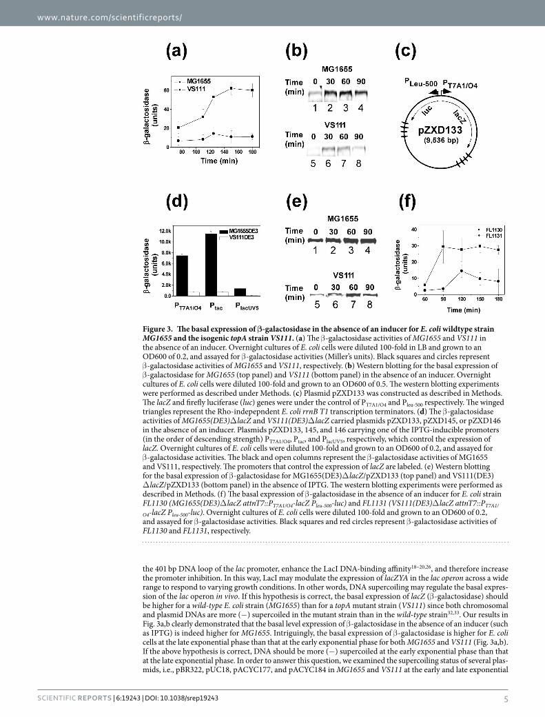
the 401 bp DNA loop of the lac promoter, enhance the LacI DNA-binding affinity18–20,26, and therefore increase the promoter inhibition. In this way, LacI may modulate the expression of lacZYA in the lac operon across a wide range to respond to varying growth conditions. In other words, DNA supercoiling may regulate the basal expres-sion of the lac operon in vivo. If this hypothesis is correct, the basal expression of lacZ (β -galactosidase) should be higher for a wild-type E. coli strain (MG1655) than for a topA mutant strain (VS111) since both chromosomal and plasmid DNAs are more (−) supercoiled in the mutant strain than in the wild-type strain32,33. Our results in Fig. 3a,b clearly demonstrated that the basal level expression of β -galactosidase in the absence of an inducer (such as IPTG) is indeed higher for MG1655. Intriguingly, the basal expression of β -galactosidase is higher for E. coli cells at the late exponential phase than that at the early exponential phase for both MG1655 and VS111 (Fig. 3a,b). If the above hypothesis is correct, DNA should be more (−) supercoiled at the early exponential phase than that at the late exponential phase. In order to answer this question, we examined the supercoiling status of several plas-mids, i.e., pBR322, pUC18, pACYC177, and pACYC184 in MG1655 and VS111 at the early and late exponential
Figure 3. The basal expression of β-galactosidase in the absence of an inducer for E. coli wildtype strain MG1655 and the isogenic topA strain VS111. (a) The β -galactosidase activities of MG1655 and VS111 in the absence of an inducer. Overnight cultures of E. coli cells were diluted 100-fold in LB and grown to an OD600 of 0.2, and assayed for β -galactosidase activities (Miller’s units). Black squares and circles represent β -galactosidase activities of MG1655 and VS111, respectively. (b) Western blotting for the basal expression of β -galactosidase for MG1655 (top panel) and VS111 (bottom panel) in the absence of an inducer. Overnight cultures of E. coli cells were diluted 100-fold and grown to an OD600 of 0.5. The western blotting experiments were performed as described under Methods. (c) Plasmid pZXD133 was constructed as described in Methods. The lacZ and firefly luciferase (luc) genes were under the control of PT7A1/O4 and Pleu-500 respectively. The winged triangles represent the Rho-indepepndent E. coli rrnB T1 transcription terminators. (d) The β -galactosidase activities of MG1655(DE3)Δ lacZ and VS111(DE3)Δ lacZ carried plasmids pZXD133, pZXD145, or pZXD146 in the absence of an inducer. Plasmids pZXD133, 145, and 146 carrying one of the IPTG-inducible promoters (in the order of descending strength) PT7A1/O4, Ptac, and PlacUV5, respectively, which control the expression of lacZ. Overnight cultures of E. coli cells were diluted 100-fold and grown to an OD600 of 0.2, and assayed for β -galactosidase activities. The black and open columns represent the β -galactosidase activities of MG1655 and VS111, respectively. The promoters that control the expression of lacZ are labeled. (e) Western blotting for the basal expression of β -galactosidase for MG1655(DE3)Δ lacZ/pZXD133 (top panel) and VS111(DE3)Δ lacZ/pZXD133 (bottom panel) in the absence of IPTG. The western blotting experiments were performed as described in Methods. (f) The basal expression of β -galactosidase in the absence of an inducer for E. coli strain FL1130 (MG1655(DE3)Δ lacZ attnT7::PT7A1/O4-lacZ Pleu-500-luc) and FL1131 (VS111(DE3)Δ lacZ attnT7::PT7A1/
O4-lacZ Pleu-500-luc). Overnight cultures of E. coli cells were diluted 100-fold and grown to an OD600 of 0.2, and assayed for β -galactosidase activities. Black squares and red circles represent β -galactosidase activities of FL1130 and FL1131, respectively.

www.nature.com/scientificreports/
6Scientific RepoRts | 6:19243 | DOI: 10.1038/srep19243
phases. Our results are shown in Fig. 4. For MG1655, plasmid DNA templates are more (−) supercoiled at the early exponential phase than those at the late exponential phase. For instance, the superhelical density of plasmid pBR322 at the early exponential phase was determined to be − 0.076 and diminished to − 0.062 at the late expo-nential phase (Fig. 4a,b). For VS111, as expected, at the early exponential phase, 80–90% of plasmids pBR322 and pACYC184 became hypernegatively supercoiled (Fig. 4c,d), which is consistent with previously published results32,34. However, less than 50% of these two plasmids were hypernegatively supercoiled at the late exponential phase (Fig. 4c,d). These results suggest that a correlation exists between the DNA supercoiling status and the basal expression of β -galactosidase in E. coli.
In order to further study how DNA supercoiling regulates the basal expression of β -galactosidase, we con-structed two E. coli strains MG1655(DE3)Δ lacZ and VS111(DE3)Δ lacZ in which lacZ was deleted from the chro-mosome using the λ Red recombination system35. Additionally, these two E. coli strains carry the lysogenic λ DE3 that harbors the lacI gene under the control of the strong lacIq promoter to overexpress LacI. We also constructed three plasmids pZXD133, 145, and 146 carrying one of the IPTG-inducible promoters (in the order of descending strength) PT7A1/O4, Ptac, and PlacUV5, respectively, which control the expression of lacZ (Fig. 3c). We transformed MG1655(DE3)Δ lacZ and VS111(DE3)Δ lacZ with these three plasmids and tested the β -galactosidase activities using Miller’s assay and Western blotting. Our results are shown in Fig. 3d,e. Similar to above results, the basal expression of β -galactosidase in the absence of an inducer is higher for MG1655 than that for VS111. Since these IPTG-inducible promoters do not contain a CRP (catabolite repressor protein) binding site, these exper-iments ruled out the possibility that the enhanced basal expression of β -galactosidase is caused by the binding of the CRP-cAMP complex to the CRP binding site upstream from the lac promoter. Additionally, we inserted lacZ under the control of PT7A1/O4 in the attTn7 site (84 min) of the chromosome of MG1655(DE3)Δ lacZ and VS111(DE3)Δ lacZ using a transposon Tn7-based method36 and tested the β -galactosidase activities using Miller’s assay. Again, our results (Fig. 3f) showed that the basal expression of β -galactosidase in the absence of an inducer is higher for MG1655 than that for VS111.
Figure 4. Time courses of DNA supercoiling status in MG1655 (a,b) and VS111 (c,d). Overnight cultures of E. coli cells carrying plasmids pBR322, pUC18, pACYC177, or pACYC184 were diluted 100-fold in LB and grown to the time points indicated. The DNA molecules were isolated using the alkaline lysis assays using the QIAprep Spin Miniprep Kit. The DNA samples were subjected to 1% agarose gel electrophoresis in the presence of 5 μ g/mL of chloroquine. The DNA supercoiling densities were determined as detailed in Methods. The symbol of (–) represents the hypernegatively supercoiled DNA.

www.nature.com/scientificreports/
7Scientific RepoRts | 6:19243 | DOI: 10.1038/srep19243
DiscussionIn this article we demonstrate that E. coli LacI is able to form a topological barrier upon binding to the O1, O2, and O3 operators of the lac promoter at their native positions and separate a supercoiled DNA molecule into two distinct topological domains: a large 4,300 bp domain and a small 401 bp domain. The small 401 bp domain is capable of constraining 3 (−) supercoils in the LacI-mediated DNA loop for the DNA template with a superheli-cal density of ~− 0.06 (Fig. 2b and Table 2). These constrained supercoils increase the LacI’s DNA-binding affinity and therefore enhance its inhibition of the lac promoter in the absence of an inducer. We also demonstrate that the stability of LacI-mediated DNA topological barriers is proportional to its DNA binding affinity: the higher the DNA binding affinity, the more stable the LacI-mediated topological barrier (Supplementary Tables S1 and S2). Figure 5 shows a molecular model of the physical interactions between LacI and lac operators that play essential roles in the formation of the DNA topological barrier with 3 constrained (−) supercoils. As mentioned above, these constrained (−) supercoils are important for the lac operon. First, they bring O1 and O2 close to each other and enhance the probability of forming the LacI-mediated DNA loop. Second, these constrained (−) supercoils provide free energy to enhance the LacI’s DNA binding affinity and therefore form a highly stable repressorsome37 that efficiently inhibits the gene expression of the lac operon. DNA supercoiling appears to be an essential compo-nent of the classical lac operon in E. coli, a component that has been overlooked previously1,4,12. Nevertheless, it is important to understand what property or properties of LacI determine its capacity to form a topological barrier and block supercoiling diffusion. We showed previously24 that LacI is able to induce superhelicity (Δ Lk) within the LacI-lac O complexes. This LacI-induced superhelicity may be essential for forming the DNA topological barrier and blocking supercoiling diffusion.
These experiments also show that DNA supercoiling plays an essential role in the regulation of the basal expression of the lac operon in E. coli. Not only did the wild-type strain MG1655 express more β -galactosidase comparing with the isogenic topA strain VS111 (Fig. 3a,b), but the expression level of β -galactosidase of MG1655 and VS111 is higher for cells at the early exponential phase than for those at the late exponential phase as well (Fig. 3a,b,e,f). The expression level is directly correlated with the DNA supercoiling status in vivo (Fig. 4). These results strongly support our hypothesis that DNA supercoiling modulates the basal expression of the lac operon, an effect allowing E. coli cells to sense the superhelical changes through the LacI-mediated topological barrier for different growth conditions.
E. coli is a single cell organism and has developed the ability to adapt rapidly to different environmental con-ditions, such as low nutrients, high osmotic pressure, and the change of temperature38. When the main carbon source glucose is abundant, E. coli cells generate sufficient ATP for normal functions and biosynthesis. In this case, DNA gyrase is fully active and drives the chromosomal DNA to more (−) supercoiled status39,40 (Fig. 4). The excess supercoils constrained in the 401 bp DNA loop by the LacI-mediated topological barrier promote the formation of a highly stable repressorsome that prevents the wasteful expression of lacZYA of the lac operon. However, when E. coli cells live in nutrient deficient environments, the ATP/ADP ratio or energy charge is low, which significantly reduces the supercoiling activities of DNA gyrase41,42. In this way, the DNA around the lac promoter is relaxed39,40 (Fig. 4), which weakens the binding of LacI to lac operators and therefore increases the basal level expression of lacZYA. This mechanism prepares E. coli cells to respond quickly to the presence of other carbon sources, such as lactose in the nutrient deficient environments. In this way, certain amounts of lactose is transported inside cells by lactose permease and converted to allolactose by β -galactosidase, the natural inducer of the lac operon. Our results in Fig. 3 are consistent with this explanation. These results also provide a reasonable explanation for the long-time observation in which DNA supercoiling enhances the DNA binding affinity of LacI to lac operators18,19 and promotes the formation of LacI-mediated DNA loop20–22. The lac operon of E. coli uses the LacI-mediated topological barrier to sense the environmental changes through sensing the DNA topological change around the lac operon. In this way, the cells can set the basal level of lacZYA expression.
In our recent publication, we demonstrated that bacterial phage λ utilizes supercoiling as a signal to decide whether the virus adopts a lytic or lysogenic life cycle when it infects an E. coli cell31. It is likely that the virus has evolved to respond to (–) DNA supercoils by facilitating the quiescent propagation through lysogenic life cycle during favorable growth conditions for bacteria31. In this article our results also suggest that E. coli genome may have evolved to regulate the basal expression of the lac operon by using LacI-mediated topological barrier as a sensor to detect the superhelical changes of chromosomal DNA during different growth conditions. It is possible that bacteria have adopted DNA topological barriers as a common mechanism to sense superhelical change within chromosomal DNA, the most dynamic epigenetic signal for transcription regulation. Since the nucleosomes are the basic packaging units for eukaryotes in which ~146 bp of DNA wrap in 1.67 left-handed superhelical turns around the histone octamer and (−) supercoils are constrains in the nucleosomes43, it is pos-sible that also eukaryotes use topological barriers to regulate different biological functions, such as transcription and recombination.
MethodsProteins, chemicals, and reagents. E. coli LacI and mutants were purified by the method of Chen and Matthews44 (E. coli strains containing the plasmid overexpressing LacI and mutants was kindly provided by K. S. Matthews at Rice University). Restriction enzymes Nt.BbvCI, Nb.BbvCI, Nb.BtsI, T4 DNA ligase, and E. coli DNA gyrase were purchased from New England Biolabs (Beverly, MA, USA). Isopropyl β -D-1-thiogalactopyranoside (IPTG) and o-nitrophenyl-β -D-galactoside (ONPG) were obtained from Sigma-Aldrich, Inc. (St. Louis, MO). All synthetic oligonucleotides were purchased from MWG-Biotech, Inc. (Huntsville, AL). γ -32P-ATP (3000 mCi/mmol) was obtained from PerkinElmer Life and Analytical Sciences (Shelton, CT).

www.nature.com/scientificreports/
8Scientific RepoRts | 6:19243 | DOI: 10.1038/srep19243
Figure 5. LacI tetramer simultaneously binds to O1 & O2 operators of the lac promoter and forms a topological barrier that constrains three (−) supercoils in the 401 bp DNA loop. This molecular model has been constructed as described under Methods and shows that it is feasible for the LacI-mediated topological barrier to constrain three (−) DNA supercoils to the 401 bp DNA loop.

www.nature.com/scientificreports/
9Scientific RepoRts | 6:19243 | DOI: 10.1038/srep19243
Plasmid DNA templates. All plasmids except pZXD133, pZXD145, and pZXD146 are derived from plasmid pACYC184. Construction of plasmid DNA templates sometimes required DNA fusions between non-complementary cohesive termini. In this scenario, cohesive ends were converted before ligation to blunt ends by incubation of the DNA fragments with T4 DNA polymerase in the presence of dNTPs. Plasmids pCB67, pCB73, pCB107, pCB109, pCB112, and pCB115 were described previously25. Plasmid pCB106 was constructed by the insertion of a 19 bp synthetic DNA fragment containing a nicking enzyme Nt.BbvCI recognition site into the unique EagI site of pCB73. Plasmid pCB108 was created by the insertion of the 378 bp BamHI-KpnI fragment of pCB106 carrying 8 tandem copies of lac O1 operators to the unique AdhI site of pCB106. In this way, pCB108 contains 16 lac O1 operators equally distributed between two locations. Plasmid pCB116 was constructed through the insertion of the 194 bp BamHI-KpnI fragment of pCB112 carrying 4 tandem copies of lac O1 operators to the unique AdhI site of pCB112. In this way, pCB116 contains 8 lac O1 operators equally distributed between two locations. Plasmid pCB110 was constructed by the insertion of the 19 bp synthetic DNA fragment described above into the unique EagI site of pCB67. Plasmid pCB126 (or pO1O1) was made by inserting a 46 bp synthetic oligonucleotide containing a lac O1 operator into the unique AhdI site of pCB110. In this case, the shortest dis-tance between the two-lac O1 operators of pCB126 is 1.2 kb.
Plasmids pO1O2 and pO1O3 were constructed by inserting a 41 bp synthetic DNA oligonucleotide containing a lac O2 or O3 operator into the unique AhdI site of pCB110, respectively. Likewise, plasmid pO1Os was made by the insertion of a 40 bp synthetic DNA oligonucleotide containing a lac Os operator into the unique AhdI site of pCB110. Plasmid pO2O2 was created by the replacement of the 46 bp BamHI-BglII fragment of pO1O2 with a 31 bp synthetic DNA oligonucleotide carrying a lac O2 operator. Plasmid pO3O3 was constructed by replacing the 46 bp BamHI-BglII fragment of pO1O3 with a 31 bp synthetic DNA oligonucleotide carrying a lac O3 oper-ator. Plasmid pOsOs was created by replacing the 46 bp BamHI-BglII fragment of pO1Os with a 31 bp synthetic DNA oligonucleotide carrying a lac Os operator. Plasmid pO3O1O2 was constructed in two steps. Step 1 is to insert a 568 bp PCR product containing lac O1, O2, and O3 operators at their native positions on the chromosome into the BamHI-KpnI sites of pYZX43F to generate plasmid pO1O2O3-1. In the second step, a nicking enzyme Nt.BbvCI recognition site between the lac O1 and O2 operators was created to yield plasmid pO3O1O2. Plasmids pO1O2n and pO3O2n were made by PCR-directed site-mutagenesis to eliminate the lac O3 or O2 operators of pO3O1O2, respectively. Plasmids pOsOs401 and pOsOs493 were, respectively, created through converting the lac operators of pO1O2n and pO3O2n into lac Os operators by PCR-based site-directed mutagenesis.
Plasmids pZXD133, pZXD145, and pZXD146 are derivative of pBR322 and were constructed in several steps. Plasmid pZXD64 was constructed by introducing a unique AgeI site into the upstream region of the tet gene of pZXD1445 using PCR-based, site-directed mutagenesis. Then, the tet gene between the unique AgeI and BsmI sites of pZXD64 was replaced by a 3,068 bp lacZ gene DNA fragment of plasmid pYC2/CT/lacZ (Life Technologies, Grand Island, NY) to generate pZXD65. Next, four Rho-independent E. coli rrnB T1 terminators from plas-mid pLUC1 were inserted into XbaI site of pGL3 (Promega Corporation, Wisconsin, WI) to yield pZXD67. A 2,511 bp HindIII-SpeI DNA fragment of pZXD67 carrying a modified firefly (Photinus pyralis) luciferase gene (the codon usage was optimized for mammalian cells) and four Rho-independent E. coli rrnB T1 terminators was inserted between the HindIII and SpeI sites of pZXD65 to produce pZXD70. Plasmid pZXD74 was created by silently removing the EcoRI site in the downstream region of lacZ gene of plasmid pZXD70 without changing the open reading frame of lacZ gene using PCR-based, site-directed mutagenesis. Plasmid pZXD76 was gener-ated after a XbaI site was inserted into the downstream region of the luciferase gene of plasmid pZXD74. Then, a 1,688 bp DNA fragment of pZE15luc carrying a firefly (Photinus pyralis) luciferase gene (the codon usage was optimized for bacterial cells) was inserted into the HindIII and XbaI sites of pZXD76 to yield pZXD77. Plasmid pZXD97 was created by inserting a 72 bp synthetic deoxyoligonucleotide containing leu-500 promoter and a unique BamHI site into the HindIII and EcoRI sites of pZXD77. pZXD99 was produced by inserting a 53 bp syn-thetic deoxyoligonucleotide into the BamHI and EcoRI sites of pZXD97. Plasmid pZXD105 was constructed by inserting a 55 bp synthetic DNA oligonucleotide containing the strong IPTG-inducible E. coli T7A1/O4 promoter into the EcoRI and XhoI sites of pZXD99. In this way, the T7 promoter was replaced by the T7A1/O4 promoter. Plasmid pZXD133 was made by the insertion of a 3,093 bp PCR product containing the lacZ gene amplified from MG1655 genomic DNA into the AgeI and BsmI sites of pZXD105. In this case, the codon usage of lacZ was optimized for E. coli cells. Plasmid pZXD106 was constructed by inserting an 85 bp synthetic DNA oligonucle-otide containing the tac promoter into the EcoRI and XhoI sites of pZXD99. In this way, the T7 promoter was replaced by Ptac. Plasmid pZXD145 was created by the insertion of a 3,093 bp PCR product containing the lacZ gene amplified from MG1655 genomic DNA into the AgeI and BsmI sites of pZXD106. Plasmid pZXD107 was made by inserting an 87 bp synthetic DNA oligonucleotide containing the lacUV5 promoter into the EcoRI and XhoI sites of pZXD99. In this way, the T7 promoter was replaced by PlacUV5. Plasmid pZXD146 was constructed by the insertion of a 3,093 bp PCR product containing the lacZ gene amplified from MG1655 genomic DNA into the AgeI and BsmI sites of pZXD107.
Bacterial strains. Escherichia coli strains MG1655 [F−, λ −, rph-I] and VS111 [F−, λ −, rph-I, Δ topA] were obtained from the Coli Genetic Stock Collection/E. coli Genetic Resource Center (CGSC) at Yale University. MG1655(DE3) and VS111(DE3) were described previously34. E. coli strains FL1130 (MG1655(DE3) Δ lacZ attnT7::PT7A1/O4-lacZ Pleu-500-luc) and FL1131 (VS111(DE3) Δ lacZ attnT7::PT7A1/O4-lacZ Pleu-500-luc) were con-structed in two steps. First, MG1655(DE3) Δ lacZ and VS111(DE3) Δ lacZ were constructed using the λ Red recombination system35. In the next step, using a Tn7-based site-specific recombination system36, a 5.1 kb DNA fragment carrying the divergently coupled Pleu-500 and PT7A1/O4 promoters with the luc and lacZ genes was inserted to the attTn7 site of the E. coli chromosome46 (84 min of the chromosome) to generate FL1130 and FL1131. In both strains, the IPTG-inducible PT7A1/O4 controls the expression of β -galactosidase.
www.nature.com/scientificreports/
1 0Scientific RepoRts | 6:19243 | DOI: 10.1038/srep19243
The DNA-nicking method. The DNA-nicking method was described previously25 with some modifica-tions. Briefly, a typical DNA-nicking reaction mixture (320 μ L) contained 20 mM Tris-acetate (pH 7.9 at 25 °C), 10 mM magnesium acetate, 1 mM DTT, a negatively supercoiled DNA template, and LacI. Where specified, 1 mM of IPTG was also added to the DNA-nicking assays. All components were assembled on ice and incubated for 30 min at 37 °C. After the incubation, the supercoiled DNA templates were digested by either Nt.BbvCI or Nb.BbvCI at 37 °C for 30 min or various times. Then, a large excess of a double-stranded oligonucleotide con-taining Nt.BbvCI recognition site were added to the reaction mixtures to inhibit the restriction enzyme activities. The nicked DNA templates were ligated by T4 DNA ligase in the presence of 1 mM of ATP at 37 °C for 5 min and the reactions were terminated by extraction with an equal volume of phenol. The DNA samples were precipitated with ethanol and dissolved in 25 μ L of 10 mM Tris-HCl buffer (pH 8.5). The linking number of the ligated DNA products was determined with 1% agarose gel electrophoresis in the absence or presence of 0.5 μ g/ml of chloro-quine and calculated from the gel images stained with SYBR Gold using KODAK 1D Image Analysis Software.
Determining supercoiling density of plasmid DNA templates. 3 μ g of supercoiled plasmid pBR322, pUC18, pACYC177, or pACYC184 was relaxed by human DNA topoisomerase I in the presence of various con-centrations of ethidium bromide at 37 °C in 20 mM Tris-acetate (pH 7.9), 50 mM KAc, 10 mM Mg(Ac)2, and 1 mM DTT for one hour. Subsequently, the relaxation reaction was stopped by addition of an equal volume of phenol. The topological status of each DNA preparation was analyzed by electrophoresis in a 1% agarose gel in 1 × TAE buffer (40 mM Tris-acetate (pH 7.8) and 1 mM EDTA) containing different concentrations of chloro-quine. After electrophoresis, agarose gels were stained with ethidium bromide, destained, and photographed under UV light. The DNA linking number change (Δ Lk) was determined by analyzing the distributions of the topoisomers in these gel images and the supercoiling density (σ) was calculated using the following equation:
σ = ∆ =−
( )Lk
LkLk Lk
Lk 10
0
0
where Lk0 and Lk represent the DNA linking number for the relaxed and the supercoiled DNA, respectively.
Electrophoretic Mobility Shift Assay (EMSA). EMSA experiments were used to determine the appar-ent DNA binding constant of LacI and mutants. DNA oligomers containing a DNA-binding site of one of the DNA-binding proteins (the EcoRV fragment of the pBend2 derivatives) were labeled with 32P at 5′ termini by T4 polynucleotide kinase in the presence of γ -32P-ATP. The protein-DNA complexes were formed by addition of appropriate amounts of the protein to a solution containing 0.1 nM of 32P-labeled DNA in the 1 × DNA-binding buffer containing 20 mM Tris-HCl (pH 8.0), 200 mM NaCl, 0.5 mM EDTA, 1 mM DTT, 5 mM MgCl2, and 5% glycerol. After equilibration for 60 min at 22 °C, the samples were loaded on a 8% native polyacryamide gel in 0.5 × TBE buffer (0.045 M Tris-Borate (pH 8.3) and 1 mM EDTA) to separate free and bound DNA. The gels were subsequently dried and visualized by autoradiography or quantified using a Fuji FLA 3000 image analyzer. The radioactivity of the free and bound DNA was determined and used to calculate the binding ratio (R), which is equal to the ratio of the radioactivity of the bound DNA divided by the sum of the radioactivity of the bound and free DNA. The apparent DNA binding constant (Kapp) was obtained by nonlinear-least-squares fitting the follow-ing equation using the program Scientist.
=( + + / ) − ( + + / ) −
( )Ra x K a x K ax
a
1 1 4
2 2app app
2
where a and x represent the total DNA and the total protein concentration, respectively.
Atomic Force Microscopy. The LacI-DNA samples were prepared according to the DNA-nicking method as described above. After the supercoiled DNA templates were digested by Nt.BbvCI at 37 °C for 30 min, the LacI-DNA complexes (10 μ L) were deposited on a poly-L-ornithine-coated mica surface and incubated for 2 min at room temp. The droplet was rinsed away with 0.4 mL HPLC water and dried gently with compressed air. Images were acquired with a NanoScope MultiMode AFM microscope (Digital Instrument, Santa Barbara, CA) oper-ated in tapping mode using a 50–60 mV oscillation amplitude of uncoated, etched silicon tips with a resonance frequency of 75 kHz (NSC18, MirkoMasch, San Jose, CA47). Areas of 1 × 1 μ m2 were scanned at a rate of 1.2 Hz and a resolution of 512 × 512 pixels. The DNA contour lengths were estimated by using Image Analysis Software ImageJ. The LacI-mediated 401 bp DNA loops were identified by measuring the DNA contour lengths of these relaxed DNA loops and by comparing the size and volume of the LacI molecules that form the DNA loops with our previously published results25.
The expression of β-galactisidase. The expression level of β -galactosidase was measured by Miller’s assay as described48. Briefly, 100 mL of LB was inoculated with 1 mL of overnight bacterial cell culture until OD600 reaches ~0.2. 100 μ L of bacterial cell culture was mixed with 900 μ L of Z-buffer (60 mM Na2HPO4, 40 mM NaH2PO4, 10 mM KCl, 1 mM MgSO4, and 50 mM β -mercaptoethanol). Cells were lysed with 60 μ L of chloroform and 30 μ L of 0.1% SDS. After cell lysates were equilibrated at 30 °C for five minutes, 200 μ L of 4 mg/mL ONPG was added to the cell lysates. After additional 15 min incubation at 30 °C, reactions were stopped by addition of 500 μ L of 1 M Na2CO3. After cell debris was removed by centrifugation at 13,000 rpm for 1 min, OD420 and OD550 were measured in a Cary 50 spectrophotometer. β -Galactosidase activities (E) were calculated using the following equation:
www.nature.com/scientificreports/
1 1Scientific RepoRts | 6:19243 | DOI: 10.1038/srep19243
= ×− . ×× × ( )
E OD ODt v OD
1000 1 753
420 550
600
where t and v represent reaction time and cell culture volume, respectively.
Western blotting experiments. Western blotting experiments were used to monitor the basal level expression of β -galactosidase in different E. coli strains. Total protein purified from E. coli cells was analyzed by electrophoresis in a 10% SDS-PAGE gel and electrophoretically transferred to a 0.45 nm nitrocellulose membrane. The membrane blot was then blocked with a solution containing 5% nonfat skim milk in TBST (50 mM Tris-HCl, pH 8.0, 138 mM NaCl, 2.7 mM KCl, and 0.05% Tween-20) for 45 min at room temperature and incubated with the primary antibody, β -galactosidase antibody (Thermofisher Scientific, Inc.), diluted 1:1000 in TBST solution overnight at 4 °C. After the overnight incubation, the membrane blot was washed three times with TBST and blocked with a solution containing 5% nonfat skim milk in TBST for 15 min at room temperature. Membranes were incubated with secondary antibodies in blocking buffer at 1:20,000 for fluorescently conjugated antibodies purchased from Li-Cor Biosciences. Membranes were again washed three times for five minutes in 1 × TBST. Western blots were developed using fluorescence detection using the LI-COR Odyssey CLx near infrared scanner.
Molecular modeling. The DNA-loop was created with the GraphiteLifeExplorer modeling tool49. VMD software50 was used to patch and render the DNA-loop structure with the LacI-DNA complex (PDB id 1Z04), which was originally modeled by patching several NMR-structures of fragments of LacI and DNA51. Three (−) DNA supercoils were introduced to the DNA loop to match our experimental results for the LacI-mediated 401 bp DNA loop.
References1. Muller-Hill, B. The Lac Operon: A Short History of a Genetic Paradigm (Walter de Gruyter & Co., Berlin, 1996).2. Lewis, M. Allostery and the lac Operon. J. Mol. Biol. 425, 2309–2316 (2013).3. Lewis, M. et al. Crystal structure of the lactose operon repressor and its complexes with DNA and inducer. Science 271, 1247–1254
(1996).4. Oehler, S., Eismann, E. R., Kramer, H. & Muller-Hill, B. The three operators of the lac operon cooperate in repression. EMBO J. 9,
973–979 (1990).5. Gilbert, W. & Muller-Hill, B. Isolation of the lac repressor. Proc. Natl. Acad. Sci. USA 56, 1891–1898 (1966).6. Muller-Hill, B. The function of auxiliary operators. Mol. Microbiol. 29, 13–18 (1998).7. Oehler, S., Amouyal, M., Kolkhof, P., von Wilcken-Bergmann, B. & Muller-Hill, B. Quality and position of the three lac operators of
E. coli define efficiency of repression. EMBO J. 13, 3348–3355 (1994).8. Shanblatt, S. H. & Revzin, A. The binding of catabolite activator protein and RNA polymerase to the Escherichia coli galactose and
lactose promoters probed by alkylation interference studies. J. Biol. Chem. 261, 10885–10890 (1986).9. Liu, M. et al. Kinetics of transcription initiation at lacP1. Multiple roles of cyclic AMP receptor protein. J. Biol. Chem. 278,
39755–39761 (2003).10. Beckwith, J., Grodzicker, T. & Arditti, R. Evidence for two sites in the lac promoter region. J. Mol. Biol. 69, 155–160 (1972).11. Malan, T. P. & McClure, W. R. Dual promoter control of the Escherichia coli lactose operon. Cell 39, 173–180 (1984).12. Kuhlman, T., Zhang, Z., Saier, M. H., Jr. & Hwa, T. Combinatorial transcriptional control of the lactose operon of Escherichia coli.
Proc. Natl. Acad. Sci. USA 104, 6043–6048 (2007).13. Inada, T., Kimata, K. & Aiba, H. Mechanism responsible for glucose-lactose diauxie in Escherichia coli: challenge to the cAMP
model. Genes Cells 1, 293–301 (1996).14. Kimata, K., Takahashi, H., Inada, T., Postma, P. & Aiba, H. cAMP receptor protein-cAMP plays a crucial role in glucose-lactose
diauxie by activating the major glucose transporter gene in Escherichia coli. Proc. Natl. Acad. Sci. USA 94, 12914–12919 (1997).15. Bates, A. D. & Maxwell, A. DNA Topology(Oxford University Press, Oxford, UK, 2005).16. Cozzarelli, N. R. & Wang, J. C. DNA Topology and Its Biological Effects(Cold Spring Harbor Laboratory Press, Cold Spring Harbor,
NY, 1990).17. James C Wang Untangling the Double Helix: DNA Entanglement and the Action of the DNA Topoisomerases(Cold Spring Harbor
Laboratory Press,2009).18. Wang, J. C., Barkley, M. D. & Bourgeois, S. Measurements of unwinding of lac operator by repressor. Nature 251, 247–249 (1974).19. Whitson, P. A., Hsieh, W. T., Wells, R. D. & Matthews, K. S. Influence of supercoiling and sequence context on operator DNA
binding with lac repressor. J. Biol. Chem. 262, 14592–14599 (1987).20. Whitson, P. A., Hsieh, W. T., Wells, R. D. & Matthews, K. S. Supercoiling facilitates lac operator-repressor-pseudooperator
interactions. J. Biol. Chem. 262, 4943–4946 (1987).21. Borowiec, J. A., Zhang, L., Sasse-Dwight, S. & Gralla, J. D. DNA supercoiling promotes formation of a bent repression loop in lac
DNA. J. Mol. Biol. 196, 101–111 (1987).22. Eismann, E. R. & Muller-Hill, B. lac repressor forms stable loops in vitro with supercoiled wild-type lac DNA containing all three
natural lac operators. J. Mol. Biol. 213, 763–775 (1990).23. Kim, R. & Kim, S. H. Direct measurement of DNA unwinding angle in specific interaction between lac operator and repressor. Cold
Spring Harb. Symp. Quant. Biol. 47 Pt 1, 451–454 (1983).24. Chen, B., Xiao, Y., Liu, C., Li, C. & Leng, F. DNA linking number change induced by sequence-specific DNA-binding proteins.
Nucleic Acids Res. 38, 3643–3654 (2010).25. Leng, F., Chen, B. & Dunlap, D. D. Dividing a supercoiled DNA molecule into two independent topological domains. Proc. Natl.
Acad. Sci. USA 108, 19973–19978 (2011).26. Kramer, H., Amouyal, M., Nordheim, A. & Muller-Hill, B. DNA supercoiling changes the spacing requirement of two lac operators
for DNA loop formation with lac repressor. EMBO J. 7, 547–556 (1988).27. Crut, A., Koster, D. A., Seidel, R., Wiggins, C. H. & Dekker, N. H. Fast dynamics of supercoiled DNA revealed by single-molecule
experiments. Proc. Natl. Acad. Sci. USA 104, 11957–11962 (2007).28. van Loenhout, M. T., de Grunt, M. V. & Dekker, C. Dynamics of DNA supercoils. Science 338, 94–97 (2012).29. Sadler, J. R., Sasmor, H. & Betz, J. L. A perfectly symmetric lac operator binds the lac repressor very tightly. Proc. Natl. Acad. Sci. USA
80, 6785–6789 (1983).30. Falcon, C. M. & Matthews, K. S. Glycine insertion in the hinge region of lactose repressor protein alters DNA binding. J. Biol. Chem.
274, 30849–30857 (1999).
www.nature.com/scientificreports/
1 2Scientific RepoRts | 6:19243 | DOI: 10.1038/srep19243
31. Ding, Y. et al. DNA supercoiling: A regulatory signal for the lambda repressor. Proc. Natl. Acad. Sci. USA 111, 15402–15407 (2014).32. Pruss, G. J. DNA topoisomerase I mutants. Increased heterogeneity in linking number and other replicon-dependent changes in
DNA supercoiling. J. Mol. Biol. 185, 51–63 (1985).33. Stupina, V. A. & Wang, J. C. Viability of Escherichia coli topA mutants lacking DNA topoisomerase I. J. Biol. Chem 280, 355–360
(2005).34. Samul, R. & Leng, F. Transcription-coupled hypernegative supercoiling of plasmid DNA by T7 RNA polymerase in Escherichia coli
topoisomerase I-deficient strains. J. Mol. Biol. 374, 925–935 (2007).35. Datsenko, K. A. & Wanner, B. L. One-step inactivation of chromosomal genes in Escherichia coli K-12 using PCR products. Proc.
Natl. Acad. Sci. USA 97, 6640–6645 (2000).36. McKenzie, G. J. & Craig, N. L. Fast, easy and efficient: site-specific insertion of transgenes into enterobacterial chromosomes using
Tn7 without need for selection of the insertion event. BMC. Microbiol. 6, 39 (2006).37. Semsey, S., Virnik, K. & Adhya, S. Three-stage regulation of the amphibolic gal operon: from repressosome to GalR-free DNA. J.
Mol. Biol. 358, 355–363 (2006).38. Foster, P. L. Stress-induced mutagenesis in bacteria. Crit Rev. Biochem. Mol. Biol. 42, 373–397 (2007).39. Jensen, P. R., Loman, L., Petra, B., van der Weijden, C. & Westerhoff, H. V. Energy buffering of DNA structure fails when Escherichia
coli runs out of substrate. J. Bacteriol. 177, 3420–3426 (1995).40. van, W. M. et al. DNA supercoiling depends on the phosphorylation potential in Escherichia coli. Mol. Microbiol. 20, 351–360
(1996).41. Westerhoff, H. V., O’Dea, M. H., Maxwell, A. & Gellert, M. DNA supercoiling by DNA gyrase. A static head analysis. Cell Biophys.
12, 157–181 (1988).42. Hsieh, L. S., Rouviere-Yaniv, J. & Drlica, K. Bacterial DNA supercoiling and [ATP]/[ADP] ratio: changes associated with salt shock.
J. Bacteriol. 173, 3914–3917 (1991).43. Luger, K., Mader, A. W., Richmond, R. K., Sargent, D. F. & Richmond, T. J. Crystal structure of the nucleosome core particle at 2.8 A
resolution. Nature 389, 251–260 (1997).44. Chen, J. & Matthews, K. S. Deletion of lactose repressor carboxyl-terminal domain affects tetramer formation. J. Biol. Chem. 267,
13843–13850 (1992).45. Zhi, X. & Leng, F. Dependence of transcription-coupled DNA supercoiling on promoter strength in Escherichia coli topoisomerase
I deficient strains. Gene 514, 82–90 (2013).46. Waddell, C. S. & Craig, N. L. Tn7 transposition: recognition of the attTn7 target sequence. Proc. Natl. Acad. Sci. USA 86, 3958–3962
(1989).47. Wang, H., Finzi, L., Lewis, D. E. & Dunlap, D. AFM studies of lambda repressor oligomers securing DNA loops. Curr. Pharm.
Biotechnol. 10, 494–501 (2009).48. Miller, J. H. Experiments in Molecular Genetics (Cold Spring Harbor Laboratory, Cold Spring Harbor, NY, 1972).49. Hornus, S., Levy, B., Lariviere, D. & Fourmentin, E. Easy DNA modeling and more with GraphiteLifeExplorer. PLos ONE 8, e53609
(2013).50. Humphrey, W., Dalke, A. & Schulten, K. VMD: visual molecular dynamics. J. Mol. Graph. 14, 33–38 (1996).51. Balaeff, A., Mahadevan, L. & Schulten, K. Structural basis for cooperative DNA binding by CAP and lac repressor. Structure. 12,
123–132 (2004).
AcknowledgementsWe thank Kathleen S. Matthews for providing us with E. coli strains containing plasmids overexpressing E. coli lac repressor and mutants. We also thank Dr. Alberto Martin at the University of Toronto for providing us with E. coli strain MG1655(DE3). We are grateful to Dr. Bo Chen for technical support. This work was supported by grants from the National Institutes of Health: 1SC1HD063059-04 and 1R15GM109254-01A1 (to F.L.) and RGM084070A (to Laura Finzi at Emory University).
Author ContributionsF.L. designed research; G.F., S.D., X.Z., B.C. and D.D. performed research; P.C. and B.G. constructed the molecular model; F.L. analyzed data; F.L. wrote the paper.
Additional InformationSupplementary information accompanies this paper at http://www.nature.com/srepCompeting financial interests: The authors declare no competing financial interests.How to cite this article: Fulcrand, G. et al. DNA supercoiling, a critical signal regulating the basal expression of the lac operon in Escherichia coli. Sci. Rep. 6, 19243; doi: 10.1038/srep19243 (2016).
This work is licensed under a Creative Commons Attribution 4.0 International License. The images or other third party material in this article are included in the article’s Creative Commons license,
unless indicated otherwise in the credit line; if the material is not included under the Creative Commons license, users will need to obtain permission from the license holder to reproduce the material. To view a copy of this license, visit http://creativecommons.org/licenses/by/4.0/