dna damage repair and cancer: the role of rad51 protein
TRANSCRIPT

3
DNA Damage Repair and Cancer: The Role of RAD51 Protein and Its Genetic Variants
Augusto Nogueira1,4, Raquel Catarino1 and Rui Medeiros1,2,3,4 1Molecular Oncology & Virology - Portuguese Institute of Oncology
2CEBIMED, Faculty of Health Sciences of Fernando Pessoa University 3ICBAS, Abel Salazar Institute for the Biomedical Sciences
4Portuguese League Against Cancer (LPCC-NRNorte) Portugal
1. Introduction
Genomes are continually attacked by both endogenous and exogenous agents that damage DNA. DNA damage in the form of DNA breaks can lead to chromosome translocations, cell cycle arrest, and apoptosis. Homologous recombination is an essential biological process that ensures the accurate repair of DNA breaks and thereby contributes to genomic integrity (Kuzminov 1999; Paques and Haber 1999). DNA double-strand breaks (DSBs) are considered the principal lethal DNA damage resulting from ionizing radiation and cross-linking drugs. In addition, DSB can arise from endogenous processes, such as replication fork stalling during attempted replication over a single strand-break and topoisomerase poisons that are common therapies in the treatment of human cancers (Arnaudeau, Lundin et al. 2001). It is of great importance that cells recognise DSBs and act upon them rapidly and efficiently, because cell death or impaired cell function can occur if these are left unrepaired or are repaired inaccurately. In addition to DNA repair mechanisms, cell cycle checkpoint activation processes are initiated in response to DNA damage. More specifically, DNA damage signals to arrest cell cycle progression, giving the cell more time to repair what might otherwise be a fatal lesion (Henning and Sturzbecher 2003).
2. DNA damage – repair pathways
Faithful genome transmission requires the co-ordination of a network of pathways such as cell cycle checkpoint, DNA replication, DNA repair/recombination and programmed cell death. In response to DNA damage, cells arrest their cell cycle progression, thus providing time for repair, or activate programmed cell death – both responses preventing transmission of genetic instability (Khanna and Jackson 2001). Thus, maintenance of the genomic integrity by DNA repair genes is an essential step in normal cellular growth and differentiation (Hoeijmakers 2001). Failure to repair DNA lesions such as DSBs can lead to mutations, genomic instability, and cell death. Due to the severe consequences of DSBs, cells have developed two major repair pathways: homologous recombination (HR) and non-homolgous end joining (NHEJ) (Helleday, Lo et al. 2007).
www.intechopen.com

DNA Repair and Human Health
74
HR takes advantage of large sequence homologies to repair DSBs. These homologous sequences can be found on homologous chromosome or in DNA repeats. HR refers to several processes, the two most documented in mammalian cells being single-strand annealing (SSA) and gene conversion associated or not with crossing over (Lin, Sperle et al. 1984). SSA process occurs between direct repeat sequences (Lin, Sperle et al. 1984) and is initiated by homologous pairing, except that—unlike HR—the homology is between short stretches of single-stranded DNA at staggered DSBs, and pairing precedes re-ligation, not strand exchange. It is a non-conservative process and error-prone because sequence information can be lost or rearranged when ends overlapping by as little as 30 bp are unsuitably joined (Henning and Sturzbecher 2003). Homologous recombination requires a homologous intact sequence and results in gene conversion associated or not with crossing over (Szostak, Orr-Weaver et al. 1983). Gene conversion is a conservative, generally error-free process, although it can also generate genetic variability. Gene conversion is involved in meiosis and molecular evolution (Daboussi, Dumay et al. 2002). The mitotic cell cycle is an important determinant in the choice of the right repair pathway for a given physiological situation. Repair by HR predominates during S/G phases of the cell cycle, when sister chromatids, the preferred substrate for error-free exchange, are present (Henning and Sturzbecher 2003). Non-homologous end joining (NHEJ) ligates the two broken DNA ends and does not require extensive sequence homologies between the two recombining DNA molecules. During the process, limited degradation of the DNA ends or DNA capture can lead to deletion or insertion of nucleotides or DNA fragments. It is thus a potentially error-prone process (Smith and Jackson 1999).
3. Role of mammalian protein RAD51
Mammalian RAD51 protein is a structural, biochemical and genetic homologue of the bacterial RecA and of the yeast RAD51 recombination proteins. Interestingly, overexpression of RAD51 alone is sufficient to stimulate gene conversion in mammalian cells (Vispe, Cazaux et al. 1998; Arnaudeau, Helleday et al. 1999; Lambert and Lopez 2000). In contrast, expression of dominant negative forms of RAD51 is enough to abolish almost totally gene conversion between tandem repeat sequences (Lambert and Lopez 2000). These data suggest that RAD51 plays a pivotal role in gene conversion regulation. In vitro, RAD51 protein promotes DNA homologous pairing and strand exchange, in association with other proteins of the gene conversion complex (Benson, Baumann et al. 1998). In cultured mammalian cells, RAD51 is involved in spontaneous gene conversion as well as in HR induced by ┛-rays (Lambert and Lopez 2000) , alkylating agents, UV-C and replication elongation inhibitors (Saintigny, Delacote et al. 2001). More precisely, RAD51 controls DSB repair via gene conversion leading to gene conversion associated or not with crossing over (Lambert and Lopez 2000). Finally, RAD51 partly participates in induced sister chromatid exchange in mammalian cells (Lambert and Lopez 2001). These roles of mammalian RAD51 in gene conversion are very similar to the roles of yeast RAD51. However, there are important differences between yeast and vertebrate RAD51 (Daboussi, Dumay et al. 2002). The RAD51 gene consists of 10 exons and spans at least 30 kb. All exon-intron boundaries follow the GT-AC rule. Further sequencing of the region 5’ of the first exon revealed that nonconding exon 1 contained a CpG island that was approximately 900 bp in size. This
www.intechopen.com

DNA Damage Repair and Cancer: the Role of RAD51 Protein and Its Genetic Variants
75
putative promoter region contains several recognition sites for Sp1 transcription factors but lacks a TATA box (Schmutte, Tombline et al. 1999). The presence of several putative Sp1 promoter binding sites is consistent with the observed cell cycle-dependent expression of RAD51 (Johnson 1992). The translation start codon is located in exon 2, and the average size of the coding exons is 112 bp (Schmutte, Tombline et al. 1999). RAD51 gene encodes a highly conserved well-characterized DNA repair protein (Liu, Lamerdin et al. 1998) (Table 1). RAD51 gene is located at chromosome position 15q15.1 (Takahashi, Matsuda et al. 1994), a region that exhibits loss of heterozygosity in a large of cancers, including those of the lung, the colorectum and the breast (Wick, Petersen et al. 1996).
RAD51
Gene symbol hRAD51 Molecular Weight (Da) 36966 Gene type Protein coding (RAD51) Function Involved in the homologous recombination and repair of
DNA Gene Map Locus 15q15.1 Localization primary Nucleus Protein interactions BRCA1, RAD51C, ABL, P53, BRCA2, RAD54-like protein,
RAD54B, RAD52, ERCC2, Cell cycle checkpoint kinase (CHEK1/CHK1), ATM, RAD51C, XRCC3, XRCC2, RAD51B
Described polymorphisms
5’ UTR G135C and 5’ UTR G172T
Table 1. Characteristics of the gene RAD51.
When cells are exposed to genotoxic agents or irradiation, such as mitomycin C, UV, and ionizing radiation, RAD51 protein is recruited to sites of DNA damage where it mediates the search for a homologous sequence during homologous recombination (Buchhop, Gibson et al. 1997; Vispe, Cazaux et al. 1998). It has been revealed that the RAD51 nuclear foci are the sites of repair of DNA damage (Tashiro, Kotomura et al. 1996). This protein is therefore required for meiotic and mitotic recombination and plays a central role in homology-dependent recombinational repair of DSBs (Levy-Lahad, Lahad et al. 2001). Upon its regulated recruitment to sites of DNA breaks, RAD51 forms a nucleoprotein filament by polymerizing onto single-stranded DNA at the processed break. This filament catalyses DNA strand exchange with an undamaged sister chromatid or homologous chromosome, which serve as templates for the restoration of missing genetic information (Li and Heyer 2008; San Filippo, Sung et al. 2008). So when HR is used for repair, in eukaryotes it is promoted by the recombinase RAD51, which binds to 3’-tailed single strands at the end of DSBs in a helical fashion and promotes pairing with homologous DNA sequences as a prelude to strand invasion and repair of the DSBs (Sung and Klein 2006) (Figure 1). In response to DNA damage, RAD51 is translocated from the cytosol to the nucleus (Haaf, Golub et al. 1995). In the nucleus, RAD51 sequesters into foci together with other proteins involved in homologous recombination, e. g., RAD52 (Liu and Maizels 2000). RAD51 is required for the resistance to ionising radiation (Ohnishi, Taki et al. 1998), and high levels of RAD51 have been correlated with resistance to chemotherapeutics agents
www.intechopen.com

DNA Repair and Human Health
76
(Maacke, Jost et al. 2000; Slupianek, Hoser et al. 2002). Importantly, RAD51 is also essential for embryonic survival in the absence of exogenous DNA damaging agents and has a role in the repair of spontaneously occurring chromosome breaks in proliferating cells of higher eukaryotes (Sonoda, Sasaki et al. 1998). This protein has been shown to be involved in the repair of different kinds of DNA lesions during replication (Lundin, Schultz et al. 2003). Thus, RAD51 is likely to promote genomic stability in eukaryotic cells (Orre, Falt et al. 2006). However, despite its role in maintaining genomic integrity, it has been proposed that the aberrant increase in RAD51 expression found in tumour cells may contribute to genomic instability by stimulating aberrant recombination between short repetitive elements and homologous sequences (Xia, Shammas et al. 1997; Flygare, Falt et al. 2001).
Fig. 1. Mechanism of DNA repair by RAD51 (homologous recombination).
The regulation of RAD51 appears to occur through at least two ways: (a) transcriptionally, by genes that confer a proliferative potential, as well as by checkpoint signaling pathways that regulate DNA damage responses; and (b) at the protein level, where interactions with other molecules leads to distinct cellular localization in RAD51 nuclear foci. It is possible
www.intechopen.com
DNA Damage Repair and Cancer: the Role of RAD51 Protein and Its Genetic Variants
77
that RAD51 regulation may occur at the transcriptional level in a cell type-, cell cycle-, or damage response-coordinated manner. RAD51 gene expression is controlled by a variety of transcriptional activators and repressors (Hasselbach, Haase et al. 2005; Arias-Lopez, Lazaro-Trueba et al. 2006), but is not affected by DNA damage (Henson, Tsai et al. 2006). The accurate functioning of the DNA-repair proteins is a crucial step in maintaining genomic homeostasis and preventing carcinogenesis.
4. Levels of RAD51 protein expression
RAD51 expression is cell cycle-regulated, being lowest in resting cells. In proliferating cells, RAD51 expression peaks in the S/G2 phases of the cell cycle (Flygare, Benson et al. 1996; Yamamoto, Taki et al. 1996), indicating a role of the protein in intrachromosomal recombinational repair (Flygare, Falt et al. 2001). Several studies have shown RAD51 protein expression levels to be elevated in immortalized cells and a wide variety of human cancer cell lines (Xia, Shammas et al. 1997; Raderschall, Stout et al. 2002). Given this high RAD51 expression, it is possible that RAD51 overexpression followed by hyperrecombination may contribute to genomic instability and malignant transformation (Vispe, Cazaux et al. 1998; Yanez and Porter 1999). Moreover, a growing body of literature suggests that RAD51 overexpression can increase cellular resistance to radiation and some chemotherapeutic drugs (Maacke, Jost et al. 2000; Henning and Sturzbecher 2003; Qiao, Wu et al. 2005). This could be of clinical importance for the treatment of cancer patients with radio- and/or chemotherapy (Flygare, Falt et al. 2001). Aberrant overexpression of RAD51 protein could confer several advantages to tumor cells. First, the DNA repair function of RAD51 may protect cells from DNA damage and apoptosis. Secondly, overstimulation of homologous recombination and chromatid exchange mechanisms by RAD51 protein (Xia, Shammas et al. 1997; Arnaudeau, Helleday et al. 1999) may contribute to genomic instability and genetic diversity of tumour cells (Raderschall, Stout et al. 2002). Hasselbach and co-workers (Hasselbach, Haase et al. 2005) identified three separate cis-acting sequence elements within the RAD51 transcriptional promoter, one ensuring basal levels of expression and two elements limiting expression to relatively low levels. The characterisation of transcription factor binding might help to explain high-level expression of RAD51 in a variety of solid tumours. The mechanisms underlying the observed radioresistance accompanying RAD51 overexpression are poorly understood. It is possible that an increased DSB repair capacity following RAD51 upregulation is responsible for the increased radioresistance. Alternatively, the overexpression of RAD51 might affect other cellular processes influencing cell survival, e.g., cell cycle progression (Flygare, Falt et al. 2001). The tumour suppressors p53 (Buchhop, Gibson et al. 1997) and BRCA2 (Patel, Yu et al. 1998; Yuan, Lee et al. 1999) associate with RAD51 and play roles in DNA repair and cell cycle checkpoint pathways (Dasika, Lin et al. 1999). The reasons for RAD51 overexpression in cancer cells are not entirely understood. It is not the result of gene duplication or protein stability, but is thought to occur at the level of transcriptional regulation in the promoter region (Raderschall, Stout et al. 2002). Since many tumours exhibit resistance to therapeutic drugs that damage DNA, it is important to understand the molecular mechanisms causing this DNA damage resistance. In
www.intechopen.com

DNA Repair and Human Health
78
several cases it appears that the resistance is from enhanced RAD51 expression, due to oncogene-induced expression of RAD51 and inhibition of pathways that limit RAD51 protein levels. Cells that express the oncogenes BCR/ABL have increased levels of RAD51 protein (Slupianek, Schmutte et al. 2001). This occurs through STAT5-dependent transcription of RAD51 and inhibition of RAD51 protein cleavage by caspase-3. These cells have increased resistance to cisplatin and mitomycin C, drugs whose damage requires RAD51 for repair. Additionally, double strand break-induced homologous recombination is elevated in these cells (Klein 2008). Therefore, another level of regulation of RAD51 activity occurs through protein modification. RAD51 is a target of the BCR/ABL kinase and is phosphorylated on tyrosine 315. Mutation of this tyrosine residue to phenylalanine resulted in increased sensitivity to cisplatin and mitomycin C, suggesting that RAD51 recombinational repair of DNA crosslink damage is controlled trough RAD51 tyrosine 315 (Klein 2008). Thus, upon DNA damage, RAD51 is phosphorylated by c-Abl. In addition, RAD51 is cleaved by capase-3 during apoptosis (Slupianek, Schmutte et al. 2001; Klein 2008). Together, these findings suggest that the cellular level of the RAD51 protein is important for the control of homologous recombination and survival after DNA damage (Hansen, Lundin et al. 2003). A study demonstrated that RAD51 expression levels in human cell lines were modulated by introducing various fusion tyrosine kinase (FTK) proteins. All of the FTK's, except one, elevated RAD51 expression levels (5- to 8-fold) relative to the parental cell line. The RAD51 expression levels correlated with cellular resistance to cisplatin, and this resistance was partially reversed by blocking RAD51 expression with an anti-sense strategy (Slupianek, Hoser et al. 2002). Earlier studies found that due to its central role in recombination, RAD51 was likely to be a target for regulatory factors that coordinate DNA repair, transcription, replication and cell-cycle progression. The tumour-suppressor protein p53 is one of several factors that could interact directly with human RAD51 (Buchhop, Gibson et al. 1997). The p53 protein has a well-established role in linking progression through the cell cycle with genome integrity. This function is likely to require contact with the DNA-repair machinery, and RAD51 is therefore a potential target. There are some indications that the presence of TP53 affects the activities of RAD51 (Levine 1997). Overexpression of the c-myc, ┚-catenin or human papilloma virus E7 oncogenes results in induction of RAD51 and increased protein levels (Pauklin, Kristjuhan et al. 2005). RAD51 induction is dependent on the ATM and ATR kinases acting on p53 phosphorylation and downregulation of RAD51 levels. Increased RAD51 levels are correlated with an induction of the DNA damage response when oncogenes are overexpressed, using formation of ┛-H2AX foci as a marker of DNA damage. Further studies suggested that the ARF tumor suppressor pathway also regulated RAD51 levels through p53 activation (Klein 2008). Therefore, the tumor suppressor protein p53, which is frequently mutated in cancer, interacts with the RAD51 core promoter and RAD51 protein to inhibit both its expression and activity (Linke, Sengupta et al. 2003; Arias-Lopez, Lazaro-Trueba et al. 2006), while the transcription factor STAT5 has been shown to stimulate the expression of RAD51 (Slupianek, Schmutte et al. 2001; Slupianek, Hoser et al. 2002). There are only a few studies that have investigated RAD51 expression levels in human tumors. Since increased levels of RAD51 have been correlated with elevated recombination rates (Xia, Shammas et al. 1997; Vispe, Cazaux et al. 1998), but also
www.intechopen.com

DNA Damage Repair and Cancer: the Role of RAD51 Protein and Its Genetic Variants
79
increased genomic instability, the consequences of increased RAD51 expression were studied in human cells. Elevated RAD51 protein levels have been detected in human pancreatic adenocarcinoma cell lines (Maacke, Jost et al. 2000) and in cells derived from a patient with Bloom’s syndrome (Magnusson, Sandstrom et al. 2000), a disorder that confers pronounced genomic instability. It has also been shown that elevated levels of RAD51 correlate with increased invasiveness of breast cancer (Maacke, Opitz et al. 2000) and can be used as an independent prognostic marker for mean survival time in patients with non-small cell lung cancer (Qiao, Wu et al. 2005). It has already been demonstrated that increased RAD51 protein expression leads to a perturbation of the cellular state of equilibrium, reflected in alterations of gene expression patterns detectable at the mRNA level. Up-regulation of p53 and auto-regulated decrease of RAD51 protein indicate that high RAD51 protein levels may have induced stress responses in our system (Orre, Falt et al. 2006). However, the resulting RAD51 protein level remained higher than that observed in cells expressing RAD51 and was similar to the degree of RAD51 up-regulation observed in many cancer cell lines (Raderschall, Stout et al. 2002). However, additional studies will be required to determine at which point during the multistage process of tumorigenesis RAD51 up-regulation occurs and to understand its clinical significance. Raderschall and co-workers (Raderschall, Stout et al. 2002) showed possible diagnostic and therapeutic applications. Firstly, RAD51 could serve as a diagnostic/prognostic marker to improve tumour classification. More importantly, down-regulation of RAD51 protein by RAD51 antisense oligonucleotides (Ohnishi, Taki et al. 1998) or RAD51-inhibitory drugs could be used to sensitize tumours to radiation or chemotherapy. Therefore, various studies of DNA damage and repair in cancer are important, because they can give not only deeper insight into molecular mechanisms of carcinogenesis, but may also yield information on risk markers for cancer and help to improve cancer therapy as well as fight its hindrances (Synowiec, Stefanska et al. 2008).
5. Genetic variants and cancer
In the process of generating a draft sequence of the human genome, it has become clear that the extent of genetic variation is much larger than previously estimated (Lander, Linton et al. 2001; Venter, Adams et al. 2001). The most common variations in human genome are single nucleotide polymorphisms (SNPs), which are polymorphisms with only one nucleotide substitution. By definition, SNPs are single base pair positions in genomic DNA at which different sequence alternatives (alleles) exist in normal individuals in some population(s), wherein the least frequent allele has an abundance of 1% or greater (Brookes 1999; Risch 2000). These genetic variants are defined as low penetrance susceptibility alleles, providing an altered risk for cancer development. This risk appears to be influenced by individual SNPs profile in key genes for cancer susceptibility (Brookes 1999). The association between exposure factor (polymorphism) and the disease is evaluated by relative risk (RR) estimation, indicating the probability of disease development in the group of polymorphic variant carriers. The great majority of molecular epidemiology studies on cancer are of case-control type; therefore RR is evaluated through Odds Ratio (OR). OR represents an association magnitude and supplies helpful information on causality and
www.intechopen.com

DNA Repair and Human Health
80
definition of attributable risk, which is the proportion of all cases that is attributed to the risk factor (Knudsen, Loft et al. 2001). SNPs and haplotype analysis in cancer research may contribute to the determination of high risk groups and help cancer prevention and development of new therapeutic orientations. Several studies have reported that variations in genes involved in DNA repair and in the maintenance of genome integrity may be responsible in the increase of cancer risk (Jara, Acevedo et al. 2007). Thus, there is increasing volume of data supporting the hypothesis that genetic polymorphisms in various DNA repair genes result in reduced DNA repair capacity, in this way, being associated with increased susceptibility to various human solid tumours (Qiao, Spitz et al. 2002; Au, Salama et al. 2003; Hung, Hall et al. 2005). Presence of polymorphisms in DNA repair genes could change the DNA-repair capacity and subsequently modulate the response to DNA-damaging agents and alter an individual’s susceptibility to cancer (Hu, Mohrenweiser et al. 2002).
5.1 RAD51 gene polymorphisms (G135C and G172T) Two single-nucleotide polymorphisms (SNPs) polymorphisms have been described in the 5’- untranslated region (5’-UTR) of RAD51 gene , a G to C substitution at position +135 bp, and a G to T substitution at position +172 bp from the start of the cDNA sequence (Levy-Lahad, Lahad et al. 2001; Wang, Spurdle et al. 2001; Rollinson, Smith et al. 2007). Promoter activity is significantly enhanced by substituting G at the polymorphic positions +135 and +172 for C and T, respectively (Hasselbach, Haase et al. 2005). The biological effect of these polymorphisms is yet to be elucidated and will be important to investigate (Blasiak, Przybylowska et al. 2003). However, RAD51 G135C polymorphism could affect mRNA splicing, regulation of transcription, translation efficiency or mRNA stability by association of 5’UTR region with regulatory elements (Gray 1998), leading to altered polypeptide product levels, which could affect the function of the final product – the RAD51 protein (Poplawski, Arabski et al. 2006). Because a guanine-to-cytosine substitution at position +135 of the RAD51 is a gain-of-function mutation, it is expected to result in increased activity of RAD51. This effect is opposite to those found for most of the other genetic variations in DNA repair genes, which result in the decrease of function (Chistiakov, Voronova et al. 2008). Human RAD51, known to function in DNA repair, interacts with a number of proteins implicated in breast cancer, including BRCA1 and BRCA2. Few studies have investigated the role of RAD51 gene variations in familial breast cancer (Jara, Acevedo et al. 2007). However, some authors hypothesize that several polymorphisms of DNA repair genes could modify either DNA capacity or fidelity, which may contribute to familial and sporadic breast cancer susceptibility (Costa, Pinto et al. 2007). These genes involved in DNA repair, especially those that interact with the product of the BRCA1 or BRCA2 genes, are of particular interest as cancer risk modifiers in BRCA1/2 mutation carriers. Both BRCA1 and BRCA2 participate in DNA double-strand break repair through homologous recombination (Venkitaraman 2002). Therefore, the problem of genetic variability of the RAD51 gene in breast cancer is worth studying for at least two reasons: (1) the involvement of RAD51 in the stability of the genome and (2) its potential to modify the penetrance of BRCA1/BRCA2 mutations, which can increase susceptibility for breast cancer (Blasiak, Przybylowska et al. 2003). It was reported that the G135C polymorphism of the RAD51 gene is a clinically significant modifier of BRCA2 penetrance, specifically in raising breast cancer risk at younger ages (Levy-Lahad, Lahad et al. 2001).
www.intechopen.com

DNA Damage Repair and Cancer: the Role of RAD51 Protein and Its Genetic Variants
81
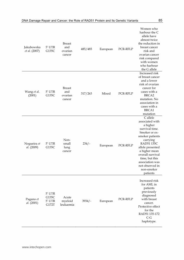
Previous studies have linked the RAD51 135C allele with altered susceptibility to both breast cancer and ovarian cancer. In breast cancer, although a study found no association for the genetic variant (Kuschel, Auranen et al. 2002), Wang et al. (Wang, Spurdle et al. 2001) reported an increased risk of breast cancer and a lower risk of ovarian cancer amongst cases also possessing a BRCA2 mutation, however, no association was seen for individuals known to have a BRCA1 mutation. Apparently conflicting results have been reported by Jakubowska et al. (Jakubowska, Gronwald et al. 2007). These researchers investigated the role of the RAD51 G135C polymorphism in breast and ovarian cancer in case-control populations of Polish women matched for BRCA1 mutation and age. The results revealed that women who harboured the C allele had almost two times reduced risk of breast and ovarian cancer risk compared with women who harboured only the G allele. Moreover, it was shown in this study that the site of the BRCA1 mutation did not influence the effect of the RAD51 C allele, indicating that this polymorphism contributes to prevention of the disease among BRCA1 carriers. These differences in associated risk among for BRCA1 mutation carriers may be due to chance, but also could be explained by the nature of the BRCA1 mutations reported in the two studies. The most common mutation seen in the Jakubowska study was the 5382insC, which results in a truncated protein but which retains an intact RAD51 binding site (Jakubowska, Narod et al. 2003). The primary mutation reported in the Wang study was the 185delAG, which also results in a truncated protein but abolishes the BRCA1-RAD51 binding site. This suggests that for a protective effect to be seen in BRCA1 mutation carriers, the RAD51 interaction site must be present, enabling the RAD51 135C allele to enhance the activity of mutant BRCA1 (Jakubowska, Narod et al. 2003). Another study showed an elevated breast cancer risk associated with the RAD51 135C allele in BRCA2 mutation carriers, but not in BRCA1 mutation carriers (Levy-Lahad, Lahad et al. 2001; Wang, Spurdle et al. 2001). Synowiec et al. (Synowiec, Stefanska et al. 2008) showed previously that the G135C polymorphism was not an independent marker in breast cancer, but it could be associated with an increased breast cancer risk in BRCA2 mutation carriers (Blasiak, Przybylowska et al. 2003; Sliwinski, Krupa et al. 2005), confirming similar results from other studies (Levy-Lahad, Lahad et al. 2001; Antoniou, Sinilnikova et al. 2007). They also observed a protective effect against breast cancer occurrence for the G/C genotype of this polymorphism (OR 0.25; 95% CI 0.10-0.63). The results from a combined analysis of 19 studies revealed an increased risk of breast cancer in the C/C homozygotes with BRCA2 mutation [41]. Jara et al. (Jara, Acevedo et al. 2007) proposed that RAD51 G135C polymorphism presents an increased risk of familial breast cancer in women with age < 50 years at diagnosis, and this polymorphism may be a breast cancer risk variant. This finding should be confirmed in other populations. BRCA2 is required for the orderly assembly of RAD51 on single stranded DNA ends. In the absence of BRCA2, initiation of accurate HR is impaired and repair errors will rapidly accumulate (Powell, Willers et al. 2002). The increased risk associated with the RAD51 135C allele suggests an increase in repair errors. The biological explanation for this is uncertain but may reflect the use of an alternative pathway such as NHEJ (Moynahan, Pierce et al. 2001), or may be a result of error prone HR (Tutt, Bertwistle et al. 2001). Costa et al. (Costa, Pinto et al. 2007) in a case–control study, showed an association of RAD51 135C allele and increased breast cancer risk only among women with family history of breast cancer, suggesting that this polymorphism contributed to the familial breast cancer in
www.intechopen.com

DNA Repair and Human Health
82
the Portuguese population, in opposition to reported results in a Brazilian population (Dufloth, Costa et al. 2005). Concerning sporadic breast cancer risk, similar results to Costa and co-workers findings were obtained by other studies in Australian women (Webb, Hopper et al. 2005) and in the Anglo-Saxon population (Kuschel, Auranen et al. 2002), where no association was obtained. In order to confirm these results, Kadouri et al. (Kadouri, Easton et al. 2001) evaluated the effect of the RAD51 G135C polymorphism on breast cancer risk in BRCA1/2 mutation carriers and in non-carrier breast cancer cases, mainly of Ashkenazi origin. These researchers reported a modifying effect for the RAD51 G135C polymorphism in BRCA2 carriers, similar to the effect shown in two previous studies. This is the first modifier gene identified in BRCA2 carriers. The clinical implication of these findings is still limited; however, it hints at differences in molecular mechanisms involved in tumour development in BRCA1 and BRCA2 carriers. The study of polymorphisms in other DNA repair genes could further elucidate the mechanism of tumorigenesis in BRCA1 and BRCA2 carriers. Recent structural studies suggest a mechanism for the regulation of RAD51 activity by BRCA2, and cancer-associated mutations affecting the domain where RAD51 binds to BRCA2 or reduced level of the protein itself disrupt this interaction leading to impaired DNA repair via HR (Galkin, Esashi et al. 2005; Martin, Winkelmann et al. 2005). Because mutations in the BRCA2 gene may be associated with breast and ovarian cancer and results from multi-site cancer phenotype, genetic variation in the RAD51 gene may contribute to cancer (Martinez, Herzog et al. 2004). Some studies suggest that the G135C polymorphism of the RAD51 gene may have a phenotypic effect, manifested in the changes in the extent of oxidative DNA damage. Recently, HR has been implicated in the repair of stalled replication forks (Michel, Grompone et al. 2004). This type of cellular events can occur as a consequence of oxidation of DNA. DNA double strand breaks (DSBs), which are the main substrate for HR, can arise directly from reactive oxygen species (ROS) (Galli, Piroddi et al. 2005). A study in gastric cancer suggested that that the G135C polymorphism of the RAD51 gene may be linked with gastric cancer by the modulation of the cellular response to oxidative stress. In this work, the authors correlated the genetic constitution expressed by genotypes of the G135C polymorphism with susceptibility to DNA damage and efficacy of DNA repair in human lymphocytes of gastric cancer patients (Poplawski, Arabski et al. 2006). The results of this study suggest that the variants of the G135C polymorphism of the RAD51 gene can be associated with the occurrence of gastric cancer in individuals with a high level of oxidative DNA damage or impaired repair of such damage, which can be a consequence of another genetic variation or/and environmental factor(s). Therefore, this polymorphism can be considered as an additional marker in gastric cancer. However, this study had mainly preliminary character and further research, performed on a larger group, is needed to establish a correlation between gastric cancer and the G/C polymorphism of the RAD51 gene (Poplawski, Arabski et al. 2006). It is known that in humans, inherited defects in HR pathways are known to predispose to acute myeloid leukaemia (AML), an example of this, Fanconi anemia (FA) (Bogliolo, Cabre et al. 2002) is characterized by spontaneous and mutagen-induced chromosome instability. Recently BRCA2, was identified as an FA protein, linking this pathway to HR through the interaction of BRCA2 with RAD51 (Godthelp, Artwert et al. 2002). There is a study that highlights the importance of the link between RAD51, BRCA1, BRCA2 and a risk for AML. An increased risk for AML has been noted in patients previously diagnosed with breast cancer (Pagano, Pulsoni et al. 2001) . Rollinson et al. (Rollinson, Smith et al. 2007) observed a
www.intechopen.com

DNA Damage Repair and Cancer: the Role of RAD51 Protein and Its Genetic Variants
83
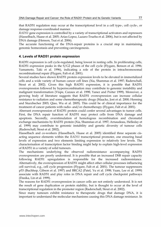
protective effect for the RAD51 135–172 C–G haplotype suggesting that it may be associated with increased RAD51 expression, modulating HR and protecting the cells against aberrant DNA repair events, thus reducing the risk of AML. Therapy-related acute myeloid leukemia (t-AML) is a devastating complication of chemotherapy and/or radiotherapy for a primary cancer. The risk of the development of t-AML was found to be associated with the G-to-C polymorphism at –135 of the 5' untranslated region (135G/C-5'UTR) of RAD51 (Seedhouse and Russell 2007). The promoter activity of the RAD51 gene is enhanced by the G-to-C substitution (135G/C-5'UTR), resulting in high levels of RAD51 expression in individuals with the variation. RAD51's role in t-AML was also supported by an indirect finding that RAD51 was upregulated in mismatch repair-deficient murine embryonic stem cells. This process can be recapitulated by treatment with alkylating agents. Mismatch repair deficiency has been proposed to play an early role in therapy-related carcinogenesis. These data indicate that high levels of RAD51 not only confer resistance to DNA-damaging agents but also contribute to the development of therapy-related cancers (Miyagawa 2008). We previously reported a study evaluating the prognostic and predictive role of RAD51 G135C polymorphism in non-small lung cancer (NSCLC) patients treated with combined platinum taxanes/gemcitabine first line chemotherapy (Nogueira, Catarino et al. 2009). In this study, our results demonstrated that the C allele is associated with a higher survival time, conferring a better prognosis than the GG genotype carrier patients. Thus, individuals carrying the C allele showed a longer overall survival after chemotherapy, compared with individuals carrying the allele G. This study also indicates that the influence of RAD51 G135C polymorphism in treatment response of NSCLC patients seems to be modulated by smoking history. Our results demonstrate that smoker or ex-smoker patients carriers of RAD51 135C allele present a higher mean overall survival time (Nogueira, Catarino et al. 2009). According to the results obtained, we believe that RAD51 genotypes could be useful molecular markers for predicting the clinical outcome of NSCLC patients. The following table shows the main characteristics of some association studies between polymorphisms in the RAD51 gene and risk for cancer (table 2).
6. Conclusion
New factors and pathways with the ability to recognize and repair DSBs are being discovered and studied. DSB production is now recognized as a general occurrence in cells, and these lesions frequently arise through endogenous and exogenous events. As a consequence of evolution from prokaryotes to eukaryotes, cells have developed complex mechanisms which can recognize and repair this type of severe damage rapidly and correctly. Cells have been exposed to many types of environmental stresses, and these stresses can sometimes lead to sub-lethal damage. In order to survive and function under adverse conditions, it is necessary to repair or eliminate DNA damage, and as a consequence, cells have developed a number of complex repair systems to enable their survival and functioning. Knowledge and understanding of these complex systems will make contributions to biology and medicine (Ohnishi, Mori et al. 2009). A recent series of findings established a connection between apoptosis, HR regulation and tumorigenesis. Regulation of RAD51 activity appears to be essential in these regulation networks. It is questionable whether other kinases or signalling processes can affect RAD51 regulation. These data should enhance understanding of the general mechanisms
www.intechopen.com

DNA Repair and Human Health
84
Authors RAD51
SNP Tumoral
model Population (case/control)
Ethnicity Genotyping
methods Results
Synowiec et al. (2008)
5’ UTR G135C
Breast cancer
41/48 European PCR-RFLP
Polymorphism was not an
independent marker in
breast cancer, but it could be associated with
an increased breast cancer
risk in BRCA2 mutation carriers
Costa et al. (2007)
5’ UTR G135C
Breast cancer
285/442 European PCR-RFLP
Association of RAD51 135C
allele and increased breast cancer risk only among women
with family history of
breast cancer. No association
for sporadic breast cancer
risk
Kadouri et al. (2001)
5’ UTR G135C
Breast cancer
309/152 Jewish PCR-RFLP
Elevated risk for breast cancer in carriers of
BRCA2 mutations who also carry a 135
C allele. No association for
BRCA1 carriers. No association
for BRCA1 non-carriers
Jara et al. (2007)
5’ UTR G135C
Breast cancer
143/ 247 South
American PCR-RFLP
Increased risk of familial
breast cancer in women with
age < 50 years at diagnosis
Blasiak et al. (2003)
5’ UTR G135C
Breast cancer
46/60 European PCR-RFLP
No association between the
polymorphism and appearance
and progression of breast cancer
www.intechopen.com
DNA Damage Repair and Cancer: the Role of RAD51 Protein and Its Genetic Variants
85
Jakubowska et al. (2007)
5’ UTR G135C
Breast and
ovarian cancer
485/485 European PCR-RFLP
Women who harbour the C
allele have almost twice
the reduction in breast cancer
risk and ovarian cancer risk compared with women who harbour the G allele
Wang et al. (2001)
5’ UTR G135C
Breast and
ovarian cancer
317/263 Mixed PCR-RFLP
Increased risk of breast cancer
and a lower risk of ovarian
cancer for cases with a
BRCA2 mutation. No association in cases with a
BRCA1 mutation
Nogueira et al. (2009)
5’ UTR G135C
Non-small lung
cancer
234/-
European PCR-RFLP
C allele associated with
a higher survival time. Smoker or ex-
smoker patients carrying
RAD51 135C allele presented a higher mean
overall survival time, but this
association was not observed in
non-smoker patients
Pagano et al. (2001)
5’ UTR G135C 5’ UTR G172T
Acute myeloid
leukaemia 3934/- European
PCR-RFLP
Increased risk for AML in
patients previously diagnosed with breast
cancer. Protective effect
for the RAD51 135-172
C-G haplotype.
www.intechopen.com

DNA Repair and Human Health
86
Rollinson et al. (2007)
5’ UTR G135C 5’ UTR G172T
Acute myeloid leukaemi
a
479/952 European Real-Time
PCR
Protective effect for the RAD51 135-172 C-G haplotype,
reducing the risk of AML.
Poplawski et al.(2006)
5’ UTR G135C
Gastric cancer
30/30 European PCR-RFLP
The variants of the G135C
polymorphism can be
associated with the occurrence
of gastric cancer in
individuals with a high
level of oxidative DNA
damage or impaired repair
of such damage.
Table 2. Main characteristics of some studies included in this review
controlling genome stability, their connections with cell cycle control, apoptosis regulation
and more generally predisposition to tumour development (Daboussi, Dumay et al. 2002).
Most of case-control studies searching for the contribution of genetic alterations within
DNA repair genes to susceptibility to radiation-related cancer have been focused on genes
involved in HR. Additional efforts are needed to find novel genetic variants of DNA repair
genes involved in HR that confer susceptibility to radiation-induced cancer as well as to
confirm already discovered disease-associated variants. To date, significant advances have
been achieved in evaluating the role of genetic variations within DNA repair genes in
clinical radiosensitivity in cancer. RAD51 gene polymorphisms have been suggested to be
associated with radiosensitivity in cancer (Chistiakov, Voronova et al. 2008).
Recently, several national and international clinical research projects have been initiated
to find markers of genetic predisposition to radiation-induced cancer and clinical
radiosensitivity in tumour tissues. However, over the next few years, a considerable
molecular characterization of large-scale cohorts of individuals who show therapeutic
radiation sensitivity is likely to be achieved. The construction and use of genetic-risk profiles
may provide significant improvements in the efficacy of population-based programs
of intervention for cancers. This also should help in predicting radiosensitivity that
will eventually allow individual tailoring of treatment and reduce the risk of developing
acute reactions in anticancer radiotherapy (Kuhne, Riballo et al. 2004; Chistiakov, Voronova
et al. 2008).
7. Acknowledgment
The authors thank the Portuguese League Against Cancer (LPCC-NRNorte) for their support.
www.intechopen.com

DNA Damage Repair and Cancer: the Role of RAD51 Protein and Its Genetic Variants
87
8. References
Antoniou, A. C., O. M. Sinilnikova, et al. (2007). "RAD51 135G-->C modifies breast cancer risk among BRCA2 mutation carriers: results from a combined analysis of 19 studies." Am J Hum Genet 81(6): 1186-200.
Arias-Lopez, C., I. Lazaro-Trueba, et al. (2006). "p53 modulates homologous recombination by transcriptional regulation of the RAD51 gene." EMBO Rep 7(2): 219-24.
Arnaudeau, C., T. Helleday, et al. (1999). "The RAD51 protein supports homologous recombination by an exchange mechanism in mammalian cells." J Mol Biol 289(5): 1231-8.
Arnaudeau, C., C. Lundin, et al. (2001). "DNA double-strand breaks associated with replication forks are predominantly repaired by homologous recombination involving an exchange mechanism in mammalian cells." J Mol Biol 307(5): 1235-45.
Au, W. W., S. A. Salama, et al. (2003). "Functional characterization of polymorphisms in DNA repair genes using cytogenetic challenge assays." Environ Health Perspect 111(15): 1843-50.
Benson, F. E., P. Baumann, et al. (1998). "Synergistic actions of Rad51 and Rad52 in recombination and DNA repair." Nature 391(6665): 401-4.
Blasiak, J., K. Przybylowska, et al. (2003). "Analysis of the G/C polymorphism in the 5'-untranslated region of the RAD51 gene in breast cancer." Acta Biochim Pol 50(1): 249-53.
Bogliolo, M., O. Cabre, et al. (2002). "The Fanconi anaemia genome stability and tumour suppressor network." Mutagenesis 17(6): 529-38.
Brookes, A. J. (1999). "The essence of SNPs." Gene 234(2): 177-86. Buchhop, S., M. K. Gibson, et al. (1997). "Interaction of p53 with the human Rad51 protein."
Nucleic Acids Res 25(19): 3868-74. Chistiakov, D. A., N. V. Voronova, et al. (2008). "Genetic variations in DNA repair genes,
radiosensitivity to cancer and susceptibility to acute tissue reactions in radiotherapy-treated cancer patients." Acta Oncol 47(5): 809-24.
Costa, S., D. Pinto, et al. (2007). "DNA repair polymorphisms might contribute differentially on familial and sporadic breast cancer susceptibility: a study on a Portuguese population." Breast Cancer Res Treat 103(2): 209-17.
Daboussi, F., A. Dumay, et al. (2002). "DNA double-strand break repair signalling: the case of RAD51 post-translational regulation." Cell Signal 14(12): 969-75.
Dasika, G. K., S. C. Lin, et al. (1999). "DNA damage-induced cell cycle checkpoints and DNA strand break repair in development and tumorigenesis." Oncogene 18(55): 7883-99.
Dufloth, R. M., S. Costa, et al. (2005). "DNA repair gene polymorphisms and susceptibility to familial breast cancer in a group of patients from Campinas, Brazil." Genet Mol Res 4(4): 771-82.
Flygare, J., F. Benson, et al. (1996). "Expression of the human RAD51 gene during the cell cycle in primary human peripheral blood lymphocytes." Biochim Biophys Acta 1312(3): 231-6.
Flygare, J., S. Falt, et al. (2001). "Effects of HsRad51 overexpression on cell proliferation, cell cycle progression, and apoptosis." Exp Cell Res 268(1): 61-9.
Galkin, V. E., F. Esashi, et al. (2005). "BRCA2 BRC motifs bind RAD51-DNA filaments." Proc Natl Acad Sci U S A 102(24): 8537-42.
www.intechopen.com

DNA Repair and Human Health
88
Galli, F., M. Piroddi, et al. (2005). "Oxidative stress and reactive oxygen species." Contrib Nephrol 149: 240-60.
Godthelp, B. C., F. Artwert, et al. (2002). "Impaired DNA damage-induced nuclear Rad51 foci formation uniquely characterizes Fanconi anemia group D1." Oncogene 21(32): 5002-5.
Gray, N. K. (1998). "Translational control by repressor proteins binding to the 5'UTR of mRNAs." Methods Mol Biol 77: 379-97.
Haaf, T., E. I. Golub, et al. (1995). "Nuclear foci of mammalian Rad51 recombination protein in somatic cells after DNA damage and its localization in synaptonemal complexes." Proc Natl Acad Sci U S A 92(6): 2298-302.
Hansen, L. T., C. Lundin, et al. (2003). "The role of RAD51 in etoposide (VP16) resistance in small cell lung cancer." Int J Cancer 105(4): 472-9.
Hasselbach, L., S. Haase, et al. (2005). "Characterisation of the promoter region of the human DNA-repair gene Rad51." Eur J Gynaecol Oncol 26(6): 589-98.
Helleday, T., J. Lo, et al. (2007). "DNA double-strand break repair: from mechanistic understanding to cancer treatment." DNA Repair (Amst) 6(7): 923-35.
Henning, W. and H. W. Sturzbecher (2003). "Homologous recombination and cell cycle checkpoints: Rad51 in tumour progression and therapy resistance." Toxicology 193(1-2): 91-109.
Henson, S. E., S. C. Tsai, et al. (2006). "Pir51, a Rad51-interacting protein with high expression in aggressive lymphoma, controls mitomycin C sensitivity and prevents chromosomal breaks." Mutat Res 601(1-2): 113-24.
Hoeijmakers, J. H. (2001). "Genome maintenance mechanisms for preventing cancer." Nature 411(6835): 366-74.
Hu, J. J., H. W. Mohrenweiser, et al. (2002). "Symposium overview: genetic polymorphisms in DNA repair and cancer risk." Toxicol Appl Pharmacol 185(1): 64-73.
Hung, R. J., J. Hall, et al. (2005). "Genetic polymorphisms in the base excision repair pathway and cancer risk: a HuGE review." Am J Epidemiol 162(10): 925-42.
Jakubowska, A., J. Gronwald, et al. (2007). "The RAD51 135 G>C polymorphism modifies breast cancer and ovarian cancer risk in Polish BRCA1 mutation carriers." Cancer Epidemiol Biomarkers Prev 16(2): 270-5.
Jakubowska, A., S. A. Narod, et al. (2003). "Breast cancer risk reduction associated with the RAD51 polymorphism among carriers of the BRCA1 5382insC mutation in Poland." Cancer Epidemiol Biomarkers Prev 12(5): 457-9.
Jara, L., M. L. Acevedo, et al. (2007). "RAD51 135G>C polymorphism and risk of familial breast cancer in a South American population." Cancer Genet Cytogenet 178(1): 65-9.
Johnson, L. F. (1992). "G1 events and the regulation of genes for S-phase enzymes." Curr Opin Cell Biol 4(2): 149-54.
Kadouri, L., D. F. Easton, et al. (2001). "CAG and GGC repeat polymorphisms in the androgen receptor gene and breast cancer susceptibility in BRCA1/2 carriers and non-carriers." Br J Cancer 85(1): 36-40.
Khanna, K. K. and S. P. Jackson (2001). "DNA double-strand breaks: signaling, repair and the cancer connection." Nat Genet 27(3): 247-54.
Klein, H. L. (2008). "The consequences of Rad51 overexpression for normal and tumor cells." DNA Repair (Amst) 7(5): 686-93.
www.intechopen.com

DNA Damage Repair and Cancer: the Role of RAD51 Protein and Its Genetic Variants
89
Knudsen, L. E., S. H. Loft, et al. (2001). "Risk assessment: the importance of genetic polymorphisms in man." Mutat Res 482(1-2): 83-8.
Kuhne, M., E. Riballo, et al. (2004). "A double-strand break repair defect in ATM-deficient cells contributes to radiosensitivity." Cancer Res 64(2): 500-8.
Kuschel, B., A. Auranen, et al. (2002). "Variants in DNA double-strand break repair genes and breast cancer susceptibility." Hum Mol Genet 11(12): 1399-407.
Kuzminov, A. (1999). "Recombinational repair of DNA damage in Escherichia coli and bacteriophage lambda." Microbiol Mol Biol Rev 63(4): 751-813, table of contents.
Lambert, S. and B. S. Lopez (2000). "Characterization of mammalian RAD51 double strand break repair using non-lethal dominant-negative forms." Embo J 19(12): 3090-9.
Lambert, S. and B. S. Lopez (2001). "Role of RAD51 in sister-chromatid exchanges in mammalian cells." Oncogene 20(45): 6627-31.
Lander, E. S., L. M. Linton, et al. (2001). "Initial sequencing and analysis of the human genome." Nature 409(6822): 860-921.
Levine, A. J. (1997). "p53, the cellular gatekeeper for growth and division." Cell 88(3): 323-31. Levy-Lahad, E., A. Lahad, et al. (2001). "A single nucleotide polymorphism in the RAD51
gene modifies cancer risk in BRCA2 but not BRCA1 carriers." Proc Natl Acad Sci U S A 98(6): 3232-6.
Li, X. and W. D. Heyer (2008). "Homologous recombination in DNA repair and DNA damage tolerance." Cell Res 18(1): 99-113.
Lin, F. L., K. Sperle, et al. (1984). "Homologous recombination in mouse L cells." Cold Spring Harb Symp Quant Biol 49: 139-49.
Lin, F. L., K. Sperle, et al. (1984). "Model for homologous recombination during transfer of DNA into mouse L cells: role for DNA ends in the recombination process." Mol Cell Biol 4(6): 1020-34.
Linke, S. P., S. Sengupta, et al. (2003). "p53 interacts with hRAD51 and hRAD54, and directly modulates homologous recombination." Cancer Res 63(10): 2596-605.
Liu, N., J. E. Lamerdin, et al. (1998). "XRCC2 and XRCC3, new human Rad51-family members, promote chromosome stability and protect against DNA cross-links and other damages." Mol Cell 1(6): 783-93.
Liu, Y. and N. Maizels (2000). "Coordinated response of mammalian Rad51 and Rad52 to DNA damage." EMBO Rep 1(1): 85-90.
Lundin, C., N. Schultz, et al. (2003). "RAD51 is involved in repair of damage associated with DNA replication in mammalian cells." J Mol Biol 328(3): 521-35.
Maacke, H., K. Jost, et al. (2000). "DNA repair and recombination factor Rad51 is over-expressed in human pancreatic adenocarcinoma." Oncogene 19(23): 2791-5.
Maacke, H., S. Opitz, et al. (2000). "Over-expression of wild-type Rad51 correlates with histological grading of invasive ductal breast cancer." Int J Cancer 88(6): 907-13.
Magnusson, K. P., M. Sandstrom, et al. (2000). "p53 splice acceptor site mutation and increased HsRAD51 protein expression in Bloom's syndrome GM1492 fibroblasts." Gene 246(1-2): 247-54.
Martin, J. S., N. Winkelmann, et al. (2005). "RAD-51-dependent and -independent roles of a Caenorhabditis elegans BRCA2-related protein during DNA double-strand break repair." Mol Cell Biol 25(8): 3127-39.
Martinez, S. L., J. Herzog, et al. (2004). "Loss of five amino acids in BRCA2 is associated with ovarian cancer." J Med Genet 41(2): e18.
www.intechopen.com

DNA Repair and Human Health
90
Michel, B., G. Grompone, et al. (2004). "Multiple pathways process stalled replication forks." Proc Natl Acad Sci U S A 101(35): 12783-8.
Miyagawa, K. (2008). "Clinical relevance of the homologous recombination machinery in cancer therapy." Cancer Sci 99(2): 187-94.
Moynahan, M. E., A. J. Pierce, et al. (2001). "BRCA2 is required for homology-directed repair of chromosomal breaks." Mol Cell 7(2): 263-72.
Nogueira, A., R. Catarino, et al. (2009). "Influence of DNA repair RAD51 gene variants in overall survival of non-small cell lung cancer patients treated with first line chemotherapy." Cancer Chemother Pharmacol.
Ohnishi, T., E. Mori, et al. (2009). "DNA double-strand breaks: their production, recognition, and repair in eukaryotes." Mutat Res 669(1-2): 8-12.
Ohnishi, T., T. Taki, et al. (1998). "In vitro and in vivo potentiation of radiosensitivity of malignant gliomas by antisense inhibition of the RAD51 gene." Biochem Biophys Res Commun 245(2): 319-24.
Orre, L. M., S. Falt, et al. (2006). "Rad51-related changes in global gene expression." Biochem Biophys Res Commun 341(2): 334-42.
Pagano, L., A. Pulsoni, et al. (2001). "Acute myeloid leukemia in patients previously diagnosed with breast cancer: experience of the GIMEMA group." Ann Oncol 12(2): 203-7.
Paques, F. and J. E. Haber (1999). "Multiple pathways of recombination induced by double-strand breaks in Saccharomyces cerevisiae." Microbiol Mol Biol Rev 63(2): 349-404.
Patel, K. J., V. P. Yu, et al. (1998). "Involvement of Brca2 in DNA repair." Mol Cell 1(3): 347-57.
Pauklin, S., A. Kristjuhan, et al. (2005). "ARF and ATM/ATR cooperate in p53-mediated apoptosis upon oncogenic stress." Biochem Biophys Res Commun 334(2): 386-94.
Poplawski, T., M. Arabski, et al. (2006). "DNA damage and repair in gastric cancer--a correlation with the hOGG1 and RAD51 genes polymorphisms." Mutat Res 601(1-2): 83-91.
Powell, S. N., H. Willers, et al. (2002). "BRCA2 keeps Rad51 in line. High-fidelity homologous recombination prevents breast and ovarian cancer?" Mol Cell 10(6): 1262-3.
Qiao, G. B., Y. L. Wu, et al. (2005). "High-level expression of Rad51 is an independent prognostic marker of survival in non-small-cell lung cancer patients." Br J Cancer 93(1): 137-43.
Qiao, Y., M. R. Spitz, et al. (2002). "Modulation of repair of ultraviolet damage in the host-cell reactivation assay by polymorphic XPC and XPD/ERCC2 genotypes." Carcinogenesis 23(2): 295-9.
Raderschall, E., K. Stout, et al. (2002). "Elevated levels of Rad51 recombination protein in tumor cells." Cancer Res 62(1): 219-25.
Risch, N. J. (2000). "Searching for genetic determinants in the new millennium." Nature 405(6788): 847-56.
Rollinson, S., A. G. Smith, et al. (2007). "RAD51 homologous recombination repair gene haplotypes and risk of acute myeloid leukaemia." Leuk Res 31(2): 169-74.
Saintigny, Y., F. Delacote, et al. (2001). "Characterization of homologous recombination induced by replication inhibition in mammalian cells." Embo J 20(14): 3861-70.
www.intechopen.com

DNA Damage Repair and Cancer: the Role of RAD51 Protein and Its Genetic Variants
91
San Filippo, J., P. Sung, et al. (2008). "Mechanism of eukaryotic homologous recombination." Annu Rev Biochem 77: 229-57.
Schmutte, C., G. Tombline, et al. (1999). "Characterization of the human Rad51 genomic locus and examination of tumors with 15q14-15 loss of heterozygosity (LOH)." Cancer Res 59(18): 4564-9.
Seedhouse, C. and N. Russell (2007). "Advances in the understanding of susceptibility to treatment-related acute myeloid leukaemia." Br J Haematol 137(6): 513-29.
Sliwinski, T., R. Krupa, et al. (2005). "Polymorphisms of the BRCA2 and RAD51 genes in breast cancer." Breast Cancer Res Treat 94(2): 105-9.
Slupianek, A., G. Hoser, et al. (2002). "Fusion tyrosine kinases induce drug resistance by stimulation of homology-dependent recombination repair, prolongation of G(2)/M phase, and protection from apoptosis." Mol Cell Biol 22(12): 4189-201.
Slupianek, A., C. Schmutte, et al. (2001). "BCR/ABL regulates mammalian RecA homologs, resulting in drug resistance." Mol Cell 8(4): 795-806.
Smith, G. C. and S. P. Jackson (1999). "The DNA-dependent protein kinase." Genes Dev 13(8): 916-34.
Sonoda, E., M. S. Sasaki, et al. (1998). "Rad51-deficient vertebrate cells accumulate chromosomal breaks prior to cell death." Embo J 17(2): 598-608.
Sung, P. and H. Klein (2006). "Mechanism of homologous recombination: mediators and helicases take on regulatory functions." Nat Rev Mol Cell Biol 7(10): 739-50.
Synowiec, E., J. Stefanska, et al. (2008). "Association between DNA damage, DNA repair genes variability and clinical characteristics in breast cancer patients." Mutat Res 648(1-2): 65-72.
Szostak, J. W., T. L. Orr-Weaver, et al. (1983). "The double-strand-break repair model for recombination." Cell 33(1): 25-35.
Takahashi, E., Y. Matsuda, et al. (1994). "Chromosome mapping of the human (RECA) and mouse (Reca) homologs of the yeast RAD51 and Escherichia coli recA genes to human (15q15.1) and mouse (2F1) chromosomes by direct R-banding fluorescence in situ hybridization." Genomics 19(2): 376-8.
Tashiro, S., N. Kotomura, et al. (1996). "S phase specific formation of the human Rad51 protein nuclear foci in lymphocytes." Oncogene 12(10): 2165-70.
Tutt, A., D. Bertwistle, et al. (2001). "Mutation in Brca2 stimulates error-prone homology-directed repair of DNA double-strand breaks occurring between repeated sequences." Embo J 20(17): 4704-16.
Venkitaraman, A. R. (2002). "Cancer susceptibility and the functions of BRCA1 and BRCA2." Cell 108(2): 171-82.
Venter, J. C., M. D. Adams, et al. (2001). "The sequence of the human genome." Science 291(5507): 1304-51.
Vispe, S., C. Cazaux, et al. (1998). "Overexpression of Rad51 protein stimulates homologous recombination and increases resistance of mammalian cells to ionizing radiation." Nucleic Acids Res 26(12): 2859-64.
Wang, W. W., A. B. Spurdle, et al. (2001). "A single nucleotide polymorphism in the 5' untranslated region of RAD51 and risk of cancer among BRCA1/2 mutation carriers." Cancer Epidemiol Biomarkers Prev 10(9): 955-60.
Webb, P. M., J. L. Hopper, et al. (2005). "Double-strand break repair gene polymorphisms and risk of breast or ovarian cancer." Cancer Epidemiol Biomarkers Prev 14(2): 319-23.
www.intechopen.com

DNA Repair and Human Health
92
Wick, W., I. Petersen, et al. (1996). "Evidence for a novel tumor suppressor gene on chromosome 15 associated with progression to a metastatic stage in breast cancer." Oncogene 12(5): 973-8.
Xia, S. J., M. A. Shammas, et al. (1997). "Elevated recombination in immortal human cells is mediated by HsRAD51 recombinase." Mol Cell Biol 17(12): 7151-8.
Yamamoto, A., T. Taki, et al. (1996). "Cell cycle-dependent expression of the mouse Rad51 gene in proliferating cells." Mol Gen Genet 251(1): 1-12.
Yanez, R. J. and A. C. Porter (1999). "Gene targeting is enhanced in human cells overexpressing hRAD51." Gene Ther 6(7): 1282-90.
Yuan, S. S., S. Y. Lee, et al. (1999). "BRCA2 is required for ionizing radiation-induced assembly of Rad51 complex in vivo." Cancer Res 59(15): 3547-51.
www.intechopen.com

DNA Repair and Human HealthEdited by Dr. Sonya Vengrova
ISBN 978-953-307-612-6Hard cover, 792 pagesPublisher InTechPublished online 26, October, 2011Published in print edition October, 2011
InTech EuropeUniversity Campus STeP Ri Slavka Krautzeka 83/A 51000 Rijeka, Croatia Phone: +385 (51) 770 447 Fax: +385 (51) 686 166www.intechopen.com
InTech ChinaUnit 405, Office Block, Hotel Equatorial Shanghai No.65, Yan An Road (West), Shanghai, 200040, China
Phone: +86-21-62489820 Fax: +86-21-62489821
Over the past decades, great advances have been made in understanding the cellular DNA repair pathways.At the same time, a wealth of descriptive knowledge of human diseases has been accumulated. Now, thebasic research of the mechanisms of DNA repair is merging with clinical research, placing the action of theDNA repair pathways in the context of the whole organism. Such integrative approach enables understandingof the disease mechanisms and is invaluable in improving diagnostics and prevention, as well as designingbetter therapies. This book highlights the central role of DNA repair in human health and well-being. Thereviews presented here, contain detailed descriptions of DNA repair pathways, as well as analysis of a largebody of evidence addressing links between DNA damage repair and human health. They will be of interest to abroad audience, from molecular biologists working on DNA repair in any model system, to medicalresearchers.
How to referenceIn order to correctly reference this scholarly work, feel free to copy and paste the following:
Augusto Nogueira, Raquel Catarino and Rui Medeiros (2011). DNA Damage Repair and Cancer: The Role ofRAD51 Protein and Its Genetic Variants, DNA Repair and Human Health, Dr. Sonya Vengrova (Ed.), ISBN:978-953-307-612-6, InTech, Available from: http://www.intechopen.com/books/dna-repair-and-human-health/dna-damage-repair-and-cancer-the-role-of-rad51-protein-and-its-genetic-variants

© 2011 The Author(s). Licensee IntechOpen. This is an open access articledistributed under the terms of the Creative Commons Attribution 3.0License, which permits unrestricted use, distribution, and reproduction inany medium, provided the original work is properly cited.