dithiocarbamate-protected ruthenium nanoparticles...
TRANSCRIPT

A
sseoccaTa©
K
1
tocadOsmcia
0d
Available online at www.sciencedirect.com
Electrochimica Acta 53 (2007) 1150–1156
Dithiocarbamate-protected ruthenium nanoparticles: Synthesis,spectroscopy, electrochemistry and STM studies
Wei Chen, Debraj Ghosh, Jia Sun, Moony C. Tong,Fengjun Deng, Shaowei Chen ∗
Department of Chemistry and Biochemistry, University of California,1156 High Street, Santa Cruz, CA 95064, USA
Received 10 October 2006; received in revised form 22 January 2007; accepted 27 January 2007Available online 13 February 2007
bstract
Stable ruthenium nanoparticles were synthesized in a biphasic system with a protecting monolayer of dithiocarbamate derivatives. The coreize of the resulting Ru particles was found to vary with the initial ligand–metal feed ratio. UV–vis spectroscopic measurements showed a Miecattering profile, with no obvious surface-plasmon resonance. The size and crystal structures of the particles were characterized by transmissionlectron microscopic (TEM) measurements. A significant fraction of the nanoparticles was found within the size range of 2–4 nm in diameter andf spherical shape from the TEM measurements. Clear lattice fringes could be observed in high-resolution TEM images with the fringe spacingonsistent with the Ru(1 0 1) lattice planes. Electrochemical studies of Ru particles with different core size exhibited the solution-phase quantizedharging of the particle double layers, analogous to those reported for gold and other transition-metal particles. The potential spacing between
djacent quantized charging peaks was found to vary with the particle core size, corresponding to the variation of the particle molecular capacitance.hese charge-transfer properties were very consistent with the STM measurements of isolated nanoparticles which exhibit clear Coulomb blockadend staircase features.2007 Published by Elsevier Ltd.
ulomb
szpih[aia(
eywords: Ruthenium nanoparticle; Dithiocarbamate; Quantized charging; Co
. Introduction
It has been very well documented that owing to the quan-um mechanical effects, metal nanoparticles in the size rangef 1–10 nm display electronic structures that render their physi-al and chemical properties different from those of bulk metalsnd molecular compounds [1,2]. Numerous protocols have beeneveloped for the synthesis of novel functional nanomaterials.f these, the biphasic method reported by Brust et al. [3] repre-
ents an effective route to the preparation of organically capped
etal nanoparticles. In this synthetic protocol, a metal salt pre-ursor is reduced into metallic form by a strong reducing agentn the presence of appropriate surfactant ligands. The dimensionnd structure of the resulting nanoparticles is the combined con-
∗ Corresponding author. Tel.: +1 831 459 5841; fax: +1 831 459 2935.E-mail address: [email protected] (S. Chen).
td[amdtli
013-4686/$ – see front matter © 2007 Published by Elsevier Ltd.oi:10.1016/j.electacta.2007.01.082
blockade; TEM; STM
equence of at least two competing processes [4], nucleation ofero-valence metals in the formation of the particle cores andassivation of the surfactant molecules onto the core surface thatmpede the growth of the particles. A variety of nanoparticlesave been prepared, e.g., Au [5–7], Pt [8–10], Pd [11–13], Ag14–16], Cu [17–20], and even alloys [21,22]. In addition, whilelkanethiols have been commonly used as the ligands of choicen the stabilization of the nanoparticles because of their strongffinity to transition metal surfaces forming metal–thiolateM–S) bonds, other surfactant molecules with unique func-ional groups have also been used, for instance, isocyanideerivatives have been used to prepare stable Pt nanoparticles23]; Au and Ag nanoparticles have also been prepared by
protecting monolayer of arenethiols [24] and dithiocarba-ates [25], respectively. More recently, it was reported that
iazonium [26] and diazo [27] derivatives could also be usedo stabilize nanoparticles by the formation of metal–carboninkages. The diverse nature of the particle surface protect-ng layers can then be exploited for the ready control of the

ica A
pm
nulctghcIbpfftmbiata
nRitTotcni[tRcotoei
fabtaawBsbttd
ofo
2
2
bbSw(Aawa
2
(1mdRtSscwt3cf22cdwptand then at room temperature overnight. The toluene phase wasthen collected and dried in a rotary evaporator. The Ru particleswere washed several times with Nanopure water and methanol,and then redissolved in dichloromethane (CH2Cl2).
W. Chen et al. / Electrochim
hysical and chemical properties of the resulting nanoparticleaterials.One of the unique properties of these organically capped
anoparticle is the quantized charging to the particle molec-lar capacitance [11,28–30]. With nanometer-sized cores andow dielectric protecting shells, the particles exhibit a molecularapacitance (CMPC) of the order of attofarad (aF), which rendershe energetic barrier for a single electron transfer (e2/2CMPC)reater than the thermal kinetic energy (kBT). Such behaviorsave been observed in STM measurements of isolated nanoparti-les, resulting in the so-called Coulomb staircase charging [31].n solutions, a series of well-defined voltammetric waves cane observed which are ascribed to the discrete charging to thearticle double-layer capacitance [30]. Moreover, it has beenound that when the MPC dimensions decrease, the chargingeatures exhibit a transition from bulk double-layer chargingo molecular-like redox behaviors [30]. It should be noted that
ost of the work on electrochemical quantized charging haseen focused on Au nanoparticles thus far. More recently, sim-lar responses were also observed with Pd [11,12,32], Cu [17],nd Ag [25] particles, thanks to the rapid advance in the syn-hetic end where ultrasmall nanoparticles now become readilyvailable.
In this paper, a series of dithiocarbamate-protected rutheniumanoparticles were prepared by the modified Brust protocol [3].uthenium is well renowned for their catalytic activities. For
nstance, Ru–Pt alloys have been found to be the most effec-ive electrocatalysts used in direct methanol fuel cells [33].hus, an understanding of their electron transfer properties isf critical importance in the optimization of their applications inhese diverse electrocatalytic reaction processes. Several chemi-al methods have been reported for the preparation of rutheniumanoparticles, for instance, by the reduction of ruthenium saltsn water with a strong reducing agent at room temperature34] or by thermolysis in refluxing alcohols [35–37]. In thehermolysis route, it was observed that highly monodisperseu nanoparticles could be obtained with the particle core sizeontrolled by the refluxing temperature and the concentrationf the stabilizing ligands. However, the resulting Ru nanopar-icles were found to be insoluble in a number of commonrganic solvents and the Ru nanoparticles tended to agglom-rate after extensive rinsing, rendering it difficult for furthernvestigations.
In order to overcome this problem, in a recent study [27], weound that by using diazo derivatives which exhibited a strongffinity to freshly prepared Ru surfaces forming Ru-carbene �onds, the resulting Ru nanoparticles could then be transferredo an organic phase (e.g., toluene, hexane and dichlomethane)nd they were stable for an extended period of time withoutpparent variation of the structure and properties. In this paper,e described a complementary approach based on the modifiedrust protocol [3] to prepare stable Ru nanoparticles by the pas-
ivation of dithiocarbamate derivatives. This is partly motivated
y the strong chemisorption of the bidentate moieties on transi-ion metal surfaces [38], which is anticipated to further stabilizehe resulting particles and helps reduce the particle dimensionsuring the particle growth, as observed recently in the synthesiscta 53 (2007) 1150–1156 1151
f nanometer-sized Au [38,39] and Ag [25] nanoparticles. Ourocus here will be on the optical and electrochemical propertiesf these functional nanomaterials.
. Experimental section
.1. Chemicals
Ruthenium chloride (RuCl3, 99+%, ACROS), sodiumorohydride (NaBH4, 99+%, ACROS), tetraoctylammoniumromide (TOABr, 98%, Aldrich), carbon disulfide (CS2, Fishercientific, spectroanalyzed), and didecylamine (98%, Aldrich)ere all used as received. Tetrabutylammonium perchlorate
TBAP, 98%, ACROS) was recrystallized twice prior to use.ll solvents were obtained from typical commercial sources
nd were used without further treatment except for CH2Cl2,hich was freshly distilled prior to use. Water was supplied byBarnstead Nanopure water system (18.3 M�).
.2. Synthesis of ruthenium nanoparticles
The method to prepare didecylamine dithiocarbamateDTC10) has been described by Zhao et al. [38]. Briefly, a0 wt% solution of CS2 was prepared in ethanol, in which oneolar equivalent of didecylamine dissolved in ethanol was added
ropwise under magnetic stirring for 1 min. DTC10 protectedu nanoparticles (Ru-DTC10) were then synthesized according
o the modified Brust reaction [3], which was summarized incheme 1. In a typical reaction, 0.5 mmol RuCl3 was first dis-olved in 0.5 M HCl under vigorous stirring (forming RuCl4−omplexes). A total of 50 ml of toluene with 1.1 g of TOABras then added into the solution where the RuCl4− ions were
ransferred from the aqueous phase to the organic phase after0 min of stirring. The aqueous phase was removed and a cal-ulated amount of DTC10 ligands was added in a dropwiseashion into the solution. The solution was stirred for about0 min in an ice bath and then 0.39 g of NaBH4 dissolved in0 ml H2O was added quickly into the solution. The solutionolor changed rapidly from dark red to dark green and then toark brown. The color change signified that Ru(III) (dark red)as finally reduced to Ru(0) (dark brown), in consistence withrevious observations in the preparation of ruthenium nanopar-icles [34–37]. The solution was stirred for 30 min in the ice bath
Scheme 1. Synthetic procedure of DTC-stabilized ruthenium nanoparticles.

1 ica A
Rtiai
T(
Fe
152 W. Chen et al. / Electrochim
The resulting solution was stirred overnight and a transparentu colloid dispersed in CH2Cl2 was obtained without precipi-
ation. The resulting Ru-DTC10 nanoparticles were very stable
n apolar organic solvents even after extensive washing by waternd methanol. Three batches of particles were prepared with thenitial feed ratio of Ru to DTC10 varied from 1:3, 1:1.5 to 1:0.75.Nsf
ig. 1. Transmission electron micrographs of Ru-DTC10 nanoparticles: (A) (0.75×); (lectron micrographs. The corresponding core diameter histograms are shown to the
cta 53 (2007) 1150–1156
he resulting particles were therefore denoted as Ru-DTC103×), (1.5×), and (0.75×), respectively.
The purity of the particles was then examined by using proton
MR spectroscopy (Varian Unity 500 MHz) with concentratedolutions of the particles dissolved in CDCl3. The lack of sharpeatures indicated the absence of free ligands.
B) (1.5×); (C) (3×). Insets show the corresponding high-resolution transmissionright. The scale bars are all 100 nm.
ica A
2
mhtdcIsu
2
4etapssdt
2
mAoTrcdc
Ftc5
ostdtmSwtOr
3
3.1. Ruthenium nanoparticles
TEM has been a powerful tool in the determination ofnanoparticle dimensions and structures. Fig. 1 shows the TEM
W. Chen et al. / Electrochim
.3. Spectroscopies
Particle core size was measured with a JEOL 1200 EX trans-ission electron microscope (TEM), a Philips CM200/FEG
igh-resolution transmission electron microscope (HRTEM). Inhese measurements, the samples were prepared by casting arop of the particle solution in hexane onto a 200-mesh Formvar-oated copper grid. The particle size was measured by usingmageJ analysis of the TEM micrographs. UV–vis spectroscopictudies were performed with an ATI Unicam UV4 spectrometersing a 1-cm quartz cuvette with a resolution of 2 nm.
.4. Electrochemistry
Voltammetric measurements were carried out with a CHI40 electrochemical workstation. A polycrystalline gold disklectrode (sealed in a glass tubing) was used as the working elec-rode. A Ag/AgCl wire and a Pt coil were used as the referencend counter electrodes, respectively. The gold electrode was firstolished with alumina slurries of 0.05 �m and then cleansed byonication in 0.1 M HNO3, H2SO4 and Nanopure water succes-ively. Before each measurement the electrolyte solution waseaerated by bubbling ultra-high-purity N2 for 20 min and pro-ected with a nitrogen atmosphere during the entire experiments.
.5. STM studies
STM measurements were performed with an Agilent PicoLEicroscope, operated at room temperature in air. Atomically flatu(1 1 1) substrates were first prepared by thermal evaporationnto a freshly cleaved mica at 350 ◦C in vacuo (10−7 Torr).he gold film, about 150 nm in thickness, was cooled down to
oom temperature before being removed out of the depositionhamber. Then the Au substrate was immersed into 1 mMecanethiol in ethanol for 24 h, followed by rinsing withopious amount of ethanol and blow-drying in a gentle stream
ig. 2. UV–vis absorption spectra of the varied ruthenium nanoparticles andhe precursor RuCl3. Ruthenium particles were dissolved in CH2Cl2 with aoncentration of ca. 0.1 mg/ml; whereas the RuCl3 solution was prepared atmM in water.
F1ie1s
cta 53 (2007) 1150–1156 1153
f nitrogen. The particles were deposited onto the substrateurface using the dropcast method. The intercalation betweenhe alkyl chains of the self-assembled monolayers and theithiocarbamate ligands helped immobilize the particles andhus minimize the drifting in STM measurements [40]. A
echanically cut Pt/Ir tip was used in STM measurements.TM images were recorded in constant current mode. I–V dataere collected in the spectroscopy mode by parking the tip on
op of selected particles when the feedback loop was turned off.ne hundred I–V data points were collected in a typical voltage
ange of ±2 V. Every I–V curve was averaged for five times.
. Results and discussion
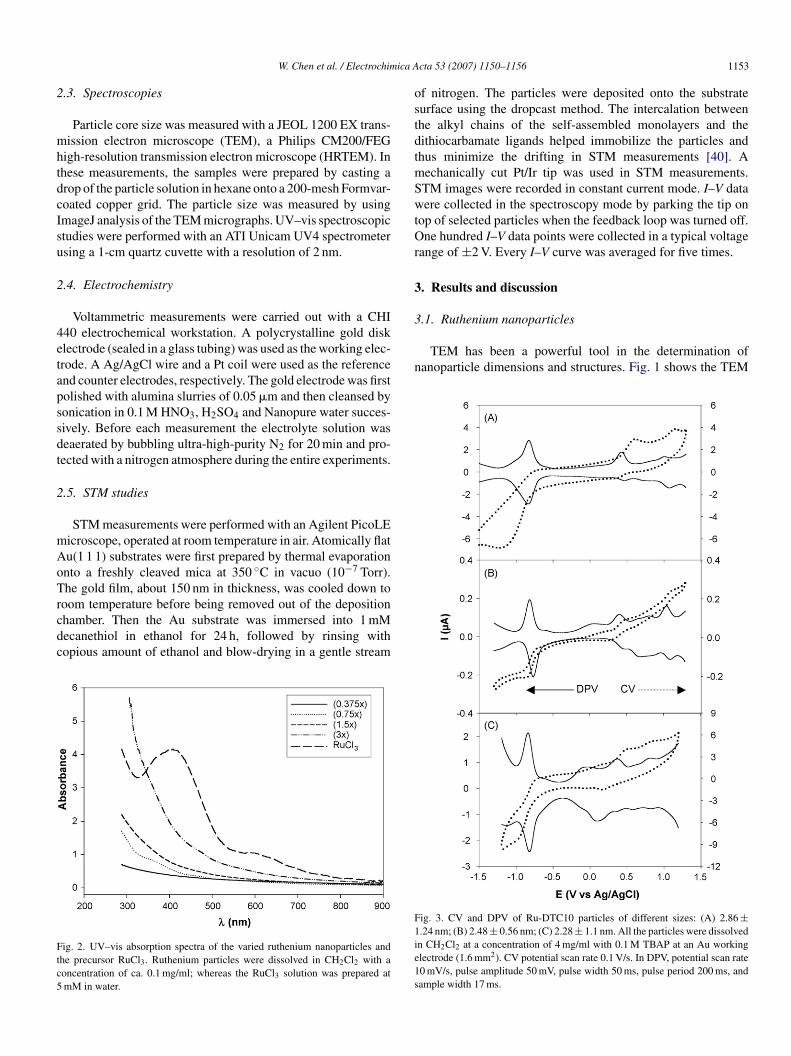
ig. 3. CV and DPV of Ru-DTC10 particles of different sizes: (A) 2.86 ±.24 nm; (B) 2.48 ± 0.56 nm; (C) 2.28 ± 1.1 nm. All the particles were dissolvedn CH2Cl2 at a concentration of 4 mg/ml with 0.1 M TBAP at an Au workinglectrode (1.6 mm2). CV potential scan rate 0.1 V/s. In DPV, potential scan rate0 mV/s, pulse amplitude 50 mV, pulse width 50 ms, pulse period 200 ms, andample width 17 ms.

1 ica A
aaicfti2oawbceismsIiat[
ttnMicwfan
3
3tFmdtwttprbCwattwto0i1f[
3
TF
C
2
2
2
154 W. Chen et al. / Electrochim
nd HRTEM micrographs of the varied Ru-DTC10 particleslong with the corresponding size histograms. From the TEMmages, we can see that in general, the prepared Ru nanoparti-les exhibit spherical shapes with the majority of the particlesalling within the range of 2–4 nm in diameter. Additionally,he particle core size exhibit a slight decrease with increas-ng DTC to Ru feed ratio: (0.75×), 2.86 ± 1.24 nm; (1.5×),.48 ± 0.56 nm; (3×), 2.28 ± 1.1 nm. This is similar to thebservation with gold nanoparticles which are passivated bylkanethiolates [41]. Yet, the core size distribution is some-hat broader than that observed with Ru particles preparedy the thermolysis route [35–37]. From HRTEM images, therystal structures of the Ru-DTC10 nanoparticles were alsoxamined (shown as the corresponding insets). In the Fig. 1Cnset, very clear lattice fringes can be observed, and the fringepacing is found to be approximately 0.206 nm, in good agree-ent with the space between the Ru(1 0 1) lattice planes. This
uggests that the nanoparticles are of single crystal ruthenium.n addition, twin structures can also be seen in the HRTEMmage (e.g., Fig. 1B inset). The two fringe spacings are 0.206nd 0.232 nm, corresponding to the Ru(1 0 1) and Ru(1 0 0) lat-ice planes. Similar observations have been reported previously42].
Fig. 2 shows the UV–vis spectra of the ruthenium nanopar-icles in dichloromethane (also included was the spectrum forhe precursor (RuCl3) in water). It can be seen that for ruthe-ium colloidal solutions, the UV–vis spectra exhibit a typicalie exponential decay profile with no obvious absorption peak,
n consistence with earlier studies [27], whereas the original pre-ursor (RuCl3) shows a broad absorption peak at about 407 nm
hich can be attributable to the ligand–metal charge trans-er. These measurements further confirm that the Ru(III) ionsre completely reduced to zero-valence ruthenium forming theanoparticles.
ad
able 1ormal potentials and capacitance of Ru-DTC10 nanoparticlesa
ore size (nm)b Ep,c (V)c Ep,a (V)c Ep (V
.86 ± 1.24 −0.863 −0.828 −0.840.464 0.424 0.440.724 0.816 0.771.072 1.008 1.04
.48 ± 0.56 −0.870 −0.820 −0.84−0.530 −0.560 −0.54−0.233 −0.202 −0.21
0.097 0.089 0.090.450 0.386 0.410.97 0.99 0.98
.28 ± 1.10 −0.925 −0.940 −0.930.132 0.102 0.110.450 0.404 0.420.808 0.794 0.80
a Peak positions were derived from differential pulse voltammetry (DPV) in Fig. 3b Core size of the particles were determined by the TEM images in Fig. 1.c Subscripts (a and c) denote anodic and cathodic peaks, Ep = (Ep,a + Ep,c)/2, �Ep =d Particle capacitances were evaluated from the average �VC (�VC = e/C).
cta 53 (2007) 1150–1156
.2. Electrochemistry
With the nanoparticle core size averaged between 2 andnm, it is anticipated that the particles in solution will exhibit
he electrochemical quantized charging characteristics [43].ig. 3 shows the cyclic (CV) and differential pulse voltam-ograms (DPV) of Ru-DTC10 particles of different sizes in
ichloromethane (DCM) containing 0.1 M TBAP. It can be seenhat for all particles, there are a series of voltammetric peaksithin the potential range of −1.5 to +1.5 V. These are ascribed
o the quantized charging to the particle molecular capaci-ance, Yet, they are less clearly defined than those observedreviously with highly monodisperse carbene-functionalizeduthenium nanoparticles [27]. From the potential spacing (�V)etween the adjacent charging peaks in DPV, the capacitanceMPC of the Ru nanoparticles can be evaluated by CMPC = e/�V,here e is the electronic charge. Table 1 summarizes the anodic
nd cathodic peak potentials, peak spacing and the capaci-ance of the varied ruthenium nanoparicles. It can be seen thathe capacitance of the Ru nanoparticles decreases somewhatith decreasing core size. For instance, the three representa-
ive samples shown in Fig. 3 exhibit a particle capacitancef 0.537 aF (2.86 nm, 0.75×), 0.507 aF (2.48 nm, 1.5×), and.468 aF (2.28 nm, 3×), respectively. In addition, from Table 1t can be seen that the typical peak splitting (�Ep) is smaller than00 mV, indicating kinetically (quasi) reversible electron trans-er reactions, akin to the observations with gold nanoparticles30].
.3. STM measurements
The nanoparticle single electron transfer properties havelso been examined in STM measurements. Fig. 4 (left panels)epicts two representative STM images of isolated Ru-DTC10
)c �Ep (V)c �Vc (V)c CMPC (aF)d
6 0.0354 0.040 1.2900 0.092 0.326 0.5370 0.064 0.270
5 0.0505 0.031 0.3008 0.031 0.327 0.5073 0.008 0.3118 0.064 0.325
0.020 0.565
3 0.0157 0.030 1.0507 0.046 0.310 0.4681 0.014 0.374
.
|Ep,a − Ep,c|, �VC is the peak spacing between two adjacent peaks.

ica A
(m1ttAwmdtH5
npartsst
Yfttwom
cRdnoaaat
Fat
W. Chen et al. / Electrochim
1.5×) nanopaticles deposited on a decanethiol self-assembledonolayer on a Au(1 1 1) surface in air. At a bias voltage of
.5 V and a small set point current of 20 pA, stable images ofhe particles were acquired at increased distance between theip and the sample [40,44]. Two isolated nanoparticles (labels
and B) were selected, and lateral sizes of 6.0 and 8.5 nmere determined by the line profiles across the particles, whichay be approximated as the particle core diameter plus two
ithiocarbamate ligand chain lengths. As the chain length ofhe dithiocarbamate ligand can be estimated to be 1.4 nm byyperchem®, the corresponding diameter of the two particles is.7 nm (particle A) and 3.2 nm (particle B), respectively.
The corresponding I–V and dI/dV curves of these two Ruanoparticles were shown in Fig. 4 (right panels). For the “A”article, the tunneling current was close to zero when the volt-ge bias varied from −0.40 to +0.40 V. Beyond this voltageange, several current steps can be seen with almost even poten-
ial spacing of 0.4 V between adjacent current steps. For themaller particle “B”, the tunneling spectroscopic measurementshowed a featureless region within the voltage range of −0.8o +0.8 V. Beyond this, some current steps can also be seen.tft
ig. 4. STM images (constant current mode) and scanning tunneling spectroscopy oft room temperature. The left panels are STM images acquired at I = 20 pA and biaswo particles labeled as “A” and “B” to the left. The initial set point is 1.5 V and 50 p
cta 53 (2007) 1150–1156 1155
et, the potential spacing between adjacent current steps areound to vary from 0.4 to 0.5 V. Furthermore, the I–V spec-rum exhibits an asymmetric shape between the currents inhe negative and positive biases. It should be mentioned thathen the STM tip was parked directly on the SAM surface,nly featureless responses were found in the corresponding I–Veasurements.The above measurements indicate that both Coulomb block-
ade and staircase features were observed with nanosizedu particles; and the Coulomb blockade gap increases withecreasing diameter of the Ru nanoparticles. For ultrasmall Ruanoparticle, the large Coulomb blockade suggests the evolutionf a significantly quantized electronic structure, for example, theppearance of a substantial gap between the highest occupiednd lowest unoccupied orbitals in the particle [30]. The observedsymmetry observed with the smaller particle might be ascribedo the nonzero residual charge residing on the particle [45].
For particle aggregates, the corresponding I–V measurementsypically exhibit (almost) linear responses, most likely arisingrom the strong electronic coupling between adjacent particleshanks to the close proximity of the particles.
Ru nanoparticle deposited on decanethiol self-assembled monolayers/Au(1 1 1)= 1.5 V. The right panels are the corresponding I–V and dI/dV profiles for the
A.

1 ica A
4
nBwrsplifp(cccoifi
A
CI
R
[[[[
[
[
[[[
[[[[[
[[
[
[
[[
[
[
[[
[
[[
[
[
[
[
[
[
156 W. Chen et al. / Electrochim
. Summary
In this study, a series of dithiocarbamate-passivated ruthe-ium nanoparticles were synthesized by using the modifiedrust protocol where the particle core size was found to increaseith decreasing feed ratio of the dithiocarbamate ligand to the
uthenium salt precursor. UV–vis spectroscopic measurementshowed a Mie scattering profile, with no obvious surface-lasmon resonance. TEM measurements indicated that a ratherarge fraction of the particles was within the range of 2–4 nmn diameter. In high-resolution TEM measurements, the latticeringes were found to be consistent with the Ru(1 0 1) latticelanes. In electrochemical measurements, quantized capacitanceCoulomb staircase) charging was observed, and the particleapacitance evaluated from the peak spacings between adjacentharging peaks was found to increase with increasing parti-le size, similar to the behaviors with gold nanoparticles. Suchbservations were also consistent with STM measurements ofndividual nanoparticles where the current–potential (I–V) pro-les varied with the particle dimensions.
cknowledgments
This work was supported in part by the NSF (CAREER AwardHE-0456130), ACS–PRF (39729-AC5M), and UC Energy
nstitute.
eferences
[1] A.P. Alivisatos, Science 271 (1996) 933.[2] M.C. Daniel, D. Astruc, Chem. Rev. 104 (2004) 293.[3] M. Brust, M. Walker, D. Bethell, D.J. Schiffrin, R. Whyman, J. Chem. Soc.
Ser. Chem. Commun. (1994) 801.[4] S.W. Chen, A.C. Templeton, R.W. Murray, Langmuir 16 (2000) 3543.[5] R. Guo, D. Georganopoulou, S.W. Feldberg, R. Donkers, R.W. Murray,
Anal. Chem. 77 (2005) 2662.[6] V.L. Jimenez, D.G. Georganopoulou, R.J. White, A.S. Harper, A.J. Mills,
D.I. Lee, R.W. Murray, Langmuir 20 (2004) 6864.[7] Y. Joseph, I. Besnard, M. Rosenberger, B. Guse, H.G. Nothofer, J.M.
Wessels, U. Wild, A. Knop-Gericke, D.S. Su, R. Schlogl, A. Yasuda, T.Vossmeyer, J. Phys. Chem. B 107 (2003) 7406.
[8] W.X. Tu, K. Takai, K. Fukui, A. Miyazaki, T. Enoki, J. Phys. Chem. B 107(2003) 10134.
[9] C. Yee, M. Scotti, A. Ulman, H. White, M. Rafailovich, J. Sokolov, Lang-muir 15 (1999) 4314.
10] S.E. Eklund, D.E. Cliffel, Langmuir 20 (2004) 6012.11] S.W. Chen, K. Huang, J.A. Stearns, Chem. Mater. 12 (2000) 540.12] F.P. Zamborini, S.M. Gross, R.W. Murray, Langmuir 17 (2001) 481.13] C.K. Yee, R. Jordan, A. Ulman, H. White, A. King, M. Rafailovich, J.
Sokolov, Langmuir 15 (1999) 3486.
[
[
[
cta 53 (2007) 1150–1156
14] K.V. Sarathy, G. Raina, R.T. Yadav, G.U. Kulkarni, C.N.R. Rao, J. Phys.Chem. B 101 (1997) 9876.
15] S.T. He, J.N. Yao, P. Jiang, D.X. Shi, H.X. Zhang, S.S. Xie, S.J. Pang, H.J.Gao, Langmuir 17 (2001) 1571.
16] M.J. Rosemary, T. Pradeep, J. Colloid Interf. Sci. 268 (2003) 81.17] S.W. Chen, J.M. Sommers, J. Phys. Chem. B 105 (2001) 8816.18] M. Aslam, G. Gopakumar, T.L. Shoba, I.S. Mulla, K. Vijayamohanan, S.K.
Kulkarni, J. Urban, W. Vogel, J. Colloid Interf. Sci. 255 (2002) 79.19] T.P. Ang, T.S.A. Wee, W.S. Chin, J. Phys. Chem. B 108 (2004) 11001.20] T.Y. Dong, H.H. Wu, M.C. Lin, Langmuir 22 (2006) 6754.21] T.P. Ang, W.S. Chin, J. Phys. Chem. B 109 (2005) 22228.22] X. Gao, K. Tam, K.M.K. Yu, S.C. Tsang, Small 1 (2005) 949.23] S.L. Horswell, C.J. Kiely, I.A. O’Neil, D.J. Schiffrin, J. Am. Chem. Soc.
121 (1999) 5573.24] S.W. Chen, R.W. Murray, Langmuir 15 (1999) 682.25] M.C. Tong, W. Chen, J. Sun, D. Ghosh, S.W. Chen, J. Phys. Chem. B 110
(2006) 19238.26] F. Mirkhalaf, J. Paprotny, D.J. Schiffrin, J. Am. Chem. Soc. 128 (2006)
7400.27] W. Chen, J.R. Davies, D. Ghosh, M.C. Tong, J.P. Konopelski, S. Chen,
Chem. Mater. 18 (2006) 5253.28] S.W. Chen, J. Electroanal. Chem. 574 (2004) 153.29] A.C. Templeton, M.P. Wuelfing, R.W. Murray, Acc. Chem. Res. 33 (2000)
27.30] S.W. Chen, R.S. Ingram, M.J. Hostetler, J.J. Pietron, R.W. Murray, T.G.
Schaaff, J.T. Khoury, M.M. Alvarez, R.L. Whetten, Science 280 (1998)2098.
31] R.S. Ingram, M.J. Hostetler, R.W. Murray, T.G. Schaaff, J.T. Khoury, R.L.Whetten, T.P. Bigioni, D.K. Guthrie, P.N. First, J. Am. Chem. Soc. 119(1997) 9279.
32] Y.G. Kim, J.C. Garcia-Martinez, R.M. Crooks, Langmuir 21 (2005) 5485.33] H.S. Liu, C.J. Song, L. Zhang, J.J. Zhang, H.J. Wang, D.P. Wilkinson, J.
Power Sources 155 (2006) 95.34] J. Yang, J.Y. Lee, T.C. Deivaraj, H.P. Too, J. Colloid Interf. Sci. 271 (2004)
308.35] S. Gao, J. Zhang, Y.F. Zhu, C.M. Che, New J. Chem. 24 (2000) 739.36] N. Chakroune, G. Viau, S. Ammar, L. Poul, D. Veautier, M.M. Chehimi,
C. Mangeney, F. Villain, F. Fievet, Langmuir 21 (2005) 6788.37] G. Viau, R. Brayner, L. Poul, N. Chakroune, E. Lacaze, F. Fievet-Vincent,
F. Fievet, Chem. Mater. 15 (2003) 486.38] Y. Zhao, W. Perez-Segarra, Q.C. Shi, A. Wei, J. Am. Chem. Soc. 127 (2005)
7328.39] M.S. Vickers, J. Cookson, P.D. Beer, P.T. Bishop, B. Thiebaut, J. Mater.
Chem. 16 (2006) 209.40] G.H. Yang, L. Tan, Y.Y. Yang, S.W. Chen, G.Y. Liu, Surf. Sci. 589 (2005)
129.41] M.J. Hostetler, J.E. Wingate, C.J. Zhong, J.E. Harris, R.W. Vachet, M.R.
Clark, J.D. Londono, S.J. Green, J.J. Stokes, G.D. Wignall, G.L. Glish,M.D. Porter, N.D. Evans, R.W. Murray, Langmuir 14 (1998) 17.
42] T.W. Hansen, J.B. Wagner, P.L. Hansen, S. Dahl, H. Topsoe, C.J.H. Jacob-sen, Science 294 (2001) 1508.
43] S.W. Chen, R.W. Murray, S.W. Feldberg, J. Phys. Chem. B 102 (1998)9898.
44] H.A. Wierenga, L. Soethout, J.W. Gerritsen, B.E.C. Vandeleemput, H.Vankempen, G. Schmid, Adv. Mater. 2 (1990) 482.
45] K. Majumdar, S. Hershfield, Phys. Rev. B 57 (1998) 11521.