disorders of the gastrointestinal system & liver university of san francisco dr. m. maag ©2003...
TRANSCRIPT

Disorders of the Gastrointestinal System & Liver
University of San Francisco
Dr. M. Maag©2003 Margaret Maag

2
Class 12 Objectives• Upon completion of this lesson, the student will be
able to– list the pathologies associated with GI motility.– determine the infectious agents associated with GI
disorders.– predict those at risk for GI bleeding and the S & S these
individuals could present.– analyze the clinical manifestations of severe liver
impairment.– state the normal clotting mechanisms and the role
vitamin K plays in blood clot formation.– state the cause(s) of DIC and list the S&S.

3
Pathologies of GI Motility• Diarrhea• Is an > in frequency, fluid and / or volume of
stool – Osmotic: the presence of nonabsorbable
substances in the intestine causing water to be drawn into the lumen by osmosis
• sorbitol-containing liquid medications; tube feedings • lactose intolerance
– Secretory: excessive mucosal secretion of fluid & electrolytes
• related to: gastroenteritis (E. Coli), rotavirus, laxative abuse, hyponatremia, fecal impaction

4
Pathologies of GI Motility
• Diarrhea• Motile: > motility is d/t stimulation caused by
inflammation or obstruction• resection of small intestine, fecal impaction, early
bowel obstruction (e.g. Bezor)
• Clinical Manifestations: • crampy abdominal pain, > bowel sounds • prolonged diarrhea leads to f & e imbalances and
dehydration • infants & elderly are at risk: check hydration & f/e
status

5
Case Study• A 72 year-old woman, who lives alone, has a
history of laxative abuse. What type of diarrhea is she at risk for? What type of fluid imbalance is she at risk for?
• What would you expect her VS to be?• Her electrolytes upon admission to the hospital
are: Na+ = 155; K+ = 3.5; Cl- = 116; Hct = 45% • Clinical manifestations? Treatment? • Which acid-base disturbance is she at risk for?
Why?

6
Pathologies of GI Motility• Constipation• Defined as infrequent or difficult defecation:
most frequently c/o digestive disorder • Etiology : functional disorder of bowel
motility • incidence is > in the elderly; diet poor in fiber &
fluids; anatomic lesions; drug therapy • d/t poor neural stimulation of GI motility, abdominal
muscle weakness, bowel obstruction• Mega colon, opiates, hypothyroidism, diabetic
neuropathy, sedentary lifestyle, low residue diet

7
Pathologies of GI Motility• GERD• Reflux of gastric contents into lower esophagus resulting in
clinical symptoms or structural alterations in the esophageal tissues (reflux esophagitis)
• 94% of the individuals have hiatal hernias • a protrusion of some part of the upper portion of
stomach through esophageal hiatus and then into the thorasic cavity
• Delayed gastric emptying is seen primarily in: • diabetics, cigarette smoking, and ETOH abuse • dysphagia, eructation, heartburn, GI bleeding,
abdominal discomfort when lying down, dyspnea may be present
• Heartburn, ulcerations, precancerous lesions

8
Fecal Incontinence
• Inadequate control of defecation in an adult due to weak pelvic floor muscles and / or weakness of the external anal sphincter
• Common causes: • Clostridium difficile responsible for nosocomial
diarrhea• Impaction, laxative abuse, hyperosmolar tube
feedings
• Risk factors: older persons in long-term care institutions (Bliss, et al., 2000)

9
Intestinal Obstructions
• Large Bowel• A large bowel obstruction is an emergency
condition that requires early & prompt surgical intervention
• Etiology: • infectious / inflammatory, neoplastic, or mechanical
pathology (colorectal cancer)
• Rotation or twisting of the cecum or sigmoid colon will cause abrupt onset of symptoms
• Immediate abdominal distention– Decreases the ability to absorb F & E

10
Intestinal Obstructions• Sigmoid volvulus usually seen in the older
individual with a hx of straining at stool • Symptoms: abdominal distention, nausea,
vomiting, and crampy abdominal pain; check history of flatus and BMs
• Abrupt onset is indicative of an acute obstruction– Sudden onset due to torsion or hernia
• A chronic hx of constipation is related to a dx of diverticulitis or carcinoma
• Obstipation (no flatus or BM) & loss of weight = carcinoma

11
Intestinal Obstructions• Paralytic ileus or “silent bowel” is most often
seen after abdominal surgery & anesthesia • bowel activity is < d/t lack of neural stimuli
(“functional”) • this can lead to “mechanical” obstruction d/t
accumulation of feces
• Hernias: a loop of bowel protrudes through abdominal wall
• inguinal canal, umbilicus, or incisional scar tissue • caused by heavy lifting, straining, or coughing

12
Disorders of GI Bleeding• Upper: includes the esophagus, stomach,
duodenum • peptic ulcer disease (PUD) or esophageal varices
• Lower: includes the jejunum, ileum, colon, rectum • colorectal cancer, polyps, hemorrhoids, IBD
• Manifestations: • hematemesis• bright red blood in the stool (“hematochezia”) • black,dark, tarry stools (“melena”) • “occult” bleeding (invisible blood in the stool)
• Tx: find the underlying cause; fluid volume replacement; endoscopy or colonoscopy; medical and /or surgical tx

13
Disorders of GI Bleeding
• Results• Shock will ensue if massive (25% EBL
within hours) bleeding occurs• Metabolic acidosis, prerenal failure,
bowel infarction will occur• < coronary & cerebral blood flow• Death
– See McCance, Figure 38-1, p. 1265

14
Peptic Ulcer Disease
• An inflammatory disorder causing deep erosion of stomach or duodenal mucosa by HCL & pepsin
• At risk: infection with H. pylori; > NSAIDS; > secretion of HCL as seen in Zollinger-Ellison syndrome
• Etiology: age, family hx– > mucolytic enzymes; may lead to pyloric obstruction,
bowel perforation and ultimately peritonitis
• Sx: hallmark sign = upper gastric pain– Emergency:hematemesis, melena, occult blood, shock

15
Peptic Ulcer Disease
• Treatment includes: – < stress – < ETOH intake – screen for H. pylori (C-urea breath test) – frequent small meals – avoid calcium based antacids d/t > gastrin
release – H2 blockers (Tagamet & Zantac) – Insert NG tube for severe bleeding and gastric
lavage

16
Gastric, Duodenal, Stress Ulcers
• Gastric– > cancer risk– Lack of remission or exacerbation periods
• Duodenal– Younger age at onset– Strong familial history– Ulcerogenic drugs used– Nocturnal pain more prevalent
• Stress– Systemic trauma, severe illness, neural injury

17
Intestinal Bowel Disorders
• Ulcerative Colitis– Inflammatory disorder with eventual
erosion of the colon
• Crohn’s Disease– Effects any part of the GI (mouth to anus)– Smoking, diet, and/or bacteria may influence– Cytokine-mediated damage

18
Gastric Cancer
• Adenocarcinoma is the primary malignant neoplasm
• 8th leading cause of mortality r/t cancer in US • Epidemiology: 55-60 year olds; 2 times greater
incidence in men vs. women • Risk factors: H. pylori, < socioeconomic class,
consumption of pickled foods, improper food storage, radiation exposure
• Etiology:chronic inflammation, dietary influences, genetic & environmental factors

19
Gastric Cancer
• Sx: Vague early sx with weight loss; indigestion; abdominal distention; mild pain induced with or without food; chronic blood loss leads to anemia; occult blood in stool
• Tx: reduce risk factors; total or partial gastrectomy; lymph node resection; chemotherapy & radiation
• 15% of cases lend a 5-year survival rate

20
Peritonitis• Etiology: Inflammation of peritoneal membrane
• sterile: rupture of biliary system; hemorrhagic pancreatitis, endometriosis, surgical procedures
• infectious: bacteria from ruptured bowel or appendix; introduction of bacteria from abdominal trauma or
invasive procedures (e.g. peritoneal dialysis)
• Sx: Circulatory volume collapse, septic shock causing a high mortality rate, absent bowel sounds, pain, abdominal distention & rigidity, > nausea & vomiting, paralytic ileus
• Tx: antibiotics, NG tube, IV fluids, surgical repair of etiology

21
Colorectal Cancer• “Patients with long-standing ulcerative colitis
have been shown to be at increased risk of developing colorectal cancer” (Medscape, 1999)
• Involves a primary malignant tumor of the rectum or colon
• 2nd leading cause of cancer death in US • > incidence in 50 year olds • > fat and poor fiber diet; > ETOH consumption;
cigarette smoking; obesity; sedentary life style
• Exact etiology unknown…> incidence with polyps

22
Colorectal Cancer
• Symptoms: – fecal occult blood or ulcerative lesions manifest
as anemia or rectal bleeding • distention, abdominal pain, vomiting, constipation
– metastatic disease: weight loss, anorexia, possible palpable mass
• Prevention: ASA may < risk; routine monitoring for guaic (+)
• Tx: colostomy repair; permanent colostomy for rectal tumors

Liver Disorders

24
Portal Hypertension
• Fibrosis of the liver structures causes an increased resistance to blood flow within the liver, therefore an elevation in the portal venous pressure– This > in pressure can cause esophageal varices
and hemorrhoids and 3rd spacing of fluid into peritoneal cavity (ascites)
• “Hepatic encephalopathy” can occur d/t the toxic effects of altered metabolism – cerebral edema & IICP can result from
severe cases

25
Ascites
• A common feature of liver failure.
• Basic mechanisms include:– an increase in portal hypertension– sodium and water retention– decreased blood oncotic pressure
secondary to a low serum albumin level

26
Cirrhosis• Focal or diffuse inflammation and liver cell
necrosis that causes severe changes in the structure and function of liver cells
• Inflamed liver cells compress the liver lobule and cause increased resistance to blood flow and portal hypertension– Liver tissue is regenerated, but not in the normal
fashion – Fibrotic changes are irreversible, causing liver
dysfunction

27
Cirrhosis
• Alcoholic: results from long-term alcohol abuse; most common cause in the USA
• Biliary: caused by a < in bile flow; commonly caused by long-term obstruction of bile ducts
• Cardiac: caused by long-term right-sided CHF– results in < oxygenation of liver cells
• Postnecrotic: result from hepatoxins, chemicals, or infection with Hepatitis B or C– massive death of liver cells & associated with
cancer

28
Viral Hepatitis • Inflammation of the liver followed by the necrosis of
hepatic cells– Caused by infection with one or more hepatoviurses
• Types: A, B, C, D, E & G• Little is known about the blood-borne “G”
– Hepatic inflammation may occur d/t toxins, autoimmunity, and metabolic disorders
• http://www.roche.com/pages/facets/5/hepatitis.htm

29
Viral Hepatitis
• HAV is found primarily in contaminated food and water– Transmitted by the enteric route (oral-fecal)– Poor hand washing or unsanitary food preparation– During the viremic phase of acute infection it can be
spread via blood exposure (unusual)– Virus infects the liver and is excreted via the feces– Most contagious before presentation of S & S– Prevalence of immunity to HAV has decreased to
< 25% of US adults (DiCarlo, 1999)

30
Hepatitis A Virus
• Hepatitis A antibodies show up in the blood 2-6 weeks following exposure & remain indefinitely in the blood
• Clinical manifestations: fever, chills, brown urine, anorexia, irritability, clay-colored feces, N&V, headache
• Liver function tests & coagulation tests are abnormal

31
Hepatitis B Virus
• HBV is transmitted via blood & body fluids– “Infected adults have a 50% chance of
developing acute symptoms, but only a 10% chance of developing chronic infection” (DiCarlo, 1999)
– In the US, 60% of hepatitis B virus infections are sexually transmitted• unprotected sex with multiple partners
– A vaccine has been available since 1982• immunity develops in more than 90%

32
Hepatitis B Virus
• Hepatitis antigen-antibody complexes can be detected from 1-10 weeks after exposure to the virus
• Incubation period for HBV can last from 6 weeks to 6 months; clinical S & S of the acute phase are the same as HAV
• Patients have an > chance of “fulimant hepatic failure”…a sudden degeneration of the liver & loss of all normal liver functions
33
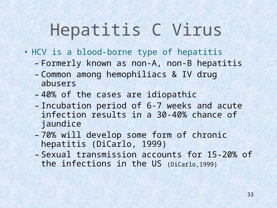
Hepatitis C Virus• HCV is a blood-borne type of hepatitis
– Formerly known as non-A, non-B hepatitis– Common among hemophiliacs & IV drug abusers– 40% of the cases are idiopathic– Incubation period of 6-7 weeks and acute
infection results in a 30-40% chance of jaundice– 70% will develop some form of chronic hepatitis
(DiCarlo, 1999)– Sexual transmission accounts for 15-20% of the
infections in the US (DiCarlo,1999)

34
Other Types of Viral Hepatitis
• HDV is also known as the “delta virus” – It is a blood-borne virus that must coexist
with HBV in order to exert its viral activity– This covirus heightens the course and
outcome of illness with HBV
• HEV is the “enteric” form of non-A, non-B hepatitis – is generally seen in underdeveloped
countries

35
Precautions• Use of gloves while handling all items
contaminated with client’s body secretions• Use of disposable patient care items, such as
thermometers, dishes, eating utensils• Use of private bathroom and room for clients
who are incontinent of feces.• Double bagging and labeling of linen or any
hospital equipment that is contaminated with feces or blood (Hartshorn, 1997, p. 462)

36
Clotting Factor Defects
• Review normal function of clotting factors• Inherited disorders: deficiencies of clotting factors
– Hemophilia's– Willebrand disease
• Acquired cases– Deficient synthesis of clotting factors by liver– Liver disease, dietary deficiency of Vitamin K
• Factor 7 is first to decline then factor 2 and 10
– Thrombocytopenia may occur due to splenomegaly liver disease and portal hypertension

37
Disseminated Intravascular Coagulation
• Acquired complex clinical syndrome– Due to > protease activity in the blood
caused by > release of thrombin– Acute, severe,life-threatening process– Massive hemorrhage and thrombosis– Becomes a chronic, low-grade condition
• Minor lab abnormalities with sub acute hemorrhage and microcirculatory thrombosis
• May involve many organs
38
References
• Bliss, D. Z., Johnson, S., Savik, Clabots, C. R., & Gerding, D. N. (2000). Fecal incontinence in hospitalized patients who are acutely ill. Nursing Research, 49(2),.101-108.
• Hansen, M. (1998). Pathophysiology: Foundations of disease and clinical intervention. Philadelphia: Saunders.
• http://www.medscape.com