diseases of the pancreas - dailymedicine.weebly.com
TRANSCRIPT
1 Diseases of Pancreas
Diseases of the Pancreas
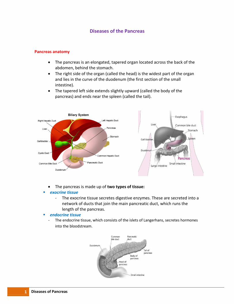
Pancreas anatomy
The pancreas is an elongated, tapered organ located across the back of the abdomen, behind the stomach.
The right side of the organ (called the head) is the widest part of the organ and lies in the curve of the duodenum (the first section of the small intestine).
The tapered left side extends slightly upward (called the body of the pancreas) and ends near the spleen (called the tail).
The pancreas is made up of two types of tissue: exocrine tissue
- The exocrine tissue secretes digestive enzymes. These are secreted into a network of ducts that join the main pancreatic duct, which runs the length of the pancreas.
endocrine tissue - The endocrine tissue, which consists of the islets of Langerhans, secretes hormones
into the bloodstream.
2 Diseases of Pancreas
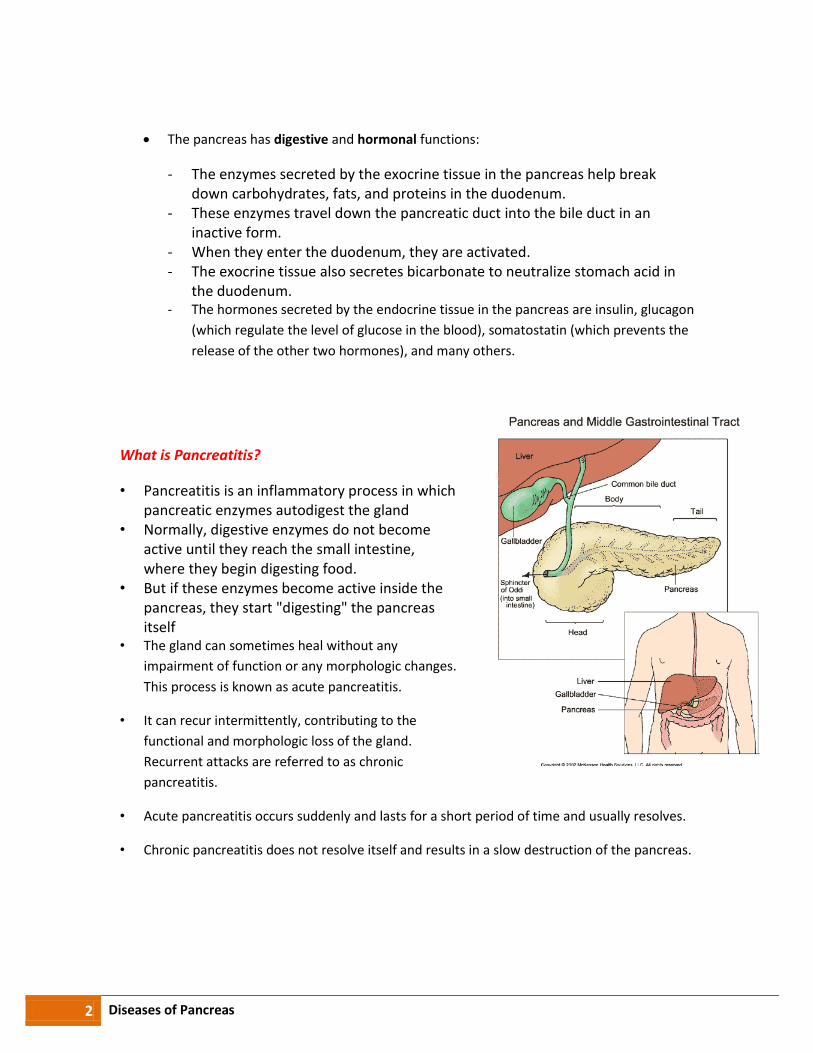
The pancreas has digestive and hormonal functions:
- The enzymes secreted by the exocrine tissue in the pancreas help break down carbohydrates, fats, and proteins in the duodenum.
- These enzymes travel down the pancreatic duct into the bile duct in an inactive form.
- When they enter the duodenum, they are activated. - The exocrine tissue also secretes bicarbonate to neutralize stomach acid in
the duodenum. - The hormones secreted by the endocrine tissue in the pancreas are insulin, glucagon
(which regulate the level of glucose in the blood), somatostatin (which prevents the
release of the other two hormones), and many others.
What is Pancreatitis?
• Pancreatitis is an inflammatory process in which pancreatic enzymes autodigest the gland
• Normally, digestive enzymes do not become active until they reach the small intestine, where they begin digesting food.
• But if these enzymes become active inside the pancreas, they start "digesting" the pancreas itself
• The gland can sometimes heal without any
impairment of function or any morphologic changes.
This process is known as acute pancreatitis.
• It can recur intermittently, contributing to the
functional and morphologic loss of the gland.
Recurrent attacks are referred to as chronic
pancreatitis.
• Acute pancreatitis occurs suddenly and lasts for a short period of time and usually resolves.
• Chronic pancreatitis does not resolve itself and results in a slow destruction of the pancreas.
3 Diseases of Pancreas
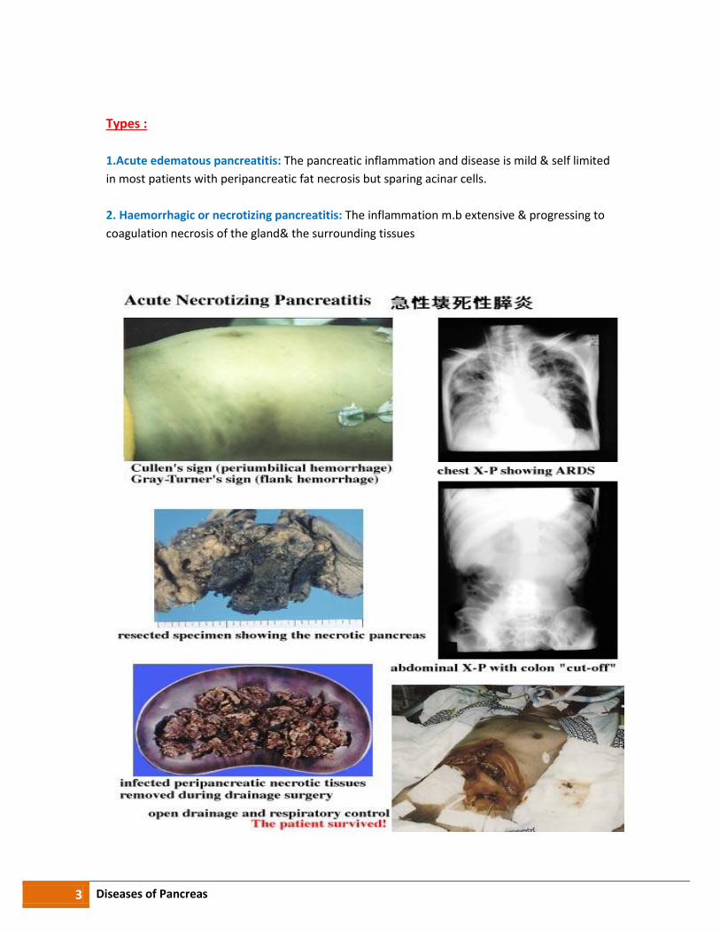
Types :
1.Acute edematous pancreatitis: The pancreatic inflammation and disease is mild & self limited
in most patients with peripancreatic fat necrosis but sparing acinar cells.
2. Haemorrhagic or necrotizing pancreatitis: The inflammation m.b extensive & progressing to
coagulation necrosis of the gland& the surrounding tissues

4 Diseases of Pancreas
Acute Pancreatitis • About 80,000 cases occur in the United States each year; some 20 percent of them
are severe. • Acute pancreatitis occurs more often in men than women.
Causes:
A) Common (90% of cases)
1. Gallstones 2. Alcohol 3. idiopathic 4. Post ERCP
B) Rare
1. Post surgical (abdominal and cardiopulmonary bypass) 2. Trauma 3. Drugs (azathioprine , thiazide diuretics) 4. Metabolic ( Ca , TG) 5. Pancreas divisum 6. Hereditary
Clinical features:
• Abdominal pain :Severe constant upper abdominal pain may radiate to back , usually build up over 15-16 mins
• N & V • Market epigastric tenderness • N.B in the early stages and in contrast to perforated P.U , guarding and rebound
tenderness are absent because the inflammation is retro peritoneal • Bowel sounds become quite or absent as paracletic ileus develops • In severe cases the patient becomes hypoxic and develops hypovolemic shock with
oliguria • Discoloration of the flanks (Grey Turner’s sign) or the periumbilical region (Cullen’s
sign) are features of Hgic pancreatitis
D.D:
1. perforated viscus 2. M.I 3. Acute cholecystitis
7. Renal failure
8. Organ transplantation (kidney ,
liver ) 9. Infection (mumps , coxsackie virus)
10. Sphincter of Oddi dysfunction
11. Severe hypothermia
12. Petrochemical exposure
This is Grey-Turner's sign with haemorrhage appearing in
both flanks. It is due to extensive retro-peritoneal bleeding
and typically occurs in haemorrhagic pancreatitis

5 Diseases of Pancreas
Complications
Diagnosis
• History • Physical exam • Lab Studies
- During acute attacks, the blood contains at least three times more amylase and lipase than usual. Amylase and lipase are digestive enzymes formed in the pancreas.
- Changes may also occur in blood levels of glucose, calcium, magnesium, sodium, potassium, and bicarbonate.
- After the pancreas improves, these levels usually return to normal. • Based on increased S. amylase or lipase + U/S or C.T evidence of pancreatic swelling
Complications Cause
(A) Systemic
1. SIRS Increased vascular permeability d.t Cytokine , Kinin release, PAF , Paralytic ileus, vomiting & RF
2. Hypoalbuminaemia Increased capillary permeability
3. Hypoxia ARDS d.t microthrombi in pulm. Vs
4. Hyperglycaemia Disruption of islets of Langerhans
5. hypocalcaemia. Sequestration of Ca in fat necrosis
(B) Pancreatic
1. Abscess tissue Infection of necrotic pancreatic .
2. Pancreatic ascites or pleural effusion
disruption of pancreatic ducts
3. Pseudocyst disruption of pancreatic ducts
(C) GIT
1. UGI bleeding Gastric or duodenal erosions
2. Variceal hge Splenic or PVT
3. Duodenal obstruction Compression by pancreatic mass
4. Obstructive jaundice Compression of CBD

6 Diseases of Pancreas
1)Serum amylase (x3 times normal)
- M.b normal after 24-48 hours of onset - So check lipase or urinary amylase : creatinine ratio - Persistent elevation = suggests pseudocyst formation - Peritoneal amylase increase in pancreatic ascites
2) U/S
- Confirm diagnosis - May also shows : gallstones , pseudocyst , biliary obstruction
3) C.T : define viability of the pancreas
4) Hyperglycemia and decreased Ca
5) S.bili , ALT , AST , alk . ph : are transiently increased
6) Serial assessment of CRP
7 Diseases of Pancreas
Ranson Scale
• Ranson developed a series of different criteria for the severity of acute pancreatitis • For the following catagories-
- answer each question regarding the patient then add up total score for
prognosis
If answer is no (o point)
If answer is yes (1 point) • Present on admission
- Older than 55 years - WBC higher than 16,000 per mcL - Blood glucose higher than 200 mg/dL - Serum lactate dehydrogenase (LDH) more than 350 IU/L - SGOT (ie, aspartate aminotransferase [AST]) greater than 250 IU/L
• Developing during the first 48 hours
- Hematocrit fall more than 10% - BUN increase more than 8 mg/dL - Serum calcium less than 8 mg/dL - Arterial oxygen saturation less than 60 mm Hg - Base deficit higher than 4 mEq/L - Estimated fluid sequestration higher than 600 mL
• A Ranson score of 0-2 has a minimal mortality rate. • A Ranson score of 3-5 has a 10%-20% mortality rate. • A Ranson score higher than 5 has a mortality rate of more than 50% and is associated
with more systemic complications
Management:
- NPO(nothing by mouth . Give parenteral nutrition - Analgesic as pethidine - Correction of hypovolemia by normal saline - CVP & urinary catheter to monitor shocked Pts. - O2 for hypoxia - ARDS may require ventilatory support - Insulin for hyperglycemia - I.V Ca if tetany occur - Nasogastric aspiration for paralytic ileus - Prophylaxis of thromboembolism low dose S.0 heparin - Pts. Who present with cholangitis or jaundice in assesment. With severe acute
Pancreatitis urgent ERCP to remove stone & drain biliary system - Prophylactic Abs

8 Diseases of Pancreas
TTT of complications:
- Necrotizing pancreatitis or pancreatic abscess urgent surgical debridement of the
pancreas
- Pancreatic pseudocysts drainage into stomach or duodenum
- Pancreatic ascites is indication for distal pancreatectomy
Lab Studies
- A complete blood count (CBC) demonstrates leukocytosis (WBC >12000) with the differential being shifted towards the segmented polymorphs.
- If blood transfusion is necessary, as in cases of hemorrhagic pancreatitis, obtain type and crossmatch.
- Measure blood glucose level because it may be elevated from B cell injury in the pancreas.
- Obtain measurements for BUN, creatine (Cr), and electrolytes (Na, K, Cl, CO2, P, Mg); a great disturbance in the electrolyte balance is usually found, secondary to third spacing of fluids
- Measure amylase levels, preferably the Amylase P, which is more specific to pancreatic pathology. Levels more than 3 times higher than normal strongly suggest the diagnosis of acute pancreatitis
- Lipase levels also are elevated and remain high for 12 days. In patients with chronic pancreatitis (usually caused by alcohol abuse), lipase may be elevated in the presence of a normal serum amylase level
Imaging Studies
A. Ultrasound
- can be used as a screening test. - If overlying gas shadows secondary to bowel distention are present, it may
not be specific. - CT scan is the most reliable imaging modality in the diagnosis of acute
pancreatitis.
B. Pancreatitis, Acute - CT Scan
9 Diseases of Pancreas
Chronic pancreatitis
Definition:
Chronic inflammatory disease caused by Fibrosis & destruction of exocrine pancreatic tissue
D.M occurs in advanced cases because The islets of Langerhans are involved.
Classification :
1. Obstructive C P 2. Calcific C P
Causes of Chronic pancreatitis
• Calcific : - Alcoholism - Tropical(? Related to malnutrition or dietary toxins)
• Obstructive: Stenosis of ampulla of vater • Cystic fibrosis & inhibitory Prt. Defects (Hereditary) • Idiopathic • Ca , trauma.
Many patients have gallstones but these do not cause chronic pancreatitis
Clinical features:
1. Abdominal pain - In 50% occurs as episodes of acute pancreatitis - In 35% occurs as slowly progressive chronic pain without acute exacerbation. - pain may relieved by leaning forwards or drinking alcohol.
2. Diarrhea & steatorrhea ( malabsorption) 3. Wt. loss d.t :
- Anorexia - Avoidance of food ( d.t PP pain) - Malabsorption - D.M
4. D.M ( 30 % of pts) 5. Epigastric tenderness.
10 Diseases of Pancreas
Complications of chronic pancreatitis
Pseudocysts and pancreatic ascites, which occur in both acute and chronic pancreatitis Extrahepatic obstructive jaundice due to a benign stricture of the CBD as it passes
through the diseased pancreas Duodenal stenosis Portal or splenic vein thrombosis leading to segmental portal hypertension and gastric
varices Peptic ulcer
Investigations:
Tests to establish diagnosis
- U/S - C.T scan: May show atrophy, calcification, ductal dilatation - Abd. X-ray: may show calcification - MRCP if non-invasive tests are –ve - Endoscopic U/S (EUS)
Tests of pancreatic function
- Collection of pure pancreatic juice after secretin injection - Pancreolauryl or PABA test ( pancreatic esterases cleave fluorescein dilaurate after
oral ingestion. Fluoroscein is absorbed & quantified in urine) - Faecal pancreatic chymotrypsin or elastase - Oral glucose tolerance test (OGTT) - Stool fat ( 72 hrs collection) …. N 10 gm/d….. Advanced insufficiency m.b 30-40gm/d
Tests of anatomy prior to surgery MRCP
Management
1. Stop alcohol & tobacco 2. Pain relief
- NSAIDs, opiate (tramadol) - Pancreatic enzymes suppress pancreatic secretions: Tab or capsule 2000
unit or 10,000-12,000 lipase units / meal - Endoscopic stenting of main pancreatic duct
- Surgical: partial pancreatic resection / pancreatico-jejunostomy 3. Steatorrhea
- Oral fat restriction - Oral pancreatic enzyme supplements + proton pump inhibitor (PPI) to
optimize duodenal pH for pancreatic enzyme activity 4. D.M:- CHO restriction & insulin therapy

11 Diseases of Pancreas
5. Management of complications Surgical or endoscopic therapy for:
- Pseudocyst - Pancreatic ascites - CBD or duodenal stricture
Diagnosis of exocrine pancreatic insufficiency
METHODS:
- for the introduction of secretin while preserving exocrine pancreatic function the amount of secretionу is increased ,the content of bicarbonate, in response to input the pankreozymin the content of enzymes is increased.
- In severe exocrine insufficiency the pathological changes of the test observed in 85-90% of cases.
1. The research of activity in feces of elastase-1. 2. Breathing tests
- . Breathing tests - during exogenous failure the production of lipase is reduced or, it is absent , and therefore the triglycerides are split a lesser extent and constitute less of 13СО2.
- amylase respiratory AP the corn-starch test – the total concentration AP at the end of the 4-o'clock research is less than 10 %, that indicating the presence of deficiency of pancreatic amylase
- Protein breathing with IZS- noticed egg white - in patients with chronic pancreatitis the total concentration of 13СО2 through 6 hours 2-3 times lower than in healthy persons, indicating a decrease in activity of trypsin.
3. Koprogram - high content of muscular fibers to digest fiber and neutral fat
STANDARD OF noninvasive diagnosis of chronic pancreatitis
Degrees of severity of external secretory of
pancreatic insufficiency
Activity of fecal pancreatic elastase -1
mild 150-200 mg / g
moderate 100 - 150 mg / g
severe less than 100 mg / g

12 Diseases of Pancreas
Ultrasound investigation.
Chronic pancreatitis
а) calcificates in the head of pancreas;
б) Virsungov’s duct;
в) pseudocyst of pancreas;
г) increase of the head of pancreas;
д) spleen vein
Chronic calcified pancreatitis
а) virsungolithiasis
б) dilated Virsungov’s duct.
Endoscopic U/S = Dilated pancreatic duct
- An endoscopic ultrasound image demonstrating a dilated pancreatic duct (markers) in a patient with advanced chronic pancreatitis
- An endoscopic ultrasound, which allows a highly detailed examination of the pancreatic parenchyma and pancreatic duct, routinely detects abnormalities in patients with chronic pancreatitis (high sensitivity), but the specificity and reproducibility of the test requires further study
13 Diseases of Pancreas
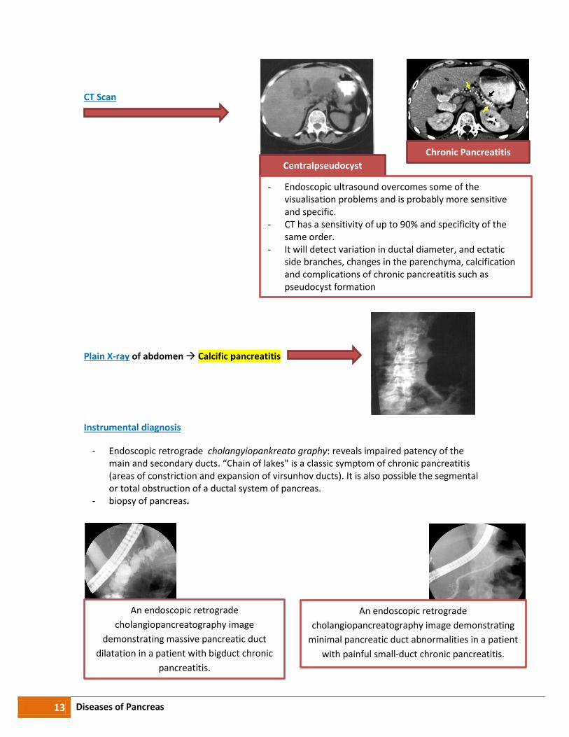
CT Scan
Plain X-ray of abdomen Calcific pancreatitis
Instrumental diagnosis
- Endoscopic retrograde cholangyiopankreato graphy: reveals impaired patency of the main and secondary ducts. “Chain of lakes" is a classic symptom of chronic pancreatitis (areas of constriction and expansion of virsunhov ducts). It is also possible the segmental or total obstruction of a ductal system of pancreas.
- biopsy of pancreas.
.
Chronic Pancreatitis
Centralpseudocyst
- Endoscopic ultrasound overcomes some of the visualisation problems and is probably more sensitive and specific.
- CT has a sensitivity of up to 90% and specificity of the same order.
- It will detect variation in ductal diameter, and ectatic side branches, changes in the parenchyma, calcification and complications of chronic pancreatitis such as pseudocyst formation
An endoscopic retrograde
cholangiopancreatography image demonstrating
minimal pancreatic duct abnormalities in a patient
with painful small-duct chronic pancreatitis.
An endoscopic retrograde
cholangiopancreatography image
demonstrating massive pancreatic duct
dilatation in a patient with bigduct chronic
pancreatitis.

14 Diseases of Pancreas
Differential Diagnosis :
- Chronic cholecystitis - Chronic gastroduodenitis - Ulcer disease - Cronic hepatitis - Bile gallstones disease - Pancreas cancer - Left-sided renal colic - Angina pectoris
Treatment
- Antisecretory drugs (H2 blocking (famotydyn,kvamatel 20mg 2 times a day); -
omeprazol 20mg,
lanzoprazol 30 mg,
pantoprazol 40 mg,
rabeprazol 20mg,
ezomehrazol 20mg-2 times a day;
Somatostatyn (sandostatyn); central action drugs (dalargin 0,001 mg intraven or -
intramus 2 times a day)
(penkreatin,kreon,pangrol,mezym) - Therapy of outersecretory enzyme deficiency - Elimination of duodenal statis,dyskinetic disorders of biliferous and pancreatic
(domperydon(motilium)10mg 3 times a day),cyzaprid) perystil )10 mg 3 times ductsa day);