diseases of mikania micrantha kuntil
TRANSCRIPT
DISEASES OF MIKANIA MICRANTHA KUNTIL
Rahiman Bin Jumat
~ Bachelor of Science With Honours 601 (Plant Resource and Science Management) 1147
2004lOOt.
P.KHIDMA TMAKLUMATAKADEMIK UIlIMAS
1111111111111111111111111 1000128270
Diseases of Mikania micrantha Kunth.
Rahiman Bin Jumat
Program Sains dan Pengurusan Sumber Tumbuhan
Fakulati Sains dan Teknologi Sumber
Universiti Malaysia Sarawak
ABSTRACT
This study was done to identify fungi associated with diseases of perennial weed, Mikania micrantha Kunth. Samples were collected from two places, Petra Jaya and Kota Semarahan. The common disease on M. micrantha was leaf spots. The disease symptoms were shown by the present of yellowish brown spots. Pathogens were isolated from spots of the infected leaves using media potato dectrose agar (PDA). After incubated at room temperature, various types of fungi were found . A total, 16 species of fungi were isolated from the leaf diseases of M. micrantha. Morphological structures of the fungi were used for identification. The fungi that had been identified were including Phomopsis sp., Colletotrichum sp., Fusarium sp. and Trichoderma sp .. Pathogenicity tests of the selected fungi were done by inoculating the mycelium of the fungi on detached leaves of M. micrantha. All of the tested fungi were able to produce disease symptoms. Observations on effect of temperature on growth of the fungi were also done. Most of the fungi grew significantly faster at P=O.05 at temperature 25°C to-30°C.
ABSTRAK
Kajian ini dijalankan adalah untuk mengenalpasti kulat yang berasosiasi dengan penyakit rumpai, Mikania micrantha. Sampel diperoleh dari dua tempat yang berbeza iaitu, Petra Jaya dan Kota Samarahan. Penyakit yang kerap ditemui pada M. micrantha ialah bintik-bintik daun. Tanda-tanda penyakit dilwljukkan melalui kehadiran bintik-bintik perang kekuningan. Patogen dipencilkan daripada tisu daun yang berpenyakit dengan menggunakan media agar dektrose kentang (PDA). Selepas tisu tersebut dieramkan pada suhu bilik, pelbagai jenis kulat ditemui. Keseluruhannya sebanyak 16 spesies kulat yang berbeza telah dipencilkan daripada daun M. micrantha yang berpenyakit. Struktur-struktur morfologi pada kulat tersebut telah digunakan untuk mengenalpastijenis kulat. Kulat-kulat yang dikenalpasti termasuklah, Phomopsis sp., Colletotrichum sp., Fusarium sp. dan Trichoderma sp. Ujian kepatogenan kulat-kulat yang terpilih telah dilakukan dengan meletakkan miselia kulat tersebut di atas keratan daun M. micrantha. Semua spesies kulat yang diuji telah menunjukkan kebolehan menghasilkan tanda-tanda penyakit. Pemerhatian kesan suhu terhadap pertumbuhan kulat juga dilakukan. Kebanyakan kulat-kulat tersebut tumbuh lebih cepat secara signifikan pada p=O.05 pada suhu 25-3(f'C berbanding pertumbuhan pada slIhu yang lebih rendah atau lebih tinggi.
1

INTRODUCTION
Weed is any plant or vegetation, excluding fungi interfering with the objectives or
requirements of people in term of plantation aspects (Zimdahl, 1999). In the other words,
plants are considered to be weeds if they are growing at a place where they are not
wanted by human (Rijn, 2000). A plant may be a weed in certain places but not in others.
For instance, the foxglove weed is grown as an ornamental in gardens and is not then
considered as a weed. Similarly, M. micrantha is often considered as weed. However, M.
micrantha has some uses in medicine. For instance, the plant can be used to treat cholera,
fever, flu and other diseases (Wan Azmulia, 1999).
In plant classification, M. micrantha is placed in family Aster or Asteraceae. The
common name for M. micrantha in Malaysia is Selaput Tunggal. Nevertheless, M.
micrantha is well known by one or more common names in different places (Ismail,
1995).
According to Rao (1992), M. micrantha has branched, pubescent to glabrous and ribbed
stems. The leaves are opposite, thin, chordate, triangular or ovate, and the blade is to 515
cm long and 2-10 cm wide on a petiole of 2-8 cm long. Leaf margins are coarsely dentate
and crenate. The leaf surface is glabrous. Inflorescence is capitula or head. The flowers
are white, 4-6 mm long, surrounded by an involucres bract on a common disc.
Fruit is linear oblong, 1.5-2 mm long, and black, with a tuft of hair. M. micrantha is
propagated by seeds and vegetative parts like old root stocks and stems.M. micrantha is
2
a creeping perennial with a shallow root system. With these characteristics, M. micrantha
can be assumed as a successful weed. With the creeping habit, M.micrantha is very
hazardous to nature and environmental condition and adversely affect a total plant
production.
Allelopathy has been reported for many crop and weed species (Zimdahl, 1999). M.
micrantha have been known to produce allelochemicals, especially during their
decomposition. Allelochemicals are very dangerous to the nearby plants because it can
retard and suppress the growth of the plants with the production of phenol, phenolic acid,
coumarin, flavonoid and others (Ismail, 1995). It is important to destroy the weeds such
as M. micrantha because they involve cost of agriculture. The weeds will take a quarter
away and this is a big loss for the agricultural industry (Ahmad Badri, 1992). Weeds
could cause 15 to 40% yield loss in tea (Rao, 1992). So, keeping the damage at
economically acceptable levels must be done.
Many researches have been done to control Mikania micrantha from suppress another
plants and give loss to the yield (Chiu et ai, 1998). The application of glyphosate as a
broadspectrum selective postmergence herbicide is widely being used for effective
control of rhizomatous and perennial weeds such asM. micrantha (Rao, 1992). But, the
use of these herbicides, chemical substances might give bad effects to other plants, soil
and environment. Other alternative must be explored to find the best way for controlling
weed.
3

,...... ,.
L l
One of the good strategy management to control weed is by biological control. With the
increased concern of conserving natural resources and reducing air, soil and water
pollution, natural or biological control of plant diseases has had increased emphasis.
Biological control by plant diseases is slow, gives few quick profits, but can be long
lasting, inexpensive and harmless to life. For instance, detached fragments of Cuscuta sp.
stem and/or tendril are capable of establishing themselves on the host plant. C.
campestris which has the same characteristics has been used in northeastern India as a
means of control the twinning of M. micrantha in tea (Parker and Riches, 1993).
Biocontrol systems neither eliminate the pathogen nor diseases but always bring them
into natural balance (Dhingra and Sinclair, 1995). Pathogenic microorganisms have not
been exhaustively used for controlling weeds. Introductions of pathogen may carry the
risk of great danger but they offer excellent possibilities (Rao, 1992). As in many other
plants, M. micrantha could be infected by several pathogens. Thus, it is possible to define
the possible biological agent of plant fungal pathogen to control the weed, M. micrantha.
The present study was done to define diseases and responsible pathogens on M.
micrantha. Effect of temperature on growth of the fungi associated with theM. micrantha
diseases were also observed.
4
MATERIALS AND METHODS
Location
Samples of M. micrantha with disease symptoms were taken from two selected area,
around the Unimas campus, Kota Samarahan and Petra Jaya, Kuching.
Description of the diseases
The symptoms of the diseases on the infected leaves of M. micrantha were described.
The plant tissues of the M. micrantha were considered infected when occurrence of
infection was established.
Isolation and identification of the pathogens
The pathogens associated with the diseased leaves were isolated. Thirty infected leaves of
the M. micrantha from each location were used. The infected leaves were cut to small
pieces about 2 mm2 between the healthy and infected tissues. After that, the cut tissues
were soaked in 10% solution of Clorox for 10 minutes. The purpose of the soaking was to
reduce bacteria and other surface microorganisms except the pathogen in the infected
tissues.
5
The cut tissues were washed twice with sterilized distilled water to remove excess
Clorox. Filter paper was used to dry the washed infected tissues. The tissues were then
transferred onto potato dextrose agar (PDA) in Petri dishes. Each Petri dish contained six
segments of the cut tissues. Five replicates for each location were prepared. All the plated
Petri dishes were incubated at room temperature.
After a week incubation at room temperature, the fungi growing on the agar plate were
observed under low magnification microscope and the mycelia were transferred to a new
PDA plate to get pure culture. The morphological characteristics of the fungi were
determined by preparing slides and observed under compound microscope.
Identifications of the fungi were based on the morphological characteristics especially the
fruiting body (Barnett, 1962 and Hall, 1993). Isolation was done twice for sample
obtained from Kota Samarahan.
Growth of fungi at different temperature
Block of agar containing mycelia of the test fungus cut from 4 to 7-day-old of pure
culture was inoculated on PDA in Petri dishes then incubated at 15, 20, 25, 30 and 35°C.
Three replicates were prepared for each fungus at each temperature. Diameter of the
fungus colony was measured everyday for seven days. The average growth diameter of
the colony was obtained from the average of two diameters of the colony, which were
perpendicular to each other. Then the average growth rate was determined.
6
Average colony diameter = a + b 2
Average growth rate = (n2-n1) + (n3-n2) + (n4-n3) + (n5-n4) + (n6-n5) + (n7-n6) n-1
n = day
a =colony diameter at axis x
b =colony diameter at axis y
Pathogenicity Test
Pathogenicity tests of selected fungi isolated from the disease of M. micrantha were done
using detached leaves. Healthy looking mature green leaves were used. The leaves were
wounded at three points on each leaf and three leaves were used for each tested fungus.
Inoculum of the fungus was inoculated on the wounded area of the leaves. The inoculated
leaves were placed in Petri dishes lined with moist sterilized cotton. All the Petri dishes
were placed at room temperature. Disease development was observed for everyday.
Percentages disease formations of the fungi on the inoculated leaves were determined.
RESULTS AND DISCUSSION
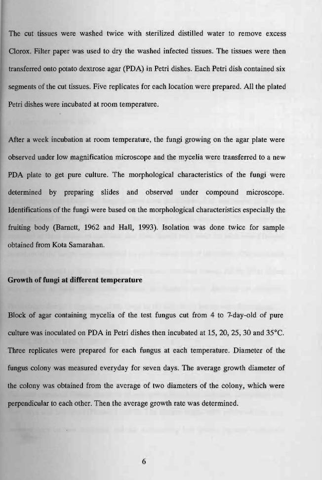
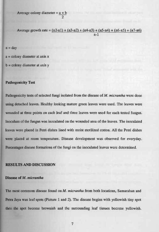
Disease ofM. micrantha
The most commom disease found on M. micrantha from both locations, Samarahan and
Petra Jaya was leaf spots (Picture 1 and 2). The disease begins with yellowish tiny spot
then the spot become brownish and the surrounding leaf tissues become yellowish.
7
Normally, numerous spots presented on the leaves. As the spot sizes increased, most part
of the leaf become yellow and then die. Similar disease symptoms were found on M.
micrantha in the second collection of the samples from Kota Samara han.
Fungi associated with the leaf spots of M. micrantha
There were different types of fungi associated with the leaf spots ofM micrantha from
both locations (Table 1). A total sixteen species of fungi were found associated with the
leaf disease of M. micrantha.
The most common fungus on sample from Kota Samarahan in the first isolation was
Fusarium sp. with total of percentage of occurrence was 36.7 followed by Pesta lotio psis
sp., Colletotrichum sp. and sterile mycelia. Each of these fungi was present on 15.6% of
the plated samples. However, the most common fungus on samples from Petra Jaya was
fungus with greyish white sterile mycelia, which was found on 12.5% of the plated
samples. Bacteria were also present and was the most common microorganism isolated
from leaf spots from this area.
During the second isolation the most common fungi found on sample from Kota
Samarahan, was Phomopsis sp. The fungus was present on 46.7% of the plated samples.
The second common fungus was Colletotrichum sp., encountered on 23% of the samples,
followed by Aspergillus sp. The occurrences of all these three fungi were much higher
compared with the percentage occurrence in the first isolation. Pestalotiopsis sp.
h !Wever, less common compared with the first isolation.
8
---~-
Picture I: Disease on leaves of M micrantha from Kota Semarahan.
Picture 2: Disease on leaves of M micrantha from Petra Jaya.
9
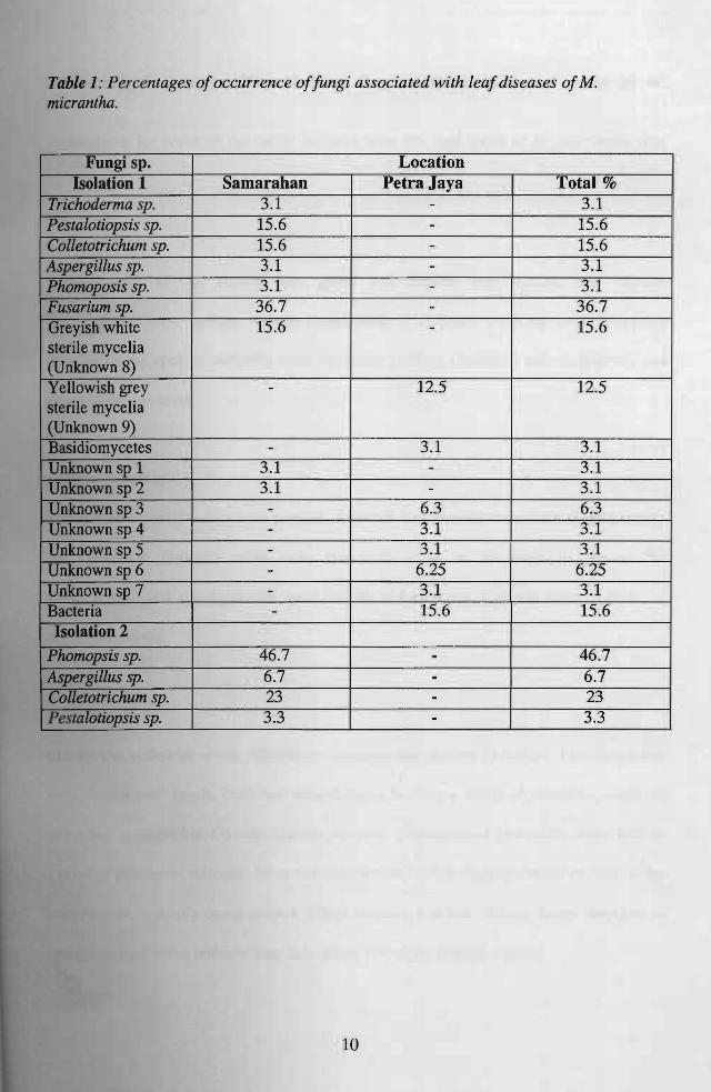
Table 1: Percentages ofoccurrence offungi associated with leaf diseases ofM. micrantha.
Fungi sp. Location Isolation 1 Samarahan Petra Jaya Total %
Trichoderma sp. 3.1 - 3.1 Pesta[otiopsis sp. 15.6 - 15.6 Colletotrichum sp. 15.6 - 15.6 Aspergillus sp. 3.1 - 3.1 Phomoposis sp. 3.1 - 3.1 Fusarium sp. 36.7 - 36.7 Greyish white sterile mycelia (Unknown 8)
15.6 - 15.6
Yellowish grey sterile mycelia (Unknown 9)
- 12.5 12.5
Basidiomycetes - 3.1 3.1 Unknown sp 1 3.1 - 3.1 Unknown sp2 3.1 - 3.1 Unknownsp3 - 6.3 6.3 Unknownsp 4 - 3.1 3.1 Unknown sp 5 - 3.1 3.1 Unknown sp 6 - 6.25 6.25 Unknown sp 7 - 3.1 3.1 Bacteria - 15.6 15.6 Isolation 2
Phomopsis sp. 46.7 - 46.7 Aspergillus sp. 6.7 - 6.7 Colletotrichum sp. 23 - 23
I Pestalotiopsis sp. 3.3 - 3.3
10

Morphological characteristics of some fungi isolated from leaf spots of M. micranthll.
Description for some of the fungi isolated from the leaf spots of M. micrantha was given.
AspergiUus sp.
Surface color of the colony was green and hyphae was hyaline and septate.
Conidiophores were upright, simple, terminating in a clavate swelling vesicle, bearing
phialides at the apex of radiating from the entire surface. Conidia 1 celled, globose, and
green colored in mass.
CoUetotrichum sp.
The surface color of colony was greyish. Averculi disc shaped or cushion shaped, waxy,
sub epidermal, typically with dark, spines or setae at the edge or among the
conidiophores and conidiophores were simple and elongate. Conidia were hyaline, 1
celled and oblong.
Fusarium sp.
Colony was yellowish white. Mycelium extensive and cottony in culture. Conidiophores
were slender and simple, branched irregularly or bearing a whorl of phialides, single or
group into sporodochia. Conidia hyaline, variable, principally of two kinds, often held in
a mass of gelatinous material. Macro conidia ~veral celled slightly curved or bent at the
pointed ends, typically canoe shaped. Micro conidia, 1 celled, oblong, borne singly or in
. . Some conidia intermediate, 2/3 celled, oblong or slightly curved
11
Phomopsis sp.
Colony was greyish white. Mycelia were septate. Pycnidia were dark, ostiolate,
immersed, erumpent, almost globose. Conidiophores were simple. Conidia hyaline, 1
cellled, of two types, ovoid to fusoid (alpha) conidia and filiform, curved or rent
stylospores (beta conidia).
Trichoderma sp.
Colony was compact to loosely floccose. Fast growing whitish mycelium gives rise
rapidly to much branched. Conidiophores hyaline, upright, much branched, not
verticillate, phialides single or in groups. Conidia hyaline, I-celled, ovoid, borne in small
terminal clusters, usually easily recognized by its rapid growth, and green patches of
cushions of conidia.
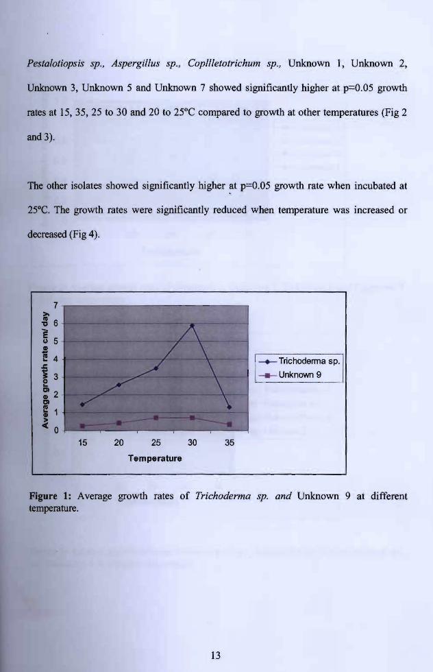
Growth of Fungi At Different Temperature
Temperature had significant effect on growth of the fungi isolated from leaf spots ofM.
micrantluz (Figure 1-3). The optimum temperature for the fungal growth was depending
on the isolates.
Trichoderma sp., and Unknown 9 (Yellowish grey sterile mycelia) showed significantly
higher at p=O.05 growth rates at 300C compared to growth at other temperatures. The
growth rates were significantly reduced at p=O.05 when the temperatures were increased
decreased (Fig 1).
12
I . _ ~_
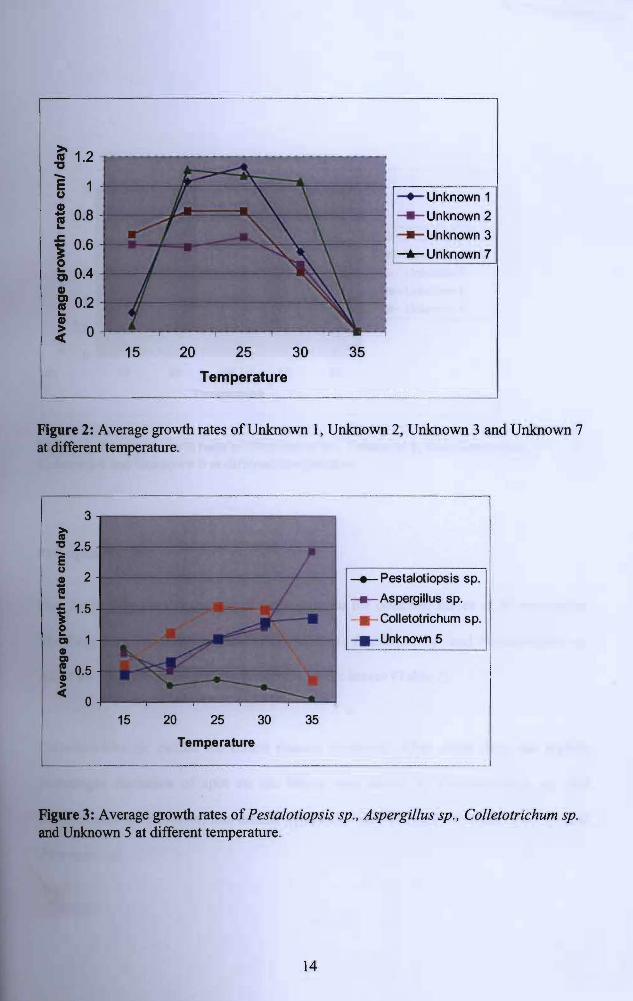
Pestalotiopsis sp., Aspergillus sp., Copllletotrichum sp., Unknown 1, Unknown 2,
Unknown 3, Unknown 5 and Unknown 7 showed significantly higher at p=O.05 growth
mtes at 15, 35, 25 to 30 and 20 to 25°C compared to growth at other temperatures (Fig 2
and 3).
The other isolates showed significantly higher at p=0.05 growth rate when incubated at
25°C. The growth rates were significantly reduced when temperature was increased or
decreased (Fig 4).
7
-+-Trichoderma sp.] ____ Unknown 9
15 20 25 30 35
Temperature
Figure 1: Average growth rates of Trichoderma sp. and Unknown 9 at different temperature.
13
-8>-1.2-E 1 u ~Unknown 1
- Unknown 2
.c
i 0.8 - Unknown 3 i 0.6 --...- Unknown 7 e
Q 0.4
•:f 0.2
l...
0 15 20 25 30 35
Temperature
Figure 2: Average growth rates of Unknown 1, Unknown 2, Unknown 3 and Unknown 7 at different temperature.
,-- ---------------- - ------
3 >I'll
" 2.5E u
~ Pestalotiopsis sp. l! .! 2
- .-Aspergillus sp.
CoUetotrichum sp.
Unknown 5___
15 20 25 30 35
Temperature
- - - - _ ._ ......_ -_._-- -- ----'
Figure 3: Average growth rates of Pestalotiopsis sp., Aspergillus sp., Colletotrichum sp. and Unknown 5 at different temperature.
14

Pusat Khidmat MaJd UNIVERSITI MALAY~~ AkademDl
94100 ~ Ia ~atn:l~~WA¥
>- 3 as
"E 2.5 u -.-Phomopsis sp . .! 2 t! ___ Basidiomycetes .c 1.5 --.-Unknown 8
_ Unknown 6 1 QI 1 4D ____ Unknown 4 QI
~ 0.5
~ o 15 20 25 30 35
Temperature
Figure 4: Average growth rates of Phomopsis sp. , Unknown 8, Basidiomycetes, Unknown 4 and Unknown 6 at different temperature.
Pathogenicity Test
Four fungi were used to test the pathogenicity on the detached leaves of M micrantha.
The fungi were Fusarium sp., Colletotrichum sp., Phomopsis sp. and Pestalotiopsis sp.
All of the fungi produced disease symptoms on the leaves (Table 2).
Colletotrichum sp. caused the fastest disease fonnation. After seven days, the highest
percentages fonnation of spot on the leaves was shown by Colletotrichum sp. and
Pestalotiopsis sp. However, less disease symptoms were shown by Pestalotiopsis sp. and
Phomopsis sp.
15

diseased leaves.
Deeded
"'
- ~ -
Table 2: Percentage of disease formation by the selected fungi inoculated onM. micrantha detached leaves after seven days. D.I.A - Day of initial disease appearance
Pathogenicity Test %Of
Diseases
Fungi D.I.A
4Fusarium sp. 66.7+
2Colletotrichum sp. + 100
4 66.7Phomopsis sp. +
Pestaiotiopsis sp. + 4 100
DISCUSSION
Successful isolation of pathogens from diseased leaves of M. micrantha depends on
careful selection of the plant parts. In the present study, 16 fungi were isolated from the
Among the common fungi, which were known as common plant
pathogens were Phomop is sp., Colletotrichum sp., Fusarium sp. and Pestaiotiopsis sp.
(Walker 1969, Sepiah el ai 2002). Bacteria was also found associated with M. micrantha
leaves and could cause disease. According to McPartland (1996), plant diseases are
always caused by fungi and rarely caused by bacteria.
For the fungi identification, pigment or colors is used to differentiate species, but care is
since pigmentation may change as cultures deteriorate (Brayford, 198~.
1UIJ"UItI colonies often form distinctive pigments such as brown, violet, white or pink.
16

Fusarium sp. can be found on a very wide range of plants. They may colonise the roots,
stems or leaves and cause serious plant disease (Brayford 1989, Blakeman and
Williamson 1994). Fusarium oxysporum attacked banana seedlings (Adeline et. AI, 2002)
and Fusarium sp. also could cause disease on papaya (Norhayati, 2002). Fusarium wilt
received attention as a potential biocontrol to eliminate illegal marijuana plantations
(Hildebrand & McCain 1978, Noviello et al. 1990).
Colletotrichum sp. also has been found attacked on the aerial parts of plants where they
caused leaf spots and anthracnose (Domsch and Gams, 1980, Noreha, 2002).
Antrachnose disease caused by Colletotrichum sp. also infected on Cannabis crops
(McPartland, 1996).
Phomopsis sp. is always found as pathogenic fungi on plant parts and responsible species
caused damages and loss in agriculture mostly to the economic value crops such as fruit
or stems of banana, carambola, citrus, cherry, durian, langsat, mango, maple, mulberry
and watermelon, and soybean (Sepiah et al. 2002, Ingold and Hudson, 1993). The species
causes damping off seedling, plant dieback, leaf blight and spots and various fruits and
seed rots (Snowdown 1991, Kaushal and Sugha, 1995). McPartland (1996) reported that
Phomopsis sp. caused a disease on leaves and stem of Cannabis crops. Phomopsis sp, has
been reported to cause cankers on Douglas fir and western hemlock seedlings in a
narthwest California nursery (Funk, 1968).
17
Pathogenicity tests can be done either with spores or mycelia inoculums. Dhingra and
Sinclair (1995) reported that in the absence of spores, mycelia of the fungi could be used
as inoculum. Several factors involve making the successful infection of the pathogens.
Penetration of the pathogen into the host is very important. The pattern of penetration
varies from fungus to fungus. Many fungi will reach the proximity of the host and will
start to grow on the ho t, but without penetration at first (Ross, 1979). The fungus that is
capable in penetration of single host may overcome any host defenses and lead to the
destruction of the entire host. Penetration of fungi occurs through stomata directly
through epidermal cells or at the junction between epidermal cells. When infection occurs
through stomata, inoculums must be applied on the surface bearing maximum stomata
and incubated under conditions that allow stomata to remain open, allowing for spore
germination and hyphal penetration (Dhingra and Sinclair, 1995).
Temperature is among the important factor to influence the growth of pathogens
(Jennings and Lysek, 1996). From the present study, different fungi associated with the
leaves disease had a different range of temperatures in which they were able to live. Ross
(1979), stated that most of fungi grow between temperatures in a range 0 to 4(J'C.
According to Dhingra and Sinclair (1995), Phomopsis sp. grow well on PDA at pH 6 and
2,8OC. In addition, temperature had been shown to have an effect on the development of
blight of plant seedling (Blakeman and Williamson, 1994). Size, shape, color and
tion of spores may be influenced by fluctuations in temperature during the fungal
growth. Most temperature studies show that requirements of sporulation or of sexuality
SOlD fungi are much narrower than those permitting vegetative growth (Ross, 1979).
18
produce disease
3a'C.
ofM. micrantha.
Some pathogens grow and sporulate well at a constant temperature and others favored by
diurnal fluctuations (Dhingra and Sinclair, 1995). According to Burrage (1971),
temperature on leaves surface and ultra violet (UV) radiation is factors infecting
pathogen! antagonists' interaction.
Since there are several fungi found associated with the leaf disease ofM. micrantha they
could be used as potential biocontrol agent to handle the weed.
CONCLUSION
The common disease on M. micrantha was leaf spots. A total 16 fungi were isolated from
the disease. The common fungi found associated with the disease were Phomopsis sp,
Fusarium sp., Colletotrichum sp., Pestaiotiopsis sp. and Aspergillus sp. Pathogenicity
tests of five selected fungi were done on detached leaves. All of the fungi were able to
symptoms. The growth tests of the fungi at different temperature
indicated that most of fungi grew significantly faster at P=O.05 at temperature between 25
Based OD this work, further study should be done to use the pathogens as a control agent
19
REFERENCES
Adeline, T. S. Y., Sariah, M., Kadir, J. and Anuar, A. R. 2002. Induction of Suppressive
Soil for Controlling Fusarium Wilt in Banana Seedlings, 175 pp. In Mulla, M.S et
al. Biopesticides: Positioning Biopesticides in Pest Management Systems. 3rd
Biopesticides Congress. Kuala Lumpur 21-26 April 2002.
Ahmad Badri, M. 1992. Keupayaan Rumpai. Sumber A lam; Potensi dan Penerokaannya.
(1), pp. 172-199. Dewan Bahasa dan Pustaka, Kuala Lumpur.
Barnett. H. L. 1962. Descriptions and Illustrations, Illustrated Genera ofImperfect Fungi,
2nd edition, pp. 306-202. Burgess Publishing company, United States of America.
Blakeman, 1. P. and Williamson, B. 1994. Fungal Interactions on Living and Necrotic
Leaves, Ecology of Plant Pathogens, pp. 321-335. CAB International,
Wallingford.
Brayford, D. 1989. Fusarium its Relatives and Their Teleomarphs, International Course
on the Identification of Fungi of Agriculture Importance. International
Mycological Institute Publication.
20

Funk. A.
Burrage, S. W. 1971. The Micro Climate at the Leaf Surface, Ecology of Leaf Surface,
pp.91-101.
Chiu, S. B., Mun, C. H. and Siow, A. 1998. Biological Control of Mikania micrantha (A
PreliminaryFiniling): The Planter. 870(74): pp. 503-510. Island and Peninsular
Publications, Malaysia.
Dhingra, O. D. and Sinclair, J. B. 1995. Culture of pathogens, Basic Plant Pathology
Methods. 2nd edition, 300 p.p, Lewis Publisher, Boca Raton.
Domsch, K. H. and Gams, W. 1980. Colletotrichum, Compendium of Soil Fungi, Volume
1, pp. 223-224, Academic Press, London.
1968. Diaporthe Iokoyae n. sp.-the perfect stage of Phomopsis Iokoyae.
Canadianlournal of Botany. 46: 601 -603.
HaU, G. 1993. Oomycetes. International Course on The Identification of Fungi of
Agriculture Importance, International Mycological Institute Publication.
debrand, D. C. and A. M. McCain. 1978. The use of various substrates for large scale
production of Fusarium oxysporum f. sp. cannabis inoculum. Phytopathology
68: 1099-1101.
21

Ingold, C. T. and Hudson, H. J. 1993. Fungi As Plant Pathogens, The Biology ofFungi,
6th edition, pp. 159-183, Chapman and Hall, London, New York.
Ismail Sahid. 1995. Alelopati: Potensinya dalam Pengurusan Rumpai. Rumpai Tropika;
Impak Biologi dan Pengurusan (1), pp. 32-37. Universiti Kebangsaan Malaysia,
Bangi.
Jennings, D. Hand Lysek, G. 1996. Withstanding Extremes of Temperature. Fungal
Biology: Understanding the Fungal Lifestyle, 1: 89-92, BOIS Scientific
Publishers.
Kausal, N. and Sugha, S: K. 1995. Role of Phomopsis vexans in damping of seedling in
egg plant and its control. Indian Journal Mycological Plant pathology 25:189
191.
McPartland, 1. M. 1996. A review of Cannabis diseases. Journal of the International
Hemp Association 3(1): 19-23.
oreha, M. 2002. Ujian Kepatogenan dan Ujian Pertumbuhan, Kulat Yang Berasosiasi
Dengan Penyakit Lepas Tuai Buah-buahan Tempatan Terpilih di Sarawak,
Laporan Tahun Akhir Bsc., Universiti Malaysia Sarawak.
22
orhayati, A. S. 2002. Ujian Kepatogenan dan Perumbuhan Kulat, Kajian Terhadap
Penyakit Lepas Tuai Buah-buahan Tempatan di Sarawak, Laporan Tahun Akhir
Bsc., Universiti Malaysia Sarawak.
Noviello, C. 1990. Lotta biological control Cannabis sativa mediante l'impiego di
Fusarium oxysporum f. sp. cannabis. Annali della Facolta di Scienze Agrarie
della Universita deg/i Studi di Napoli, Portici 24: 33-44.
Parker, C. and Riches, C. R. 1993. Biology and physiology, Parasitic Weeds of The
World (Biology and Control), pp. 203. CAB International, Oxon.
Rao, V. S. 1992. Some prominent weeds and their control, Principles of Weed Science,
pp. 357-358. Oxford and IBH Publi bing Co. PVT. LTD, New Delhi.
Rijn, P. J. V. 2000. Weed Control in Plantation Crops. Weed Management in Humid and
Sub-humid Tropics, pp. 140-143. Royal Tropic Institute, Netherlands.
I. 1979. Development and Regulation, Biology of the Fungi, pp. 270-27l.
McGraw-Hili, Inc, United States of America.
23