discriminant color texture descriptors for diabetic ...discriminant color texture descriptors for...
TRANSCRIPT

Discriminant Color Texture Descriptors for DiabeticRetinopathy Recognition
Holly H. Vo and Abhishek VermaDepartment of Computer Science
California State UniversityFullerton, California
Email: hhvo, [email protected]
Abstract—Diabetic retinopathy (DR) is a common eye diseasethat could lead to irreversible vision loss but hard to be noticedby carriers in early stages. Instead of isolating DR signs for DRrecognition, this paper examines discriminant texture featuresobtained by color multi-scale uniform local binary pattern (LBPs)descriptors on five common color spaces and two proposedhybrid color spaces. The extracted features are evaluated bythe enhanced Fisher linear discriminant, EFM. Experiments aredone on a large dataset of 35,126 training images and 53,576testing images that have been taken by different devices withhigh variance in dimensions, quality and luminance. The bestperformance is above 71.45% by HSI-LBPs, a*SI-LBPs, and bSI-LBPs descriptors.
Index Terms—diabetic retinopathy recognition, DR, texture,uniform LBPs, EFM, FLD, PCA, color space
I. INTRODUCTION
According to US Center for Disease Control and Prevention,diabetic retinopathy (DR) is the most common eye diseasewhich affects one in three Americans with diabetic and furtherleads to blurred vision and blindness. DR is caused due tohigh blood glucose level which damages small blood vesselsin retina. In consequence, blood, extra fluid, cholesterol, andother fats leak in the retina and make the macula swollenand thicken. Damaged capillaries may finally close and stopproviding nutrients and oxygen to retina. To supply sufficientblood to the area, the retina grows new abnormal fragileblood vessels called intraretinal microvascular abnormalities(IrMAs); however, the new vessels are usually accompaniedwith scar tissue which may wrinkle or detach the retina anddistort vision. In late stages, increased pressure in eye maydamage the optic nerve [1].
Unfortunately, DR carriers do not notice vision changesin the early stages. DR usually affects both eyes and causesirreversible vision loss in many cases as DR progresses whiledetecting early DR stages and timely treatment can reduce therisk of severe vision loss by over 90%. Currently, detecting DRis a time-consuming manual procedure which requires ophthal-mologists to evaluate digital fundus photography, review, andfollow up in many days and causes delayed treatment [2], [3].
A typical fundus image appears with an optic disc, bloodvessels, and a macula. Macula is the center area of the retinawhich contains color-sensitive rods and the central point ofsharpest vision. Figure 1 illustrates a fundus image withlabeled signs of diabetic retinopathy. Microaneurysms (MAs)
Fig. 1. DR signs and main structure in a retina image
are tiny bulges in blood vessels and appear as deep-red dots inthe fundus. Haemorrahages are small spots of blood discharge.Hard exudates are leakage of lipid and protein in the retina.Hard exudates typically emerge as bright, reflective, whiteor cream lesions. Hard exudates and microaneuryms aroundmacula might block vision and additionally damage the maculaand leads to permanent vision loss [3], [4].
By examining patient retina images, an ophthalmologist canidentify two types of retinopathy: nonproliferative diabeticretinopathy (NPDR) and proliferative diabetic retinopathy(PDR). NPDR is further classified into three stages based onthe presence of DR signs. Mild NPDR is formed when somemicroaneurysm appears with possible appearance of exudatesand venous loops. Moderate NPDR is developed with multi-plex microaneuryms, haemorrhages, and hard exudates. SevereNPDR stage is characterized by 4-2-1 rule. The 4-2-1 rule isdefined by the existence of hemorrhages and microaneurysmsin four quadrants, venous beading in two quadrants, and IrMAsin one quadrant. Finally, PDR is the advanced stage where newfragile blood vessels leak hemorrhages and hard exudates intothe vitreous, the gel in the center of the eye, deliver scar tissueand wrinkle to the retina, build up pressure in the retina, andlead to vision loss due to the damage of macula and opticnerve [3], [5].

This paper is a focus on robust discriminant texture featureson color spaces for a large-scale dataset. The rest of thepaper is organized as follows. Section II reviews backgroundof current feature extraction and classification techniques onDR. Section III describes the retina image dataset in thisresearch. Section IV proposes the methodology to extract colortexture features and classification techniques to identify DRstages. Experimental results and discussion present in sectionV. Section VI concludes the research with future direction.
II. RELATED WORK
With the clinical fact that MAs are the earliest signs ofdiabetic retinopathy [6], most DR papers focus on extract-ing clinical features by localizing and segmenting lesions,blood vessels, optic disks, and macula one by one. Basicpoint operators are applied to balance and enhance localcontrast, and linear filters and neighborhood operators such asmorphological operators, median filters, and Gaussian filtersare convoluted on images in pre-processing as indicated insurveys [3], [7]. Watershed transformation is applied in [8]to overcome over-segmentation caused by thresholds. Othertechniques such as active contour models and recursive region-growing technique (RRGT) are used in the domain researchesto isolate blood vessels and other interested regions [7].
In addition to segmentation, statistical texture extractionis another approach in DR recognition. Statistical textureapproach is based on the relationship between pixel intensities.In statistical approach, entropy, contrast, and correlation canbe simply calculated via gray level co-occurrence matrix.Contrast texture is extracted together with isolated areas ofMAs and HAs in [9] to classify DR. In recent years, localbinary pattern (LBP) texture is started being used for DRdetection on small retina datasets with less than 100 images in[10], [11]. In other domains such as face and scene recognition,texture descriptor such as local binary pattern texture has beenproven to contribute significant performance [12].
When sampling on a large set of images which are takenby different devices under various conditions of light andintensity, it is crucial for a robust vision system to adapt adiscriminant color space. HSI is applied for Messidor andDB-rect DR datasets in [1] to extract MAs and exudates,and selected by [13] to locate fovea. Green component inRGB is focused to extract blood vessel structure in [9], [14].All channels of RGB are separately examined in [5] withmorphology operations to extract the total area and perimetersof blood vessels, HAs, and MAs. Ram and Jayanthi [15]consider multiple color spaces such as RGB, L*u*v*, HSVand HSI to extract lesion pixel values.
In general, the aforementioned researches focus on segmen-tation of blood vessels and DR signs for feature extraction onsmall datasets whose sizes are ranging from several hundredsto several thousands of images. In classification stage, supportvector machine (SVM) and artificial neural network (ANN)are two popular techniques in DR problems [1], [5], [7], [9]–[11], [14]. An SVM classifier transforms the original trainingdata to a higher dimension space where it can find a linear
Fig. 2. Statistics of DR training dataset
optimal separating hyperplane. Meanwhile, multi-layer NNis built on a set of connected input and output nodes andweighted connections between nodes. The network is trainedin iterations to determine the proper connection weights, andits back propagation algorithm searches for a set of weights bygradient descent method [16]. Acharya et al. [14] applies SVMon a dataset of 331 retina images to identify five DR stageswith an overall accuracy of 86%. Back propagation neuralnetwork is applied in [5] with one hidden layer to identifyfour DR stages with an accuracy of 84% on a dataset of 124images.
III. RETINOPATHY IMAGE DATASET
In this paper, experiments are done on the retinopathy imagedataset provided by EyePACS, a free platform for retinopathyscreening, through Kaggle website. The dataset originallyconsists of 35,126 training images and 53,576 testing images.These images are taken by different models and types ofcamera under different conditions and stored in various, highresolutions. Each image has been examined on the presenceof DR by a clinician to be labeled with a DR stage from0 to 4, corresponding to no DR, mild, moderate, severe, andproliferative DR as being described in the introduction section.Within the train dataset, there are 74% images of stage 0(no DR), 7% of stage 1 (mild), 15% of stage 2 (moderate),2% of stage 3 (severe) and 2% of stage 4 (proliferate DR)approximately. Images in the test datasets are split to 5 stagesin the similar ratios as in the train dataset.
The challenges of this dataset are its large variance inresolution, intensity, and quality as shown in Figure 2. Byexamining the train dataset, image heights vary from 289 to3456 pixels, while their widths vary from 400 to 5184 pixelswith the range of ratios between height and width is from 0.66to 1.00. The average of image intensity spreads from 1 to 192around the mean of 63. Low intensity images are stored in8KB while other images can allocate up to 2MB files.
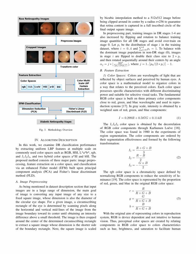
Fig. 3. Methodology Overview
IV. ALGORITHM DESCRIPTION
In this work, we examine DR classification performanceby extracting uniform LBP features at multiple scale oncommonly used color spaces such as RGB, HSI, L*a*b*, rgb,and I1I2I3, and two hybrid color spaces a*SI and bSI. Theproposed method consists of three major parts: image prepro-cessing, feature extraction on a color space, and classificationvia an enhanced Fisher model (EFM) built upon principalcomponent analysis (PCA) and Fisher’s linear discriminantmethod (FLD).
A. Image Preprocessing
As being mentioned in dataset description section that inputimages are in a large range of dimension, the main goalof image is converting any input image of any size to afixed square image, whose dimension is also the diameter ofthe circular eye shape. For a given image, a circumscribingrectangle of the eye is determined by scanning pixels alongthe horizontal and vertical mid-lines of the image from theimage boundary toward its center until obtaining an intensitydifference above a small threshold. The image is then croppedaround the center of the determined circumscribing rectangleto extract a square image whose dimension is the shorter sideof the boundary rectangle. Next, the square image is scaled
by bicubic interpolation method to a 512x512 image beforebeing clipped around its center by a radius r=256 to guaranteethat retina content is captured in a full inscribed circle of thefinal output square image.
In preprocessing part, training images in DR stages 1-4 arealso increased by flipping and rotation to balance trainingimage quantities for all DR stages and avoid over-train onstage 0. Let pi be the distribution of stage i in the trainingdataset, where i = 0..4 and
∑4i=0 pi = 1. To balance with
the dominant image population in non-DR stage (0), imagesin stage i are flipped to double their class size to 2 ∗ piand then rotated sequentially around their centers by an angleαj = j ∗ b 3600
dp0/(2∗pi)ec, where j = 1..dp0/(2 ∗ pi)e − 1.
B. Feature Extraction
1) Color Spaces: Colors are wavelengths of light that arereflected by object surfaces and perceived by human eyes. Acolor space is a mathematical model to organize colors ina way that relates to the perceived colors. Each color spacepossesses specific characteristics with different discriminatingpower and suitable for selective visual tasks. The fundamentalRGB color space is built on three primary color componentsclose to red, green, and blue wavelengths and used in repro-duction systems [17]. In gray scale, intensity is obtained by aweighted sum of red, green, and blue components:
I = 0.299R+ 0.587G+ 0.114B (1)
The I1I2I3 color space is obtained by the decorrelationof RGB color components through Karhumen Loeve [18].The color space was found in 1980 in the experiments ofregion segmentation. The color components are ordered bytheir segmentation effectiveness and formed by the followingtransformation:
I1 =R+G+B
3
I2 =R−B
2
I3 = −G+B
2
(2)
The rgb color space is a chromaticity space defined bynormalizing RGB components to reduce the sensitivity of lu-minance [19]. The color space is represented by the proportionof red, green, and blue in the original RGB color space:
r =R
R+G+B
g =G
R+G+B
b =B
R+G+B
(3)
With the original aim of representing colors in reproductionsystem, RGB is device dependent and not intuitive to humanvision. Thus, perceptual color spaces are created by relatingcomponents in RGB color space to colors characteristicssuch as hue, brightness, and saturation to facilitate human
interpretation of their components [17]. HSI is a perceptualcolor space whose components approximate the perceived hue,saturation, and intensity in order. HSI components are obtainedby the following equations:
H =
{α, if b < g
2π − α, otherwise
S = 1−min(r, g, b)
I =R+G+B
3
(4)
where
α = cos−1{
0.5 ∗ [(r − g) + (r − b)][(r − g)2 + (r − b)(g − b)]1/2
}and r, g, and b are normalized RGB components obtained byequation 3.
CIE XYZ color system was defined by the CommissionInternational de L’Eclairage (CIE) in 1931. While RGB usesvisible physical colors, XYZ is built upon imaginary primarycolors [X Y Z] to form a device-independent color space withbetter descriptive properties [17]. A color space defined inthis system, referred to as Yxy, can represent all visible colorby only positive normalized mixture of its primaries and theluminance value Y:
x =X
X + Y + Z
y =Y
X + Y + Zz = 1− x− y
(5)
L*a*b* can be directly derived from XYZ with the intentionto mimic the logarithmic response of the human vision system.The L* channel represents luminance in the range from 0to 100, while a* and b* channels represent chrominanceopponents. The red and green opponent colors are representedalong a* dimension. The yellow and blue opponent colors arerepresented along the b* dimension. L*a*b* is widely used inmany industries which requires accurate color specificationssuch as paint, dyes, and printing inks [20]. L*a*b* componentscan be derived from XYZ as the following:
L∗ = 116f(Y
Yn)− 16
a∗ = 500[X
Xn− Y
Yn]
b∗ = 200[Y
Yn− Z
Zn]
(6)
where
f(t) =
{t(1/3) if t > ( 6
29 )3
13 ( 29
6 )2t+ 429 otherwise
and Xn, Yn, and Yn are the CIE XYZ component values ofthe reference white point.
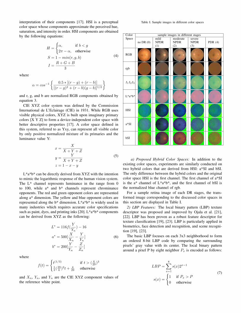
Table I. Sample images in different color spaces
ColorSpace
sample images in different stages
no DR (0)mildNPDR(1)
moderateNPDR(2)
severeNPDR(3)
PDR (4)
RGB
rgb
I1I2I3
L*a*b*
HSI
a*SI
bSI
a) Proposed Hybrid Color Spaces: In addition to theexisting color spaces, experiments are similarly conducted ontwo hybrid colors that are derived from HSI: a*SI and bSI.The only difference between the hybrid colors and the originalcolor space HSI is the first channel. The first channel of a*SIis the a* channel of L*a*b*, and the first channel of bSI isthe normalized blue channel of rgb.
For a sample retina image of each DR stages, the trans-formed image corresponding to the discussed color spaces inthis section are displayed in Table I.
2) LBP Features: The local binary pattern (LBP) texturedescriptor was proposed and improved by Ojala et al. [21],[22]. LBP has been proven as a robust feature descriptor fortexture classification [19], [23]. LBP is particularly applied inbiometrics, face detection and recognition, and scene recogni-tion [19], [23].
The basic LBP focuses on each 3x3 neighborhood to forman ordered 8-bit LBP code by comparing the surroundingpixels’ gray value with its center. The local binary patternaround a pixel P by eight neighbor Px is encoded as follows:
LBP =
8∑x=1
s(x)2x−1
s(x) =
{1 if Px > P
0 otherwise
(7)

As the operator focuses on the signed differences of grayvalues and disregards the value difference, it is invariant tochanges in mean luminance. For scale invariance improve-ment, LBP operator is extended to consider a circularlysymmetric neighbor set of P pixels on a circle of radius Rsurrounding the center pixel, denoted as LBPP,R. The topmiddle neighbor is the most significant bit in LBP code, andother neighbors are ordered clockwise. For each neighbor pointwhose coordinators are not exactly in the center of pixels, itsgray value is estimated by interpolation rather than the nearestpixel’s value.
The extension in [22] defines a so-called uniform pattern,denoted as LPBriu2
P,R , which contains at most two spatialtransitions in its circular chained binary pattern, ”1-0” and”0-1”. LPBriu2
P,R does not only improve rotation invariancebut also significantly reduces LBP dimension by preserving asingle bin for all nonuniform patterns.
In the real diabetic retinopathy recognition problem, imagesare taken by different devices under different conditions oflight and quality. In addition, besides the main structure ofa retina in the images, DR signs are fine-grain and theirgranularity diversifies. Recall that microaneurysms (MAs) aretiny red dots, hemorrhages and hard exudates can be at anysize in any unknown shape, and fragile blood are developedin undetermined directions. Thus, uniform local binary patterndetection should be applied on different scales to capturediscriminant features that are invariant to rotation, globalintensity, and scales.
In our experiments, each 512x512 retina image is dividedinto four regions. Texture features are extracted on 4 scalesby LPBriu2
8,2 , LPBriu216,4 , LPBriu2
24,6 , and LPBriu232,10 descriptors
on each 256x256 region at each color channel for a givencolor space. The extracted LBP features are standardized oneach scale. The final 1056-dimension feature vector for a colorretina image is formed by standardizing the concatenation of120 features from LPBriu2
8,2 , 216 features from LPBriu216,4 , 312
features from LPBriu224,6 , and 408 features from LPBriu2
32,10.
C. EFM Classification
The extracted LBP features on a color space will be clas-sified by the Enhanced Fisher Model (EFM), which enhancesFisher Linear Discriminant (FLD) by principal componentanalysis (PCA) [12], [19], [25].
Let X be a data matrix which consists of M feature vectors,Xi, in the space RN , where i = 1..M . The feature vectorXi may reside on a high dimensionality space. The vector isprobably composed of correlated features and contains noises.PCA is a common technique to linearly transform data to alower dimensionality space and reduce data noise [24]. Thebasic idea of PCA is diagonalizing the covariance matrix, ΣX ,of the original space, RN , to obtain eigenvectors.
ΣX = E{[X − E(X )][X − E(X )]t}ΣX = ΦΛΦt (8)
where E(X) is the expectation function, t denotes matrixtranspose operation, Φ = [φ1, φ2, . . . φN ] is the orthogonal
Fig. 4. (8,2), (16,4), (24,6), and (32,10) LBP neighborhoods
eigenvector matrix, and Λ = diag{λ1, λ2, . . . , λN} is thediagonal eigenvalue matrix in the descending order of eigen-values.
A corresponding feature vector, Yi, in the PCA reducedspace, RK , where K < N , is composed by K most dominantprincipal components and is derived by the following equation:
Yi = P tXi (9)
where P = [φ1, φ2, . . . , φK ].Although PCA is a popular technique in pattern recognition,
it is not optimized for class separability. Instead, the alternativetechnique, FLD, has been proposed to model the differencebetween classes of data [25], [26].
The Fisher linear discriminant (FLD, a.k.a. linear discrim-inant analysis LDA) is a popular discriminant criterion thatdefines a projection to reduce within-class scatter and enlargethe between-class scatter [25]. Let ωi, where i = 1..L,represent the i-th class in a domain, P (ωi) and Mi be itscorresponding priori probability and mean respectively, andM be the grand mean. The within-class and between-classscatter matrices, Sw and Sb, are defined as
Sw =
L∑i=1
P (ωi)E{(Y −Mi)(Y −Mi)t|ωi}
Sb =
L∑i=1
P (ωi)(Mi −M)(Mi −M)t
(10)
where Sw, Sb ∈ Rmxm, m < N and m < L.Let Ψ be a projection matrix. FLD method aims to opti-
mize the ratio |ΨtΣbΨ|/|ΨtΣwΨ|, which represents the classseparability in the domain. The maximized ratio is achievedwhen Ψ consists of eigenvectors of the matrix S−1w Sb [25]
S−1w SbΨ = Ψ∆ (11)
where ∆ are the eigenvalues of the matrix S−1w Sb.The FLD method encounters overfitting drawback when
there are insufficient sample data for generalization. The
Enhanced Fisher model, EFM, overcomes this issue by com-bining PCA and FLD in the proper balance of the selectedeigen features for an adequate representation of raw dataand the requirement that the eigenvalues of the within-classscatter matrix in the reduced PCA are sufficient large forgeneralization [12].
In EFM classifier, discriminant features, Z, are obtainedby projecting the PCA reduced feature vectors Y , whichare derived in Eq. 9 on the optimal projection matrix Ψ.Each discriminant feature is assigned to the nearest classby measuring its cosine distances to all class centers in ourexperiments.
Z = ΨtY (12)
δcos(x, y) = − xty
||x||||y||(13)
In this work, EFM is selected for DR recognition experi-ments on multiple colors and color channels for its simplic-ity in terms of computation and parameters and its proveneffectiveness in the domain of face recognition. Because ofthe small number of classes in DR recognition experiments,the maximum FLD feature dimension, m = L − 1 = 4, ischosen for the best generalization of within and between classrelationship. The balanced PCA criterion for the selected FLDfeature dimension is determined by 5-fold cross-validation onthe train dataset. For validation, the train dataset is equallydivided into five folds. Each fold is sequentially tested bythe EFM classifier trained on the remaining data. The optimalPCA criterion obtained in validation will be applied to trainthe final EFM classifier on the whole train dataset in the eachexperiment.
V. EXPERIMENT RESULTS
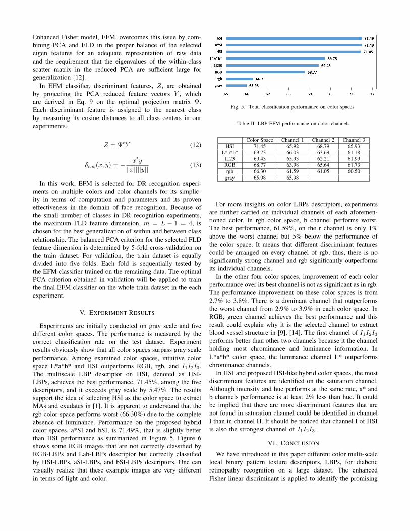
Experiments are initially conducted on gray scale and fivedifferent color spaces. The performance is measured by thecorrect classification rate on the test dataset. Experimentresults obviously show that all color spaces surpass gray scaleperformance. Among examined color spaces, intuitive colorspace L*a*b* and HSI outperforms RGB, rgb, and I1I2I3.The multiscale LBP descriptor on HSI, denoted as HSI-LBPs, achieves the best performance, 71.45%, among the fivedescriptors, and it exceeds gray scale by 5.47%. The resultssupport the idea of selecting HSI as the color space to extractMAs and exudates in [1]. It is apparent to understand that thergb color space performs worst (66.30%) due to the completeabsence of luminance. Performance on the proposed hybridcolor spaces, a*SI and bSI, is 71.49%, that is slightly betterthan HSI performance as summarized in Figure 5. Figure 6shows some RGB images that are not correctly classified byRGB-LBPs and Lab-LBPs descriptor but correctly classifiedby HSI-LBPs, aSI-LBPs, and bSI-LBPs descriptors. One canvisually realize that these example images are very differentin terms of light and color.
Fig. 5. Total classification performance on color spaces
Table II. LBP-EFM performance on color channels
Color Space Channel 1 Channel 2 Channel 3HSI 71.45 65.92 68.79 65.93
L*a*b* 69.73 66.03 63.69 61.18I123 69.43 65.93 62.21 61.99RGB 68.77 63.98 65.64 61.73rgb 66.30 61.59 61.05 60.50gray 65.98 65.98
For more insights on color LBPs descriptors, experimentsare further carried on individual channels of each aforemen-tioned color. In rgb color space, b channel performs worst.The best performance, 61.59%, on the r channel is only 1%above the worst channel but 5% below the performance ofthe color space. It means that different discriminant featurescould be arranged on every channel of rgb, thus, there is nosignificantly strong channel and rgb significantly outperformsits individual channels.
In the other four color spaces, improvement of each colorperformance over its best channel is not as significant as in rgb.The performance improvement on these color spaces is from2.7% to 3.8%. There is a dominant channel that outperformsthe worst channel from 2.9% to 3.9% in each color space. InRGB, green channel achieves the best performance and thisresult could explain why it is the selected channel to extractblood vessel structure in [9], [14]. The first channel of I1I2I3performs better than other two channels because it the channelholding most chrominance and luminance information. InL*a*b* color space, the luminance channel L* outperformschrominance channels.
In HSI and proposed HSI-like hybrid color spaces, the mostdiscriminant features are identified on the saturation channel.Although intensity and hue performs at the same rate, a* andb channels performance is at least 2% less than hue. It couldbe implied that there are more discriminant features that arenot found in saturation channel could be identified in channelI than in channel H. It should be noticed that channel I of HSIis also the strongest channel of I1I2I3.
VI. CONCLUSION
We have introduced in this paper different color multi-scalelocal binary pattern texture descriptors, LBPs, for diabeticretinopathy recognition on a large dataset. The enhancedFisher linear discriminant is applied to identify the promising

Fig. 6. Example DR images correctly classified by using HSI-LBPs, aSI-LBPs, bSI-LBPs descriptors but not by RGB-LBPs or Lab-LBP descriptors: (a) no-DRexample retina image with some luminance noise; (b) example mild DR with some microaneurysm; (c) example moderate DR with multiple microaneurysmsand hard exudates; (d) example severe DR with hemorrhages in 4 quadrants; (e) example proliferate DR retina image with hemorrhages, hard exudates, andfragile blood vessels around the fovea.
color spaces and color channel candidates to obtain the mostdiscriminant LBPs features. Results of the experiments showthat HSI-LBPs descriptor and its variances, a*SI-LBPs andbSI-LBPs descriptors outperform other color LBPs and grayLBPs descriptors.
For the future plan, the candidate color LBPs descriptorscan be combined with features from other region or gradientdetectors to improve DR performance. Other classificationand ensemble techniques will be explored to achieve betteraccuracy.
REFERENCES
[1] Jaykumar Lachure et al., ”Diabetic Retinopathy using morphologicaloperations and machine learning,” in Advance Computing Conf. (IACC),2015 IEEE Int., Banglore, 2015, pp. 617-622.
[2] Sohini Roychowdhury et al., ”Dream: Diabetic retinopathy analysis usingmachine learning,” IEEE J. Biomedical and Health Informatics, vol. 18,pp. 1717-1728, Dec. 2013.
[3] O. Faust et al., ”Algorithms for the automated detection of diabeticretinopathy using digital fundus images: a review,” J. Medical Syst., vol.36, pp. 145-157, Feb. 2012.
[4] Saiprasad Ravishankar et al., ”Automated feature extraction for earlydetection of diabetic retinopathy in fundus images,” in IEEE Conf.Computer Vision and Pattern Recognition, Miami, FL, 2009, pp 210-217.
[5] Wong Li Yun, et al., ”Identification of different stages of diabeticretinopathy using retinal optical images,” Inform. Sci., vol. 178, pp. 106-121, Jan. 2008.
[6] Balint Antal and Andras Hajdu, ”An ensemble-based system for mi-croaneurysm detection and diabetic retinopathy grading,” IEEE Trans.Biomed. Eng., vol. 59, pp. 1720-1726, Jun. 2012.
[7] Muthu Rama Krishnan Mookiah et al., ”Computer-aided diagnosis ofdiabetic retinopathy: A review.” Comput. in Biology and Medicine, vol.43, pp. 2136-2155, Dec. 2013.
[8] Thomas Walter and Jean-Claude Klein, ”Segmentation of color fundusimages of the human retina: Detection of the optic disc and the vasculartree using morphological techniques,” in 2nd Int. Symp., ISMDA, Madrid,Spain, 2001, pp. 282-287.
[9] Jagadish Nayak et al., ”Automated identification of diabetic retinopathystages using digital fundus images,” J. Medical Syst., vol. 32, pp. 107-115,Apr. 2008.
[10] Jorge de la Calleja et al., ”LBP and Machine Learning for DiabeticRetinopathy Detection,” in Intelligent Data Engineering and AutomatedLearningIDEAL 2014, Salamanca, Spain, 2014, pp. 110-117.
[11] Muhammad Nadeem Ashraf et al., ”Texture Feature Analysis of DigitalFundus Images for Early Detection of Diabetic Retinopathy,” in 11thInt. IEEE Conf. Computer Graphics, Imaging and Visualization (CGIV),Singapore, 2014, pp. 57-62.
[12] Chengjun Liu and Harry Wechsler, ”Robust coding schemes for indexingand retrieval from large face databases,” IEEE Trans. Image Process., vol.9, no. 1, pp. 132-137, Jan. 2000.
[13] Chanjira Sinthanayothin et al., ”Automated localisation of the opticdisc, fovea, and retinal blood vessels from digital colour fundus images,”British J. Ophthalmology, vol. 83, no. 8, pp. 902-910, Feb. 1999.
[14] U. R. Acharya et al., ”Computer-based detection of diabetes retinopathystages using digital fundus images,” in Proc. Institution of MechanicalEngineers, Part H: J. Engineering in Medicine, vol. 223, no. 5, pp. 545-553, Jul. 2009.
[15] K. Ram and S. Jayanthi, ”Multi-space clustering for segmentation ofexudates in retinal color photographs,” in 2009 Annu. Int. Conf. IEEEEngineering in Medicine and Biology Society, Minneapolis, MN, 2009,pp. 1437-1440.
[16] Jiawei Han and Micheline Kamber, ”Classification and Prediction,” inData mining: concepts and techniques, 2nd ed., San Francisco: MorganKaufmann, 2006, ch. 6, pp. 285-378.
[17] Mark S. Nixon and Alberto S. Aguado, ”Color images,” in Featureextraction & image processing for computer vision, 3rd ed., London,UK: Academic Press, 2012, ch. 13, pp. 541-599.
[18] Yu-Ichi Ohta et al., ”Color information for region segmentation,” Com-put. Graph. and Image Process., vol. 13, pp. 222-241, Jul. 1980.
[19] Sugata Banerji et al., ”Novel color LBP descriptors for scene and imagetexture classification,” in 15th Int. Conf. Image Processing, ComputerVision, and Pattern Recognition, Las Vegas, NV, 2011, pp. 537-543.
[20] P. J. Baldevbhai and R. S. Anand, ”Color image segmentation formedical images using L* a* b* color space,” IOSR J. Electron. andCommun. Eng. (IOSRJECE), vol. 1, pp. 24-45, May 2012.
[21] T. Ojala et al., ”A comparative study of texture measures with classifi-cation based on featured distributions,” Pattern recognition, vol. 29, pp.51-59, Jan. 1996.
[22] T. Ojala et al., ”Multiresolution Gray Scale and Rotation InvariantTexture Classification With Local Binary Patterns,” IEEE Trans. PatternAnal. Mach. Intell., vol. 24, pp. 971-987, Jul. 2002.
[23] Timo Ahonen, et al., ”Face description with local binary patterns:Application to face recognition,” IEEE Trans. Pattern Anal. Mach. Intell.,vol. 28, pp. 2037-2041, Dec. 2006.
[24] Ian H. Witten and Eibe Frank, ”Transformations: Engineering the inputand output,” in Data Mining: Practical machine learning tools andtechniques, 2nd ed., San Francisco: Morgan Kaufmann, 2005, ch. 7, sec.3, pp. 305-311.
[25] Chengjun Liu and Harry Wechsler, ”Gabor feature based classificationusing the enhanced fisher linear discriminant model for face recognition,”IEEE Trans. Image Process., vol. 11, pp. 467-476, Apr 2002.
[26] Daniel L. Swets and John Juyang Weng. ”Using discriminant eigenfea-tures for image retrieval,” IEEE Trans. Pattern Anal. Mach. Intell., vol.18, pp. 831-836, Aug. 1996.