disbiosis y endotoxemia metabólica inducidas por la dieta rica en … · rev 1792 dysbiosis and...
TRANSCRIPT

REV 1792
Dysbiosis and metabolic endotoxemia induced by high-fat diet
Disbiosis y endotoxemia metabólica inducidas por la dieta rica en grasa
Thalita Lin Netto Cândido¹, Josefina Bressan² and Rita de Cássia Gonçalves Alfenas²
1Candidate for doctoral degree in Nutrition Science. Federal University of Viçosa.
Minas Gerais, Brazil. 2Department of Nutrition and Health. Federal University of Viçosa.
Minas Gerais, Brazil
Received: 21/01/2018
Accepted: 03/04/2018
Correspondence: Department of Nutrition and Health. Federal University of Viçosa. Ed.
Biological Sciences Center II University Campus, s/n. 36570.900 Viçosa, MG. Brazil
e-mail: [email protected]
DOI: 10.20960/nh.1792
ABSTRACT
Introduction: diet plays a decisive role in the prevention and treatment of diseases
such as obesity, diabetes, allergies and inflammatory diseases. In addition to this, there
are numerous investigations about the role of the microbiota in the genesis of
metabolic diseases, especially obesity and its comorbidities.
Objective: the aim of this review is to discuss the influence of high-fat diets on
dysbiosis and metabolic endotoxemia.
Results and discussion: the intestinal microbial ecosystem has been shown to be
essential in the performance of functions in the host organism, however, several
factors can lead to an imbalance in the homeostasis of the microbiota, known as
dysbiosis. High-fat diets are associated with a reduction in intestinal bacterial diversity,
changes in membrane integrity, inducing increased permeability and
increased lipopolysaccharide (LPS) translocation, changes in the immune system, and
generation of low-intensity systemic inflammation. The installed endotoxemia can be

considered as a causal factor of subclinical inflammation related to several chronic
diseases, and as a result of this, it is essential to know the real impact of hyperlipidic
diets on the intestinal microbiota. Thus, it becomes essential to identify dietary
strategies that can minimize the inflammatory effects generated from changes in the
intestinal microbiota.
Key words: Endotoxemia. Intestinal microbiota. Lipopolysaccharides. Endotoxin.
Dietary fat.
RESUMEN
Introducción: la dieta juega un papel determinante en la prevención y el tratamiento
de enfermedades como la obesidad, la diabetes, las alergias y las enfermedades
inflamatorias. Agregado a ello, son innumerables las investigaciones acerca del papel
de la microbiota en la génesis de las enfermedades metabólicas, principalmente la
obesidad y sus comorbilidades.
Objetivo: el objetivo de esta revisión es analizar la influencia de las dietas ricas en
grasas sobre la disbiosis y la endotoxemia metabólica.
Resultados y discusión: se ha demostrado que el ecosistema microbiano intestinal es
esencial en el desempeño de funciones en el organismo del huésped, sin embargo,
varios factores pueden conducir a un desequilibrio en la homeostasis de la microbiota,
conocido como disbiosis. Las dietas ricas en grasas están asociadas a una reducción en
la diversidad bacteriana intestinal, alteraciones en la integridad de la membrana que
inducen un aumento de la permeabilidad y mayor translocación de lipopolisacáridos
(LPS), alteraciones en el sistema inmunológico y generación de inflamación sistémica
de baja intensidad. La endotoxemia instalada puede ser considerada un factor causal
de la inflamación subclínica relacionada con diversas enfermedades crónicas y, en
consecuencia, es imprescindible el conocimiento del impacto real de las dietas
hiperlipídicas sobre la microbiota intestinal. Así, es esencial la identificación de
estrategias dietéticas que puedan minimizar los efectos inflamatorios generados a
partir de alteraciones en la microbiota intestinal.

Palabras clave: Endotoxemia. Microbiota intestinal. Lipopolisacáridos. Endotoxinas.
Grasa dietética.
INTRODUCTION
In recent years, interest in the intestinal microbiota and its interactions as the host has
increased (1). In particular, investigations on the role of microbiota in health regulation
and onset of diseases such as inflammatory diseases, allergies, diabetes and obesity
(2). It is estimated that the human intestinal microbiota is composed of 10 to 100
trillion microorganisms and possesses about 150 times more genes than the human
genome (3).
With the advancement of sequencing techniques, metagenomic analyses of 16S rRNA
have demonstrated a large number of bacterial genes that inhabit the human gut (4).
The predominance of Firmicutes and Bacteroidetes and restricted anaerobic genera
such as Bacteroides, Eubacterium, Clostridium, Ruminococcus, Peptococcus,
Bifidobacterium and Fusobacterium were observed in relation to facultative anaerobes
such as Lactobacillus, Escherichia, Enterobacter, Enterococcus, Klebsiella and Proteus.
(4,5). However, there is no accuracy in the composition of the microbiota in humans
(6), being influenced by the age, genetic and environmental factors, diet and structure
of the intestinal wall of the host (7).
The intestinal microbial ecosystem has been shown essential in the performance of
functions such as preservation of intestinal mucosal integrity, nutrient absorption and
energy homeostasis, as well as being directly linked to the immune and nervous
system (8,9). It has recently been suggested that the microbiota may play a significant
role in the pathogenesis of obesity and its comorbidities (8,10-13). This is partly due to
an imbalance in the homeostasis of the microbiota, known as dysbiosis, which is
characterized by changes in diversity, toxin production, increased permeability, and
hormonal and immunological changes, culminating in low-grade inflammatory
state (14).
Lipopolysaccharide (LPS), a constituent of intestinal bacteria, can be an important
inducer of the inflammatory response (15). Detected in the intestinal lumen, under
normal conditions, it does not represent human health problems. However, when
there is an imbalance this can be easily transferred to the circulatory system. This can
Comentario [MPG1]: El estilo de fuente para taxonomias no estaba unificado. He dejado géneros y familias en cursiva y filos/clases en redonda. ¿Sería correcto?
lead to high plasma levels of LPS, a condition known as metabolic endotoxemia (16). In
addition, there is evidence that the type of diet consumed, especially diets rich in fats,
may contribute to endotoxemia (17).
Much has been discussed about the role of diet in the regulation and composition of
the intestinal microbiota (14,18,19). High-fat diets are associated with a reduction in
intestinal bacterial diversity (20), changes in intestinal membrane integrity, inducing
increased permeability and increased LPS translocation (21), changes in the immune
system and generation of systemic inflammation (22).
Therefore, the purpose of this review is to examine papers from the scientific
literature in order to discuss the mechanisms and interactions between diets rich in
fats, intestinal microbiota and metabolic endotoxemia.
METHODS
The research was performed in the Medline/PubMed and Scopus databases. The
following terms were used in English: endotoxemia; endotoxins; lipopolysaccharides;
gut microbiota and dietary fat. The literature search was conducted from January to
July 2017. All articles selected and included in this article were published between
2007 and 2017. They were read and critically grouped according to their thematic and
scientific relevance. From this, the sections presented in this article were created:
“Dysbiosis induced by high-fat diet” and “Diet rich in lipids and metabolic
endotoxemia” (Fig. 1).
HIGH-FAT DIET-INDUCED DYSBIOSIS
Well-established scientific evidence reports that excessive intake of fats and refined
carbohydrates are strongly associated with obesity and metabolic diseases. However,
it is recent discoveries that may also influence the composition of the intestinal
microbiota of the host (23). It is known that the human intestine has trillions of
microorganisms, containing more genes than the human genome itself. These
microorganisms have evolved and are capable of performing specific and unique
biochemical and metabolic functions to the microbial species (3).
The role of the intestinal microbiota in the human disease health process has been
gaining even more focus and attention, especially on the etiology of obesity and its
Comentario [MPG2]: Revisar redacción. No es comprensible.

comorbidities (24). It is suggested that intestinal bacteria are involved in energy
homeostasis and body weight control, being able to extract calories from indigestible
nutrients and produce short chain fatty acids, amino acids and vitamins (25). In
addition, they participate in the regulation and expression of genes that signal
metabolic pathways of absorption and storage of nutrients, such as fats, in the host
(26).
The interaction microorganisms and host is under a complex interface with the
gastrointestinal mucosa. Its outer layer offers conditions for colonization by bacteria
and may suffer direct influence of the diet (27) since bacteria that degrade mucus are
influenced by high-fat diets. Thus, it is evident that diet plays an important role in the
modulation of the microbial ecosystem (28).
Studies have reported that the consumption of diets with high-fat contents may alter
the composition and functionality of the intestinal microbiota in mice and humans (29-
35) (Table I and II). Short-term dietary interventions (24 h) have been shown to rapidly
change the intestinal microbiota; on the other hand, long-term dietary patterns are
able to modulate the composition of the microbiota, despite the detection of
resilience after induction of dietary changes (36). Factors such as age, diet time,
function, and properties of the different intestinal segments may also influence
microbial diversity, and when the cecum and colon mucosa are compared, the latter is
more resistant to variations (25).
Theoretically, dietary fat that reaches the colon can be metabolized by bacteria;
cholesterol is degraded to form the metabolite coprostanol that is excreted in the
feces (37). In addition, high-fat diets can stimulate the production of bile acids, which
have antimicrobial activity and eventually select the species capable of metabolizing
bile acids in the intestine (1). However, the use of fat as a source of energy for the
growth of bacteria remains unclear, since fat metabolization is not performed in
anaerobiosis, a condition common to most of the bacteria that inhabit the human
intestine (38).
The influence of the diet rich in fat was analyzed in a study with mice fed a hyperlipid
diet for three months, which showed a decrease of Bacteroidetes and increase of
Firmicutes, Proteobacteria and Actinobacteria (29). Although long-term observations
concisely present changes in the microbiota, it has been shown that short-term (five

weeks) interventions are also capable of modifying the microbial ecosystem of mice
(25). In addition, it has been reported that the diversity and composition of the
microbiota, after being altered by the high-fat diet, can be reestablished with the
intake of low fat diet.
Increased phylum Firmicutes was observed in wild-type mice receiving a high fat diet,
and reduction of Bacteroidetes in genetically obese (ob/ob) resistant leptin mice (30).
Thus, the authors suggested that a hyperlipid diet rather than the obese ob/ob
genotype exerted a greater influence on the composition of the microbiota, which was
also observed in another study (29). Thus, they emphasize that the microbiota changes
observed in mice fed a high fat diet were probably attributable to changes in diet.
Similarly, the provision of a high-fat diet caused a general decrease in microbiota
diversity and an increase in the ratio Firmicutes:Bacteroidetes in several studies with
mice (25,32,39). Based on these findings, it was suggested that the microbiota of the
obese has metabolic pathways that are highly efficient in extracting energy and
favoring lipogenesis. This fact was supported by a study that, when transplanting the
microbiota from obese mice to germ free mice, presented higher fat gain than animals
transplanted with microflora from lean mice (40).
Despite reports of high concentrations of short chain fatty acids in the stools of obese
individuals, these changes were not associated with a higher proportion of Firmicutes
(41). This result generated research on the characteristics of the obesogenic
microbiota, since it seems unlikely that only Firmicutes:Bacteroidetes are the only ones
involved in the pathogenesis of obesity (30).
In a study with pairs of monozygotic and dizygotic twins, concordant for thinness or
obesity and their mothers, Turnbaugh et al. (42) demonstrated that the microbial
ecosystem is shared among family members, but that each individual has a specific
bacterial composition. The authors also reported that obesity is associated with
changes in phylum level in the microbiota, reduction of bacterial diversity and
alteration of genes and metabolic pathways. A greater abundance of Actinobacteria,
added to the Firmicutes phylum is reported, suggesting that the action of other phyla
in the mechanisms that involve the microbiota and obesity interaction should be
investigated.
Comentario [MPG3]: Revisar redacción. No se entiende.

In addition, mice fed a high-fat diet showed an increase in Clostridia XIV and
Enterobacteriales and a decrease in Bifidobacterium spp (43). Similar results were also
observed in the analysis of the microbiota after ingestion of a hyperlipic diet rich in
palm oil and lard (33). Both diets were associated with increased relative abundance of
Clostridiales spp and decrease of Bacteroidal.
The reduction in gram-positive bacteria such as bifidobacteria directly and negatively
affects the integrity of the intestinal membrane, since they are responsible for
maintaining and improving the intestinal barrier function, preventing the passage of
bacteria and toxins (15). The same function can be attributed to the bacteria
Akkermansia muciniphila, belonging to the phylum Verrucomicrobia, associated with
stimulation of the immune system with anti-inflammatory properties (44). The
increase in the abundance of Clostridiales spp may be related to mechanisms of
metabolic pathways of cholesterol and levels of bile acids (33).
Some studies have reported that the fatty acid profile is also able to modulate the
composition of the intestinal microbiota and contribute to the induction of low
intensity inflammation (Table III) (23,35,39,43). However, this subject requires more
research and more in-depth knowledge (45).
The impact of different types of fats on the host’s health, metabolism and microbiota
was analyzed (23). Using mice fed a high-fat diet containing palm oil, olive oil,
safflower oil and flaxseed oil/fish for 16 weeks compared to mice fed a low fat diet, it
was found that mice populations fed palm had relatively lower populations of
Bacteroidetes at the phylum level, compared to olive oil diet; this in turn showed an
increase in the family Bacteroidaceae. However, mice fed flax/fish oil showed a
significant increase in the concentrations of eicosapentaenoic acid (EPA) and
docosahexaenoic acid (DHA) and in the intestinal abundance of Bifidobacterium. The
findings suggest that the impact on the microbiota is due to both changes in the
quantity and quality of fat ingested and that fats such as flaxseed oil positively impact
the microbial ecosystem of the host.
Similarly, changes in the intestinal microbiota of humans were different according to
the intake of different fatty acids, monounsaturated fatty acids (MUFA), omega-3
polyunsaturated fatty acids (PUFA) and omega-6 PUFA. While consumption of MUFA
and omega-6 PUFA was inversely associated with an increase in the number of

Bifidobacterium, an increase in omega-3 PUFA intake was directly associated with a
higher number of bacteria in the Lactobacillus group (35).
In addition, in a study with middle aged (12 months) mice fed with saturated fat (lard),
monounsaturated (soybean oil) and polyunsaturated (fish oil), different responses in
the microbiota were observed. The group fed with fish oil showed higher relative
abundance of the Proteobacteria phylum and the genus Desulfovibrio in the cecal and
colonic contents. On the other hand, the diet based on saturated fatty acids conferred
a higher Firmicutes:Bacteroidetes and more abundant presence of Verrucomicrobia
and Tenericutes (46).
RICH DIET IN LIPIDS AND METABOLIC ENDOTOXEMIA
Obesity, diabetes and insulin resistance are associated with a low intensity systemic
inflammation caused by multiple factors and whose triggering agents are not fully
elucidated. However, the intestinal microbiota has been largely associated with this
scenario (8,24). This evidence was obtained from studies that demonstrated the
existence of endotoxemia, the passage of bacterial lipopolysaccharide (LPS) into the
bloodstream during consumption of fat-rich diets by mice (47,48). This effect has also
been confirmed in human studies (17,49-51).
Naturally the microbiota is a reservoir of LPS, since it is one of the components of the
outer cell wall of gram-negative bacteria, and it is possible to detect more than 1 g of
LPS in the intestinal lumen. Therefore, under normal conditions LPS is not harmful to
the host and appears to be involved in immune regulation, such as increased
phagocytic capacity, lymphocyte proliferation and lymphokine secretion. However, in
situations of dysbiosis, which can be caused by the type of diet consumed, the transfer
of LPS to the circulatory system can be increased and thus generate what we call
metabolic endotoxemia (15).
Diet plays an important role in the regulation of endotoxemia. This evidence is
supported by the fact that an increase in plasma LPS occurs in mice fed high-energy
diets, whether rich in carbohydrates or fats, for four weeks. However, the high-fat diet
proved to be more efficient in favor of LPS transfer from the intestinal lumen to the
bloodstream (48).

An increase in postprandial serum endotoxin concentration was observed in healthy
adults on high-fat diets, especially those on a saturated fat diet, when compared to
subjects who received diets high in polyunsaturated fats (51). Similarly, men who
received high-lipid meals also had elevated postprandial LPS levels when compared to
fasted individuals (50). A possible explanation for this fact is the ability of LPS to be
incorporated into micelles, absorbed and added to chylomicrons, due to the presence
of a fraction insoluble in its molecular structure (52).
In addition, there may be an increase in local pressure and loosening of junctional
complexes between enterocytes, or even basement membrane rupture, due to excess
chylomicrons generated from the hyperlipidic diet. After the lesion caused during fat
absorption the intestinal barrier may be compromised, increasing intestinal
permeability, mainly to LPS (15).
Thus, consumption of high-fat diets leads to increased intestinal permeability and
reduced expression of genes encoding tight or tight junction proteins, such as
claudina-1, caludina-3, occludin, and junctional adhesion molecule 1 (53). This is due to
the regulation of permeability by mast cells, through the secretion of mediators such
as tumor necrosis factor α (TNF-α), IL-1b, IL-4 and IL-13, receptor-2, favoring LPS
translocation (54).
The fatty acid profile of the ingested lipid diet has been shown to be an important
modulator of the microbiota, triggering endotoxemia (15). In a study with mice fed
omega-6-rich meals, they exhibited conditions that characterized elevated levels of
metabolic endotoxemia and low-intensity inflammation (55), while mice fed omega-3
fatty acids had decreased LPS production and permeability, with a significant reduction
in metabolic endotoxemia.
In another study, it was observed that diet rich in fats (45% of lipids), when compared
to control diet (13% of lipids), increased adiposity and plasma levels of
lipopolysaccharide binding protein (LBP) ingestion for three and six weeks. However,
with only one week of ingestion, an immediate but reversible increase in paracellular
permeability and decreased expression of IL-10 was observed. In addition, a reduction
of the abundance of genera within the class Clostridia in the ileum was observed (32).
Increased dietary fat content may influence phylum Actinobacteria, which may reduce
the number of gram-positive Bifidobacterium species, considered to be beneficial. This

may lead to an increase in LPS plasma concentrations, induction of low grade
inflammation and maintenance of obesity (56). Likewise, inflammation can also occur
with the increase of gram-negative bacteria, for example Desulfovibrio, capable of
producing endotoxins, reduce sulfate to H2S and impair the intestinal barrier (20).
In addition, habitual intake of saturated fatty acids, derived from diets rich in fats and
calories, can directly stimulate TLR4 cells. Alternatively, there is also a higher
concentration of LPS by the increase of gram-negative bacteria, which can stimulate
TLR4 and induce the expression of several cytokines, resulting in a state of low
intensity inflammation and insulin resistance. In addition, increasing concentrations of
circulating fatty acids may further increase nitric oxide production and decrease insulin
sensitivity due to impaired lipoprotein lipase (LPL) activity and increased lipolysis (4).
An issue that deserves to be highlighted is the increase in bile production caused by
the ingestion of high-fat diets, which acts selectively in relation to colonization of the
intestine, being important in the modulation of the microbiota and in the role that it
exerts on the permeability and production of endotoxins (57).
CONCLUSION
There is increasing scientific evidence that high-fat diets can modulate intestinal
microbiota composition, enhancing LPS uptake and affecting mucosal integrity,
resulting in metabolic endotoxemia. Installed endotoxemia is a causal factor of
subclinical inflammation related to several chronic diseases and, as a result of this, it is
essential to know the real impact of hyperlipidic diets on the microbiota.
REFERENCIAS
1. Alou MT, Lagier JC, Raoult D. Diet influence on the gut microbiota and dysbiosis
related to nutritional disorders. Human Microb Jour 2016;1:3-11.
2. Isolauri E, Sherman PM, Walker WA. Intestinal microbiome: functional aspects
in health and disease. Nestlé Nutr Inst Workshop Ser 2017;88:95-105.
3. Qin J, Li R, Raes J, Arumugam M, Burgdorf KS, Manichanh C, et al. A human gut
microbial gene catalogue established by metagenomic sequencing. Nature
2010;464:59-65.

4. Hemalatha R. Diet and gut microbiota in human health. Proc Indian Natn Sci
Acad 2016;82(5):1437-47.
5. Caporaso JG, Lauber CL, Costello EK, Berg-Lyons D, González A, Stombaugh J, et
al. Moving pictures of the human microbiome. Genome Biol 2011;12:1-8.
6. Phillips ML. Gut reaction: environmental effects on the human microbiota.
Environ Health Perspect 2009;117(5):A198-A205.
7. Graf D, Di Cagno R, Fak F, Flint HJ, Nyman M, Saarela M, et al. Contribution of
diet to the composition of the human gut microbiota. Microb Ecol Health Dis
2015;26:261-4.
8. Arslan N. Obesity, fatty liver disease and intestinal microbiota. World J
Gastroenterol 2014;20(44):16452-63.
9. Li D, Wang P, Wang P, Hu X, Chen F. The gut microbiota: a treasure for human
health. Biotechnol Adv 2016;34:1210-24.
10. Tagliabue A, Elli M. The role of gut microbiota in human obesity: recent findings
and future perspectives. Nutr Metab Cardiovasc Dis 2013;23:160-8.
11. Zhao L. The gut microbiota and obesity: from correlation to causality. Nat Rev
Microbiol 2013;11(9):639-47.
12. Martínez KB, Pierre JF, Chang EB. The gut microbiota: the gateway to improved
metabolism. Gastroenterol Clin N Am 2016;45:601-14.
13. Tang WH, Kitai T, Hazen SL. Gut microbita in cardiovascular health and disease.
Circ Res 2017;120(7):1183-96.
14. Proctor C, Thiennimitr P, Chattipakorn N, Chattipakorn SC. Diet, gut microbiota
and cognition. Metab Brain Dis 2017;32(1):1-17.
15. Moreira APB, Teixeira TFS, Peluzio MCG, Alfenas RCG. Gut microbiota and the
development of obesity. Nutr Hosp 2012;27(5):1408-14.
16. Luche E, Cousin B, Garidou L, Serino M, Waget A, Barreau C, et al. Metabolic
endotoxemia directly increases the proliferation of adipocyte precursors at the onset
of metabolic diseases through a CD14-dependent mechanism. Mol Metab
2013;2(3):281-91.
17. Laugerette F, Vorsa C, Géloën A, Chauvind MA, Soulageb C, Lambert-Porcheron
S, et al. Emulsified lipids increase endotoxemia: possible role in early postprandial low-
grade inflammation. J Nutr Biochem 2011;22:53-9.

18. Singh RK, Chang HW, Yan D, Lee KM, Ucmak D, Wong K, et al. Influence of diet
on the gut microbiome and implications for human health. J Transl Med 2017;15(73):1-
17.
19. Tan H, O’toole PW. Impact of diet on the human intestinal microbiota. J Food
Sci 2015;2:71-7.
20. Zhang C, Zhang M, Wang S, Han R, Cao Y, Hua W, et al. Interactions between
gut microbiota, host genetics and diet relevant to development of metabolic
syndromes in mice. ISME J 2010;4:232-41.
21. Ji Y, Sakata Y, Tso P. Nutrient-induced inflammation in the intestine. Curr Opin
Clin Nutr Metab Care 2011;14:315-21.
22. Wall R, Ross RP, Fitzgerald GF, Stanton C. Fatty acids from fish: the anti-
inflammatory potential of long-chain omega-3 fatty acids. Nutr Rev 2010;68:280-9.
23. Patterson E, O’Doherty RM, Murphy EF, Wall R, O’Sullivan O, Nilaweera K, et al.
Impact of dietary fatty acids on metabolic activity and host intestinal microbiota
composition in C57BL/6J mice. Br J Nutr 2014;111:1905-17.
24. Bouter KE, Van Raalte DH, Groen AK, Nieuwdorp M. Role of the gut microbiome
in the pathogenesis of obesity and obesity-related metabolic dysfunction.
Gastroenterology 2017;152:1671-8.
25. Shang Y, Khafipour E, Derakhshani H, Sarna LK, Woo CW, Siow YL, et al. Short
term high fat diet induces obesity‑ enhancing changes in mouse gut microbiota that
are partially reversed by cessation of the high fat diet. Lipids 2017;52(6):499-511.
26. Ursell LK, Haiser HJ, Treuren WV, Garg N, Reddivari L, Vanamala J, et al. The
intestinal metabolome: an intersection between microbiota and host.
Gastroenterology 2014;146:1470-6.
27. Nehra V, Allen JM, Mailing LJ, Kashyap PC, Woods JA. Gut microbiota:
modulation of host physiology in obesity. Physiology 2016;31:327-35.
28. Cotillard A, Kennedy SP, Kong LC, Prifti E, Pons N, Le Chatelier E, et al. Dietary
intervention impact on gut microbial gene richness. Nature 2013;500:585-8.
29. Hildebrandt MA, Hoffmann C, Sherrill-Mix SA, Keilbaugh SA, Hamady M, Chen
YY, et al. High-fat diet determines the composition of the murine gut microbiome
independently of obesity. Gastroenterology 2009;137:1716-24.
30. Murphy EF, Cotter PD, Healy S, Marques TM, O’Sullivan O, Fouhy F, et al.
Composition and energy harvesting capacity of the gut microbiota: relationship to diet,
obesity and time in mouse models. Gut 2010;59:1635-42.
31. Daniel H, Gholami AM, Berry D, Desmarchelier C, Hahne H, Loh G, et al. High-fat
diet alters gut microbiota physiology in mice. ISME J 2014;8:295-08.
32. Hamilton MK, Boudry G, Lemay DG, Raybould HE. Changes in intestinal barrier
function and gut microbiota in high-fat diet-fed rats are dynamic and region
dependent. Am J Physiol Gastrointest Liver Physiol 2015;308:G840-G51.
33. Kübeck R, Bonet-Ripoll C, Hoffmann C, Walker A, Müller VM, Schüppel VL, et al.
Dietary fat and gut microbiota interactions determine diet-induced obesity in mice.
Mol Metab 2016;5:1162-74.
34. Wu GD, Chen J, Hoffmann C, Bittinger K, Chen YY, Keilbaugh SA, et al. Linking
Long-term dietary patterns with gut microbial enterotypes. Science
2011;334(6052):105-8.
35. Simões CD, Maukonen J, Kaprio J, Rissanen A, Pietilainen KH, Saarela M.
Habitual dietary intake is associated with stool microbiota composition in monozygotic
twins. J Nutr 2013;143:417-23.
36. Portune KJ, Benítez-Páez A, Del Pulgar EMG, Cerrudo V, Sanz Y. Gut microbiota,
diet, and obesity-related disorders - The good, the bad, and the future challenges. Mol
Nutr Food Res 2017;61(1):1-17.
37. Gerard P. Metabolism of cholesterol and bile acids by the gut microbiota.
Pathogens 2014;3(1):14-24.
38. Blaut M, Klaus S. Intestinal microbiota and obesity. Handb Exp Pharmacol
2012;209:251-73.
39. De Wit N, Derrien M, Bosch-Vermeulen H, Oosterink E, Keshtkar S, Duval C, et
al. Saturated fat stimulates obesity and hepatic steatosis and affects gut microbiota
composition by an enhanced overflow of dietary fat to the distal intestine. AJP
Gastrointest Liver Physiol 2012;303:G589-G99.
40. Turnbaugh PJ, Backhed F, Fulton L, Gordon JI. Marked alterations in the distal
gut microbiome linked to diet induced obesity. Cell Host Microbe 2008;3:213-23.
41. Schwiertz A, Taras D, Schäfer K, Beijer S, Bos NA, Donus C, et al. Microbiota and
SCFA in lean and overweight healthy subjects. Obesity 2010;18(1):190-5.

42. Turnbaugh PJ, Hamady M, Yatsunenko T, Cantarel BL, Duncan A, Ley RE, et al. A
core gut microbiome in obese and lean twins. Nature 2009;457:481-5.
43. Mujico JR, Baccan GC, Gheorghe A, Díaz LE, Marcos A. Changes in gut
microbiota due to supplemented fatty acids in diet-induced obese mice. Br J Nutr
2013;110:711-20.
44. Everard A, Belzer C, Geurts L, Ouwerkerk JP, Druart C, Bindels LB, et al. Cross-
talk between Akkermansia muciniphila and intestinal epithelium controls diet-induced
obesity. Proc Natl Acad Sci 2013;110(22):9066-71.
45. Kashtanova D, Popenko AS, Tkacheva ON, Tyakht AB, Alexeev DG, Boytsov AS.
Association between the gut microbiota and diet: fetal life, early childhood, and
further life. Nutrition 2016;32:620-7.
46. Li H, Zhu Y, Zhao F, Song S, Li Y, Xu X, et al. Fish oil, lard and soybean oil
differentially shape gut microbiota of middle-aged rats. Nature 2017;7(826):1-12.
47. Cani PD, Neyrinck AM, Fava F, Knauf C, Burcelin RG, Tuohy KM, et al. Selective
increases of bifidobacteria in gut microflora improve high-fat-diet-induced diabetes in
mice through a mechanism associated with endotoxaemia. Diabetologia 2007;50:2374-
83.
48. Flint HJ, Scott KP, Louis P, Duncan SH. The role of the gut microbiota in nutrition
and health. Nat Rev Gastroenterol Hepatol 2012;9(10):577-89.
49. Amar J, Burcelin R, Ruidavets JB, Cani PD, Fauvel J, Alessi MC, et al. Energy
intake is associated with endotoxemia in apparently healthy men. Am J Clin Nutr
2008;87:1219-23.
50. Erridge C, Attina T, Spickett CM, Webb DJ. A high-fat meal induces low-grade
endotoxemia: evidence of a novel mechanism of postprandial inflammation. Am J Clin
Nutr 2007;86:1286-92.
51. Lyte JM, Gabler NK, Hollis JH. Postprandial serum endotoxin in healthy humans
is modulated by dietary fat in a randomized, controlled, cross-over study. Lipids Health
Dis 2016:15(186):1-10.
52. Ghoshal S, Witta J, Zhong J, Villiers W, Eckhardt E. Chylomicrons promote
intestinal absorption of lipopolysaccharides. J Lipid Res 2009;50:90-7.

53. Cani PD, Bibiloni R, Knauf C, Waget A, Neyrinck AM, Delzenne NM, et al.
Changes in gut microbiota control metabolic endotoxemia-induced inflammation in
high-fat diet-induced obesity and diabetes in mice. Diabetes 2008;57:1470-81.
54. De La Serre CB, Ellis CL, Lee J, Hartman AL, Rutledge JC, Raybould HE.
Propensity to high-fat diet-induced obesity in rats is associated with changes in the gut
microbiota and gut inflammation. Am J Physiol Gastrointest Liver Physiol
2010;299:G440-G8.
55. Kaliannan K, Wang B, Li XY, Kim KJ, Kang JX. A host-microbiome interaction
mediates the opposing effects of omega-6 and omega-3 fatty acids on metabolic
endotoxemia. Sci Rep 2015;5(11276):1-17.
56. Cani PD, Delzenne NM. The gut microbiome as therapeutic target. Pharmacol
Ther 2011;130:202-12.
57. Suzuki T, Hara H. Dietary fat and bile juice, but not obesity, are responsible for
the increase in small intestinal permeability induced through the suppression of tight
junction protein expression in LETO and OLETF rats. Nutr Metab 2010;12:7-19.
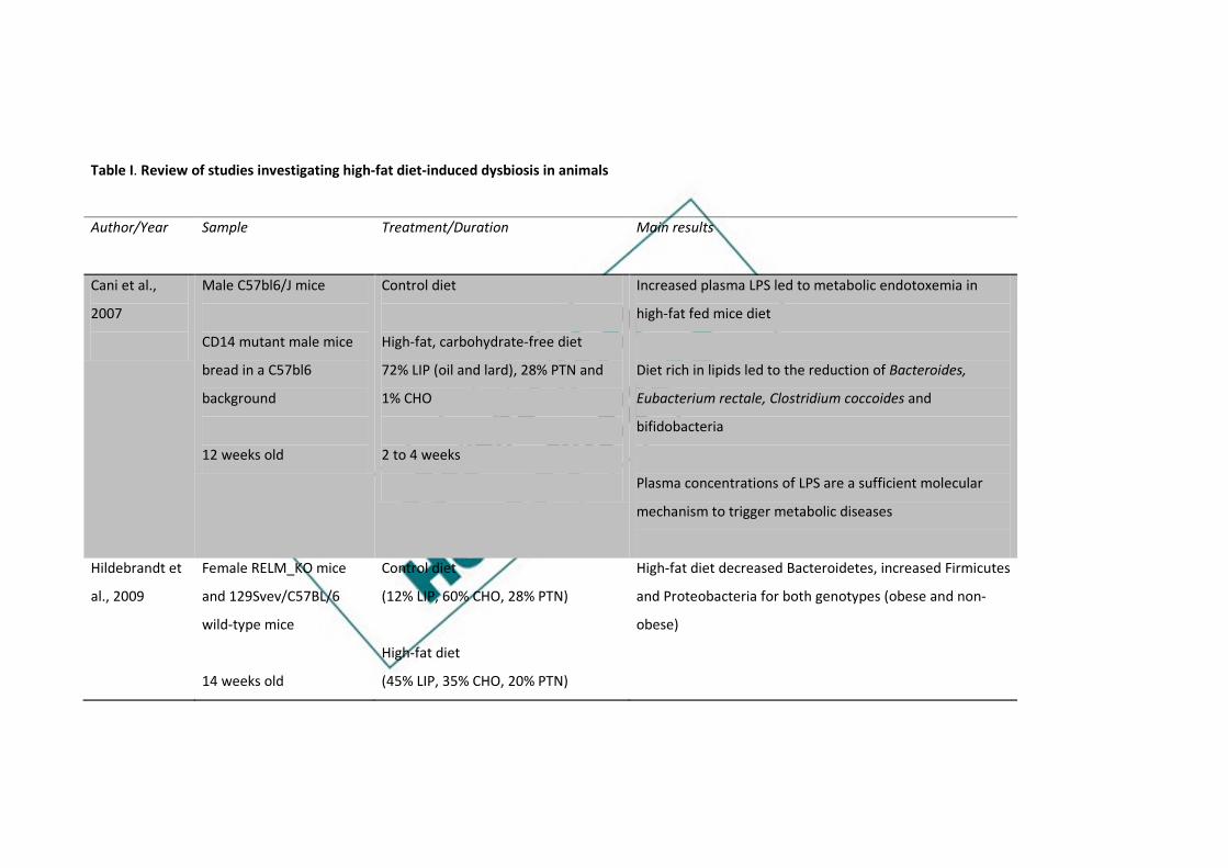
Table I. Review of studies investigating high-fat diet-induced dysbiosis in animals
Author/Year Sample Treatment/Duration
Main results
Cani et al.,
2007
Male C57bl6/J mice
CD14 mutant male mice
bread in a C57bl6
background
12 weeks old
Control diet
High-fat, carbohydrate-free diet
72% LIP (oil and lard), 28% PTN and
1% CHO
2 to 4 weeks
Increased plasma LPS led to metabolic endotoxemia in
high-fat fed mice diet
Diet rich in lipids led to the reduction of Bacteroides,
Eubacterium rectale, Clostridium coccoides and
bifidobacteria
Plasma concentrations of LPS are a sufficient molecular
mechanism to trigger metabolic diseases
Hildebrandt et
al., 2009
Female RELM_KO mice
and 129Svev/C57BL/6
wild-type mice
14 weeks old
Control diet
(12% LIP, 60% CHO, 28% PTN)
High-fat diet
(45% LIP, 35% CHO, 20% PTN)
High-fat diet decreased Bacteroidetes, increased Firmicutes
and Proteobacteria for both genotypes (obese and non-
obese)
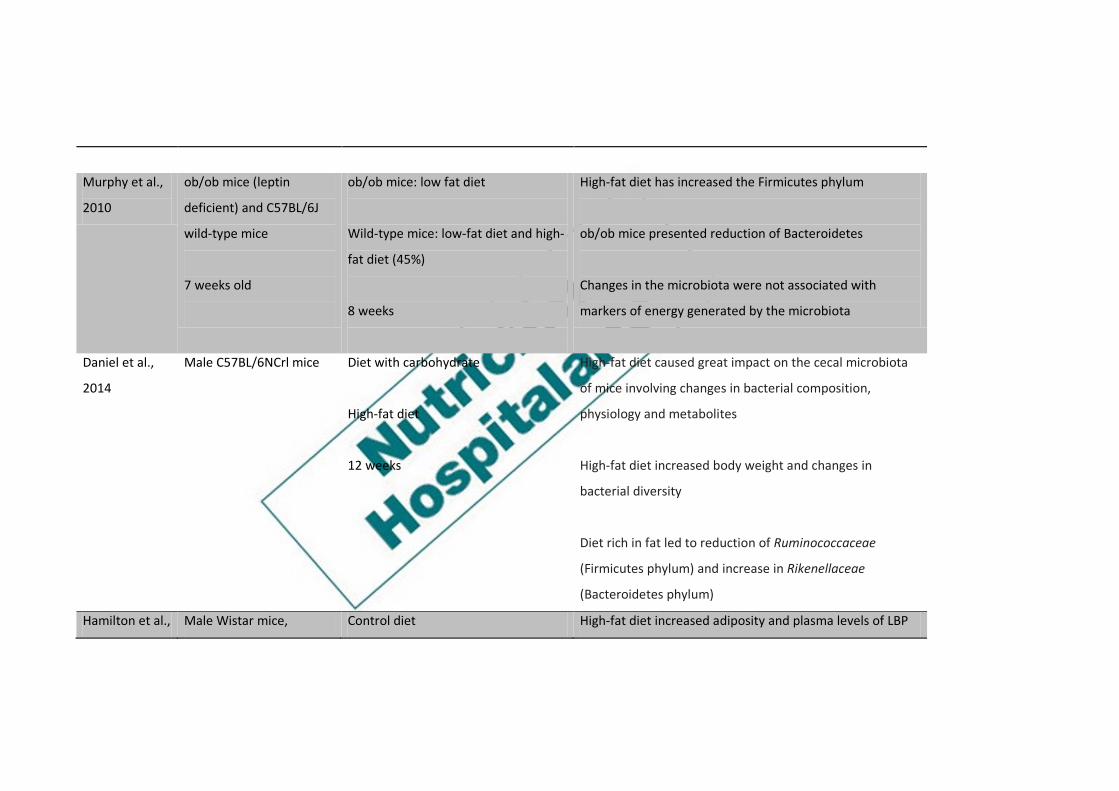
Murphy et al.,
2010
ob/ob mice (leptin
deficient) and C57BL/6J
wild-type mice
7 weeks old
ob/ob mice: low fat diet
Wild-type mice: low-fat diet and high-
fat diet (45%)
8 weeks
High-fat diet has increased the Firmicutes phylum
ob/ob mice presented reduction of Bacteroidetes
Changes in the microbiota were not associated with
markers of energy generated by the microbiota
Daniel et al.,
2014
Male C57BL/6NCrl mice
Diet with carbohydrate
High-fat diet
12 weeks
High-fat diet caused great impact on the cecal microbiota
of mice involving changes in bacterial composition,
physiology and metabolites
High-fat diet increased body weight and changes in
bacterial diversity
Diet rich in fat led to reduction of Ruminococcaceae
(Firmicutes phylum) and increase in Rikenellaceae
(Bacteroidetes phylum)
Hamilton et al., Male Wistar mice, Control diet High-fat diet increased adiposity and plasma levels of LBP

2015 9 to 10 weeks old (13% LIP, 23% PTN)
High-fat diet
(45% LIP, 20% PTN)
1, 3 or 6 weeks
at 3 and 6 weeks
After one week, there was an immediate but reversible
increase in paracellular permeability, decreased IL-10
expression, and reduced abundance of genera within the
Clostridia class in the ileum in mice fed a high-fat diet
High-fat diet has increased Firmicutes:Bacteroidetes ratio
in both the small and large intestine
With a high-fat diet, Deferribacteres increased in the
cecum
Control diet increased abundance of Cyanobacteria in the
ileum
Kubeck et al.,
2016
Germ free male mice and
specific pathogen free
male C57BL/6N
Control diet
(5% soybean oil, 12% LIP)
High-fat diet was associated with increased relative
abundance of Clostridiales spp and decrease of Bacteroidal

8 weeks old High-fat diet
(48% LIP, with palm oil and lard)
Dietary cholesterol may affect the binding between
microbiota and host metabolism
Shang et al.,
2017
Male C57BL/6J mice
6 weeks old
Control diet: low in fat (10%)
High-fat diet (60%) for 7 weeks
High-fat diet for 5 weeks followed by
low fat diet for 2 weeks
Body weight, blood glucose, hepatic triglycerides were
higher in the high-fat diet group, regardless of the time
Observed significant difference in diversity and functional
properties between group with high-fat diet and control
diet
High-fat diet reduced the ratio Bacteroidetes:Firmicutes
High-fat diet followed by diet control restored the diversity
and composition of the microbiota in the cecum
CHO: carbohydrate; LIP: lipids; PTN: protein; LPS: lipopolysaccharide; LBP: lipopolysaccharide binding protein; IL-10: Interleukin-10.

Table II. Review of studies investigating the dysbiosis induced by a high-fat diet in humans
Author/Year Sample Treatment/Duration
Main results
Amar et al.,
2008
Healthy men
1,015 people randomly
recruited in France
Three-day food record We found a link between food intake and plasma LPS
Experimental data suggest that fat was more efficient in
transporting bacterial LPS from the intestinal lumen into
the bloodstream
The results of this study add to the knowledge of
mechanisms responsible for the relationships between
food intake and metabolic diseases
Wu et al., 2011 Cross-sectional study in
healthy adults
n = 98
Food frequency questionnaire
Food recall
Bacteroidetes and Actinobacteria were positively
associated with fat, whereas Firmicutes and
Proteobacteria showed a negative association
Enterotypes were strongly associated with long-term
diets, particularly proteins and animal fat (Bacteroides)

versus carbohydrates (Prevotella)
Simões et al.,
2013
Healthy adult twins Cross-sectional data were evaluated
in pairs of monozygotic twins of
different body weight and body fat
score was assessed for habitual
daily intake and fecal microbiota
Co-twins with similar daily energy intake had similar
numbers of Bacteroides spp when compared with those
with different energy intake
Higher MUFA intake was associated with lower numbers
of Bifidobacterium and slightly larger numbers of
Bacteroides spp
Co-twins who ingested identical levels of SFA had very
similar Bacteroides spp
Intake of n3-PUFA resulted in a significant positive
association with abundance of Lactobacillus
Ingestion of n6-PUFA was associated with decreased
numbers of Bifidobacterium

Lyte et al., 2016 Healthy adults (n = 20,
mean age 25 ± 3.2 years)
Control diet (olive oil - 20%)
High-fat diet with n-3
(fish oil) (35%)
High-fat diet with n-6
(grape seed oil) (35%)
Diet rich in saturated fat
(coconut oil) (35%)
Serum endotoxin concentration was increased during the
postprandial period after consumption of a high-saturated
fat meal but decreased after the meal with n-3
The n-6 meal did not affect the postprandial endotoxin
concentration in relation to the control meal
There was no postprandial effect on inflammatory
biomarkers after meals
Postprandial serum triglycerides were significantly
elevated after the n-6 meal compared to the n-3 meal.
The non-esterified fatty acids were significantly increased
after eating the meal with saturated fat compared to the
other treatments.
LPS: lipopolysaccharide; MUFA: monounsaturated fatty acids; PUFA: polyunsaturated fatty acids; SFA: saturated fatty acids.

Table III. Review of studies investigating the dysbiosis induced by different types of fats in animals
Author/Year Sample Treatment/Duration
Main results
De La Serre et
al., 2010
Male Sprague-Dawley
mice showing obesity-
prone (DIO-P) or obesity-
resistant phenotype (DIO-
R)
Low fat diet
70% CHO, 20% PTN, 10% LIP (25.1%
SFA, 34.7% MUFA, 40.2% PUFA)
High-fat diet
35% CHO, 20% PTN, 45% LIP (36.3%
SFA, 45.3% MUFA, 18.5% PUFA)
8 to 12 weeks
High-fat diet led to reduction of total bacterial density
and the proportion of Bacteroidal and Clostridial orders
in both phenotypes
High-fat diet increased intestinal permeability, plasma
LPS, ileal inflammation associated with TLR4 activation
and decreased intestinal alkaline phosphatase, an
enzyme that detoxifies LPS in DIO-P mice
De Wint et al.,
2012
Male C57BL/6J mice
9 weeks old
Low fat diet made with palm oil (10%
LIP)
High-fat diet (45%) made with palm oil
(SFA), olive oil (MUFA) and safflower
oil (PUFA)
Rich saturated fat diet reduced the diversity of the
microbiota and raised the ratio
Firmicutes:Bacteriodetes
Diets with MUFA and PUFA did not present significant
changes in the composition of the microbiota in
relation to the low fat diet
High-fat diet with palm oil induced greater body weight

8 weeks
gain and triglyceride content in the liver
High-fat diet with palm oil induced elevation of genes
related to lipid metabolism in the distal small intestine
Mujico et al.,
2013
Female (CD-1) mice
8 weeks old
Control diet
High-fat diet (60%)
High-fat diet supplemented with oleic
acid component
High-fat diet supplemented with the
combination of n-3 fatty acids EPA and
DHA
19 weeks
High-fat diet increased the clostridial cluster XIVa and
Enterobacteriales and decreased Bifidobacterium spp
High-fat diet induced weight gain, which was reduced
by supplementation with oleic acid component and
restored the density of the microbiota
Supplementation with combination of n-3 fatty acids
EPA and DHA significantly increased the amounts of
Firmicutes (especially the Lactobacillus group)
Body weight positively correlated with the Firmicutes
phylum and clostridial cluster XIVa, and negatively with
the phylum Bacteroidetes

Hidalgo et al.,
2014
Male Webster ICR (CD-1)
mice
Standard diet (3% lip)
High-fat diet with refined olive oil
(20%)
High-fat diet with extra virgin olive oil
(20%)
Fat-rich diet with butter (20%)
0, 6 and 12 weeks
Different diets rich in fats have different effects on
intestinal microbiota
After 6 weeks the microbiota from butter fed mice was
significantly altered
Differences in diversity in all groups were more evident
after 12 weeks
Marques et al.,
2015
Male C57BL/6 mice
7 to 8 weeks old
Control diet
Diet supplemented with conjugated
linoleic acid (t10c12-CLA) (0.5%)
Linoleic acid decreased visceral fat mass, but did not
reduce body weight, increased cecal concentrations of
acetate, isobutyrate, and propionate
Supplementation revealed lower proportions of
Firmicutes and higher proportions of Bacteroidetes,
including bacteria Porphyromonadaceae

Kaliannan et
al., 2015
Fat-1 transgenic and wild-
type mice
Standard diet
Diet rich in n-6 PUFA
(10% corn oil)
Diet rich in n-3 PUFA
(5% corn oil and 5% fish oil)
Diet with n-6 exhibited elevated levels of metabolic
endotoxemia and low grade inflammation
High levels of n-3 fatty acids in the tissue increase the
production and secretion of intestinal alkaline
phosphatase that induces changes in the microbiota
High levels of n-3 fatty acids in the tissue decrease the
production of lipopolysaccharides and intestinal
permeability, with reduction of metabolic endotoxemia
Lam et al.,
2015
Female C57BL/6J mice
6 weeks old
Control diet
High-fat SFA diet (34%)
Fat-rich diet n-6 PUFA (31%)
Fat-rich diet n-3 PUFA (37%)
Diet rich in saturated fat and n-6 led to similar weight
gain
Diet rich in saturated fat increased the HOMA-IR insulin
resistance index, permeability and inflammation of the
mesenteric mass

Mice supplemented with fish oil and resolvin D1
restored barrier function and reduced inflammation in
the colon
Li et al., 2017 Male Sprague-Dawley
mice
12 months old
High-fat diet with soybean oil
High-fat diet with fish oil
High-fat diet with lard
3 months
The structure of the intestinal microbiota in the fish oil
group was substantially different from the soybean oil
and lard
The group fed with fish oil presented higher relative
abundance of the phylum Proteobacteria and the genus
Desulfovibrio in the cecal and colonic contents
The fish oil-fed group had levels of inflammatory
biomarkers in the colon, higher IL-1β, IL-6, IL-17, IL-18
and TNF-α
CHO: carbohydrate; LIP: lipids; PTN: protein; MUFA: monounsaturated fatty acids; PUFA: polyunsaturated fatty acids; SFA: saturated fatty acids;
LPS: lipopolysaccharide; EPA: eicosapentaenoic acid; DHA: docosahexaenoic acid; HOMA-IR: insulin resistance index; IL-1β: interleukin-1β; IL-6:
interleukin-6; IL-17: interleukin-17; IL-18: interleukin-18; TNF- α: tumor necrosis factor alpha.

Fig. 1. Study selection process.
Research in the database PubMed/Medline (n = 26)
Scopus (n = 80)
106 records identified in the databases research
Full text analysis
11 articles included in the tables
Duplicate (n = 27) Article in Chinese (n = 1)
Not related to lipid intake (n = 25)
Review articles (n = 32)
Unrelated characteristics (n = 10)