direttore m.ragno studio venoso cerebrale (metodologia ... · ospedale “madonna del soccorso”...
TRANSCRIPT

Unità Operativa di Neurologia Ospedale “Madonna del Soccorso”
San Benedetto del Tronto (AP) Direttore M.Ragno
Neurosonologia Stroke-Unit
Dr. Sandro Sanguigni
STUDIO VENOSO
CEREBRALE
(Metodologia ultrasonora e
vene)
Perché il TCCD è
diventato un esame
insostituibile nella
gestione del paz con
patologia cerebrale?
San Benedetto del Tronto
6-8 Novembre 2017

Vene Cerebrali

Alterazione della “direzione” del flusso venoso
Patterns sonologici - Criteri Indiretti -
3
4
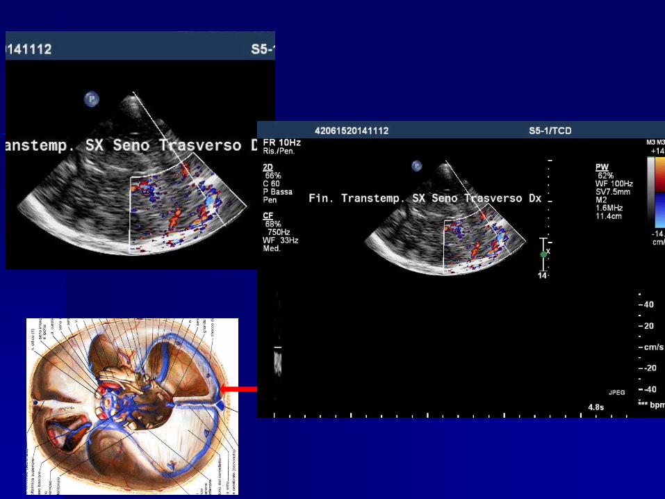
“COLOR-CODING” DEL CIRCOLO VENOSO POSTERO-BASALE
1) Seno Trasverso omol.
2) Seno Trasverso contr.
3) Seno Retto
4) Seno Sigmoideo
5) Seno Petroso sup.
6) Seno Petroso infer.
7) Seno Sfenoparietale

SENO SFENOPARIETALE
3°

Vena di Galeno
Piano Mesencefalico Superiore

Alterazione della “direzione” del flusso venoso
Alterazione della “velocità” del flusso venoso
Patterns sonologici - Criteri Indiretti -

Tuttavia l’ampia variabilità anatomica e l’estesa rete
anastomotica rende spesso molto più complessa la
dinamica venosa.
Test dinamici:
-compressione della giugulare (Franceschi)
-manovra di Valsalva
-studio a 0° e 90°
-respiro profondo
Non è sufficiente inoltre il solo pattern “color” :
Ma occorre dimostrare e studiare anche
CRITERI INDIRETTI:
-Direzione
-Velocità
Considerazioni emodinamiche

Quando i reperti venosi al TCCD/TCD diventano patologici?
A livello cerebrale sono assenti le valvole:
contano esclusivamente i gradienti pressori
Velocità venosa patologica: se > della V. media +2DS
Regola generale:
Per le vene cerebrali: FV >30-35 cm/sec
Per i seni: FV > 40-45 cm/sec
Direzione di flusso patologica:
se il flusso è invertito rispetto alla normale direzione.

«In case of hypoplasia of the sigmoid sinuous or the proximal portion
of the transvers sinous
is generally coupled with compensatory re-routing of the venous
blood into prominent mastoid emissary or posterior condilar emissary
veins»
Knott 1881; Knott JF. On the cerebral sinuses and their variations
J.Anatom.Physiol. 16,27-42 (1881)
Laff 1939;
Pitfall

Alterazione della “velocità” del flusso venoso
take home :Patterns sonologici

Patologia? Insonazione temporale Fisiologia Direzione alterata
Direzione normale
Alterazione della “direzione” del flusso venoso

•Difficile insonazione(sfenoide)
•Area compartimentata a bassissime
velocità (doppler shift troppo basso)
Seno Cavernoso

Take home per Neurosonologo:
OGNI MODIFICAZIONE ACUTA E/O PROGRESSIVA DEL
PATTERN FLUSSIMETRICO VENOSO
DEL SENO SFENOPARIETALE DEVE ESSERE ATTENTAMENTE
VALUTATA E STUDIATA
POICHE’ ESSA CI PUO’ FORNIRE IMPORTANTI INFORMAZIONI INDIRETTE RIGUARDANTI LO STATO CIRCOLATORIO A LIVELLO DEL SENO CAVERNOSO.

A
B
C
D ??
MODIFICAZIONI DEL PATTERN
FLUSSIMETRICO AL SENO
SFENOPARIETALE SX EVIDENZIATE
CON TCCD :
θ=52
θ=52
θ=34
θ=52
In order to identify the zones of convergence of the medullary veins of the
cerebral white matter, gelatin-mixed barium sulfate was injected into normal
brains at autopsy. A catheter was inserted into the internal jugular veins or the
carotid and vertebral arteries. Serial soft tissue roentgenograms of whole
brains and brain slices were used to determine the zones of convergence. The
deep medullary veins had four zones of covergence before draining into the
subependymal veins:
-the first (superficial),
-second (candelabra),
-third (palmate),
-fourth (subependymal).
Okudera T. et al
Neuropathology

Okudera et al. (1999) Neuropathology 19: 93-111

In order to identify the zones of convergence of the medullary veins of the
cerebral white matter, gelatin-mixed barium sulfate was injected into normal
brains at autopsy. A catheter was inserted into the internal jugular veins or the
carotid and vertebral arteries. Serial soft tissue roentgenograms of whole
brains and brain slices were used to determine the zones of convergence. The
deep medullary veins had four zones of covergence before draining into the
subependymal veins:
-the first (superficial),
-second (candelabra),
-third (palmate),
-fourth (subependymal).
Okudera T. et al
Neuropathology
The zones of various venous convergence within the white
matter were due to the crossing of nerve fiber tracts (e.g. the
pes of the corona radiata, the radiation of the corpus callosum,
the superior occipitofrontal fasciculus, the tapetum and the
sagittal strata).

Our ongoing experiences

Donna di 25 anni
Non obesa
Anamnesi negativa (pillola, farmaci ecc)
Cefalea da alcuni mesi


Giunge al PS: TAC negativa !

IJV sx
IJV dx


Vena di Labbe
Neuroradiologie Masson eds

Seno trasverso prox dx

Seno trasverso sx

ODX

OSX

RMN:
parenchima ndr
Angio arteriosa ndr


ESISTE ANCORA L’IPERTENSIONE
INTRACRANICA IDIOPATICA ( IIH) ???
XIV° Congresso Nazionale SINV
27-29 ottobre 2005
Bassano del Grappa

San Benedetto del Tronto
GRAZIE PER L’ATTENZIONE