dilated cardiomyopathy secondary to hypothyroidism: case ... · a reversible form of dilated...
TRANSCRIPT
32
IntroductionA reversible form of dilated cardiomyopathy (DCM) can be
developed from alcohol drinking, pregnancy, chronic uncon-trolled tachycardia, hypothyroidism, hyperthyroidism, drug use and other endocrine dysfunctions.1)2) Thyroid hormone has a great effect on the heart and vascular system.1) The heart is sensitive to changes in thyroid hormones, and cardiac disorders are commonly associated with both hyper- and hypothyroid-ism.3)4) Hemodynamic changes caused by hyperthyroidism lead to classic hyperdymamic cardiovascular state, and they are asso-ciated with increase in cardiac output and reduction in periph-eral vascular resistance.5) On the other hand, hypothyroidism is associated with bradycardia, mild diastolic hypertension, nar-row pulse pressure and slightly increased mean arterial pres-sure.6) According to a review of literatures, diastolic dysfunction is the most common finding seen in patients with hypothyroid-ism.7) In addition, it is commonly encountered that the left ven-tricular systolic function is minimally decreased with slightly reduced ejection fraction and stroke volume.8) DCM is a rare presentation of hypothyroidism.9)
We experienced a case of a 36-year-old man with DCM ac-companied by undiagnosed primary hypothyroidism. Here,
we report our case with a review of literatures.
CaseA 36-year-old man presented to the emergency room with
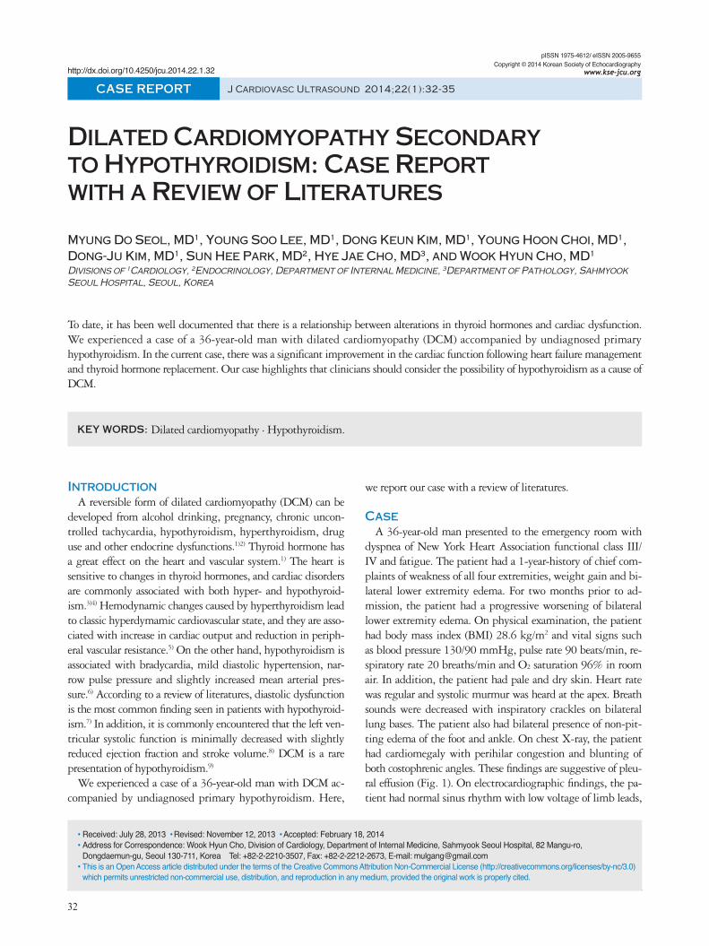
dyspnea of New York Heart Association functional class III/IV and fatigue. The patient had a 1-year-history of chief com-plaints of weakness of all four extremities, weight gain and bi-lateral lower extremity edema. For two months prior to ad-mission, the patient had a progressive worsening of bilateral lower extremity edema. On physical examination, the patient had body mass index (BMI) 28.6 kg/m2 and vital signs such as blood pressure 130/90 mmHg, pulse rate 90 beats/min, re-spiratory rate 20 breaths/min and O2 saturation 96% in room air. In addition, the patient had pale and dry skin. Heart rate was regular and systolic murmur was heard at the apex. Breath sounds were decreased with inspiratory crackles on bilateral lung bases. The patient also had bilateral presence of non-pit-ting edema of the foot and ankle. On chest X-ray, the patient had cardiomegaly with perihilar congestion and blunting of both costophrenic angles. These findings are suggestive of pleu-ral effusion (Fig. 1). On electrocardiographic findings, the pa-tient had normal sinus rhythm with low voltage of limb leads,
pISSN 1975-4612/ eISSN 2005-9655 Copyright © 2014 Korean Society of Echocardiography
www.kse-jcu.orghttp://dx.doi.org/10.4250/jcu.2014.22.1.32
CASE REPORT J Cardiovasc Ultrasound 2014;22(1):32-35
Dilated Cardiomyopathy Secondary to Hypothyroidism: Case Report with a Review of Literatures
Myung Do Seol, MD1, Young Soo Lee, MD1, Dong Keun Kim, MD1, Young Hoon Choi, MD1, Dong-Ju Kim, MD1, Sun Hee Park, MD2, Hye Jae Cho, MD3, and Wook Hyun Cho, MD1
Divisions of 1Cardiology, 2Endocrinology, Department of Internal Medicine, 3Department of Pathology, Sahmyook Seoul Hospital, Seoul, Korea
To date, it has been well documented that there is a relationship between alterations in thyroid hormones and cardiac dysfunction. We experienced a case of a 36-year-old man with dilated cardiomyopathy (DCM) accompanied by undiagnosed primary hypothyroidism. In the current case, there was a significant improvement in the cardiac function following heart failure management and thyroid hormone replacement. Our case highlights that clinicians should consider the possibility of hypothyroidism as a cause of DCM.
KEY WORDS: Dilated cardiomyopathy · Hypothyroidism.
•Received: July 28, 2013 •Revised: November 12, 2013 •Accepted: February 18, 2014 •Address for Correspondence: Wook Hyun Cho, Division of Cardiology, Department of Internal Medicine, Sahmyook Seoul Hospital, 82 Mangu-ro, Dongdaemun-gu, Seoul 130-711, Korea Tel: +82-2-2210-3507, Fax: +82-2-2212-2673, E-mail: [email protected]•This is an Open Access article distributed under the terms of the Creative Commons Attribution Non-Commercial License (http://creativecommons.org/licenses/by-nc/3.0) which permits unrestricted non-commercial use, distribution, and reproduction in any medium, provided the original work is properly cited.
DCM and Hypothyroidism | Myung Do Seol, et al.
33
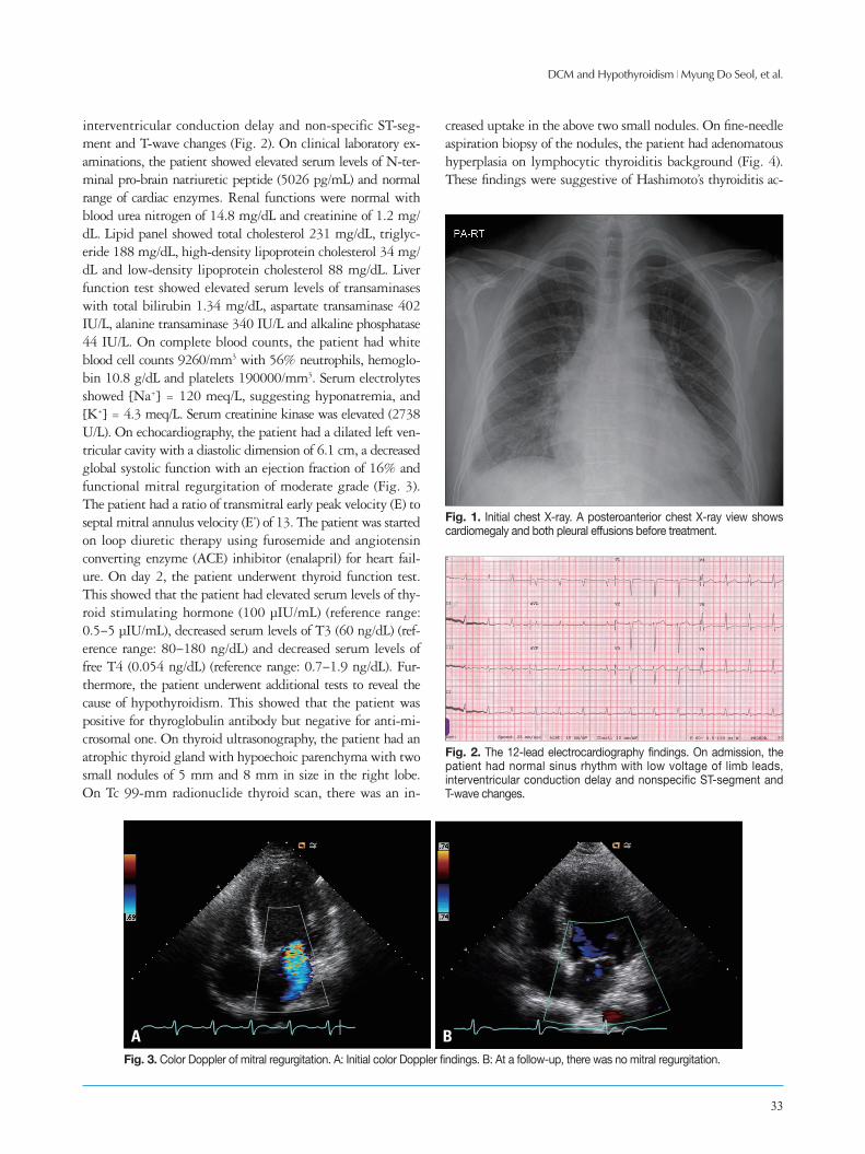
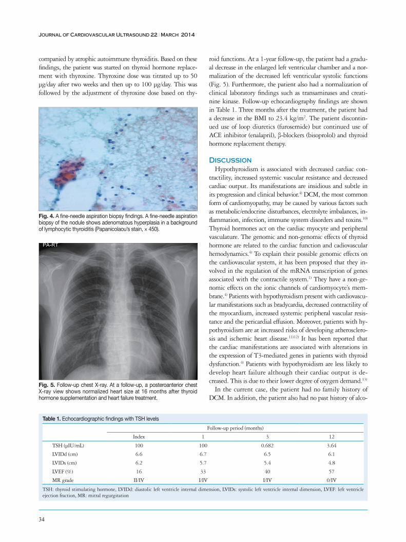
interventricular conduction delay and non-specific ST-seg-ment and T-wave changes (Fig. 2). On clinical laboratory ex-aminations, the patient showed elevated serum levels of N-ter-minal pro-brain natriuretic peptide (5026 pg/mL) and normal range of cardiac enzymes. Renal functions were normal with blood urea nitrogen of 14.8 mg/dL and creatinine of 1.2 mg/dL. Lipid panel showed total cholesterol 231 mg/dL, triglyc-eride 188 mg/dL, high-density lipoprotein cholesterol 34 mg/dL and low-density lipoprotein cholesterol 88 mg/dL. Liver function test showed elevated serum levels of transaminases with total bilirubin 1.34 mg/dL, aspartate transaminase 402 IU/L, alanine transaminase 340 IU/L and alkaline phosphatase 44 IU/L. On complete blood counts, the patient had white blood cell counts 9260/mm3 with 56% neutrophils, hemoglo-bin 10.8 g/dL and platelets 190000/mm3. Serum electrolytes showed [Na+] = 120 meq/L, suggesting hyponatremia, and [K+] = 4.3 meq/L. Serum creatinine kinase was elevated (2738 U/L). On echocardiography, the patient had a dilated left ven-tricular cavity with a diastolic dimension of 6.1 cm, a decreased global systolic function with an ejection fraction of 16% and functional mitral regurgitation of moderate grade (Fig. 3). The patient had a ratio of transmitral early peak velocity (E) to septal mitral annulus velocity (E’) of 13. The patient was started on loop diuretic therapy using furosemide and angiotensin converting enzyme (ACE) inhibitor (enalapril) for heart fail-ure. On day 2, the patient underwent thyroid function test. This showed that the patient had elevated serum levels of thy-roid stimulating hormone (100 μIU/mL) (reference range: 0.5–5 μIU/mL), decreased serum levels of T3 (60 ng/dL) (ref-erence range: 80–180 ng/dL) and decreased serum levels of free T4 (0.054 ng/dL) (reference range: 0.7–1.9 ng/dL). Fur-thermore, the patient underwent additional tests to reveal the cause of hypothyroidism. This showed that the patient was positive for thyroglobulin antibody but negative for anti-mi-crosomal one. On thyroid ultrasonography, the patient had an atrophic thyroid gland with hypoechoic parenchyma with two small nodules of 5 mm and 8 mm in size in the right lobe. On Tc 99-mm radionuclide thyroid scan, there was an in-
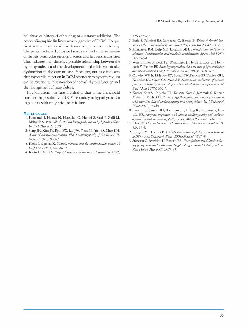
creased uptake in the above two small nodules. On fine-needle aspiration biopsy of the nodules, the patient had adenomatous hyperplasia on lymphocytic thyroiditis background (Fig. 4). These findings were suggestive of Hashimoto’s thyroiditis ac-
Fig. 1. Initial chest X-ray. A posteroanterior chest X-ray view shows cardiomegaly and both pleural effusions before treatment.
Fig. 2. The 12-lead electrocardiography findings. On admission, the patient had normal sinus rhythm with low voltage of limb leads, interventricular conduction delay and nonspecific ST-segment and T-wave changes.
Fig. 3. Color Doppler of mitral regurgitation. A: Initial color Doppler findings. B: At a follow-up, there was no mitral regurgitation.
A B
Journal of Cardiovascular Ultrasound 22 | March 2014
34
companied by atrophic autoimmune thyroiditis. Based on these findings, the patient was started on thyroid hormone replace-ment with thyroxine. Thyroxine dose was titrated up to 50 μg/day after two weeks and then up to 100 μg/day. This was followed by the adjustment of thyroxine dose based on thy-
roid functions. At a 1-year follow-up, the patient had a gradu-al decrease in the enlarged left ventricular chamber and a nor-malization of the decreased left ventricular systolic functions (Fig. 5). Furthermore, the patient also had a normalization of clinical laboratory findings such as transaminases and creati-nine kinase. Follow-up echocardiography findings are shown in Table 1. Three months after the treatment, the patient had a decrease in the BMI to 23.4 kg/m2. The patient discontin-ued use of loop diuretics (furosemide) but continued use of ACE inhibitor (enalapril), β-blockers (bisoprolol) and thyroid hormone replacement therapy.
DiscussionHypothyroidism is associated with decreased cardiac con-
tractility, increased systemic vascular resistance and decreased cardiac output. Its manifestations are insidious and subtle in its progression and clinical behavior.4) DCM, the most common form of cardiomyopathy, may be caused by various factors such as metabolic/endocrine disturbances, electrolyte imbalances, in-flammation, infection, immune system disorders and toxins.10) Thyroid hormones act on the cardiac myocyte and peripheral vasculature. The genomic and non-genomic effects of thyroid hormone are related to the cardiac function and cadiovascular hemodynamics.4) To explain their possible genomic effects on the cardiovascular system, it has been proposed that they in-volved in the regulation of the mRNA transcription of genes associated with the contractile system.1) They have a non-ge-nomic effects on the ionic channels of cardiomyocyte’s mem-brane.4) Patients with hypothyroidism present with cardiovascu-lar manifestations such as bradycardia, decreased contractility of the myocardium, increased systemic peripheral vascular resis-tance and the pericardial effusion. Moreover, patients with hy-pothyroidism are at increased risks of developing atherosclero-sis and ischemic heart disease.11)12) It has been reported that the cardiac manifestations are associated with alterations in the expression of T3-mediated genes in patients with thyroid dysfunction.4) Patients with hypothyroidism are less likely to develop heart failure although their cardiac output is de-creased. This is due to their lower degree of oxygen demand.13)
In the current case, the patient had no family history of DCM. In addition, the patient also had no past history of alco-
Table 1. Echocardiographic findings with TSH levels
Follow-up period (months)
Index 1 3 12
TSH (µIU/mL) 100 100 0.682 3.64
LVIDd (cm) 6.6 6.7 6.5 6.1
LVIDs (cm) 6.2 5.7 5.4 4.8
LVEF (%) 16 33 40 57
MR grade II/IV I/IV I/IV 0/IV
TSH: thyroid stimulating hormone, LVIDd: diastolic left ventricle internal dimension, LVIDs: systolic left ventricle internal dimension, LVEF: left ventricle ejection fraction, MR: mitral regurgitation
Fig. 5. Follow-up chest X-ray. At a follow-up, a posteroanterior chest X-ray view shows normalized heart size at 16 months after thyroid hormone supplementation and heart failure treatment.
Fig. 4. A fine-needle aspiration biopsy findings. A fine-needle aspiration biopsy of the nodule shows adenomatous hyperplasia in a background of lymphocytic thyroiditis (Papanicolaou’s stain, × 450).
DCM and Hypothyroidism | Myung Do Seol, et al.
35
hol abuse or history of other drug or substance addiction. The echocardiographic findings were suggestive of DCM. The pa-tient was well responsive to hormone replacement therapy. The patient achieved euthyroid status and had a normalization of the left ventricular ejection fraction and left ventricular size. This indicates that there is a possible relationship between the hypothyroidism and the development of the left ventricular dysfunction in the current case. Moreover, our case indicates that myocardial function in DCM secondary to hypothyroidism can be reversed with restoration of normal thyroid function and the management of heart failure.
In conclusion, our case highlights that clinicians should consider the possibility of DCM secondary to hypothyroidism in patients with congestive heart failure.
References1. Khochtali I, Hamza N, Harzallah O, Hamdi S, Saad J, Golli M,
Mahjoub S. Reversible dilated cardiomyopathy caused by hypothyroidism. Int Arch Med 2011;4:20.
2. Sung JK, Kim JY, Ryu DW, Lee JW, Youn YJ, Yoo BS, Choe KH. A case of hypocalcemia-induced dilated cardiomyopathy. J Cardiovasc Ul-trasound 2010;18:25-7.
3. Klein I, Ojamaa K. Thyroid hormone and the cardiovascular system. N Engl J Med 2001;344:501-9.
4. Klein I, Danzi S. Thyroid disease and the heart. Circulation 2007;
116:1725-35.5. Fazio S, Palmieri EA, Lombardi G, Biondi B. Effects of thyroid hor-
mone on the cardiovascular system. Recent Prog Horm Res 2004;59:31-50.6. McAllister RM, Delp MD, Laughlin MH. Thyroid status and exercise
tolerance. Cardiovascular and metabolic considerations. Sports Med 1995; 20:189-98.
7. Wieshammer S, Keck FS, Waitzinger J, Henze E, Loos U, Hom-bach V, Pfeiffer EF. Acute hypothyroidism slows the rate of left ventricular diastolic relaxation. Can J Physiol Pharmacol 1989;67:1007-10.
8. Crowley WF Jr, Ridgway EC, Bough EW, Francis GS, Daniels GH, Kourides IA, Myers GS, Maloof F. Noninvasive evaluation of cardiac function in hypothyroidism. Response to gradual thyroxine replacement. N Engl J Med 1977;296:1-6.
9. Kumar Kota S, Tripathy PR, Krishna Kota S, Jammula S, Kumar Meher L, Modi KD. Primary hypothyroidism: uncommon presentation with reversible dilated cardiomyopathy in a young subject. Int J Endocrinol Metab 2012;10:440-3.
10. Kuethe F, Sigusch HH, Bornstein SR, Hilbig K, Kamvissi V, Fig-ulla HR. Apoptosis in patients with dilated cardiomyopathy and diabetes: a feature of diabetic cardiomyopathy? Horm Metab Res 2007;39:672-6.
11. Ichiki T. Thyroid hormone and atherosclerosis. Vascul Pharmacol 2010; 52:151-6.
12. François M, Delemer B. [What’s new in the couple thyroid and heart in 2008?]. Ann Endocrinol (Paris) 2008;69 Suppl 1:S37-43.
13. Stanescu C, Branidou K, Ranetti EA. Heart failure and dilated cardio-myopathy associated with severe longstanding untreated hypothyroidism. Rom J Intern Med 2007;45:77-83.