diagnostic imaging of lung cancer
TRANSCRIPT
Diagnostic imaging of lung cancer
• Carcinoma of the bronchus is the most common malignancy in theWestern world. It is also the leading cause of cancer-related death accounting for 32% of all cancer deaths in males and 25% in females.
Radiological characteristics by cell type
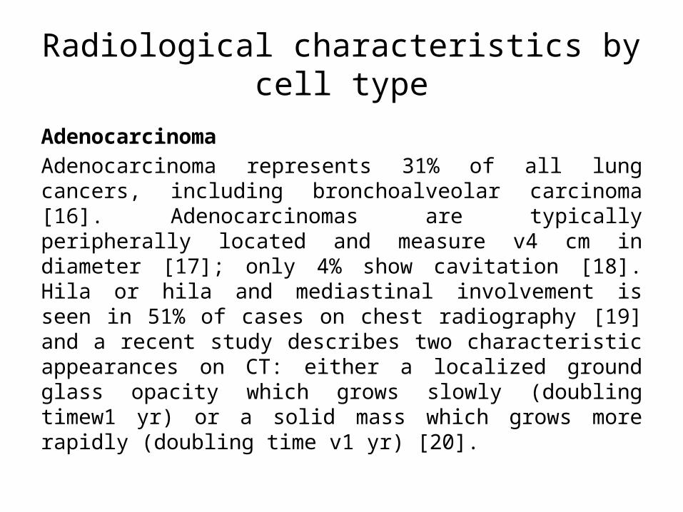
AdenocarcinomaAdenocarcinoma represents 31% of all lung cancers, including bronchoalveolar carcinoma [16]. Adenocarcinomas are typically peripherally located and measure v4 cm in diameter [17]; only 4% show cavitation [18]. Hila or hila and mediastinal involvement is seen in 51% of cases on chest radiography [19] and a recent study describes two characteristic appearances on CT: either a localized ground glass opacity which grows slowly (doubling timew1 yr) or a solid mass which grows more rapidly (doubling time v1 yr) [20].
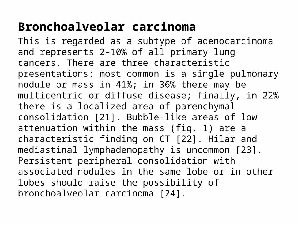
Bronchoalveolar carcinomaThis is regarded as a subtype of adenocarcinoma and represents 2–10% of all primary lung cancers. There are three characteristic presentations: most common is a single pulmonary nodule or mass in 41%; in 36% there may be multicentric or diffuse disease; finally, in 22% there is a localized area of parenchymal consolidation [21]. Bubble-like areas of low attenuation within the mass (fig. 1) are a characteristic finding on CT [22]. Hilar and mediastinal lymphadenopathy is uncommon [23]. Persistent peripheral consolidation with associated nodules in the same lobe or in other lobes should raise the possibility of bronchoalveolar carcinoma [24].
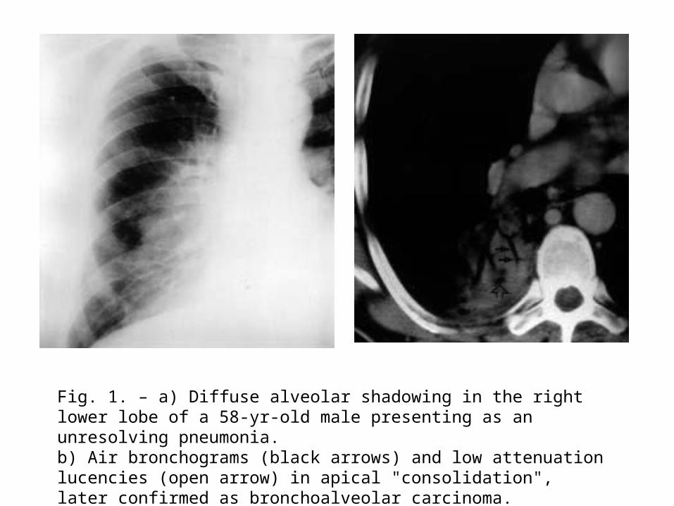
Fig. 1. – a) Diffuse alveolar shadowing in the right lower lobe of a 58-yr-old male presenting as an unresolving pneumonia. b) Air bronchograms (black arrows) and low attenuation lucencies (open arrow) in apical "consolidation", later confirmed as bronchoalveolar carcinoma.
Adenosquamous carcinomaAdenosquamous carcinoma represents 2% of all lung cancers. This cell type is typically identified as a solitary, peripheral nodule. Over one-half are 1–3 cm in size and cavitation is seen in 13%. Evidence of parenchymal scars or fibrosis in or next to the tumour is seen in 50% .
Squamous cell carcinomaSquamous cell carcinoma represents 30% of all lungcancers. These tumours are more often centrallylocated within the lung and may grow much larger than 4 cm in diameter. Cavitation (fig. 2) is seen in up to 82%. They commonly cause segmental or lobar lung collapse due to their central location and relative frequency.
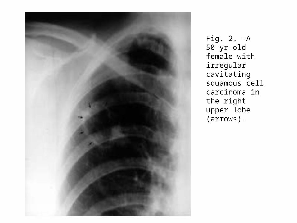
Fig. 2. –A 50-yr-old female with irregular cavitating squamous cellcarcinoma in the right upper lobe (arrows).
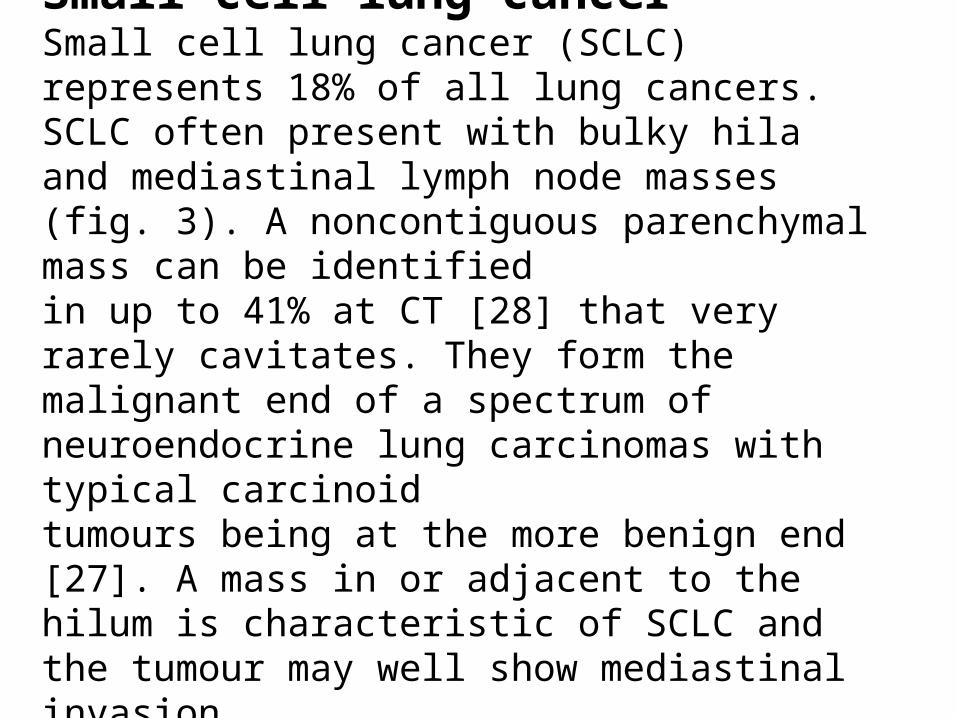
Small cell lung cancerSmall cell lung cancer (SCLC) represents 18% of all lung cancers. SCLC often present with bulky hila and mediastinal lymph node masses (fig. 3). A noncontiguous parenchymal mass can be identifiedin up to 41% at CT [28] that very rarely cavitates. They form the malignant end of a spectrum of neuroendocrine lung carcinomas with typical carcinoidtumours being at the more benign end [27]. A mass in or adjacent to the hilum is characteristic of SCLC and the tumour may well show mediastinal invasion.

b)a)
Fig. 3. – a) A 55-yr-old dyspnoeic female. Chest radiograph demonstrating widened mediastinum particularly on the right with reduced vascularity of the right lung. b) Contrast enhanced computed tomography showing central mediastinal mass invading the right pulmonary artery. Small cell carcinoma was confirmed on percutaneous biopsy.
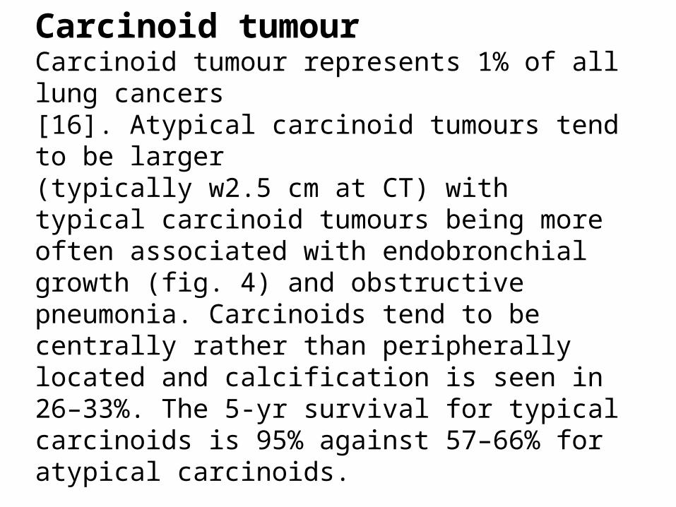
Carcinoid tumourCarcinoid tumour represents 1% of all lung cancers[16]. Atypical carcinoid tumours tend to be larger(typically w2.5 cm at CT) with typical carcinoid tumours being more often associated with endobronchial growth (fig. 4) and obstructive pneumonia. Carcinoids tend to be centrally rather than peripherally located and calcification is seen in 26–33%. The 5-yr survival for typical carcinoids is 95% against 57–66% for atypical carcinoids.
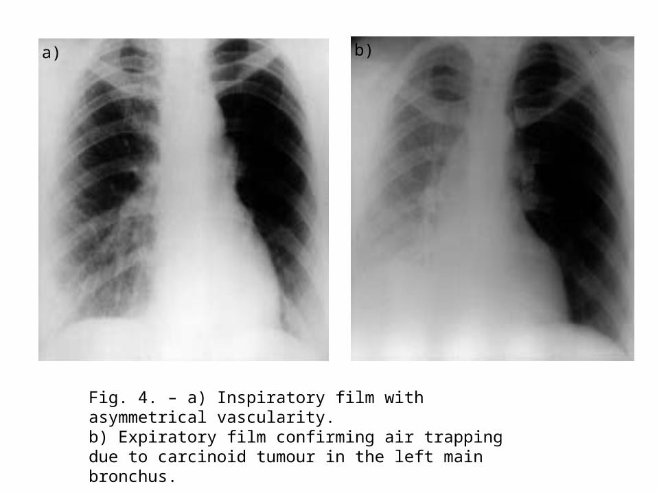
Fig. 4. – a) Inspiratory film with asymmetrical vascularity. b) Expiratory film confirming air trapping due to carcinoid tumour in the left main bronchus.
a) b)
Large cell carcinomaLarge cell carcinoma represents 9% of all lung cancers. Large or giant cell carcinoma is a poorly differentiated nonsmall cell carcinoma (NSCLC) and is diagnosed histologically after exclusion of adenocarcinomatousor squamous differentiation. It may grow extremely rapidly [30] to a large size but metastasizes early to the mediastinum and brain.
Imaging techniquesChest radiography• Due to its widespread availability, including to primary care
physicians, the chest radiograph is often the first imaging modality to suggest the diagnosis of bronchogenic carcinoma. Lung cancer may present as a straightforward spiculated mass but its presence may also be inferred from other appearances such as an unresolving pneumonia or lobar collapse (fig. 5).
• In some situations, no further imaging will be necessary when bulky contralateral mediastinal adenopathy is present or when an obvious bony lesion is identified. However, CT scanning of the chest is often needed because of the lack of sensitivity of the chest radiographs in detecting mediastinal lymph node metastases and chest wall and mediastinal invasion.
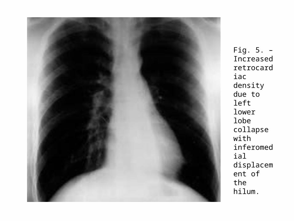
Fig. 5. – Increased retrocardiac density due to left lower lobecollapse with inferomedial displacement of the hilum.
Computed tomography
CT can identify specific features in lung nodules that are diagnostic, e.g. arteriovenous fistulae, rounded atelectasis, fungus balls, mucoid impaction and infarcts. High-resolution scanning further refines diagnostic process.The ability of CT scanning to evaluate the entire thorax at the time of nodule assessment is of further benefit. Spiral or helical CT is advantageous as small nodules are not missed between slices as may happen on older, nonspiral machines. It also increases the detection rate of nodules v5 mm in diameter, especially when viewed in cine-format on a workstation. The acquisition of continuous volume data sets permits three-dimensional image reconstruction and multiplanar (i.e. nonaxial) reformatting (fig. 6).
Computed tomography
• These techniques have been shown to improve the detection of pleural invasion by tumour and clarify the origin of peridiaphragmatic tumours respectively. Further manipulation of raw data sets enables the technique of virtual bronchoscopy.
• An interactive, simulated bronchoscopy can be performed with the added benefit of simultaneous information on adjacent mediastinal structures.
• This technique has far reaching potential both as a teaching tool and as a means of evaluating patients’ thoracic and bronchial anatomy prior to interventional procedures and stent placement.
Computed tomography
The recent advent of multislice scanners has seen advances in image resolution with a substantial reduction in both tube loading and scanning time as up to four slices can be acquired simultaneously. Both spiral and multislice machines suffer less from respiratory motion artefact due to their shorter scanning times.
Computed tomography
Spiral CT with a bolus injection of intravenous iodinated contrast medium affords "dynamic scanning". A recent study of 84 patients with NSCLC found no difference in radiological stage when noncontrast enhanced scans were compared with contrast enhanced scans in 80 patients (95%), recommending that nonenhanced CT through the thorax and adrenals was sufficient for staging patients with newlydiagnosed NSCLC. However, another study of 50 patients comparing both techniques found an 11% higher detection rate of enlarged mediastinal nodes after contrast enhancement and recommended its routine administration (figs. 7 and 8).
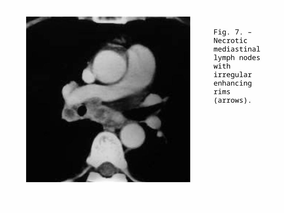
Fig. 7. – Necrotic mediastinal lymph nodes with irregular enhancingrims (arrows).
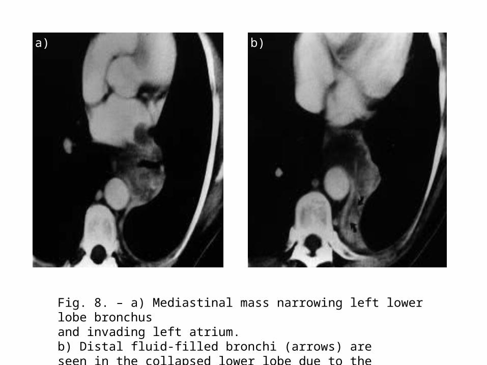
Fig. 8. – a) Mediastinal mass narrowing left lower lobe bronchusand invading left atrium. b) Distal fluid-filled bronchi (arrows) areseen in the collapsed lower lobe due to the proximal tumour.
a) b)
Computed tomography
Many centres perform hepatic and adrenal scans having given intravenous contrast. Slice thickness and interval should be ¡10 mm and extend from the lung apices to the adrenal glands. It is now common practice to perform 5-mm slices through the hila and aortopulmonary regions to improve delineation of local lymph nodes and the origins of the lobar bronchi. The field of view should include the contiguous chest wall.
Magnetic resonance imaging
Magnetic resonance imaging (MRI) is becoming more available but pressure on MRI scanning time is so intense that it is usually used for problem solving and where administration of contrast media is contraindicated. MRI can be more accurate than CT in separating stage IIIa (resectable) from IIIb (generally unresectable) tumours in selected patients due to its ability to detect invasion of major mediastinal structures, i.e. T4 disease.
Magnetic resonance imagingThe advantages MRI has over CT include: better soft tissue contrast, multiplanar imaging capability, and therefore useful for superior sulcus tumours and evaluation of the aortopulmonary window (fig. 9), and cardiac gating which enables excellent delineation of the heart and great vessels and removes cardiac pulsation artefact. MRI is also useful in the assessment of mediastinal and chest wall invasion by virtue of its ability to determine fat-stripe invasion (fig. 10) and involvement of the diaphragm and spinal canal. In addition, it has been shown to aid in differentiating lymph nodes from hila vessels due to the "flow void" phenomenon.
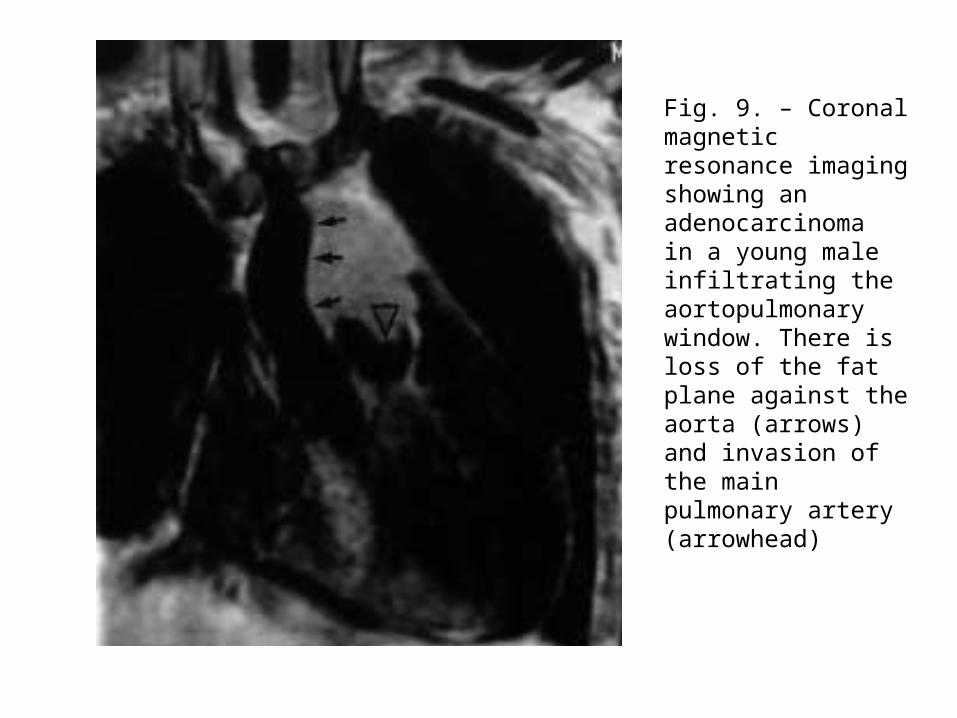
Fig. 9. – Coronal magnetic resonance imaging showing an adenocarcinomain a young male infiltrating the aortopulmonarywindow. There is loss of the fat plane against the aorta (arrows)and invasion of the main pulmonary artery (arrowhead)
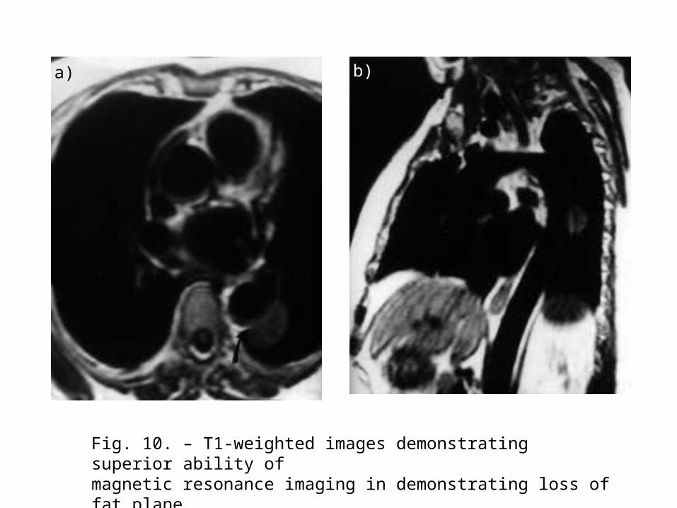
Fig. 10. – T1-weighted images demonstrating superior ability ofmagnetic resonance imaging in demonstrating loss of fat plane(arrow) in a) axial and b) sagittal planes.
a) b)
Staging nonsmall cell lung cancer
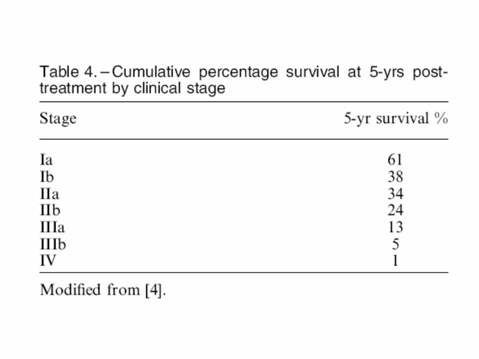
The revised international system for staging lung cancer incorporates the tumour, node, metastasis (TNM) subset system (tables 2 and 3) and shows improved survival rates with more accurate staging and appropriate selection of patients for definitive surgical treatment by distinguishing the IIIa from the IIIb group (table 4).
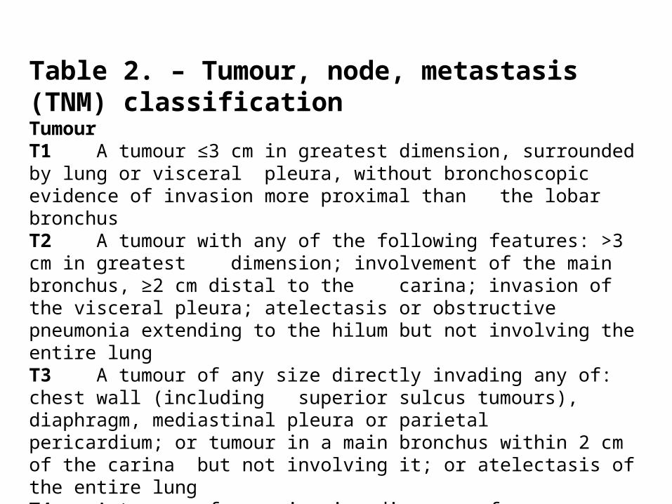
Table 2. – Tumour, node, metastasis (TNM) classificationTumourT1 A tumour ≤3 cm in greatest dimension, surrounded by lung or visceral pleura, without bronchoscopic evidence of invasion more proximal than the lobar bronchusT2 A tumour with any of the following features: >3 cm in greatest dimension; involvement of the main bronchus, ≥2 cm distal to the carina; invasion of the visceral pleura; atelectasis or obstructive pneumonia extending to the hilum but not involving the entire lungT3 A tumour of any size directly invading any of: chest wall (including superior sulcus tumours), diaphragm, mediastinal pleura or parietal pericardium; or tumour in a main bronchus within 2 cm of the carina but not involving it; or atelectasis of the entire lungT4 A tumour of any size invading any of: mediastinum, heart, great vessels,
trachea, oesophagus, vertebral body or carina; or tumour with a malignant pleural or pericardial effusion or with satellite tumour nodules within the ipsilateral primary-tumour lobe of the lung
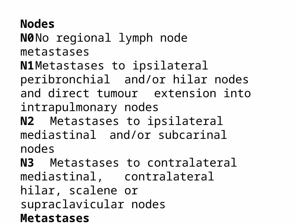
NodesN0 No regional lymph node metastasesN1 Metastases to ipsilateral peribronchial
and/or hilar nodes and direct tumour extension into intrapulmonary nodesN2 Metastases to ipsilateral mediastinal and/or subcarinal nodesN3 Metastases to contralateral mediastinal,
contralateral hilar, scalene or supraclavicular nodesMetastasesM0 No distant metastasesM1 Distant metastases present
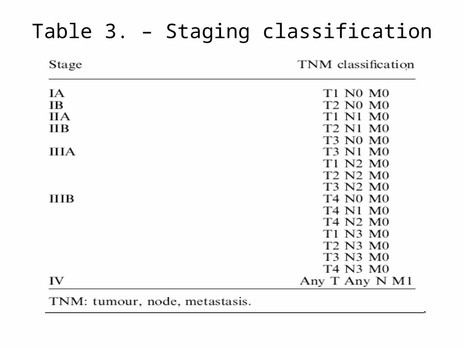
Table 3. – Staging classification
Staging small cell lung cancer
SCLC is distinguished from NSCLC by its rapid tumour doubling time, development of early widespread metastases and almost exclusive occurrence in smokers [130]. It is divided into two stages: limited disease, which is confined to the ipsilateral hemithorax within a single, tolerable radiotherapy port and extensive disease which covers all other disease including distant metastases.