diagnostic approach to the hiv infected patient with...
TRANSCRIPT
Diagnostic approach to the HIV infected patient with
altered mental state
Kennedy Nyamande
Department of Internal Medicine
University of KwaZulu-Natal
Plan
Definitions
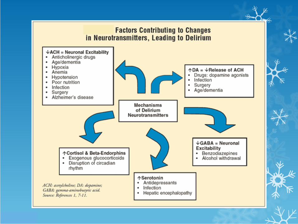
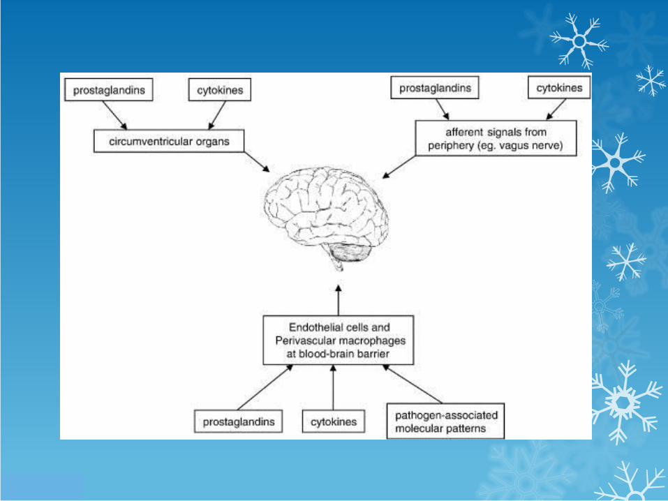
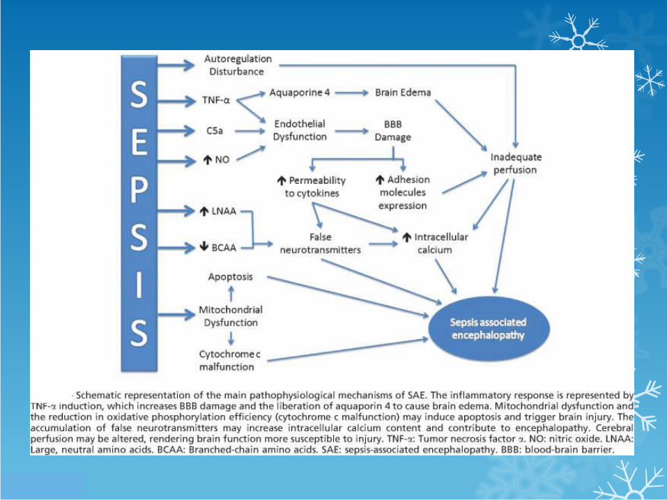
Pathogenesis
Risk factors
History
Examination
Investigations
Algorithms
Definitions
Confusion: inability to maintain a coherent stream of thought or action
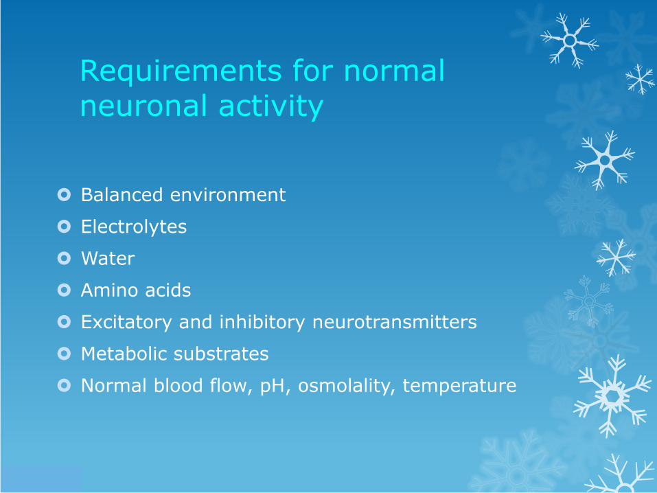
Requirements for normal neuronal activity
Balanced environment
Electrolytes
Water
Amino acids
Excitatory and inhibitory neurotransmitters
Metabolic substrates
Normal blood flow, pH, osmolality, temperature
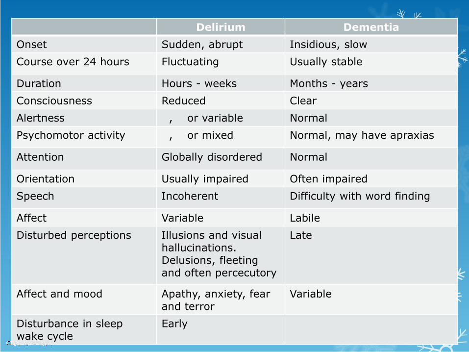
Delirium Dementia
Onset Sudden, abrupt Insidious, slow
Course over 24 hours Fluctuating Usually stable
Duration Hours - weeks Months - years
Consciousness Reduced Clear
Alertness , or variable Normal
Psychomotor activity , or mixed Normal, may have apraxias
Attention Globally disordered Normal
Orientation Usually impaired Often impaired
Speech Incoherent Difficulty with word finding
Affect Variable Labile
Disturbed perceptions Illusions and visual hallucinations. Delusions, fleeting and often percecutory
Late
Affect and mood Apathy, anxiety, fear and terror
Variable
Disturbance in sleep wake cycle
Early
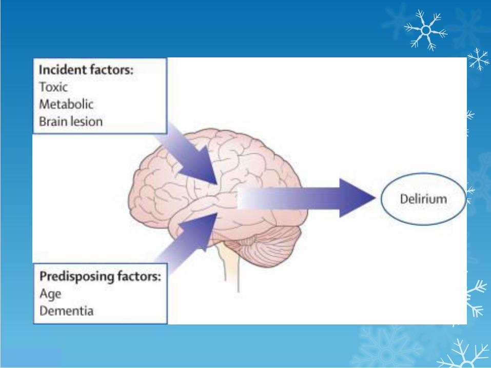
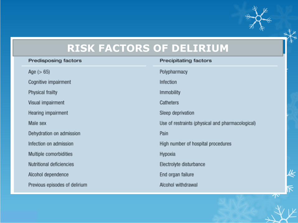
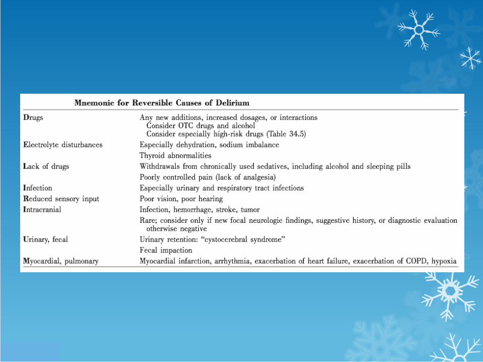
RISK FACTORS OF DELIRIUM
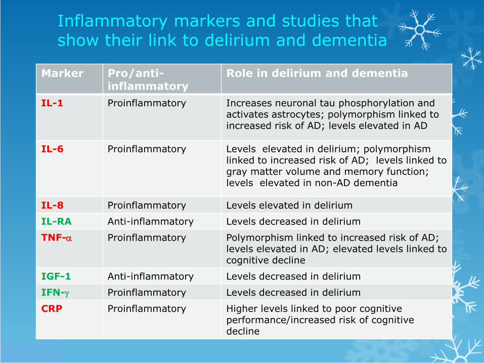
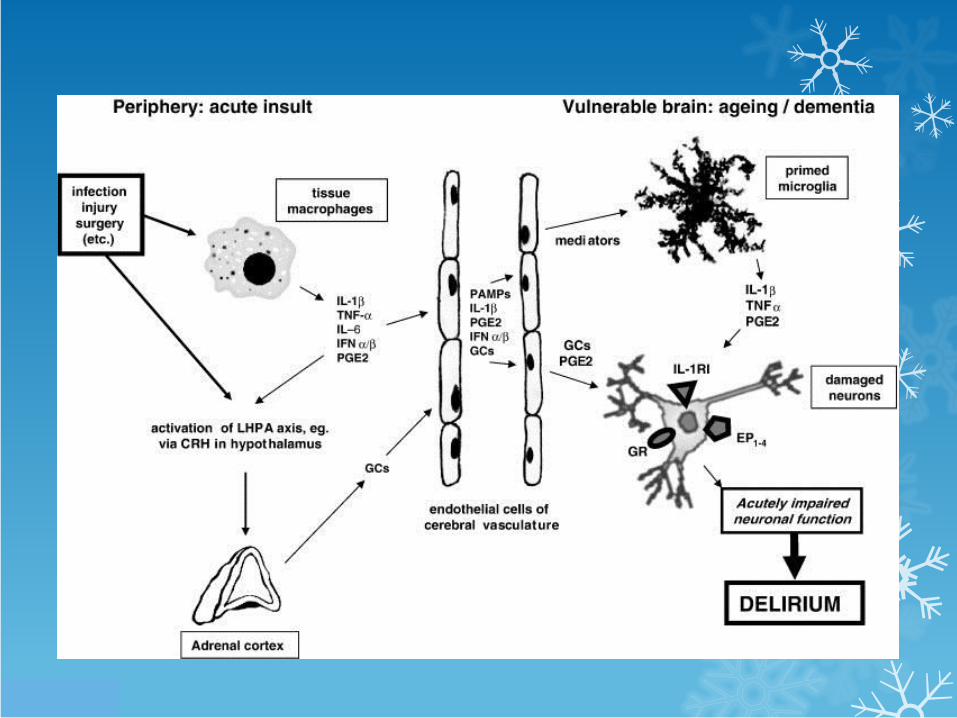
Inflammatory markers and studies that show their link to delirium and dementia
Marker Pro/anti-inflammatory
Role in delirium and dementia
IL-1 Proinflammatory Increases neuronal tau phosphorylation and activates astrocytes; polymorphism linked to increased risk of AD; levels elevated in AD
IL-6 Proinflammatory Levels elevated in delirium; polymorphism linked to increased risk of AD; levels linked to gray matter volume and memory function; levels elevated in non-AD dementia
IL-8 Proinflammatory Levels elevated in delirium
IL-RA Anti-inflammatory Levels decreased in delirium
TNF- Proinflammatory Polymorphism linked to increased risk of AD; levels elevated in AD; elevated levels linked to cognitive decline
IGF-1 Anti-inflammatory Levels decreased in delirium
IFN- Proinflammatory Levels decreased in delirium
CRP Proinflammatory Higher levels linked to poor cognitive performance/increased risk of cognitive decline

History
The Patient
The wife/partner
The relative
The friend
The neighbour
The work mate

Mental status examination
Impaired attention
Decreased alertness
Hallucinations
Impaired memory
Disorientation
Apathetic, withdrawn
Anxious, agitated, fearful
LOC reflects severity of underlying condition
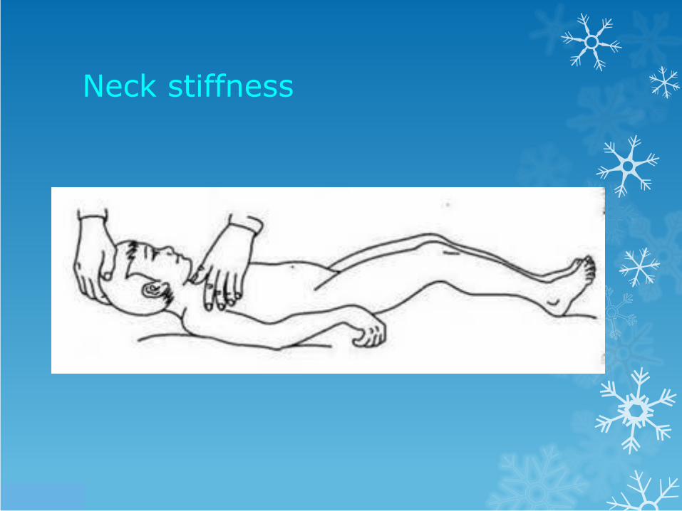
Physical examination
Complete
General
All systems
Pupils
Hemiparesis/plegia
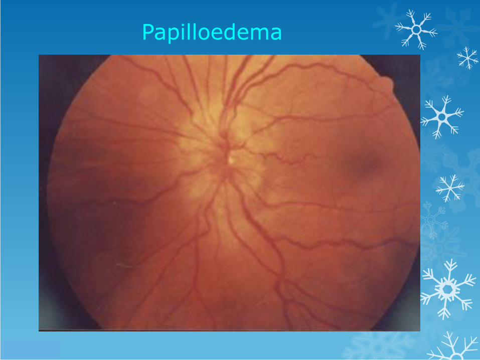
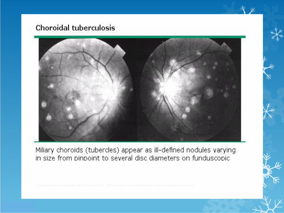
Fundi
Urine dipstix
Investigations
Glucose
Urea and electrolytes, Ca, Phos, Mag, LFTs
Serum osmolality
ABG
Toxicology screen
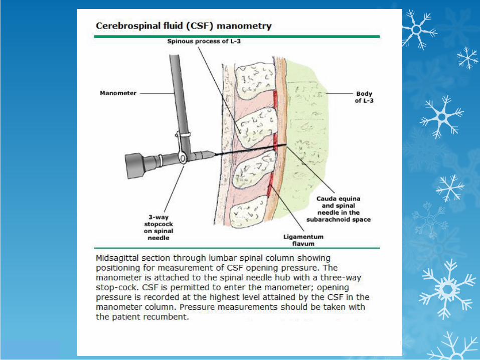
CSF examination
TFTs, Vit B12, serum cortisol
EEG, ECG
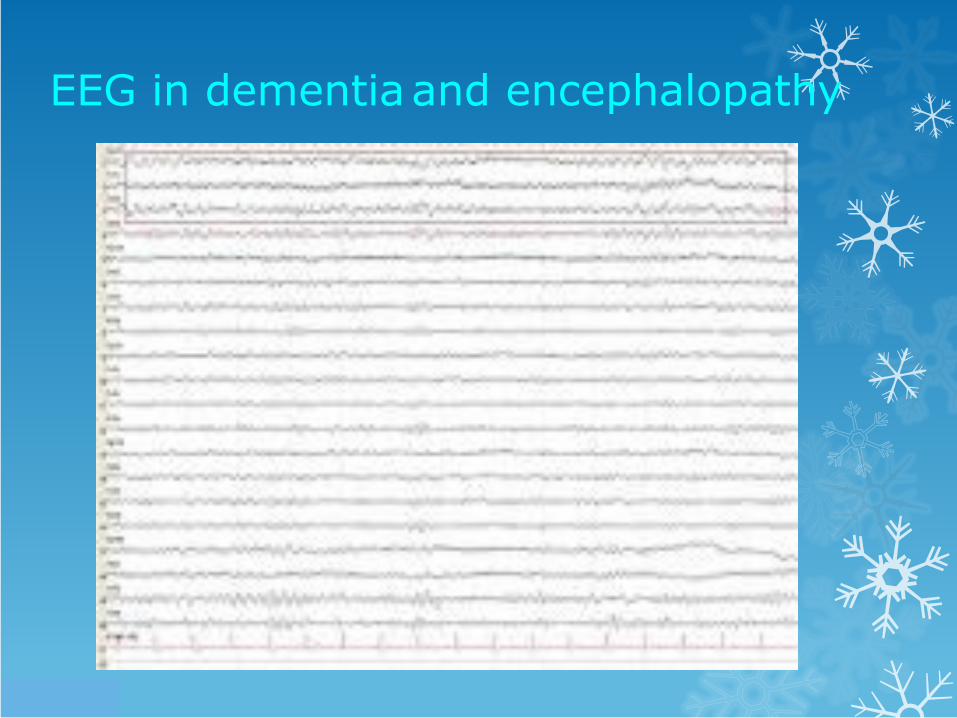
EEG in dementia and encephalopathy
Imaging
CXR
CT brain
MRI brain
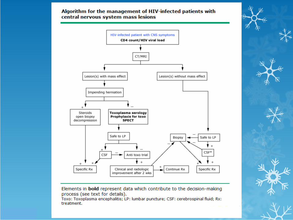
Central nervous system lesions
Degree of immune suppression
CD4 > 500: TB meningitis, brain tumours, metastases
CD4 200-500: TBM, HIV associated motor and cognitive disorders
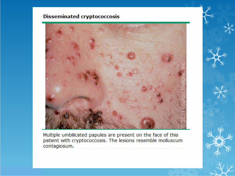
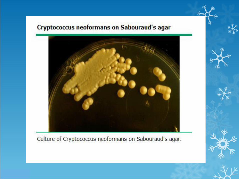
CD4 <200: Cryptococcus, TBM, Toxoplasma encephalitis, CMV encephalitis, herpes encephalitis, HIV encephalopathy, Primary CNS lymphoma, PML,
Useful radiological features
MRI
More sensitive
Determining whether a lesion is truly solitary
White matter disease
Posterior fossa lesions
Biopsy procedure: choice of lesion
Lesions with mass effect
Toxoplasmosis
TB
Primary CNS lymphoma
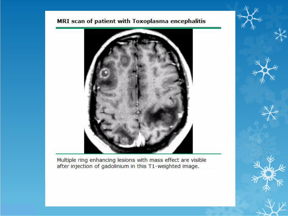
Toxoplasma encephalitis
Toxoplasma antibodies
Multiple lesions
Parietal or frontal lobes
Thalamus, basal ganglia
Cortico- medullary junction
Ring enhancement and surrounding oedema in 90 % cases
Uncommonly: diffuse encephalitis with no abscesses
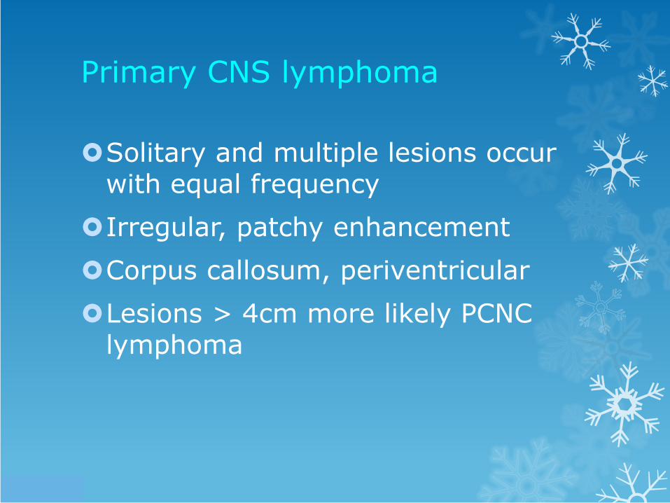
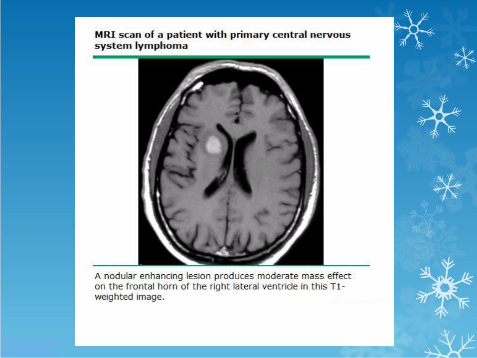
Primary CNS lymphoma
Solitary and multiple lesions occur with equal frequency
Irregular, patchy enhancement
Corpus callosum, periventricular
Lesions > 4cm more likely PCNC lymphoma
Other infections with mass effect
Tuberculomas
Brain abscesses: crypto, syphillis, neurocysticercosis, staph, strep, aspergillus, nocardia,
CNC lesions without mass effect
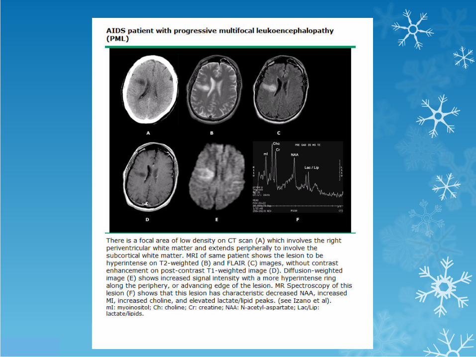
Progressive multifocal leukoencephalopathy
JC virus
Demyelination
Multifocal areas, bilateral,asymetrical
Periventricular areas, subcortical matter
Not contrast enhancing
No surrounding oedema
Exception: IRIS
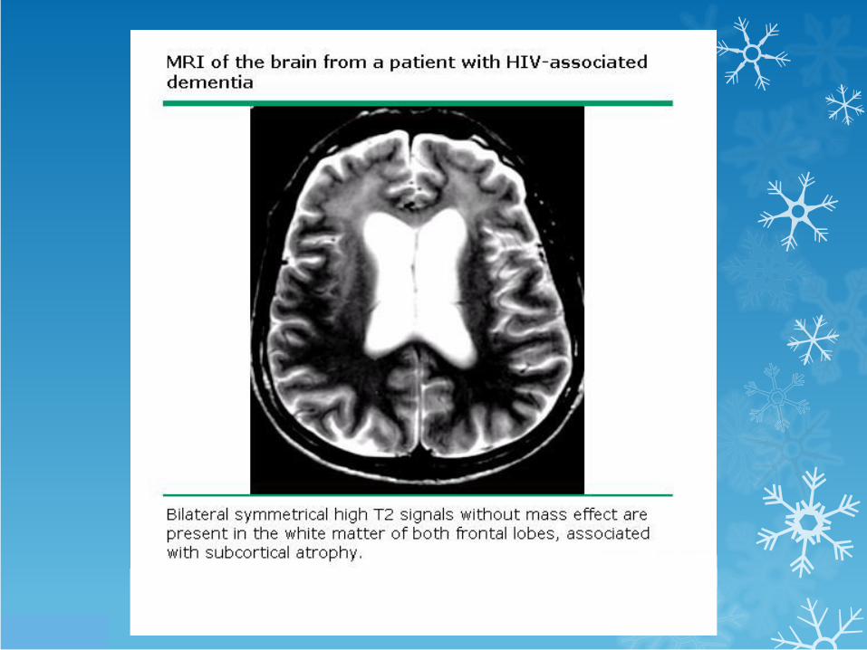
HIV encephalopathy
Classic triad
Memory and psychomotor speed imparment
Depression
Movement disorders
Multiple non enhancing lesions
Bilateral
Subcortical
Symmetrical, less well demarcated
CMV encephalitis
CD4 < 50
Micronodular encephalitis or ventriculoencephalitis
Cortex, basal ganglia, brain stem, cerebellum,
Rarely ring enhancing mass lesions with oedema
Ancillary imaging studies
Thallium 201 SPECT: lymphoma vs toxo
Perfusion MRI: Toxo vs PCNSL
MR spectroscopy
Positron emission tomography (PET): Toxo vs PCNSL
CSF
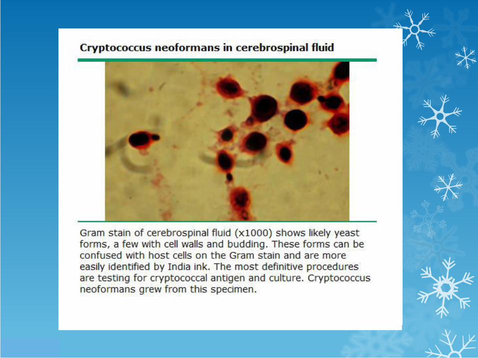
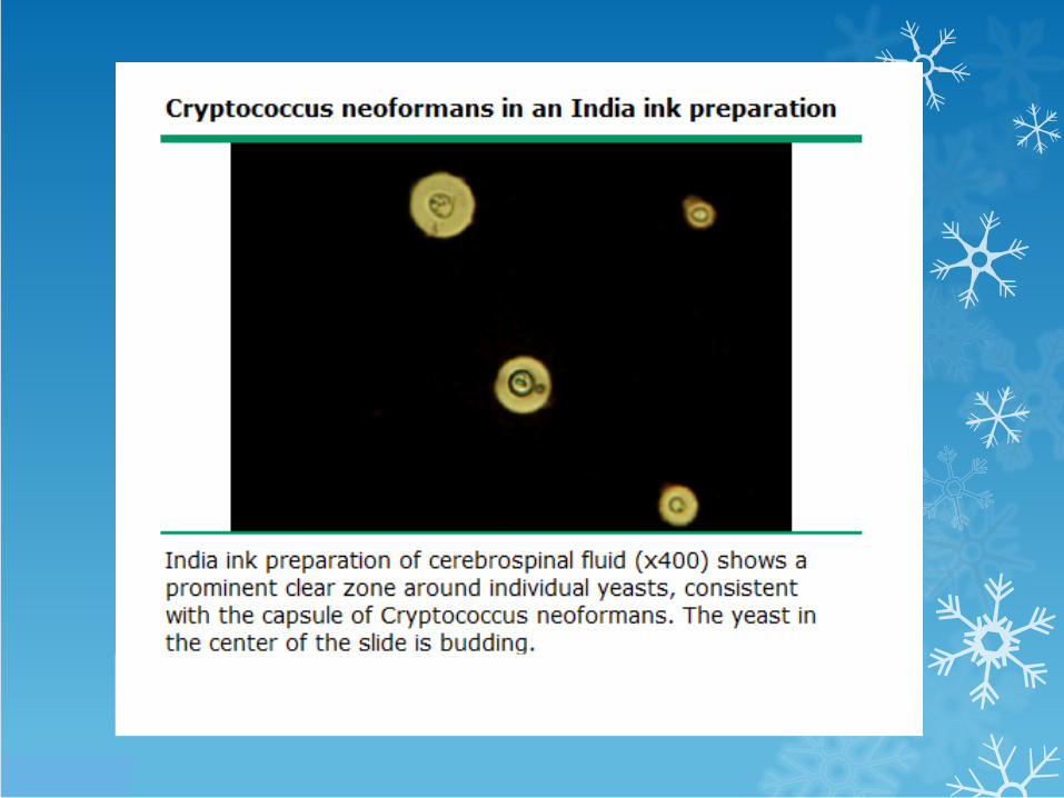
TBM, Cryptococcus, bacterial meningitis
In mass lesions, utility limited
Pleocytosis, elevated protein non specific
CNS lymphoma: cytology diagnostic in 15 %
CSF PCR
DNA of JC virus, Toxo, EB virus
JC virus: sensitivity 74-93 %, specificity 92-100
EBV DNA less clear
Toxo: sensitivity 50 %; specificity 96-100 %
CMV encephalitis: sensitivity > 80 %; specificity > 90 %
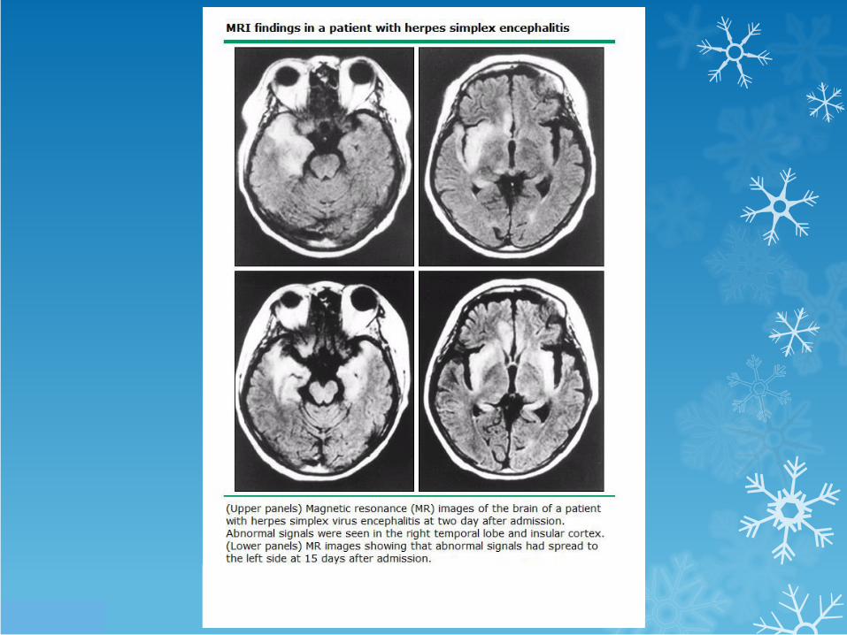
Herpes simplex encephalitis
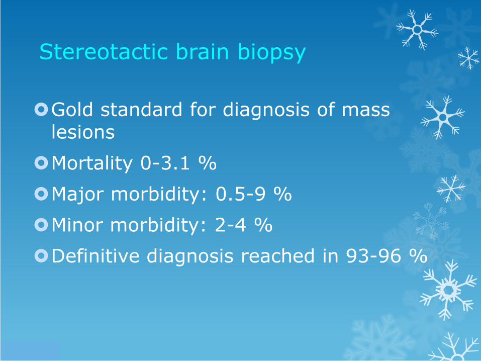
Stereotactic brain biopsy
Gold standard for diagnosis of mass lesions
Mortality 0-3.1 %
Major morbidity: 0.5-9 %
Minor morbidity: 2-4 %
Definitive diagnosis reached in 93-96 %
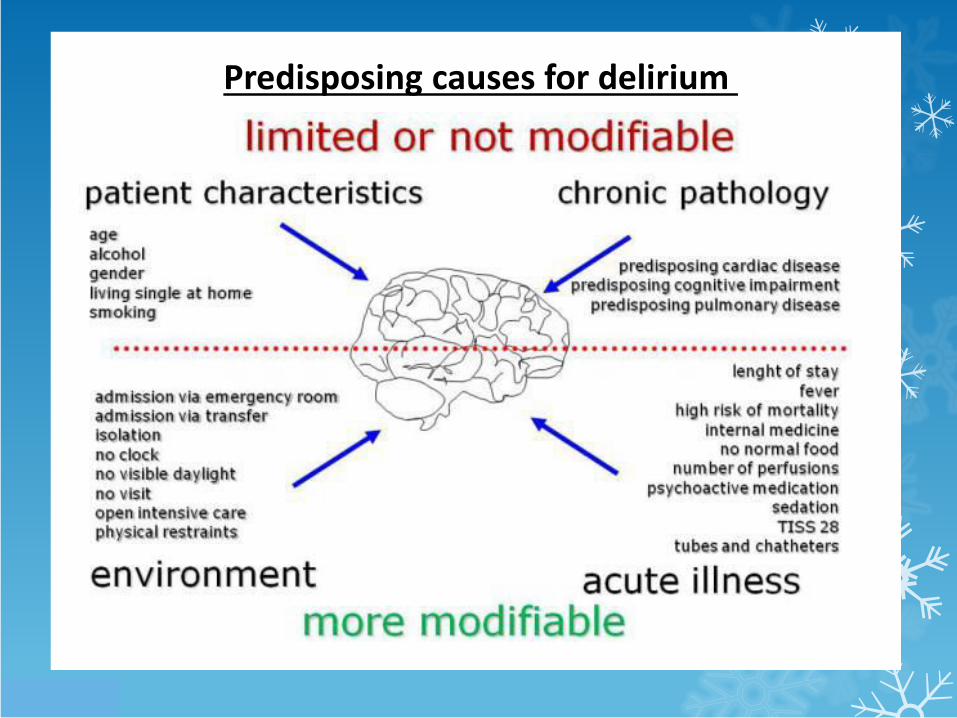
Predisposing causes for delirium