diabetic foot infections-state_of_the_art
TRANSCRIPT

review
article
Diabetes, Obesity and Metabolism 2013.© 2013 John Wiley & Sons Ltdreview article
Diabetic foot infections: state-of-the-artI. Uckay1,2, K. Gariani3, Z. Pataky4 & B. A. Lipsky1,5
1Service of Infectious Diseases, Geneva University Hospitals and Faculty of Medicine, Geneva, Switzerland2Orthopaedic Surgery Service, Geneva University Hospitals and Faculty of Medicine, Geneva, Switzerland3Service of Internal Medicine, Geneva University Hospitals and Faculty of Medicine, Geneva, Switzerland4Division of Therapeutic Education for Chronic Diseases, Geneva University Hospitals and Faculty of Medicine, Geneva, Switzerland5Department of Medicine, University of Oxford, Oxford, UK
Foot infections are frequent and potentially devastating complications of diabetes. Unchecked, infection can progress contiguously to involve thedeeper soft tissues and ultimately the bone. Foot ulcers in people with diabetes are most often the consequence of one or more of the following:peripheral sensory neuropathy, motor neuropathy and gait disorders, peripheral arterial insufficiency or immunological impairments. Infectiondevelops in over half of foot ulcers and is the factor that most often leads to lower extremity amputation. These amputations are associatedwith substantial morbidity, reduced quality of life and major financial costs. Most infections can be successfully treated with optimal woundcare, antibiotic therapy and surgical procedures. Employing evidence-based guidelines, multidisciplinary teams and institution-specific clinicalpathways provides the best approach to guide clinicians through this multifaceted problem. All clinicians regularly seeing people with diabetesshould have an understanding of how to prevent, diagnose and treat foot infections, which requires familiarity with the pathophysiology of theproblem and the literature supporting currently recommended care.Keywords: diabetes complications, diabetes mellitus, foot complications, infections
Date submitted 11 June 2013; date of first decision 5 July 2013; date of final acceptance 11 July 2013
IntroductionDiabetic foot infections (DFIs) are defined as a clinicalsyndrome characterized by local findings of inflammation orpurulence (sometimes accompanied by systemic manifesta-tions of sepsis) occurring in a site below the malleoli in a personwith diabetes. Estimates of the incidence DFIs range from alifetime risk of 4% in all persons with diabetes to 7% yearly inpatients treated in a diabetic foot centre [1]. Most DFIs occurin a neuropathic or neuroischaemic ulcer, which serves as apoint of entry for pathogens. With the exception of erysipelasand posttraumatic (including postsurgical) infection [2], DFIsare almost always epiphenomena, i.e. the consequence ofprogressive peripheral polyneuropathy, with associated lossof protective sensation coupled with gait disorders, anteriordisplacement of weight-bearing during walking [3] withreduced mobility, and arterial insufficiency in a mostly elderlypatient population [4]. Vascular disease, mostly in the form ofocclusive atherosclerotic disease of the arteries below the knee,sometimes accompanied by small vessel dysfunction [5], cancause ischaemic ulcers and may contribute to elevated plantarpressures and to prolonged duration of foot-to-floor contact[6]. Figure 1 shows the major steps in the pathophysiological‘chain’ ultimately leading to DFI [7], and the role of differenthealthcare workers who may help reverse or postpone theprogression of infection and lower extremity amputation.
Correspondence to: Prof. Benjamin A. Lipsky, MD, FACP, FIDSA, FRCP, Department ofMedicine, University of Oxford, 79 Stone Meadow, Oxford, Oxfordshire OX2 6TD, UK.E-mail: [email protected]
Developing a DFI is often the pivotal event leading to lowerextremity amputation. Diabetes is the leading cause of non-traumatic lower extremity amputation worldwide. Diabetes-related amputations at various levels (from toe to above-knee)are responsible for about 60% of all amputations in developedcountries [8] and confer a high burden of financial cost,morbidity and mortality. In high-income countries, treatmentcosts (published in 2000) for a DFI range between US $30 000without amputation and US $58 000 with amputation [9].Diabetes is also associated with a significantly higher rate ofpostoperative stump dehiscence compared to amputations forpurely ischaemic reasons [10]. The presence of osteomyelitisfurther raises the costs for hospitalization because of the needfor additional diagnostic studies, prolonged medical treatmentand surgeries; specifically, the use of antibiotics is at leastdoubled [11]. When amputation is needed, a high level (i.e.transtibial) procedure is more often indicated because ofirreversible ischaemia than because of uncontrolled infection[12]. Most amputations, however, reflect the multimodalfoot problems related to diabetes, emphasizing the needfor a multidisciplinary approach (figure 1) [7]. All cliniciansregularly seeing persons with diabetes should have anunderstanding of how to prevent, diagnose and treat DFIs.Because of the burgeoning research in this area, this reviewaims to help these clinicians be aware of the developments inthis field of science.
Literature Search MethodologySeveral systematic reviews of the literature have beenconducted in recent years [13]. To update and expand

review article DIABETES, OBESITY AND METABOLISM
Figure 1. Overview of causes and management of diabetic foot complications.
on these, we conducted a non-systematic literature searchthrough June 2013 using the PubMed database with the MeSHterms ‘diabetic’, ‘foot’ and ‘infection’ in English and Frenchlanguages. We concentrated on in vivo human data publishedwithin the last 10 years, and excluded basic experimentalpublications, studies performed in animals, papers lackingoriginal human clinical data (other than guidelines), thosehighlighting only surgical techniques or radiological diagnosesand those with non-clinical data. We selected papers initiallyby reading their abstracts, then obtaining the full contents ofrelevant sources. We also reviewed the references of retrievedarticles seeking any additional references. In this review, wewill concentrate on the epidemiology and medical treatmentof DFIs, rather than their pathophysiology.
Risk Factors for InfectionFew studies have specifically assessed factors associated withthe occurrence of a DFI. One prospective, multicenter study
compared 150 diabetic patients with DFI with 97 who did notdevelop infection [14]. Factors significantly associated with DFIwere bone contact on probing; foot ulcer duration of longerthan 30 days; a history of recurrent foot ulcers; traumaticaetiology of the ulcer; and, peripheral vascular disease. Anotherretrospective review of 112 patients with a severe DFI found thatrisk factors for infection were previous amputation, peripheralvascular disease and neuropathy [15]. Other studies haveidentified walking barefoot as a risk for infection. Of note,these risk variables are similarly associated with recurrence ofulcers [16] or reinfection after successful treatment.
Definition and Classification of InfectionMultiple classification schemes have been promulgated fordiabetic foot complications, for most of which the infectioninformation is a subsection of broader ulcer classifications. Incontrast, the Infectious Diseases Society of America (IDSA)and the International Working Group on the Diabetic Foot
2 Uckay et al. 2013

DIABETES, OBESITY AND METABOLISM review article(IWGDF) developed guidelines specifically aimed to defineand classify DFI, and thereby guide the therapy. The IWGDF-PEDIS-classification (an acronym standing for perfusion,extent [size], depth, infection and sensation/neuropathy)suggests a semi-quantitative 4-point scale to describe infectionthat can be used for including patients in research studies, butalso appears to help predict the outcome of a DFI [17].
Because all open wounds will be colonized with microorgan-isms, occasionally even virulent bacteria, culture results alonecannot define infection. Some favour using quantitative micro-biology (e.g. the presence of ≥105 colony forming units/gramof tissue) to differentiate colonization from infection, but nodata support this criterion in the diabetic foot and very fewclinical microbiology laboratories offer this procedure. Thus,most authorities suggest that DFI be diagnosed based on clinicalfindings, i.e. the classical signs and symptoms of inflammation:redness, warmth, induration, pain or tenderness and purulence.Unfortunately, these findings are somewhat subjective. Forexample, foot ischaemia, gout, pyoderma gangrenosum [18]or Charcot neuro-osteoarthropathy may mimic the inflamma-tion of infection. Furthermore, pain may be mitigated by (orattributed to) peripheral neuropathy, and ischaemia may limiterythema, warmth and induration. Therefore, some wound-healing authorities suggest using ‘secondary’ findings to diag-nose infection, such as wound friability, undermining or poorgranulation tissue, foul odour or unexpectedly slow healing.Systemic inflammatory signs (such as fever, chills, hypotension,delirium), elevated serological inflammatory markers (such asleukocytosis, elevated sedimentation rate, C-reactive proteinor procalcitonin levels) [19] or positive blood cultures defineserious infections, but are infrequent in DFI.
The results of microbiological tests, such as a Gram-stainedsmear or culture from diabetic foot wound specimens, mustbe interpreted with reference the clinical situation. There isno evidence that treating a clinically uninfected wound withantimicrobials has any value in either preventing infectionor improving ulcer healing. When there are clinical signs ofinfection, however, obtaining an appropriate sample for cultureand sensitivity testing helps guide antibiotic therapy. Specimensshould be taken after cleansing and debriding the wound fromdeep, non-necrotic tissue or pus, to lessen the chance ofisolating colonizing species. Superficial cultures obtained withcotton swabs are easily collected, but are less reliable thantissue biopsies and should be avoided. Most studies have foundthat swab specimens have more isolates (likely contaminatingor colonizing flora) than aseptically obtained deep tissuespecimens and also may miss true pathogens, especiallyanaerobic or fastidious species. In particular, most studies withdiabetic foot osteomyelitis have found that neither superficialnor deep soft tissue cultures correlate well with those of bonespecimens [20]. One study suggested, however, that repeatedbone surface swabbing yields similar results compared withbone culture in patients with a wound with underlying clinicalosteomyelitis [21], but this needs to be confirmed in larger trials.
Diagnosis of Osteomyelitis
Infection of bone underlying a diabetic foot ulcer should besuspected in the presence of a large or deep wound, especially
if it is chronic and overlying bone, or if a toe is red and swollen.Most blood tests are of limited value in diagnosing osteomyeli-tis, but an erythrocyte sedimentation rate of over 70 is highlysuggestive. Of note is that one study found that a physician’sclinical judgment about the presence of osteomyelitis had apositive likelihood ratio of 5.5 and a negative likelihood ratio of0.54 [22]. The probe-to-bone test, in which a hard gritty struc-ture is palpated with a sterile, blunt metal probe, is both easy toperform and useful. A negative probe-to-bone test in a patientin whom the pretest probability of osteomyelitis is low is reas-suring [23], but does not rule out osteomyelis [24]. On the otherhand, a positive test in a patient in whom clinical suspicion ishigh (especially if the plain X-ray is suggestive of osteomyelitis)has a high predictive value for bone infection [25].
The gold standard for diagnosing osteomyelitis remains thecombination of microbiological culture and histopathologicalexamination of bone [26]. Bone specimens may be obtained atsurgery or by transcutaneous biopsy. Although needle punctureof deep soft tissue near bone does not reliably predict theresults of bone cultures [27], puncture of the bone itself maybe an easy way to obtain bone culture at the bedside [28].The first test to consider when osteomyelitis is suspected isplain radiography, but early infection maybe missed becauseit takes several weeks for the findings of bone infection tobe detected. Characteristic features of osteomyelitis on plainX-rays include periosteal elevation and erosions of the osseousborders, but interobserver reproducibility of detecting thesesigns is poor, especially among inexperienced observers [29].The reported sensitivity of plain radiography in diagnosingosteomyelitis ranges from 28 to 75%, with one review citinga pooled sensitivity among four studies of 0.54 and specificity0.68 [22]. Repeating and comparing foot X-rays over time ismore likely to detect osteomyelitis than a single series.
In some patients, advanced imaging is needed to detectosteomyelitis. In these situations, magnetic resonance imaging(MRI) is considered the best available technique, not only fordiagnosing osteomyelitis but also for better visualizing deep softtissue infection or sinus tracts. One meta-analysis, reported apooled sensitivity of 0.90, and the diagnostic odds ratio was24.4 [30]. In another meta-analysis the pooled sensitivity was77–100%, but the specificity was only 40% [31]. Undoubtedly,the value of this (like most tests) varies with the skill andexperience of the interpreting radiologist, and MRI does notneed to be routinely obtained [32]. Computed tomography(CT) scans, more readily available in some centres, are usuallyless expensive and may be useful when MRI is contraindicated.Nuclear medicine scintigraphic examinations are certainlymore sensitive than plain X-rays for detecting osteomyelitis,but bone scans have a low specificity. If scintigraphy is needed,leukocyte scans are superior to bone scans, but we think thesetests should usually be reserved for long-bone osteomyelitis orprosthetic joint infections. Newer procedures, such as SPECT-CT, PET/CT or PET/MRI, show promise and may be even moreaccurate than MRI, but to date there have been only a limitednumber of studies in DFI. Of note, in patients undergoingpercutaneous bone biopsy for suspicion of osteomyelitis whohave a negative culture, one of four will develop osteomyelitisin the next 2 years [33].
2013 doi:10.1111/dom.12190 3
review article DIABETES, OBESITY AND METABOLISM
Microbiology of DFIs Around the WorldIn Western developed countries, mild community-acquiredinfections in patients who have not recently been treatedwith antibiotics are mainly caused by aerobic Gram-positivecocci, especially Staphylococcus aureus and, to a lesser degree,by β-streptococci (usually group B) or coagulase-negativestaphylococci. One study using molecular microbiologicalmethods found that ulcer depth is directly correlated withthe presence of S. aureus [34]. In chronic wounds, especiallythose in a patient who has been treated with antibiotics,infections are more often polymicrobial, including aerobicGram-negative and obligate anaerobic bacteria. Recently,epidemiological surveys from subtropical, less-developedcountries have reported that S. aureus is less prevalent thannoted in developed countries (30% vs. 75%) while thereis a considerably higher prevalence of Gram-negative rods,especially Pseudomonas aeruginosa (Table 1). The reasons forthis geographical difference has not been elucidated, but may berelated to differences in specimen types, laboratory techniques,prior antibiotic use, availability of non-prescription (over-the-counter) antibiotic agents, foot sweating and washingor reporting bias. Of note, most of these reports emanatefrom countries in arid and hot areas, especially India [35].It might also be that the microbiology of DFIs is evolvingslowly towards more Gram-negative microorganisms [35]in some regions, whereas in other regions of the samecountry infections with S. aureus may still be dominant[36]. Areas of the southwest of the USA have also reporteda relatively low proportion of DFIs caused by S. aureus [37]and isolation of P. aeruginosa is more frequent in nosocomialDFIs [38].
Traumatic wound infections [39,40] and cultures of deepwounds with moderate to severe infections, especially in pre-viously antibiotic-treated patients, are usually polymicrobialwith mixed Gram-positive cocci (vide supra), Gram-negativerods (e.g. Escherichia coli, Proteus, Klebsiella), sometimesincluding non-fermentative Gram-negatives (P. aeruginosa),and anaerobes (e.g. Finegoldia, Bacteroides) (Table 1). Severeinfections may harbour P. aeruginosa, especially in cases of deeppuncture wounds and in patients whose feet are frequentlyexposed to water. Fungi are rarely principal pathogens and arethus mostly highlighted in case reports [41]. When looked forcarefully by clinical laboratories, fungi have be regularly culti-vated, as have anaerobes [41–44], but the clinical importanceof these findings is unclear. Parasitic or mycobacterial DFIshave rarely been noted in published reports.
A recent problem has been the isolation of multidrugresistant organisms (MDROs) from DFIs. The predominantresistant pathogen has been methicillin-resistant S. aureus(MRSA). After many reports of this pathogen in DFIs fromthe mid-1990s to the early 2000s, more recent studies suggestthe prevalence may be decreasing in most countries. Lately,the antibiotic resistance problem of greatest concern has beenGram-negative organisms that produce extended-spectrumβ-lactamases-(ESBLs) or carbapenemases. Overall, thelikelihood of isolating MDROs from a DFI has increased overthe past decade [42,45,46].
TreatmentOrthopaedic Surgery, Podiatry and Revascularization
Most DFIs require both medical and surgical interventions.Surgery is particularly important for dealing with abscesses,necrotizing fasciitis and a substantial proportion of osteomyeli-tis cases (e.g. when there is necrotic bone [47]). Many DFIswill require debridement or incision and drainage, and somepatients will benefit from revascularization or procedures tocorrect anatomic problems or gait disorders. Details regardingthe surgical approach to DFIs are important, but beyond thescope of this review.
Podiatric care is especially aimed at debridement of callusand necrotic tissue, treatment of blisters, caring for nails andselecting proper footwear. Repeated removal of calluses isespecially important, as emphasized by the fact that theirpresence may cause 18 tons of excess plantar pressure eachday [48]. Surgical treatment for DFI without concomitantantimicrobial therapy is possible [49], but the available reportsfrom the pre-antibiotic era demonstrate high mortality rates,despite the fact that most patients underwent major (oftenabove the knee) amputations. While urgent surgery is neededfor most deep severe infections, some orthopaedic or vascularprocedures may best be delayed until infection is bettercontrolled. Some procedures, such as the correction of footdeformities, arthrodesis [50] or combination of correction anddebridement for infection [51], may also serve to prevent DFIs.For example, flexor tenotomy, a surgical intervention with alow surgical site infection rate [52], may be highly efficacious(>90% success within 5–8 weeks) for prevention and healingof distal toe ulcers [52,53].
All patients with a diabetic foot wound require a vascularassessment. Those with clinically compromising arterialinsufficiency of the foot require revascularization, if feasible.This may be done by either endovascular or open methods [54].Contrary to what some profess, infrapopliteal endovascularrevascularization, even in patients with long-standing diabetes,is possible with modern techniques, at least in resource-richsettings [55,56].
Antimicrobial Therapy
Antibiotic therapy is almost always necessary, but often notsufficient, to cure DFIs. It must usually be combined with oneor more surgical procedures, pressure off-loading and properwound care. Initial antibiotic therapy for most patients mustbe selected empirically, and should be based on the presentingclinical features, knowledge of the local antibiotic resistancepatterns and an assessment of infection severity. Severalprinciples may help to avoid selecting either an unnecessarilybroad or an inappropriately narrow regimen [57]. Antibioticcoverage should always include S. aureus, the commonestpathogen in most situations. If the prevalence of methicillin-resistance among S. aureus isolates is known to be high, or if theinfection is more than mild, anti-MRSA therapy is advisable.Therapy should be broadened to target Gram-negativepathogens in all severe and many moderate infections, orif the patient has failed to respond to prior narrower-spectrumantibiotic therapy. In these latter cases, it is especially important
4 Uckay et al. 2013

DIABETES, OBESITY AND METABOLISM review articleTable 1. Results of selected reports from around the world over the past decade of the microbiology of diabetic foot wounds.
Percentage of isolates from wound culture
First author[Reference] Country Year
Types ofwounds
No. ofpatients Staphylococci Streptococci
Gram-positive
Gram-negative Ps. aeruginosa Anaerobes
Carvalho [109] Brazil 2003 Infections 141 20 4 29 59 7 12Candel [110] Spain 2003 Infections 27 49 15 78 22 1 2Anandi [111] India 2004 Infections 107 14 — — — — 4Unachukwu [112] Nigeria 2005 Gangrene 60 56 — — — — —Senneville [113] France 2005 Bone 76 52 12 — 18 2 5Abdulrazak [114] Kuwait 2005 Infections 86 38 17 74 26 18 11Shankar [115] India 2005 Infections 77 — 3 42 58 30 6Yoga [116] Malaysia 2006 Infections 44 20 — — — 14 —Gadepalli [72] India 2006 Ulcers 80 20 0 33 51 10 15Sharma [117] Nepal 2006 Ulcers — 38 — — — 18 —Ormen [118] Turkey 2007 Bone 50 — — 40 60 — —Raja [119] Malaysia 2007 Infections 194 44 25 45 52 25 —Cetin [120] Turkey 2007 Infections 65 18 6 59 41 8 3Dowd [121] USA 2008 Ulcers 40 8 37 — — 15 18Umadevi [122] India 2008 Infections 105 17 0 29 71 17 0Khoharo [123] Pakistan 2009 Infections 60 20 3 27 73 48 2Ramakant [35] India 2010 Ulcers 447 19 3 31 57 17 1Zubair [124] India 2010 Infections 60 31 0 38 62 11 0Ozer [43] Turkey 2010 Infections 78 17 7 38 56 19 —Mendes [125] Portugal 2011 Infections 49 84 4 85 19 1 14Hayat [126] Pakistan 2011 Infections 85 18 5 27 68 27 2Pappu [127] India 2011 Infections 104 21 4 — >67 23 0Malone [128] Saudi-Arabia 2011 Toe bone 34 33 9 57 29 9 0Aziz [129] Singapore 2011 Infections 100 40 21 — — 26 30Dezfulian [138] Iran 2011 Infections 69 43 5 55 45 6 5Aamir [36] Pakistan 2011 Wounds 114 55 5 — — 4 —Tiwari [130] India 2012 Infections 62 — — 32 68 — —Swarna [131] India 2012 Infections 62 30 0 44 56 20 0Pai [38] India 2012 Infections 55 7 — 24 65 2 —Widatella [47] Sudan 2012 Bone 330 33 — — — 32 —Parvez [132] India 2012 Bone 60 9 0 14 70 7 16Banashankari [133] India 2012 Infections 202 19 — 32 66 13 —Al Benwan [134] Kuwait 2012 Infections 440 20 — 32 51 17 15Shanmugam [135] India 2013 Infections 50 13 — 35 65 16 —Anjali [136] India 2013 Infections 100 — — 42 58 19 —Osariemen [137] Nigeria 2013 Infections 150 38 0 38 62 8 0Djahmi [45] Algeria 2013 Infections 128 43 1 45 55 8 0
Percentage values do not sum up to 100% because of mixed infections and reporting of only selected isolates.
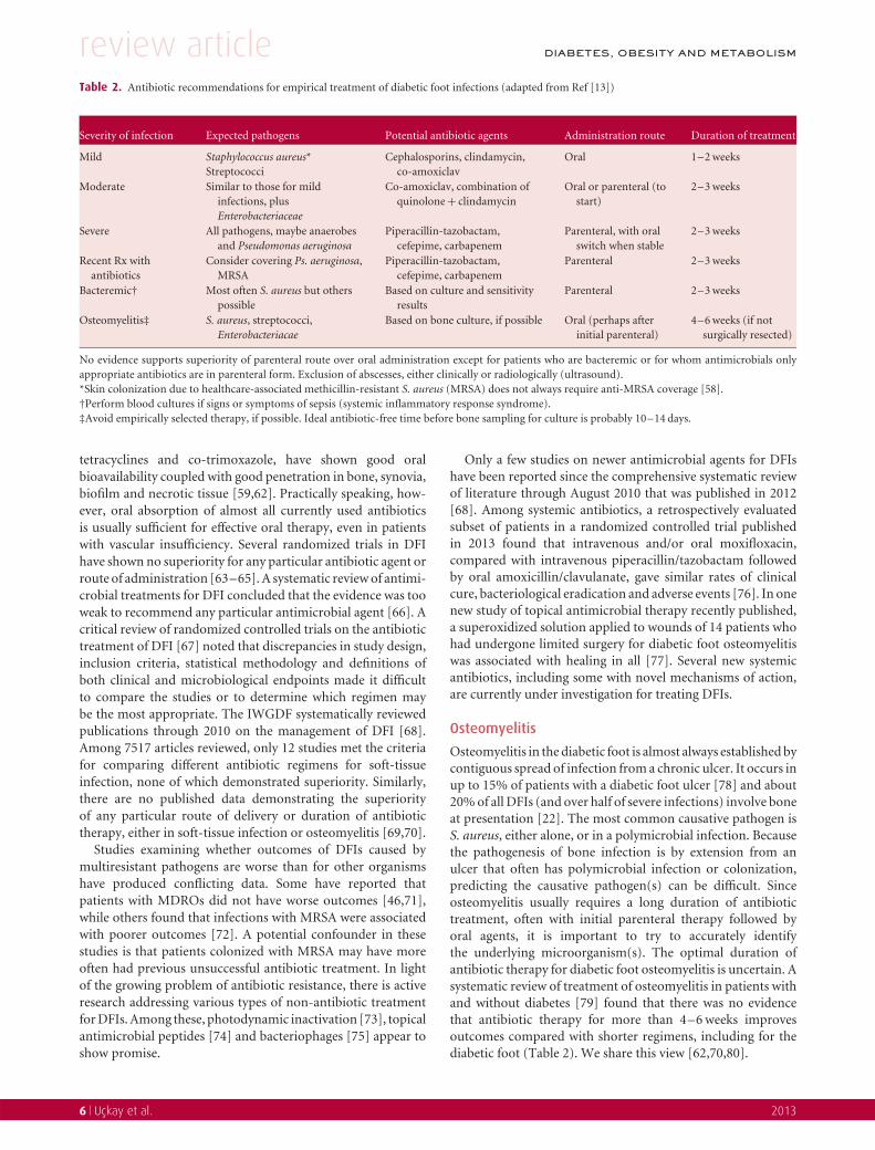
to obtain optimal specimens for culture and to initiate anempiric regimen different from the failing one. Adding agentsthat are specifically active against obligate anaerobes is usuallyneeded only if the wound is gangrenous or if there is afoetid odour. Finally, most severe infections require initialparenteral, broad-spectrum therapy [57]. Table 2 displayssuggested antibiotic regimens (most often used at GenevaUniversity Hospitals), based on the recent IDSA guidelines [13].
Using the results of appropriately obtained specimens forculture allows more targeted therapy, which usually can benarrower in scope than the empiric regimen. When culturesyield multiple organisms deciding which isolates need to becovered depends on the quality of the specimen sent forculture and the specific organisms isolated. If the specimenwas aseptically obtained deep soft tissue or bone, coveringall isolates may be prudent. In most situations, however, it
may be sufficient to treat just the likeliest pathogens, such asS. aureus, streptococci and any Enterobacteriaceae present inlarge numbers. Skin commensals, such as coagulase-negativestaphylococci, corynebacteria or Bacillus spp., in the absenceof an infection involving osteosynthetic material or hardware[59,60] can usually be dismissed. Similarly, the mere presenceof skin colonization with healthcare-associated MRSA doesnot oblige the clinician to empirically cover this organism [58],even in the presence of underlying osteosynthetic material [61].
Because most DFIs occur in the setting of some degree ofperipheral arterial disease, some have raised concerns abouthow well various antibiotic agents penetrate, especially in thepresence of bone infection. Several studies have shown thatat standard doses most β-lactam antibiotics achieve relativelylow (albeit likely therapeutic) tissue levels, but clindamycin,fluoroquinolones, linezolid, rifampin, and to some degree,
2013 doi:10.1111/dom.12190 5
review article DIABETES, OBESITY AND METABOLISM
Table 2. Antibiotic recommendations for empirical treatment of diabetic foot infections (adapted from Ref [13])
Severity of infection Expected pathogens Potential antibiotic agents Administration route Duration of treatment
Mild Staphylococcus aureus*Streptococci
Cephalosporins, clindamycin,co-amoxiclav
Oral 1–2 weeks
Moderate Similar to those for mildinfections, plusEnterobacteriaceae
Co-amoxiclav, combination ofquinolone + clindamycin
Oral or parenteral (tostart)
2–3 weeks
Severe All pathogens, maybe anaerobesand Pseudomonas aeruginosa
Piperacillin-tazobactam,cefepime, carbapenem
Parenteral, with oralswitch when stable
2–3 weeks
Recent Rx withantibiotics
Consider covering Ps. aeruginosa,MRSA
Piperacillin-tazobactam,cefepime, carbapenem
Parenteral 2–3 weeks
Bacteremic† Most often S. aureus but otherspossible
Based on culture and sensitivityresults
Parenteral 2–3 weeks
Osteomyelitis‡ S. aureus, streptococci,Enterobacteriacae
Based on bone culture, if possible Oral (perhaps afterinitial parenteral)
4–6 weeks (if notsurgically resected)
No evidence supports superiority of parenteral route over oral administration except for patients who are bacteremic or for whom antimicrobials onlyappropriate antibiotics are in parenteral form. Exclusion of abscesses, either clinically or radiologically (ultrasound).*Skin colonization due to healthcare-associated methicillin-resistant S. aureus (MRSA) does not always require anti-MRSA coverage [58].†Perform blood cultures if signs or symptoms of sepsis (systemic inflammatory response syndrome).‡Avoid empirically selected therapy, if possible. Ideal antibiotic-free time before bone sampling for culture is probably 10–14 days.
tetracyclines and co-trimoxazole, have shown good oralbioavailability coupled with good penetration in bone, synovia,biofilm and necrotic tissue [59,62]. Practically speaking, how-ever, oral absorption of almost all currently used antibioticsis usually sufficient for effective oral therapy, even in patientswith vascular insufficiency. Several randomized trials in DFIhave shown no superiority for any particular antibiotic agent orroute of administration [63–65]. A systematic review of antimi-crobial treatments for DFI concluded that the evidence was tooweak to recommend any particular antimicrobial agent [66]. Acritical review of randomized controlled trials on the antibiotictreatment of DFI [67] noted that discrepancies in study design,inclusion criteria, statistical methodology and definitions ofboth clinical and microbiological endpoints made it difficultto compare the studies or to determine which regimen maybe the most appropriate. The IWGDF systematically reviewedpublications through 2010 on the management of DFI [68].Among 7517 articles reviewed, only 12 studies met the criteriafor comparing different antibiotic regimens for soft-tissueinfection, none of which demonstrated superiority. Similarly,there are no published data demonstrating the superiorityof any particular route of delivery or duration of antibiotictherapy, either in soft-tissue infection or osteomyelitis [69,70].
Studies examining whether outcomes of DFIs caused bymultiresistant pathogens are worse than for other organismshave produced conflicting data. Some have reported thatpatients with MDROs did not have worse outcomes [46,71],while others found that infections with MRSA were associatedwith poorer outcomes [72]. A potential confounder in thesestudies is that patients colonized with MRSA may have moreoften had previous unsuccessful antibiotic treatment. In lightof the growing problem of antibiotic resistance, there is activeresearch addressing various types of non-antibiotic treatmentfor DFIs. Among these, photodynamic inactivation [73], topicalantimicrobial peptides [74] and bacteriophages [75] appear toshow promise.
Only a few studies on newer antimicrobial agents for DFIshave been reported since the comprehensive systematic reviewof literature through August 2010 that was published in 2012[68]. Among systemic antibiotics, a retrospectively evaluatedsubset of patients in a randomized controlled trial publishedin 2013 found that intravenous and/or oral moxifloxacin,compared with intravenous piperacillin/tazobactam followedby oral amoxicillin/clavulanate, gave similar rates of clinicalcure, bacteriological eradication and adverse events [76]. In onenew study of topical antimicrobial therapy recently published,a superoxidized solution applied to wounds of 14 patients whohad undergone limited surgery for diabetic foot osteomyelitiswas associated with healing in all [77]. Several new systemicantibiotics, including some with novel mechanisms of action,are currently under investigation for treating DFIs.
Osteomyelitis
Osteomyelitis in the diabetic foot is almost always established bycontiguous spread of infection from a chronic ulcer. It occurs inup to 15% of patients with a diabetic foot ulcer [78] and about20% of all DFIs (and over half of severe infections) involve boneat presentation [22]. The most common causative pathogen isS. aureus, either alone, or in a polymicrobial infection. Becausethe pathogenesis of bone infection is by extension from anulcer that often has polymicrobial infection or colonization,predicting the causative pathogen(s) can be difficult. Sinceosteomyelitis usually requires a long duration of antibiotictreatment, often with initial parenteral therapy followed byoral agents, it is important to try to accurately identifythe underlying microorganism(s). The optimal duration ofantibiotic therapy for diabetic foot osteomyelitis is uncertain. Asystematic review of treatment of osteomyelitis in patients withand without diabetes [79] found that there was no evidencethat antibiotic therapy for more than 4–6 weeks improvesoutcomes compared with shorter regimens, including for thediabetic foot (Table 2). We share this view [62,70,80].
6 Uckay et al. 2013

DIABETES, OBESITY AND METABOLISM review articleEstablished wisdom has held that diabetic foot osteomyelitis,
like most chronic bone infections, requires surgical debride-ment or resection of necrotic and infected bone. For example, ina study of 50 patients with chronic toe osteomyelitis, patientswho underwent surgical resection had a significantly lowerrelapse rate [81]. There are, however, hundreds of reports ofapparently successful treatment without surgery, with mostseries reporting remission rates of 60% to 70% [82,83]. Thus,when the patient or the medical team prefer to avoid surgery,a trial of exclusively antibiotic therapy may be reasonable. But,the advantages of surgical therapy (especially in case of toeamputations), including the relatively short lengths of hos-pital stay, reduced antibiotic consumption and likely higherremission rates, should be weighed against the potential risks.
Hyperbaric Oxygen Therapy and Stimulating Factors
The value of hyperbaric oxygen therapy for non-infected dia-betic ulcers is a question of ongoing debate [80,84–86]. A 2012Cochrane systematic review concluded that hyperbaric oxygentherapy significantly increased ulcer healing in the short termbut not the long term, but because of the flawed trials they werenot confident in the results [87]. Some suggest that hyperbaricoxygen decreases rates of lower extremity amputation inpatients with diabetic foot ulcers or postsurgical amputationwounds in persons with diabetes, and facilitates ulcer healing[88]. There are, however, no published data directly related tothe effect of hyperbaric oxygen therapy for infectious aspects(either soft tissue or bone) of the diabetic foot [84].
Several studies have examined the usefulness of adjunctivetreatment of DFIs with granulocyte-colony stimulating factors.A Cochrane systematic review of the five eligible trialsconcluded that these treatments did not increase the likelihoodof resolution of infection but did appear to reduce the need forsurgical interventions, especially amputations, and the durationof hospitalization. Studies of platelet-derived [89] and othergrowth factors and skin substitutes have not shown any specificbenefit regarding resolution or prevention of infection [90,91].
Antiseptic Dressings and Other Topical Treatments
Several studies have assessed topical treatments in patients withdiabetic foot ulcers and, to a lesser extent, for DFI. For themajority, ulcer healing rather than resolution or preventionof infection was the primary outcome of interest. Most of thestudies evaluated topical antiseptic agents (e.g. silver, povidoneiodine, hypochlorite, peroxide, zinc oxide) as adjuncts to otherstandard treatments. None of these agents has been proven toprovide superior outcomes compared to other non-antisepticdressings [92]. Similarly, recent systematic reviews have failedto detect a superiority of various other dressings, such as foam[93,94], hydrocolloid [95] and alginate [96], for ulcer healing orresolution of infection. A review of topical antimicrobial ther-apy for treating chronic wounds concluded that there are fewproven indications for any of the currently available agents [91].
Two topical antimicrobial agents have been investigated forDFIs. A large randomized controlled trial in patients with amild DF showed that treatment with a topical antimicrobialpeptide (pexiganan) produced clinical outcomes similar to
those of an oral antibiotic (ofloxacin) [74]. In an open-labelstudy, daily application of a gentamicin-collagen sponge for upto 28 days in combination with systemic antibiotic therapy wascompared to systemic antibiotic therapy alone in the treatmentof moderate DFIs [97]. Although this study failed to meet itsprimary endpoint (percent of patients with an outcome ofclinical cure at day 7 of treatment), the gentamicin-collagensponge showed superior efficacy in eradicating baselinepathogens and achieving clinical cure at the final visit [97].
Vacuum-Assisted Negative-Pressure Therapy
While widely used for accelerating wound healing, there arelimited published data on the effectiveness of vacuum-assistednegative-pressure therapy on DFI, including osteomyelitis [98].Concerning diabetic foot ulcers, a systematic review identifiedfour randomized trials [99]. While all, including a multicentrestudy that enrolled 342 patients [100], found that vacuum-assisted therapy was more effective than conventional dressings,the quality of each of the studies was weak and the outcomesstudied and patient selection were divergent [99,101].
Off-Loading
Off-loading pressure is a critical part of the treatment of almostall diabetic foot ulcers, including those that are infected [102].While the principle of off-loading is easy to understand, inpractice it greatly depends on near total compliance on the partof the patient. The criterion standard for off-loading is the totalcontact cast, which is associated with ulcer healing rates of over90% [102]. The main advantage of this device may be that thepatient cannot easily remove it. Given the high recurrence ratesof neuropathic foot ulcers, new approaches include helpingpatients to modify their walking pattern over the long term,perhaps with feedback-based approaches [103].
Clinical Pathways, Guidelines and BundleInterventionsDFIs exemplify a multifaceted problem that particularly bene-fits from a multidisciplinary approach [7]. In the past decade,several evidence-based DFI guidelines have been publishedthat provide an approach to optimize outcomes [7,104] and toavoid amputations [105]. All consistently address the criticalrole of multidisciplinary teams involving specialists such as dia-betologists, orthopaedic/podiatric surgeons, infectious diseasesspecialists, vascular surgeons, angiologists, interventional radi-ologists, specialized nurses and physiotherapists; such teamshave been established in many large hospitals in resource-richcountries all over the world [7,13]. These teams have beenshown to be beneficial in avoiding adverse outcomes in bothinpatients and outpatients in many studies. They are, however,hampered by several logistic problems: (a) it is often difficultto bring the members of the multidisciplinary team togetheroutside of a fixed meeting time; (b) the number of patientsrequiring evaluation often exceeds the capacity of fixed mul-tidisciplinary meetings; and (c) members of the team oftenturnover. For example, at Geneva University Hospitals, thereis a 45-min diabetic foot team meeting weekly, but we have
2013 doi:10.1111/dom.12190 7
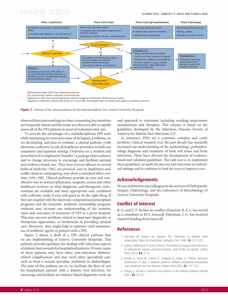
review article DIABETES, OBESITY AND METABOLISM
Figure 2. Schema of the clinical pathway for the infected diabetic foot, Geneva University Hospitals.
observed that team meetings are time-consuming, key membersare frequently absent and the teams are often not able to rapidlyassess all of the DFI patients in need of evaluation and care.
To provide the advantages of a multidisciplinary DFI teamwhile attempting to overcome some of its logistic problems, weare developing, and plan to evaluate, a clinical pathway (withelectronic ordersets) to aid all healthcare providers in both ouroutpatient and inpatient settings. Ordersets are a modern andpowerful tool to implement ‘bundles’ (a package of procedures)and to change processes to encourage and facilitate optimaland evidence-based care. They have proven efficacy in severalfields of medicine [106], are practical, easy to implement and,unlike classical campaigning, may show a sustained effect overtime [104–108]. Clinical pathways provide an easy and cost-effective way to instruct physicians, surgeons, nurses and otherhealthcare workers on what diagnostic and therapeutic inter-ventions are available and most appropriate and, combinedwith ordersets, make it easy and quick to do the right thing. Ifthey are coupled with the electronic computerized prescriptionprograms and the hospitals’ antibiotic stewardship program,ordersets may increase our understanding of the number,types and outcomes of treatment of DFI in a given hospital.This may uncover problems related to improper diagnostic ortherapeutic approaches, or bottlenecks in providing optimalcare. Moreover, they might help to optimize (and minimize)use of antibiotic agents in patients with a DFI.
Figure 2 shows a draft of a DFI clinical pathway thatwe are implementing at Geneva University Hospitals. Thispathway provides guidance for dealing with infectious aspectsof diabetic foot wounds for hospitalized patients. Of note, manyof these patients may have other non-infectious diabetes-related complications and may need other specialized care,such as from a wound specialist, podiatrist or diabetologist.The aims of this pathway are to: (a) facilitate the flow of carefor hospitalized patients with a diabetic foot infection; (b)encourage and facilitate an evidence-based diagnostic work-up
and approach to treatment, including avoiding unnecessaryexaminations and therapies. This schema is based on theguidelines developed by the Infectious Diseases Society ofAmerica for diabetic foot infections [13].
In summary, DFIs are a common, complex and costlyproblem. Clinical research over the past decade has markedlyincreased our understanding of the epidemiology, pathophys-iology diagnosis and treatment of both soft tissue and boneinfections. These have allowed the development of evidence-based and validated guidelines. The task now is to implementthese guidelines, to audit the process and outcomes in individ-ual settings and to continue to look for ways to improve care.
AcknowledgementsWe are indebted to our colleagues in the services of OrthopaedicSurgery, Diabetology, and the Laboratory of Microbiology ofGeneva University Hospitals.
Conflict of InterestK. G. and Z. P. declare no conflict of interest. B. A. L. has servedas a consultant to KCI, Innocoll, Dipexium. I. U. has receivedresearch funding from Innocoll.
References
1. Pecoraro RE, Reiber GE, Burgess EM. Pathways to diabetic limbamputation. Basis for prevention. Diabetes Care 1990; 13: 513–521.
2. Uckay I, Hoffmeyer P, Lew D, Pittet D. Prevention of surgical site infectionsin orthopaedic surgery and bone trauma: state-of-the-art update. J HospInfect 2012; 84: 5–12.
3. Pataky Z, Assal JP, Conne P, Vuagnat H, Golay A. Plantar pressuredistribution in type 2 diabetic patients without peripheral neuropathyand peripheral vascular disease. Diabet Med 2005; 22: 762–767.
4. Pataky Z, Vischer U. Diabetic foot disease in the elderly. Diabetes Metab2007; 33: 56–65.
8 Uckay et al. 2013
DIABETES, OBESITY AND METABOLISM review article5. Peeters P, Verbist J, Keirse K, Callaert J, Deloose K, Bosiers M. Endovascular
procedures and new insights in diabetic limb salvage. J Cardiovasc Surg(Torino) 2012; 53: 31–37.
6. Pataky Z, Golay A, Bounameaux H, Bobbioni-Harsch E, Assal JP.Relationship between peripheral vascular disease and high plantarpressures in diabetic neuro-ischaemic patients. Diabetes Metab 2003;29: 489–495.
7. Darbellay P, Uckay I, Dominguez D et al. Diabetic foot infection: amultidisciplinary approach. Rev Med Suisse 2011; 7: 894–897.
8. Prompers L, Schaper N, Apelqvist J et al. Prediction of outcome inindividuals with diabetic foot ulcers: focus on the differences betweenindividuals with and without peripheral arterial disease. Diabetologia2008; 51: 747–755.
9. Tennvall GR, Apelqvist J, Eneroth M. Costs of deep foot infections inpatients with diabetes mellitus. Pharmacoeconomics 2000; 18: 225–238.
10. Dunkel N, Belaieff W, Assal M et al. Wound dehiscence and stumpinfection after lower limb amputation: risk factors and association withantibiotic use. J Orthop Sci 2012; 17: 588–594.
11. Mutluoglu M, Sivrioglu AK, Eroglu M et al. The implications of the presenceof osteomyelitis on outcomes of infected diabetic foot wounds. Scand JInfect Dis 2013; 45: 497–503.
12. Faglia E, Clerici G, Caminiti M, Curci V, Somalvico F. Influence ofosteomyelitis location in the foot of diabetic patients with transtibialamputation. Foot Ankle Int 2013; 34: 222–227.
13. Lipsky BA, Berendt AR, Cornia PB et al. Infectious Diseases Society ofAmerica clinical practice guideline for the diagnosis and treatment ofdiabetic foot infections. Clin Infect Dis 2012; 54: 132–173.
14. Lavery LA, Armstrong DG, Wunderlich RP, Mohler MJ, Wendel CS, LipskyBA. Risk factors for foot infections in individuals with diabetes. DiabetesCare 2006; 29: 1288–1293.
15. Peters EJ, Lavery LA, Armstrong DG. Diabetic lower extremity infection:influence of physical, psychological, and social factors. J DiabetesComplications 2005; 19: 107–112.
16. Dubsky M, Jirkovska A, Bem R et al. Risk factors for recurrence of diabeticfoot ulcers: prospective follow-up analysis of a Eurodiale subgroup. IntWound J 2012; DOI: 10.1111/j.1742-481X.2012.01022.x [Epub ahead ofprint].
17. Schaper NC. Diabetic foot ulcer classification system for researchpurposes: a progress report on criteria for including patients in researchstudies. Diabetes Metab Res Rev 2004; 20: 90–95.
18. Lee HS, Choi YR, Ha SH, Jeong JJ. Pyoderma gangrenosum mimicking adiabetic foot infection: a case report. J Foot Ankle Surg 2013; 52: 67–71.
19. Uckay I, Garzoni C, Ferry T et al. Postoperative serum pro-calcitonin andC-reactive protein levels in patients with orthopedic infections. SwissMed Wkly 2010; 140: 13124.
20. Senneville E, Melliez H, Beltrand E et al. Culture of percutaneousbone biopsy specimens for diagnosis of diabetic foot osteomyelitis:concordance with ulcer swab cultures. Clin Infect Dis 2006; 42: 57–62.
21. Bernard L, Assal M, Garzoni C, Uckay I. Predicting the pathogen of diabetictoe osteomyelitis by two consecutive ulcer cultures with bone contact.Eur J Clin Microbiol Infect Dis 2011; 30: 279–281.
22. Newman LG, Waller J, Palestro CJ et al. Unsuspected osteomyelitis indiabetic foot ulcers. Diagnosis and monitoring by leukocyte scanningwith indium in 111 oxyquinoline. JAMA 1991; 266: 1246–1251.
23. Lavery LA, Peters EJ, Armstrong DG, Wendel CS, Murdoch DP, Lipsky BA.Risk factors for developing osteomyelitis in patients with diabetic footwounds. Diabetes Res Clin Pract 2009; 83: 347–352.
24. Butalia S, Palda VA, Sargeant RJ, Detsky AS, Mourad O. Does this patientwith diabetes have osteomyelitis of the lower extremity? JAMA 2008;299: 806–813.
25. Aragon-Sanchez J, Lipsky BA, Lazaro-Martinez JL. Diagnosing diabeticfoot osteomyelitis: is the combination of probe-to-bone test and plainradiography sufficient for high-risk inpatients? Diabet Med 2011; 28:191–194.
26. Lipsky BA, Peters EJ, Senneville E et al. Expert opinion on the managementof infections in the diabetic foot. Diabetes Metab Res Rev 2012; 28:163–178.
27. Senneville E, Morant H, Descamps D et al. Needle puncture andtranscutaneous bone biopsy cultures are inconsistent in patients withdiabetes and suspected osteomyelitis of the foot. Clin Infect Dis 2009;48: 888–893.
28. Aslangul E, M’Bemba J, Caillat-Vigneron N et al. Diagnosing diabeticfoot osteomyelitis in patients without signs of soft tissue infectionby coupling hybrid 67Ga SPECT/CT with bedside percutaneous bonepuncture. Diabetes Care 2013; 36: 2203–2210.
29. Alvaro-Afonso FJ, Lazaro-Martinez JL, Aragon-Sanchez J, Garcia-Morales E,Cecilia-Matilla A, Beneit-Montesinos JV. Interobserver and intraobserverreproducibility of plain X-rays in the diagnosis of diabetic footosteomyelitis. Int J Low Extrem Wounds 2013; 12: 12–15.
30. Dinh MT, Abad CL, Safdar N. Diagnostic accuracy of the physicalexamination and imaging tests for osteomyelitis underlying diabeticfoot ulcers: meta-analysis. Clin Infect Dis 2008; 47: 519–527.
31. Kapoor A, Page S, Lavalley M, Gale DR, Felson DT. Magnetic resonanceimaging for diagnosing foot osteomyelitis: a meta-analysis. Arch InternMed 2007; 167: 125–132.
32. Vartanians VM, Karchmer AW, Giurini JM, Rosenthal DI. Is there a rolefor imaging in the management of patients with diabetic foot? SkeletalRadiol 2009; 38: 633–636.
33. Senneville E, Gaworowska D, Topolinski H et al. Outcome of patientswith diabetes with negative percutaneous bone biopsy performed forsuspicion of osteomyelitis of the foot. Diabet Med 2012; 29: 56–61.
34. Gardner SE, Hillis SL, Heilmann K, Segre JA, Grice EA. The neuropathicdiabetic foot ulcer microbiome is associated with clinical factors. Diabetes2013; 62: 923–930.
35. Ramakant P, Verma AK, Misra R et al. Changing microbiological profileof pathogenic bacteria in diabetic foot infections: time for a rethink onwhich empirical therapy to choose? Diabetologia 2011; 54: 58–64.
36. Aamir AH, Nasir A, Jadoon MZ, Mehmood K, Ali SS. Diabetic foot infectionsand their management in a tertiary care hospital. J Ayub Med CollAbbottabad 2011; 23: 58–62.
37. Dowd SE, Wolcott RD, Sun Y, McKeehan T, Smith E, Rhoads D.Polymicrobial nature of chronic diabetic foot ulcer biofilm infectionsdetermined using bacterial tag encoded FLX amplicon pyrosequencing.PLoS One 2008; 3: 3326.
38. Pai SA, Vijaykumar H, Sreevathsa MR, Parag D. Comparative study ofcommunity and hospital acquired infections in diabetic foot. Int J Microbiol2012; 10: 1–4.
39. Aragon-Sanchez J, Lipsky BA, Lazaro-Martinez JL. Gram-negative diabeticfoot osteomyelitis: risk factors and clinical presentation. Int J Low ExtremWounds 2013; 12: 63–68.
40. Di Benedetto C, Lew D, Uckay I. Post-traumatic septic arthritis. EurMusculoskelet Rev 2012; 7: 38–42.
41. Bader M, Jafri AK, Krueger T, Kumar V. Fusarium osteomyelitis of the footin a patient with diabetes mellitus. Scand J Infect Dis 2003; 35: 895–896.
42. Boyanova L, Mitov I. Antibiotic resistance rates in causative agents ofinfections in diabetic patients: rising concerns. Expert Rev Anti Infect Ther2013; 11: 411–420.
43. Ozer B, Kalaci A, Semerci E, Duran N, Davul S, Yanat AN. Infections andaerobic bacterial pathogens in diabetic foot. Afr J Microbiol Res 2010; 4:2153–2160.
2013 doi:10.1111/dom.12190 9
review article DIABETES, OBESITY AND METABOLISM
44. Missoni EM, Kalenic S, Vukelic M et al. Role of yeasts in diabetic footulcer infection. Acta Med Croatica 2006; 60: 43–50.
45. Djahmi N, Messad N, Nedjai S et al. Molecular epidemiology ofStaphylococcus aureus strains isolated from inpatients with infecteddiabetic foot ulcers in an Algerian University Hospital. Clin MicrobiolInfect 2013; 19: E398–404.
46. Byren I, Peters EJ, Hoey C, Berendt A, Lipsky BA. Pharmacotherapyof diabetic foot osteomyelitis. Expert Opin Pharmacother 2009; 10:3033–3047.
47. Widatalla AH, Mahadi SE, Shawer MA, Mahmoud SM, AbdelmageedAE, Ahmed ME. Diabetic foot infections with osteomyelitis: efficacy ofcombined surgical and medical treatment. Diabet Foot Ankle 2012; 3:18809–18815.
48. Pataky Z, Golay A, Faravel L et al. The impact of callosities on themagnitude and duration of plantar pressure in patients with diabetesmellitus. A callus may cause 18,600 kilograms of excess plantar pressureper day. Diabetes Metab 2002; 28: 356–361.
49. Tian M, Jiang YZ, Niu YW, Xiao YR, Lu SL, Wang XQ. A severely infecteddiabetic foot treated successfully without using systemic antibiotics. Int JLow Extrem Wounds 2012; 11: 296–298.
50. Assal M, Ray A, Stern R. Realignment and extended fusion with use ofa medial column screw for midfoot deformities secondary to diabeticneuropathy. Surgical technique. J Bone Joint Surg Am 2010; 92: 20–31.
51. Pinzur MS, Gil J, Belmares J. Treatment of osteomyelitis in charcot footwith single-stage resection of infection, correction of deformity, andmaintenance with ring fixation. Foot Ankle Int 2012; 33: 1069–1074.
52. Kearney TP, Hunt NA, Lavery LA. Safety and effectiveness of flexortenotomies to heal toe ulcers in persons with diabetes. Diabetes Res ClinPract 2010; 89: 224–226.
53. van Netten JJ, Bril A, van Baal JG. The effect of flexor tenotomy on healingand prevention of neuropathic diabetic foot ulcers on the distal end ofthe toe. J Foot Ankle Res 2013; 6: 3.
54. Schaper NC, Andros G, Apelqvist J et al. Diagnosis and treatment ofperipheral arterial disease in diabetic patients with a foot ulcer. Aprogress report of the International Working Group on the Diabetic Foot.Diabetes Metab Res Rev 2012; 28: 218–224.
55. Soderstrom M, Alback A, Biancari F, Lappalainen K, Lepantalo M, VenermoM. Angiosome-targeted infrapopliteal endovascular revascularization fortreatment of diabetic foot ulcers. J Vasc Surg 2013; 57: 427–435.
56. Alexandrescu V, Hubermont G. Primary infragenicular angioplasty fordiabetic neuroischemic foot ulcers following the angiosome distribution:a new paradigm for the vascular interventionist? Diabetes Metab SyndrObes 2011; 4: 327–336.
57. Lipsky BA. Empirical therapy for diabetic foot infections: are thereclinical clues to guide antibiotic selection? Clin Microbiol Infect 2007;13: 351–353.
58. Reber A, Moldovan A, Dunkel N et al. Should the methicillin-resistantStaphylococcus aureus carriage status be used as a guide to treatmentfor skin and soft tissue infections? J Infect 2012; 64: 513–519.
59. Uckay I, Pittet D, Vaudaux P, Sax H, Lew D, Waldvogel F. Foreignbody infections due to Staphylococcus epidermidis. Ann Med 2009; 41:109–119.
60. Richard JL, Sotto A, Jourdan N et al. Risk factors and healing impact ofmultidrug-resistant bacteria in diabetic foot ulcers. Diabetes Metab 2008;34: 363–369.
61. Uckay I, Teterycz D, Ferry T et al. Poor utility of MRSA screening to predictstaphylococcal species in orthopaedic implant infections. J Hosp Infect2009; 73: 89–91.
62. Uckay I, Jugun K, Gamulin A, Wagener J, Hoffmeyer P, Lew D. Chronicosteomyelitis. Curr Infect Dis Rep 2012; 14: 566–575.
63. Lipsky BA, Armstrong DG, Citron DM, Tice AD, Morgenstern DE,Abramson MA. Ertapenem versus piperacillin/tazobactam for diabeticfoot infections: prospective, randomised, controlled, double-blinded,multicentre trial. Lancet 2005; 366: 1695–1703.
64. Lipsky BA, Stoutenburgh U. Daptomycin for treating infected diabeticfoot ulcers: evidence from a randomized, controlled trial comparing dap-tomycin with vancomycin or semi-synthetic penicillins for complicatedskin and skin-structure infections. J Antimicrob Chemother 2005; 55:240–245.
65. Schaper NC, Dryden M, Kujath P et al. Efficacy and safety ofIV/PO moxifloxacin and IV piperacillin/tazobactam followed by POamoxicillin/clavulanic acid in the treatment of diabetic foot infections:results of the RELIEF study. Infection 2013; 41: 175–186.
66. Nelson EA, O’Meara S, Golder S, Dalton J, Craig D, Iglesias C. Systematicreview of antimicrobial treatments for diabetic foot ulcers. Diabet Med2006; 23: 348–359.
67. Crouzet J, Lavigne JP, Richard JL, Sotto A. Diabetic foot infection: a criticalreview of recent randomized clinical trials on antibiotic therapy. Int JInfect Dis 2011; 15: 601–610.
68. Peters EJ, Lipsky BA, Berendt AR et al. A systematic review of theeffectiveness of interventions in the management of infection in thediabetic foot. Diabetes Metab Res Rev 2012; 28: 142–162.
69. Embil JM, Rose G, Trepman E et al. Oral antimicrobial therapy for diabeticfoot osteomyelitis. Foot Ankle Int 2006; 27: 771–779.
70. Spellberg B, Lipsky BA. Systemic antibiotic therapy for chronicosteomyelitis in adults. Clin Infect Dis 2012; 54: 393–407.
71. Dang CN, Prasad YD, Boulton AJ, Jude EB. Methicillin-resistantStaphylococcus aureus in the diabetic foot clinic: a worsening problem.Diabet Med 2003; 20: 159–161.
72. Gadepalli R, Dhawan B, Sreenivas V, Kapil A, Ammini AC, Chaudhry R. Aclinico-microbiological study of diabetic foot ulcers in an Indian tertiarycare hospital. Diabetes Care 2006; 29: 1727–1732.
73. Kashef N, Esmaeeli Djavid G, Siroosy M, Taghi Khani A, Hesami Zokai F,Fateh M. Photodynamic inactivation of drug-resistant bacteria isolatedfrom diabetic foot ulcers. Iran J Microbiol 2011; 3: 36–41.
74. Lipsky BA, Holroyd KJ, Zasloff M. Topical versus systemic antimicrobialtherapy for treating mildly infected diabetic foot ulcers: a randomized,controlled, double-blinded, multicenter trial of pexiganan cream. ClinInfect Dis 2008; 47: 1537–1545.
75. Chhibber S, Kaur T, Sandeep K. Co-therapy using lytic bacteriophageand linezolid: effective treatment in eliminating methicillin resistantStaphylococcus aureus (MRSA) from diabetic foot infections. PloS One2013; 8: e56022.
76. Schaper NC, Dryden M, Kujath P et al. Efficacy and safety ofIV/PO moxifloxacin and IV piperacillin/tazobactam followed by POamoxicillin/clavulanic acid in the treatment of diabetic foot infections:results of the RELIEF study. Infection 2013; 41: 175–86.
77. Aragon-Sanchez J, Lazaro-Martınez JL, Quintana-Marrero Y, Sanz-CorbalanI, Hernandez-Herrero MJ, Cabrera-Galvan JJ. Super-oxidized solution(dermacyn wound care) as adjuvant treatment in the postoperativemanagement of complicated diabetic foot osteomyelitis: preliminaryexperience in a specialized department. Int J Low Extrem Wounds 2013;12: 130–7.
78. Ramsey SD, Newton K, Blough D et al. Incidence, outcomes, and cost offoot ulcers in patients with diabetes. Diabetes Care 1999; 22: 382–387.
79. Lazzarini L, Lipsky BA, Mader JT. Antibiotic treatment of osteomyelitis:what have we learned from 30 years of clinical trials? Int J Infect Dis2005; 9: 127–138.
80. Liu R, Li L, Yang M, Boden G, Yang G. Systematic review of theeffectiveness of hyperbaric oxygenation therapy in the management ofchronic diabetic foot ulcers. Mayo Clin Proc 2013; 88: 166–175.
10 Uckay et al. 2013

DIABETES, OBESITY AND METABOLISM review article81. Simpson AH, Deakin M, Latham JM. Chronic osteomyelitis. The effect of
the extent of surgical resection on infection-free survival. J Bone JointSurg Br 2001; 83: 403–407.
82. Pittet D, Wyssa B, Herter-Clavel C, Kursteiner K, Vaucher J, LewPD. Outcome of diabetic foot infections treated conservatively: aretrospective cohort study with long-term follow-up. Arch Intern Med1999; 159: 851–856.
83. Jeffcoate WJ, Lipsky BA. Controversies in diagnosing and managingosteomyelitis of the foot in diabetes. Clin Infect Dis 2004; 39: 115–122.
84. Berendt AR. Counterpoint: hyperbaric oxygen for diabetic foot wounds isnot effective. Clin Infect Dis 2006; 43: 193–198.
85. Margolis DJ, Gupta J, Hoffstad O et al. Lack of effectiveness of hyperbaricoxygen therapy for the treatment of diabetic foot ulcer and theprevention of amputation: a cohort study. Diabetes Care 2013; 36:1961–1966.
86. Londahl M. Hyperbaric oxygen therapy as adjunctive treatment fordiabetic foot ulcers. Int J Low Extrem Wounds 2013; 12: 152–157.
87. Kranke P, Bennett MH, Martyn-St James M, Schnabel A, Debus SE.Hyperbaric oxygen therapy for chronic wounds. Cochrane Database SystRev 2012; 4: CD004123.
88. Londahl M, Katzman P, Nilsson A, Hammarlund C. Hyperbaric oxygentherapy facilitates healing of chronic foot ulcers in patients with diabetes.Diabetes Care 2010; 33: 998–1003.
89. Mulder G, Tallis AJ, Marshall VT et al. Treatment of nonhealing diabeticfoot ulcers with a platelet-derived growth factor gene-activated matrix(GAM501): results of a phase 1/2 trial. Wound Repair Regen 2009; 17:772–779.
90. Buchberger B, Follmann M, Freyer D, Huppertz H, Ehm A, Wasem J. Theevidence for the use of growth factors and active skin substitutes for thetreatment of non-infected diabetic foot ulcers (DFU): a health technologyassessment (HTA). Exp Clin Endocrinol Diabetes 2011; 119: 472–479.
91. Lipsky BA, Hoey C. Topical antimicrobial therapy for treating chronicwounds. Clin Infect Dis 2009; 49: 1541–1549.
92. Hinchliffe RJ, Andros G, Apelqvist J et al. A systematic review of theeffectiveness of revascularization of the ulcerated foot in patients withdiabetes and peripheral arterial disease. Diabetes Metab Res Rev 2012;28: 179–217.
93. Dumville JC, Deshpande S, O’Meara S, Speak K. Foam dressings for healingdiabetic foot ulcers. Cochrane Database Syst Rev 2013; 6: CD009111.
94. Jeffcoate WJ, Price PE, Phillips CJ et al. Randomised controlled trial ofthe use of three dressing preparations in the management of chroniculceration of the foot in diabetes. Health Technol Assess 2009; 13: 1–86.
95. Dumville JC, Deshpande S, O’Meara S, Speak K. Hydrocolloid dressingsfor healing diabetic foot ulcers. Cochrane Database Syst Rev 2012; 2:CD009099.
96. Dumville JC, O’Meara S, Deshpande S, Speak K. Alginate dressingsfor healing diabetic foot ulcers. Cochrane Database Syst Rev 2012; 2:CD009110.
97. Lipsky BA, Kuss M, Edmonds M, Reyzelman A, Sigal F. Topical applicationof a gentamicin-collagen sponge combined with systemic antibiotictherapy for the treatment of diabetic foot infections of moderate severity:a randomized, controlled, multicenter clinical trial. J Am Podiatr MedAssoc 2012; 102: 223–232.
98. Berendt AR, Peters EJ, Bakker K et al. Diabetic foot osteomyelitis: aprogress report on diagnosis and a systematic review of treatment.Diabetes Metab Res Rev 2008; 24: 145–161.
99. Yarwood-Ross L, Dignon AM. NPWT and moist wound dressings in thetreatment of the diabetic foot. Br J Nurs 2012; 21: 30–32.
100. Blume PA, Walters J, Payne W, Ayala J, Lantis J. Comparison ofnegative pressure wound therapy using vacuum-assisted closure with
advanced moist wound therapy in the treatment of diabetic foot ulcers:a multicenter randomized controlled trial. Diabetes Care 2008; 31:631–636.
101. Noble-Bell G, Forbes A. A systematic review of the effectiveness ofnegative pressure wound therapy in the management of diabetes footulcers. Int Wound J 2008; 5: 233–242.
102. Malacarne S, Paoli C, Philippe J. Important of off loading in the treatmentof foot diabetic ulcers. Rev Med Suisse 2011; 7: 1267–1268.
103. Pataky Z, Faravel L, Da Silva J, Assal J. A new ambulatory foot pressuredevice for patients with sensory impairment. A system for continuousmeasurement of plantar pressure and a feed-back alarm. J Biomechanics2000; 33: 1135–1138.
104. Centre for Clinical Practice. Diabetic foot problems: Inpatient manage-ment of diabetic foot problems (CG 119). London: NICE, 2011, 31pp. Available from URL: http://publications.nice.org.uk/diabetic-foot-problems-cg119. Accessed 20 August 2013.
105. Alvarsson A, Sandgren B, Wendel C, Alvarsson M, Brismar K. Aretrospective analysis of amputation rates in diabetic patients: canlower extremity amputations be further prevented? Cardiovasc Diabetol2012; 11: 18.
106. Cornia PB, Amory JK, Fraser S, Saint S, Lipsky BA. Computer-basedorder entry decreases duration of indwelling urinary catheterization inhospitalized patients. Am J Med 2003; 114: 404–407.
107. Whippy A, Skeath M, Crawford B et al. Kaiser Permanente’s performanceimprovement system, part 3: multisite improvements in care for patientswith sepsis. Jt Comm J Qual Saf 2011; 37: 483–493.
108. Haynes K, Linkin DR, Fishman NO et al. Effectiveness of an informationtechnology intervention to improve prophylactic antibacterial use in thepostoperative period. J Am Med Inform Assoc 2011; 18: 164–168.
109. Carvalho CB, Neto RM, Aragao LP, Oliveira MM, Nogueira MB, Forti AC.Diabetic foot infection. Bacteriologic analysis of 141 patients. Arq BrasEndocrinol Metabol 2004; 48: 398–405.
110. Candel Gonzalez FJ, Alramadan M, Matesanz M et al. Infections in diabeticfoot ulcers. Eur J Intern Med 2003; 14: 341–343.
111. Anandi C, Alaguraja D, Natarajan V et al. Bacteriology of diabetic footlesions. Ind J Med Mircobiol 2004; 22: 175–178.
112. Unachukwu CN, Obunge OK, Odia OJ. The bacteriology of diabetic footulcers in Port Harcourt, Nigeria. Niger J Med 2005; 14: 173–176.
113. Senneville E. Antimicrobial interventions for the management of diabeticfoot infections. Expert Opin Pharmacother 2005; 6: 263–273.
114. Abdulrazak A, Bitar ZI, Al-Shamali AA, Mobasher LA. Bacteriological studyof diabetic foot infections. J Diabetes Complications 2005; 19: 138–141.
115. Shankar EM, Mohan V, Premalatha G, Srinivasan RS, Usha AR. Bacterialetiology of diabetic foot infections in South India. Eur J Intern Med 2005;16: 567–570.
116. Yoga R, Khairul A, Sunita K, Suresh C. Bacteriology of diabetic foot lesions.Med J Malaysia 2006; 61: 14–16.
117. Sharma VK, Khadka PB, Joshi A, Sharma R. Common pathogens isolatedin diabetic foot infection in Bir Hospital. Kathmandu Univ Med J 2006; 4:295–301.
118. Ormen B, Turker N, Vardar I et al. Clinical and bacteriological analysis ofdiabetic foot infections. Infek Derg (Turk J Infect) 2007; 21: 65–69.
119. Raja NS. Microbiology of diabetic foot infections in a teaching hospital inMalaysia: a retrospective study of 194 cases. J Microbiol Immunol Infect2007; 40: 39–44.
120. Cetin M, Ocak S, Kuvandik G, Aslan B. Comparison of bacterial isolatescultured from hemodialysis patients and other patients with diabetic footand their antimicrobial resistance. Ren Fail 2007; 29: 973–978.
2013 doi:10.1111/dom.12190 11
review article DIABETES, OBESITY AND METABOLISM
121. Dowd SE, Delton Hanson J, Rees E et al. Survey of fungi and yeastin polymicrobial infections in chronic wounds. J Wound Care 2011; 20:40–47.
122. Umadevi S, Kumar S, Joseph NM et al. Microbiological study of diabeticfoot infections. Ind J Med Spec 2011; 2: 12–17.
123. Khoharo KH, Ansari S, Qureshi F. Diabetic foot ulcers; common isolatedpathogens & in vitro antimicrobial activity. Professional Med J 2009; 16:53–60.
124. Zubair M, Malik A, Ahmad J. Clinico-microbiological study andantimicrobial drug resistance profile of diabetic foot infections in NorthIndia. Foot (Edinb) 2011; 21: 6–14.
125. Mendes JJ, Marques-Costa A, Vilela C et al. Clinical and bacteriologicalsurvey of diabetic foot infections in Lisbon. Diabetes Res Clin Pract 2012;95: 153–161.
126. Hayat SA, Khan AH, Masood N, Shaikh N. Study for microbiologicalpattern and in vitro antibiotic susceptibility in patients having diabeticfoot infections at tertiary care hospital in Abbottabad. World Appl Sci J2011; 12: 123–131.
127. Pappu AP, Sinha A, Johnson A. Microbiological profile of diabetic footulcer. Calicut Med J 2011; 9: 1–4.
128. Malone M, Bowling FL, Gannass A, Jude EB, Boulton AJ. Deep woundcultures and bone biopsy in diabetic foot osteomyelitis. DiabetesMetab Res Rev 2013; DOI: 10.1002/dmrr.2425 [Epub ahead ofprint].
129. Aziz Z, Lin WK, Nather A, Huak CY. Predictive factors for lower extremityamputations in diabetic foot infections. Diabet Foot Ankle 2011; 2:7463–7469.
130. Tiwari S, Pratyush DD, Dwivedi A, Gupta SK, Rai M, Singh SK.Microbiological and clinical characteristics of diabetic foot infectionsin northern India. J Infect Dev Ctries 2012; 6: 329–332.
131. Swarna SRMR, Gomathi S, Thamaraiselvi D, Thamaraiselvi S. A studyof biofilm in diabetic foot ulcer. Int J Res Pharma Biomed Sci 2012; 3:1809–1814.
132. Parvez N, Dutta P, Ray P et al. Microbial profile and utility of soft tissue,pus, and bone cultures in diagnosing diabetic foot infections. DiabetesTechnol Ther 2012; 14: 669–674.
133. Banashankari GSRH, Harsha AH. Prevalence of gram negative bacteria indiabetic foot—a clinico-microbiological study. Al Ameen J Med Sci 2012;5: 224–232.
134. Al Benwan K, Al Mulla A, Rotimi VO. A study of the microbiology ofdiabetic foot infections in a teaching hospital in Kuwait. J Infect PublicHealth 2012; 5: 1–8.
135. Shanmugam P, Jeya M, Susan SL. The bacteriology of diabetic foot ulcers,with a special reference to multidrug resistant strains. J Clin Diagn Res2013; 7: 441–445.
136. Anjali SSP, Ghosh SJ. Detection of multidrug resistant gram negativebacilli in type ii diabetic foot infections. Int J Med Health Sci 2013; 2:186–194.
137. Osariemen IJ, Olowu SS, Adevbo E et al. Aerobic bacteria associatedwith diabetic wounds in patients attending clinic in a rural community inNigeria. Glob Res J Microbiol 2013; 3: 8–11.
138. Dezfulian A, Salehian MT, Amini V et al. Bacteriological study of diabeticfoot infections in an Iranian hospital. Iran Red Cres Med J 2011; 13:590–591.
12 Uckay et al. 2013