diabetes insipidus: a pragmatic approach to management
TRANSCRIPT
Review began 12/13/2020 Review ended 12/30/2020 Published 01/05/2021
© Copyright 2021Priya et al. This is an open access articledistributed under the terms of theCreative Commons Attribution LicenseCC-BY 4.0., which permits unrestricteduse, distribution, and reproduction in anymedium, provided the original author andsource are credited.
Diabetes Insipidus: A Pragmatic Approach toManagementGagan Priya , Sanjay Kalra , Arundhati Dasgupta , Emmy Grewal
1. Endocrinology, Fortis Hospital, Mohali, IND 2. Endocrinology, Bharti Research Institute of Diabetes andEndocrinology (BRIDE), Karnal, IND 3. Endocrinology, Rudraksh Centre, Siliguri, IND 4. Endocrinology, Max Hospital,Mohali, IND
Corresponding author: Gagan Priya, [email protected]
AbstractDiabetes insipidus (DI) is a disorder of water balance characterized by polyuria and polydipsia. It can occurdue to genetic and acquired causes that affect the secretion or action of arginine vasopressin (AVP) or
antidiuretic hormone (ADH). Markedly increased thirst and urination are not only quite distressing but alsoincreases the risk of volume depletion and hypernatremia in severe situations. A careful diagnosis of thetype of DI and its etiology is based on careful clinical evaluation, measurement of urine and serumosmolality, and water deprivation test. Management includes the correction of any water deficit and the useof specific pharmacological agents, including desmopressin, thiazides, and amiloride.
Categories: Endocrinology/Diabetes/Metabolism, Internal Medicine, NephrologyKeywords: diabetes insipidus, nephrogenic diabetes insipidus, central diabetes insipidus, desmopressin
Introduction And BackgroundDiabetes insipidus (DI) is a disorder of water balance characterized by polyuria and polydipsia. It can occurat any age, and the reported prevalence is approximately 1:25,000. It can occur due to genetic (10%) andacquired (90%) causes that affect the secretion or action of arginine vasopressin (AVP) or antidiuretichormone (ADH) [1].
While DI is rare in general practice, it is not so infrequent in the endocrine and neurosurgical units.Markedly increased thirst and urination are not only quite distressing but also increases the risk of volumedepletion and hypernatremia in severe situations. We conducted a PubMed® search using the terms‘diabetes insipidus’, ‘central diabetes insipidus’, ‘nephrogenic diabetes insipidus’, ‘psychogenic polydipsia’,‘vasopressin’ and ‘desmopressin’ for this review and selected full-text of relevant articles.
ReviewWater homeostasisWater balance is maintained by AVP, thirst mechanism, and kidneys. Daily, 170 L of urine is filtered at theglomerulus; most is reabsorbed from the renal tubules with excretion of only 1% as urine. Seventy percent ofwater reabsorption is from the proximal tubule through aquaporin-1 (AQP-1) channels, while the rest isreabsorbed from the loop of Henle and distal tubule by AQP-2, 3, and 4 channels [1].
Plasma osmolality is closely regulated to a narrow range of 285±5 mOsm/kg of water by AVP and thirstmechanism [1, 2]. Plasma osmolality is sensed by the osmoregulatory neurons in the hypothalamus. In thenormal state, plasma AVP concentration is approximately 4 pg/ml. If plasma osmolality decreases evenslightly (to 280 mOsm//kg), AVP secretion is suppressed. On the other hand, with a small increase in serumosmolality, AVP secretion increases two-fold (at 285 mOsm/kg) and thirst is also stimulated (at 290
mOsm/kg) [3].
Other stimuli that trigger AVP secretion include a decrease in circulating blood volume sensed bybaroreceptors (carotid arteries, aorta, and atria), postural hypotension, nausea, acetylcholine,cholecystokinin, and certain medications [2, 4]. Apelin, a recently discovered peptide expressed in thehypothalamus, is a potent diuretic neuropeptide that inhibits AVP secretion.
AVP, in turn, regulates water homeostasis and plasma osmolality. AVP or ADH is a small peptide hormoneproduced by the magnocellular neurons in the supra-optic and para-ventricular nuclei of the hypothalamusthat project into the posterior pituitary via the stalk. AVP is encoded by the AVP-neurophysin II gene (AVP-NPII) and is synthesized as a precursor complex that contains AVP, NPII, and copeptin. AVP reaches theposterior pituitary gland through the stalk and is then secreted into circulation.
AVP acts on vasopressin-2 receptors (V2R) in the kidney, located on the basolateral membrane of principal
1 2 3 4
Open Access ReviewArticle DOI: 10.7759/cureus.12498
How to cite this articlePriya G, Kalra S, Dasgupta A, et al. (January 05, 2021) Diabetes Insipidus: A Pragmatic Approach to Management. Cureus 13(1): e12498. DOI10.7759/cureus.12498
cells in the thick ascending limb of the loop of Henle and collecting ducts of nephrons. Resultant actionthrough Gs-adenylyl cyclase and increased cyclic adenosine monophosphate (cAMP) levels leads toactivation of protein kinase A and subsequent translocation of AQP-2 water channels on the luminalsurface. Therefore, more water diffuses into the principal cells and then into interstitial tissue via the AQP-3and 4 channels that are constitutively expressed on the basolateral membrane of these cells [4]. This resultsin increased water reabsorption across the renal medullary concentration gradient. As a result, kidneys canconcentrate urine [3].
Diabetes insipidusDiabetes insipidus derives its name from the defining feature of hypotonic, dilute, and insipid (tasteless)urine. The hallmarks of DI include polyuria (urine output of >40-50 ml/kg/day in adults and >100 ml/kg/dayin children) with the passage of dilute urine (urine osmolality <300 mOsm/L) and polydipsia. DI occurs dueto inadequate secretion of AVP or impaired response of the kidneys to AVP. Therefore, the urinaryconcentrating ability of the kidneys is impaired, and the nephrons pass large quantities of dilute urine,irrespective of hydration status. The clinic spectrum varies from partial to complete DI [5].
Various types of DI include central, nephrogenic, and gestational DI and the related condition of primarypolydipsia (PP).
Central Diabetes Insipidus
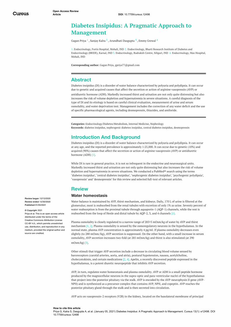
Central DI (CDI) results from defects in synthesis, transport and/or secretion of AVP due to a wide range ofcauses, including both acquired and congenital, as enlisted in Table 1. Congenital CDI (10%) occurs due tomutations in the AVP-NPII gene1 [4], or wolframin (WFS1) gene (Wolfram syndrome) [3]. Wolfram orDIDMOAD syndrome is characterized by central diabetes Insipidus, diabetes mellitus, optic atrophy andsensorineural deafness.
Causes
Congenital Autosomal dominant – mutations in AVP-NPII gene. Autosomal recessive – Wolfram syndrome, mutations in WFS1 gene,septo-optic dysplasia, Alstrom syndrome, Hartsfield syndrome X-linked recessive
Acquired
Traumatic injury to the hypothalamus or posterior pituitary – intracranial or transsphenoidal surgery, blunt or penetratinghead injury, deceleration injury. Vascular causes – intracranial hemorrhage (cerebral or hypothalamic), hypothalamicinfarction, Sheehan’s syndrome, ACA aneurysm, ligation of ACA, hypoxic encephalopathy. Tumors –craniopharyngioma, pituitary macroadenoma, meningioma, germinoma, brain metastasis. Chronic granulomatousdiseases – Langerhans cell histiocytosis, sarcoidosis, tubercular granulomas, neuro-sarcoidosis Infections – meningitis(bacterial, tubercular, cryptococcal), encephalitis, toxoplasmosis. Autoimmune – lymphocytic neurohypophysitis,xanthogranulomatous hypophysitis. Drugs and toxins – ethanol, snake venom, phenytoin Idiopathic.
TABLE 1: Causes of central diabetes insipidusAVP-NPII - arginine vasopressin-neurophysin II gene; ACA - anterior cerebral artery
Acquired DI results when more than 80% of AVP-secreting neurons have been damaged. The causes includepenetrating or blunt head injuries and pituitary surgery. A traumatic injury can result in the directdestruction of AVP-secreting neurons or lead to ischemia and hypoxia of the hypothalamic-pituitary regionas a result of vascular insult or raised intracranial pressure. The incidence of central DI after pituitarysurgery varies and can be either transient or permanent. Minimally invasive procedures such as endoscopictranssphenoidal surgery (TSS) carry a lower risk of postoperative DI [6]. Another important cause of acquiredCDI is intracranial tumors, infections, and infiltrative disorders. Langerhans cell histiocytosis (LCH) is a raredisorder that should be suspected in cases of CDI with multisystem (skeletal, pulmonary, dermatological,anterior pituitary dysfunction) involvement, but many patients may present with DI as the initialmanifestation. Idiopathic central DI comprises almost one-fourth of cases [7]. AVP cell antibodies have beendemonstrated in one-third of patients with idiopathic CDI [8].
Nephrogenic Diabetes Insipidus
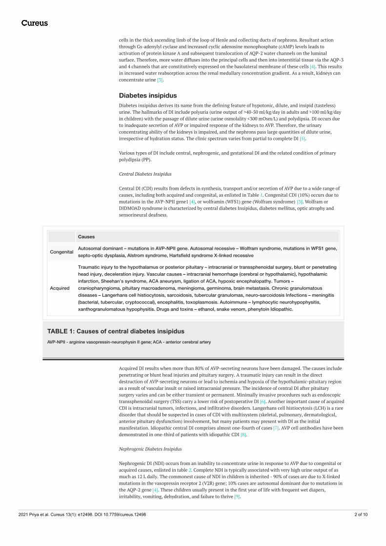
Nephrogenic DI (NDI) occurs from an inability to concentrate urine in response to AVP due to congenital oracquired causes, enlisted in table 2. Complete NDI is typically associated with very high urine output of asmuch as 12 L daily. The commonest cause of NDI in children is inherited - 90% of cases are due to X-linkedmutations in the vasopressin receptor 2 (V2R) gene; 10% cases are autosomal dominant due to mutations inthe AQP-2 gene [4]. These children usually present in the first year of life with frequent wet diapers,irritability, vomiting, dehydration, and failure to thrive [9].
2021 Priya et al. Cureus 13(1): e12498. DOI 10.7759/cureus.12498 2 of 10
Causes
Congenital Mutations in V2R – X-linked recessive. Mutations in AQP-2 – autosomal recessive Bartter syndrome polyhydramnios,megaloencephaly, symptomatic epilepsy (PSME) syndrome.
Acquired
Drugs – lithium, cisplatin, vinblastine, methoxyflurane, demeclocycline, amphotericin B, aminoglycosides, colchicine.Infiltrating disorders – sarcoidosis, amyloidosis, Sjogren’s syndrome. Metabolic disorders – hypercalcemia,hypokalemia. Hematological – multiple myeloma, sickle cell anemia. Renal disorders – acute or chronic kidney disease,obstructive uropathy, polycystic kidney disease, renal infarction.
TABLE 2: Causes of nephrogenic diabetes insipidusV2R - vasopressin receptor 2; AQP - aquaporin
In adults, acquired causes of nephrogenic DI are more common and include electrolyte abnormalities(hypokalemia and hypercalcemia), renal, hematological and infiltrative diseases and medications. Lithiumcan enter the principal cells of the collecting duct via epithelial sodium channels (EnaC) and inhibits thetranslocation of AQP-2 channels to the cell surface. Further long-term exposure may lead to downregulationof AQP-2 gene expression. In acquired nephrogenic DI, polyuria is of moderate severity (3-4 L/day).
Gestational Diabetes Insipidus
Gestational DI is rare (2-4 per 100,000 pregnancies), occurs in the third trimester, and spontaneouslyresolves 4-6 weeks after delivery [10]. The cause is increased degradation of AVP by vasopressinase,expressed by the placental trophoblasts [11]. There may be a mild underlying deficiency of AVP in thesewomen that is unmasked during pregnancy. In addition, increased prostaglandin production may also bluntthe action of AVP [10]. The risk of gestational DI is higher in women with liver disease, possibly becausevasopressinase is metabolized by the liver. However, gestational DI may remain undiagnosed as polyuria isconsidered normal in pregnancy.
Primary Polydipsia
Excessive water intake over a prolonged period of time can also result in symptoms similar to DI and isclassified as primary polydipsia (PP). This may occur as a result of an abnormal thirst mechanism orincreased thirst (dipsogenic DI). Dipsogenic DI may occur due to chronic meningitis, chronic granulomatousdiseases such as sarcoidosis, multiple sclerosis, tubercular meningitis, hypothalamic tumors, or injury.
Some patients may have underlying psychiatric illnesses such as compulsive disorders or schizophrenia andare classified as psychogenic polydipsia. Dry mouth may also result from certain drugs such asphenothiazines or anticholinergic agents. Due to excess fluid intake, serum osmolality is reduced and AVPsecretion is suppressed in PP.
Clinical PresentationThe most common presenting symptom of DI is significant polyuria, defined as urine output of >40-50ml/kg/day (at least 2.5-3 L/day, but may exceed 8-16 L) and polydipsia, defined as water intake of > 100ml/kg/day [5, 12]. Polyuria, however, should be differentiated from frequent micturition, urgency or dysuria,and other causes of polyuria as enlisted in Table 3.
2021 Priya et al. Cureus 13(1): e12498. DOI 10.7759/cureus.12498 3 of 10
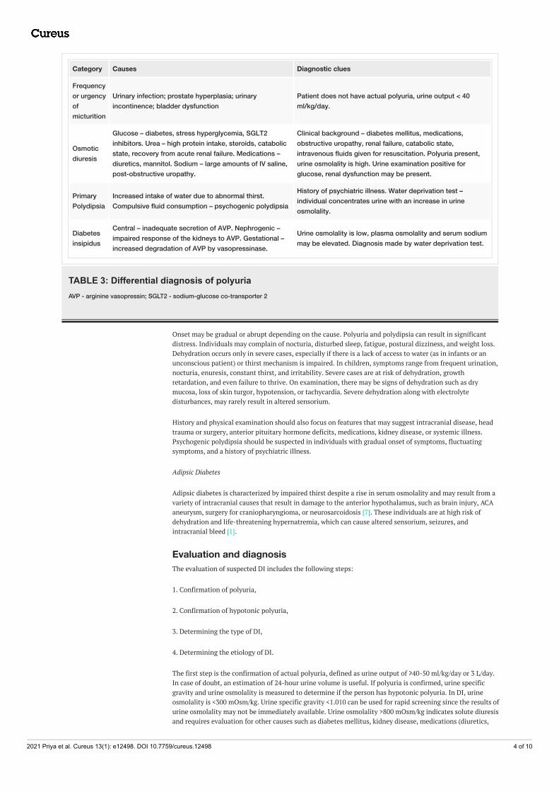
Category Causes Diagnostic clues
Frequencyor urgencyofmicturition
Urinary infection; prostate hyperplasia; urinaryincontinence; bladder dysfunction
Patient does not have actual polyuria, urine output < 40ml/kg/day.
Osmoticdiuresis
Glucose – diabetes, stress hyperglycemia, SGLT2inhibitors. Urea – high protein intake, steroids, catabolicstate, recovery from acute renal failure. Medications –diuretics, mannitol. Sodium – large amounts of IV saline,post-obstructive uropathy.
Clinical background – diabetes mellitus, medications,obstructive uropathy, renal failure, catabolic state,intravenous fluids given for resuscitation. Polyuria present,urine osmolality is high. Urine examination positive forglucose, renal dysfunction may be present.
PrimaryPolydipsia
Increased intake of water due to abnormal thirst.Compulsive fluid consumption – psychogenic polydipsia
History of psychiatric illness. Water deprivation test –individual concentrates urine with an increase in urineosmolality.
Diabetesinsipidus
Central – inadequate secretion of AVP. Nephrogenic –impaired response of the kidneys to AVP. Gestational –increased degradation of AVP by vasopressinase.
Urine osmolality is low, plasma osmolality and serum sodiummay be elevated. Diagnosis made by water deprivation test.
TABLE 3: Differential diagnosis of polyuriaAVP - arginine vasopressin; SGLT2 - sodium-glucose co-transporter 2
Onset may be gradual or abrupt depending on the cause. Polyuria and polydipsia can result in significantdistress. Individuals may complain of nocturia, disturbed sleep, fatigue, postural dizziness, and weight loss.Dehydration occurs only in severe cases, especially if there is a lack of access to water (as in infants or anunconscious patient) or thirst mechanism is impaired. In children, symptoms range from frequent urination,nocturia, enuresis, constant thirst, and irritability. Severe cases are at risk of dehydration, growthretardation, and even failure to thrive. On examination, there may be signs of dehydration such as drymucosa, loss of skin turgor, hypotension, or tachycardia. Severe dehydration along with electrolytedisturbances, may rarely result in altered sensorium.
History and physical examination should also focus on features that may suggest intracranial disease, headtrauma or surgery, anterior pituitary hormone deficits, medications, kidney disease, or systemic illness.Psychogenic polydipsia should be suspected in individuals with gradual onset of symptoms, fluctuatingsymptoms, and a history of psychiatric illness.
Adipsic Diabetes
Adipsic diabetes is characterized by impaired thirst despite a rise in serum osmolality and may result from avariety of intracranial causes that result in damage to the anterior hypothalamus, such as brain injury, ACAaneurysm, surgery for craniopharyngioma, or neurosarcoidosis [7]. These individuals are at high risk ofdehydration and life-threatening hypernatremia, which can cause altered sensorium, seizures, andintracranial bleed [1].
Evaluation and diagnosisThe evaluation of suspected DI includes the following steps:
1. Confirmation of polyuria,
2. Confirmation of hypotonic polyuria,
3. Determining the type of DI,
4. Determining the etiology of DI.
The first step is the confirmation of actual polyuria, defined as urine output of ≥40-50 ml/kg/day or 3 L/day.In case of doubt, an estimation of 24-hour urine volume is useful. If polyuria is confirmed, urine specificgravity and urine osmolality is measured to determine if the person has hypotonic polyuria. In DI, urineosmolality is <300 mOsm/kg. Urine specific gravity <1.010 can be used for rapid screening since the results ofurine osmolality may not be immediately available. Urine osmolality >800 mOsm/kg indicates solute diuresisand requires evaluation for other causes such as diabetes mellitus, kidney disease, medications (diuretics,
2021 Priya et al. Cureus 13(1): e12498. DOI 10.7759/cureus.12498 4 of 10
mannitol). Table 3 provides the differential diagnosis of polyuria.
Serum electrolytes and osmolality, calcium and renal functions are also useful. Serum osmolality iscalculated from serum sodium, blood glucose, and urea [5]:
Ø Plasma osmolality = 2 [Na+] + [blood glucose/18] + [blood urea nitrogen/2.8]
If hypotonic polyuria is documented, a careful evaluation of the type and cause of DI is needed. Thepresence of urine hypoosmolality (<300 mOsm/kg) and plasma hyperosmolality (>300 mOsm/kg) withpolyuria clinches the diagnosis of DI. Hypernatremia (Na >145 mEq/L) and high serum osmolality (>300mOsm/kg) suggest DI, while both remain normal or low in PP [2]. However, many individuals haveindeterminate urine osmolality (300-800 mOsm/kg) and normal serum osmolality and electrolytes, requiringfurther confirmatory tests. This would require dynamic tests of the AVP-kidney axis (water deprivation testor infusion of hypertonic saline) and the estimation of plasma ADH concentration [7]. Misdiagnosis andinappropriate treatment carry significant risks, e.g., if desmopressin is used for primary polydipsia, it cancause hyponatremia.
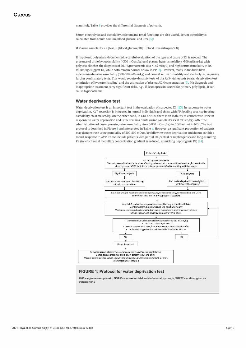
Water deprivation testWater deprivation test is an important test in the evaluation of suspected DI [13]. In response to waterdeprivation, AVP secretion is increased in normal individuals and those with PP, leading to a rise in urineosmolality >800 mOsm/kg. On the other hand, in CDI or NDI, there is an inability to concentrate urine inresponse to water deprivation and urine remains dilute (urine osmolality <300 mOsm/kg). After theadministration of desmopressin, urine osmolality rises (>800 mOsm/kg) in CDI but not in NDI. The testprotocol is described in Figure 1 and interpreted in Table 4. However, a significant proportion of patientsmay demonstrate urine osmolality of 300-800 mOsm/kg following water deprivation and do not exhibit arobust response to AVP. These include patients with partial DI (central or nephrogenic) and long-standingPP (in which renal medullary concentration gradient is reduced, mimicking nephrogenic DI) [14].
FIGURE 1: Protocol for water deprivation testAVP - arginine vasopressin; NSAIDs - non-steroidal anti-inflammatory drugs; SGLT2 - sodium glucosetransporter 2
2021 Priya et al. Cureus 13(1): e12498. DOI 10.7759/cureus.12498 5 of 10
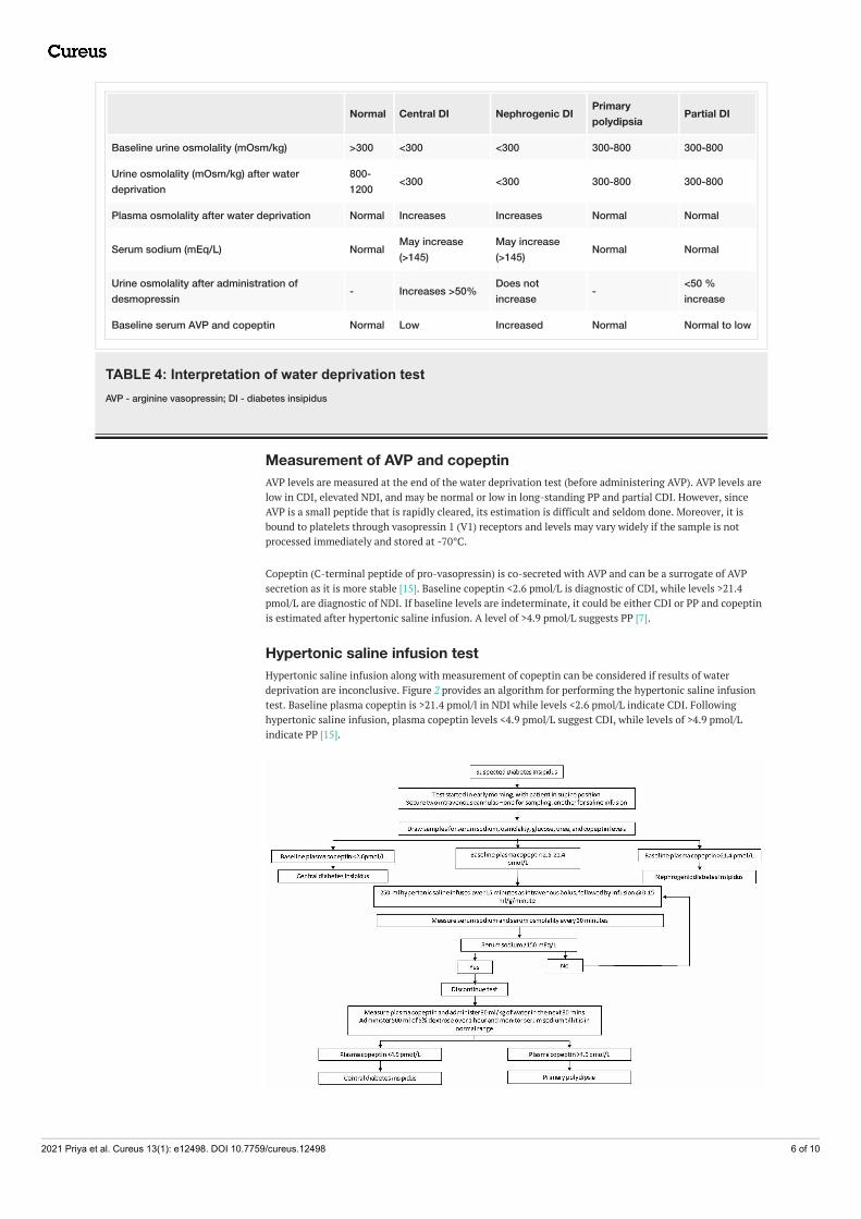
Normal Central DI Nephrogenic DI Primarypolydipsia Partial DI
Baseline urine osmolality (mOsm/kg) >300 <300 <300 300-800 300-800
Urine osmolality (mOsm/kg) after waterdeprivation
800-1200 <300 <300 300-800 300-800
Plasma osmolality after water deprivation Normal Increases Increases Normal Normal
Serum sodium (mEq/L) Normal May increase(>145)
May increase(>145) Normal Normal
Urine osmolality after administration ofdesmopressin - Increases >50% Does not
increase - <50 %increase
Baseline serum AVP and copeptin Normal Low Increased Normal Normal to low
TABLE 4: Interpretation of water deprivation testAVP - arginine vasopressin; DI - diabetes insipidus
Measurement of AVP and copeptinAVP levels are measured at the end of the water deprivation test (before administering AVP). AVP levels arelow in CDI, elevated NDI, and may be normal or low in long-standing PP and partial CDI. However, sinceAVP is a small peptide that is rapidly cleared, its estimation is difficult and seldom done. Moreover, it isbound to platelets through vasopressin 1 (V1) receptors and levels may vary widely if the sample is notprocessed immediately and stored at -70°C.
Copeptin (C-terminal peptide of pro-vasopressin) is co-secreted with AVP and can be a surrogate of AVPsecretion as it is more stable [15]. Baseline copeptin <2.6 pmol/L is diagnostic of CDI, while levels >21.4pmol/L are diagnostic of NDI. If baseline levels are indeterminate, it could be either CDI or PP and copeptinis estimated after hypertonic saline infusion. A level of >4.9 pmol/L suggests PP [7].
Hypertonic saline infusion testHypertonic saline infusion along with measurement of copeptin can be considered if results of waterdeprivation are inconclusive. Figure 2 provides an algorithm for performing the hypertonic saline infusiontest. Baseline plasma copeptin is >21.4 pmol/l in NDI while levels <2.6 pmol/L indicate CDI. Followinghypertonic saline infusion, plasma copeptin levels <4.9 pmol/L suggest CDI, while levels of >4.9 pmol/Lindicate PP [15].
2021 Priya et al. Cureus 13(1): e12498. DOI 10.7759/cureus.12498 6 of 10
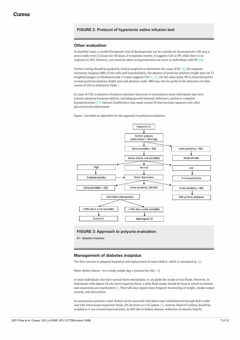
FIGURE 2: Protocol of hypertonic saline infusion test
Other evaluationIn doubtful cases, a careful therapeutic trial of desmopressin can be considered. Desmopressin 100 mcg isgiven orally every 12 hours for 48 hours; if symptoms resolve, it suggests CDI or PP; while there is noresponse in NDI. However, care must be taken as hyponatremia can occur in individuals with PP [16].
Further testing should be guided by clinical suspicion to determine the cause of DI [1]. On magneticresonance imaging (MRI) of the sella and hypothalamus, the absence of posterior pituitary bright spot (on T1weighted images) or thickened stalk (>3 mm) suggests CDI [1, 17]. On the other hand, PP is characterized bynormal posterior pituitary bright spot and pituitary stalk. MRI may also be useful in the detection of othercauses of CDI as enlisted in Table 1.
In cases of CDI, evaluation of anterior pituitary functions is warranted as many individuals may haveanterior pituitary hormone deficits, including growth hormone deficiency, partial or completehypopituitarism [17]. Adrenal insufficiency may mask central DI that becomes apparent only afterglucocorticoid replacement.
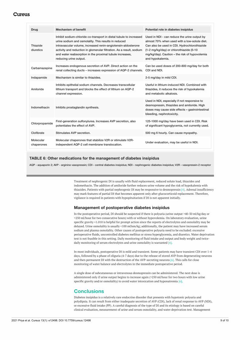
Figure 3 provides an algorithm for the approach to polyuria evaluation.
FIGURE 3: Approach to polyuria evaluationDI - diabetes insipidus
Management of diabetes insipidusThe first concern is adequate hydration and replacement of water deficit, which is calculated as [6]:
Water deficit (liters) = 0.6 x body weight (kg) x [(serum Na/140) - 1]
In most individuals who have normal thirst mechanism, it can guide the intake of oral fluids. However, inindividuals with adipsic DI who have impaired thirst, a daily fluid intake should be fixed at which euvolemiaand eunatremia are maintained [1]. They will also require more frequent monitoring of weight, intake/outputrecords, and electrolytes.
In unconscious patients, water deficit can be corrected with plain water administered through Ryle’s tubeand with intravenous hypotonic fluids (5% dextrose or 0.45 saline) [3]. Isotonic fluid (0.9 saline) should beavoided as it can worsen hypernatremia. In NDI due to kidney disease, reduction of osmotic load by
2021 Priya et al. Cureus 13(1): e12498. DOI 10.7759/cureus.12498 7 of 10

restricting the intake of sodium and proteins is important since increased solute excretion contributes tofurther increased urine output [9]. Causative agents, such as lithium should be discontinued.
In mild DI, fluid replacement is adequate, but more severe cases require pharmacological treatment [3].Pharmacological management depends on the underlying cause. Most cases of CDI can be effectivelymanaged with vasopressin or its analogue, desmopressin. Treatment of the underlying cause of CDI and NDIis also important. As follows, we describe medications used in the management of DI.
Desmopressin
Posterior pituitary extracts that contained vasopressin and oxytocin were used for the management of DI inthe early 1900s. Pitressin (vasopressin tannate in oil) later became widely available but had significantvasopressor effects. Desmopressin or 1-deamino-8-D-AVP (DDAVP), a synthetic analogue of vasopressin,became available in the 1970s [12]. Desmopressin is resistant to degradation by vasopressinase and has agreater anti-diuretic effect with 2000-fold lower vasopressor activity [1]. So, it is associated with less risk ofside effects such as vasoconstriction or hyponatremia.
Several formulations of desmopressin are available as enlisted in Table 5. With oral or nasal formulations,the maximum drug concentration is achieved within one hour; a decrease in urine output occurs in 1-2
hours and the effect lasts 6-18 hours [12]. Individual variations in clinical response exist. Therefore, the dosehas to be adjusted at a weekly interval till a stable dose is attained over one month.
Formulation Dose Advantages Disadvantages
Intranasalspray
10-40 mcg (1puff is 10 mcg) More rapid onset and longer duration of action.
Need storage in cold chain for stability.Absorption may be erratic and impaired if thereis nasal congestion, nasal discharge or chronicrhinitis.
Oral tablet100-400 mcg(tablets of 100mcg)
Easy to administer. Absorbed unaltered from thegastrointestinal tract. As effective as nasalsprays. Less risk of hyponatremia than nasalsprays.
Oraldisintegratingtablet
Same as oraltablet
Dissolves instantly in the mouth – 60% greaterbioavailability. Better quality of life. Not easily available. Higher cost.
Injectable
4 mcg(maximum 0.4mcg/kg) 4mcg/mlinjections
Intravenous, intramuscular or subcutaneous use.Used in the water deprivation test and if nasal ororal administration is not possible.
Robust effect – more risk of water intoxicationor hyponatremia.
TABLE 5: Various formulations of desmopressin
Safety concerns: The most common adverse effects are hyponatremia and water intoxication, though the riskremains low [3]. No effect on blood pressure, heart rate, or body weight has been demonstrated anddesmopressin appears to have no major safety concerns. Desmopressin also appears safe during pregnancyfor gestational DI.
Other Drugs
Several other medications, including thiazide diuretics, carbamazepine, indomethacin, amiloride, etc., areavailable for the management of DI, especially NDI. The mechanism of benefit and the potential role ofthese agents is discussed in Table 6.
2021 Priya et al. Cureus 13(1): e12498. DOI 10.7759/cureus.12498 8 of 10
Drug Mechanism of benefit Potential role in diabetes insipidus
Thiazidediuretics
Inhibit sodium-chloride co-transport in distal tubule to increasedurine sodium and osmolality. This results in reducedintravascular volume, increased renin-angiotensin-aldosteroneactivity and reduction in glomerular filtration. As a result, sodiumand water reabsorption in the proximal tubule increases,reducing urine output.
Used in NDI - can reduce the urine output byalmost 70% when used with a low-solute diet.Can also be used in CDI. Hydrochlorothiazide(1-2 mg/kg/day) or chlorothiazide (5-10mg/kg/day). Caution – the risk of hypovolemiaand hypokalemia.
Carbamazepine Increases endogenous secretion of AVP. Direct action on therenal collecting ducts – increases expression of AQP-2 channels.
Can be used doses of 200-800 mg/day for bothCDI and NDI.
Indapamide Mechanism is similar to thiazides. 2-5 mg/day in mild CDI.
AmilorideInhibits epithelial sodium channels. Decreases transcellularlithium transport and blocks the effect of lithium on AQP-2channel expression.
Useful in lithium-induced NDI. Combined withthiazides, it reduces the risk of hypokalemiaand metabolic alkalosis.
Indomethacin Inhibits prostaglandin synthesis.
Used in NDI, especially if not responsive todesmopressin, thiazides and amiloride. Highdoses may cause side effects – gastrointestinalbleeding, nephrotoxicity.
Chlorpropamide First-generation sulfonylurea. Increases AVP secretion, alsopotentiates the effect of AVP.
125-1000 mg/day have been used in CDI. Riskof significant hypoglycemia, not currently used.
Clofibrate Stimulates AVP secretion. 500 mg 6 hourly. Can cause myopathy.
Molecularchaperones
Molecular chaperones that stabilize V2R or stimulate V2R-independent AQP-2 cell membrane translocation. Under evaluation, may be useful in NDI.
TABLE 6: Other medications for the management of diabetes insipidusAQP - aquaporin 2; AVP - arginine vasopressin; CDI - central diabetes insipidus; NDI - nephrogenic diabetes insipidus; V2R - vasopressin-2 receptor
Treatment of nephrogenic DI is usually with fluid replacement, reduced solute load, thiazides andindomethacin. The addition of amiloride further reduces urine volume and the risk of hypokalemia withthiazides. Patients with partial nephrogenic DI may be responsive to desmopressin [4]. Adrenal insufficiencymay mark features of partial DI that becomes apparent only after glucocorticoid replacement. Therefore,vigilance is required in patients with hypopituitarism if DI is not apparent initially.
Management of postoperative diabetes insipidusIn the postoperative period, DI should be suspected if there is polyuria (urine output >40-50 ml/kg/day or>250 ml/hour for two consecutive hours) with or without hypovolemia. On laboratory evaluation, urinespecific gravity <1.010 is helpful for prompt action since the reports of electrolytes and osmolality may bedelayed. Urine osmolality is usually <100 mOsm/kg; additionally, the patient may have increased serumsodium and plasma osmolality. Other causes of postoperative polyuria need to be excluded: excessiveperioperative fluids, uncontrolled diabetes mellitus or stress hyperglycemia, and diuretics. Water deprivationtest is not feasible in this setting. Daily monitoring of fluid intake and output and body weight and twice-daily monitoring of serum electrolytes and urine osmolality is warranted [6].
In most individuals, postoperative DI is mild and transient. Some patients may have transient CDI over 1-4days, followed by a phase of oliguria (4-7 days) due to the release of stored AVP from degenerating neuronsand then permanent DI with the destruction of the AVP-secreting neurons [6]. This calls for closemonitoring of water balance and electrolytes in the immediate postoperative period.
A single dose of subcutaneous or intravenous desmopressin can be administered. The next dose isadministered only if urine output begins to increase again (>250 ml/hour for two hours with low urinespecific gravity and/or osmolality) to avoid water intoxication and hyponatremia [6].
ConclusionsDiabetes insipidus is a relatively rare endocrine disorder that presents with hypotonic polyuria andpolydipsia. It can result from either inadequate secretion of AVP (CDI), lack of renal response to AVP (NDI),or excessive fluid intake (PP). A careful diagnosis of the type of DI and its etiology is based on carefulclinical evaluation, measurement of urine and serum osmolality, and water deprivation test. Management
2021 Priya et al. Cureus 13(1): e12498. DOI 10.7759/cureus.12498 9 of 10
includes the correction of any water deficit and the use of specific pharmacological agents includingdesmopressin, thiazides and amiloride.
Additional InformationDisclosuresConflicts of interest: In compliance with the ICMJE uniform disclosure form, all authors declare thefollowing: Payment/services info: All authors have declared that no financial support was received fromany organization for the submitted work. Financial relationships: All authors have declared that they haveno financial relationships at present or within the previous three years with any organizations that mighthave an interest in the submitted work. Other relationships: All authors have declared that there are noother relationships or activities that could appear to have influenced the submitted work.
References1. Di Iorgi N, Napoli F, Allegri AEM, et al.: Diabetes insipidus - diagnosis and management . Horm Res Paediatr.
2012, 77:69-84. 10.1159/0003363332. Muhsin SA, Mount DB: Diagnosis and treatment of hypernatremia. Best Pract Res Clin Endocrinol Metab.
2016, 30:189-203. 10.1016/j.beem.2016.02.0143. Dabrowski E, Kadakia R, Zimmerman D, et al.: Diabetes insipidus in infants and children . Best Pract Res Clin
Endocrinol Metab. 2016, 30:317-328. 10.1016/j.beem.2016.02.0064. Schernthaner-Reiter MH, Stratakis CA, Luger A: Genetics of diabetes insipidus . Endocrinol Metab Clin N
Am. 2017, 46:305-334. 10.1016/j.ecl.2017.01.0025. Sarma KV: Algorithmic approach for the diagnosis of polyuria . Medicine Update. Muruganathan A (ed):
Jaypee Brothers Medical Publishers Ltd, New Delhi; 2013. 23:311-313.6. Lamas C, del Pozo C, Villabona C: Clinical guidelines for the management of diabetes insipidus and
syndrome of inappropriate antidiuretic hormone secretion after pituitary surgery. Endocrinol Nutr. 2014,61:15-24. 10.1016/j.endoen.2014.03.010
7. Garrahy A, Moran C, Thompson CJ: Diagnosis and management of central diabetes insipidus in adults . ClinEndocrinol. 2019, 90:23-30.
8. Gut P, Czarnywojtek A, Ziemnicka K, et al.: Incidence of pituitary autoantibodies in idiopathic diabetesinsipidus. Cent Eur J Immunol. 2018, 43:428-433. 10.5114/ceji.2018.81346
9. Kavanagh C, Uy NS: Nephrogenic diabetes insipidus . Pediatr Clin N Am. 2019, 66:227-234.10.1016/j.pcl.2018.09.006
10. Aleksandrov N, Audibert F, Bedard MJ, et al.: Gestational diabetes insipidus: a review of an underdiagnosedcondition. J Obstet Gynaecol Can. 2010, 32:225-231. 10.1016/S1701-2163(16)34448-6
11. Marques P, Gunawardana K, Grossman A: Transient diabetes insipidus in pregnancy . Endocrinol DiabetesMetab Case Rep. 2015, 2015:1-4. 10.1530/EDM-15-0078
12. Kalra S, Zargar AH, Jain SM, et al.: Diabetes insipidus: the other diabetes . Indian J Endocr Metab. 2016, 20:9-21. 10.4103/2230-8210.172273
13. Pedrosa W, Drummond JB, Soares BS, Ribeiro-Oliveira A: A combined outpatient and inpatient overnightwater deprivation test is effective and safe in diagnosing patients with polyuria-polydipsia syndrome.Endocrine Practice. 2018, 24:963-972.
14. Trimpou P, Olsson D S, Ehn O, Ragnarsson O: Diagnostic value of the water deprivation test in the polyuria-polydipsia syndrome. Hormones. 2017, 16:414-422. 10.14310/horm.2002.1762
15. Christ-Crain M, Fenske W: Copeptin in the diagnosis of vasopressin-dependent disorders of fluidhomeostasis. Nat Rev Endocrinol. 2016, 12:168-176. 10.1038/nrendo.2015.224
16. Odeh M, Oliven A: Coma and seizures due to severe hyponatremia and water intoxication in an adult withintranasal desmopressin therapy for nocturnal enuresis. J Clin Pharmacol. 2001, 41:582-584.10.1177/00912700122010320
17. Liu W, Hou J, Liu X, Wang L, Li G: Causes and follow-up of central diabetes insipidus in children . Int JEndocrinol. 2019, 2019:5303765. 10.1155/2019/5303765
2021 Priya et al. Cureus 13(1): e12498. DOI 10.7759/cureus.12498 10 of 10