development/plasticity/repair ... · pdf file800 1200 0 2 4 6 ** ** ** ** a c 2 c 3 c 4 c 1...
TRANSCRIPT
Development/Plasticity/Repair
Functional Excitatory Microcircuits in Neonatal CortexConnect Thalamus and Layer 4
Cuiping Zhao,1,2 Joseph P. Y. Kao,3,4 and Patrick O. Kanold1,2
1Department of Biology, Institute for Systems Research, and 2Program in Neuroscience and Cognitive Science, University of Maryland, College Park,Maryland 20742, and 3Medical Biotechnology Center, University of Maryland Biotechnology Institute, and 4Department of Physiology, University ofMaryland School of Medicine, Baltimore, Maryland 21201
The functional connectivity of the cerebral cortex is shaped by experience during development, especially during a critical period early inlife. In the prenatal and neonatal cortex, transient neuronal circuits are formed by a population of subplate neurons (SPNs). However,SPNs are absent in the adult cortex. While SPNs are crucial for normal development of the cerebral cortex and of thalamocorticalsynapses, little is known about how they are integrated in the developing thalamocortical circuit. We therefore investigated SPNs in vitroin thalamocortical slices of A1 and medial geniculate nucleus (MGN) in mouse from postnatal day 1 (P1) to P13. We found that SPNs canfire action potentials at P1 and that their intrinsic membrane properties are mature after P5. We find that SPNs receive functionalexcitatory inputs from the MGN as early as P2. The MGN projections to SPNs strengthen between P2 and P13 and are capable of inducingaction potentials in SPNs. Selective activation of SPNs by photostimulation produced EPSCs in layer 4 neurons, demonstrating a func-tional excitatory connection. Thus, SPNs are tightly integrated into the developing thalamocortical circuit and would be a reliable relay ofearly spontaneous and sound-evoked activity. The role of SPNs in development likely results from their strong excitatory projection tolayer 4, which might function to regulate activity-dependent processes that enable mechanisms required for the functional maturationand plasticity of the developing cortex and thereby contribute to the development of normal cortical organization.
IntroductionSubplate neurons (SPNs) are among the earliest-appearing neuronsin the cerebral cortex that are largely lost in the adult (Allendoerferand Shatz, 1994; Kanold, 2004, 2009). Subplate neurons are lo-cated in the developing white matter of all cortical regions, in-cluding auditory cortex (ACX). In rodents, SPNs exist during thefirst 3 weeks of life, but some (15–39%) remain into adulthood(Vandevelde et al., 1996; Price et al., 1997; Reep, 2000; Clancy etal., 2001; Torres-Reveron and Friedlander, 2007). Subplate deathoccurs earlier at caudal than rostral areas (Price et al., 1997), thusmore SPNs might be present in ACX than in visual cortex at agiven age. Removing SPNs in visual cortex, however, prevents thesegregation and functional maturation of thalamocortical andintracortical projections, suggesting a key role for SPNs in corti-cal development (Ghosh and Shatz, 1992; Kanold et al., 2003;Kanold and Shatz, 2006). SPNs receive input from lower braincenters, such as the thalamus, before their axons grow to theirtargets in the cortex (Friauf et al., 1990; Allendoerfer and Shatz,1994; Hanganu et al., 2002; Higashi et al., 2002; Molnar et al.,2003; Kanold, 2004). Glutamatergic SPNs project into the over-lying developing cortical plate and are thought to excite cortical
neurons (Friauf et al., 1990; Finney et al., 1998; Pinon et al.,2009). Such an excitatory function of SPNs on layer 4 neuronscombined with reliable transmission of thalamic activity throughSPNs could control activity-dependent processes in layer 4 lead-ing to mature cortical circuitry (Kanold et al., 2003; Kanold andShatz, 2006; Kanold, 2009). However, direct evidence of such anexcitatory connection has been lacking.
While SPNs are present before and during the onset of func-tional hearing [�postnatal day (P)8 –P9] (Alford and Ruben,1963; Mikaelian and Ruben, 1965; Mikaelian et al., 1965; Shnersonet al., 1983; Friauf et al., 1990; Romand and Ehret, 1990; Catalanoet al., 1991; Friauf and Shatz, 1991; Ehret and Romand, 1992;Molnar and Blakemore, 1995), little is known about the func-tional properties of SPNs in ACX. Given the potential key role ofSPNs mediating thalamic excitation to the cortical plate wetherefore investigated SPNs and their associated circuits byin vitro recordings in thalamocortical brain slices from neona-tal mouse ACX.
We found that SPNs fire action potentials as early as P1. SPNsreceive thalamic (MGN) inputs as early as tested (P2) and overpostnatal development the number of MGN inputs to SPNs in-creases. MGN inputs can elicit action potentials in SPNs, aided bythe high input resistance and depolarized resting potential ofSPNs at young ages. In addition, we find that selective SPN stim-ulation results in EPSCs in layer 4 neurons, thus SPNs provideexcitatory input to layer 4. Together, we demonstrate that SPNsare integrated into the developing thalamocortical microcircuit,receive functionally strong thalamic input, and provide a strongexcitatory feedforward excitation to developing layer 4, the target
Received Sept. 8, 2009; revised Oct. 22, 2009; accepted Oct. 31, 2009.P.O.K. is supported by National Institutes of Health (NIH) Grant R01DC009607, and by the International Cerebral
Palsy Research foundation; J.P.Y.K. is supported by NIH Grant R01 GM056481. We thank Dr. Karel Svoboda (HowardHughes Medical Institute) for assistance in implementing LSPS and Julie Zemskova and Aminah Sheikh for histolog-ical assistance.
Correspondence should be addressed to Patrick O. Kanold at the above address. E-mail: [email protected]:10.1523/JNEUROSCI.4471-09.2009
Copyright © 2009 Society for Neuroscience 0270-6474/09/2915479-10$15.00/0
The Journal of Neuroscience, December 9, 2009 • 29(49):15479 –15488 • 15479
layer of thalamic axons. This feedforward excitatory circuit mightinfluence maturational processes in layer 4, leading to maturethalamocortical and intracortical connectivity.
Materials and MethodsSlice physiology and imaging methods are as published previously(Kanold et al., 2003; Kanold and Shatz, 2006). All procedures followedthe University of Maryland College Park animal use regulations.
Slice preparation. Mice (C57BL/6) from Jackson (jax.org) are deeplyanesthetized with isofluorane (Halocarbon). A block of brain containingACX and MGN is removed and thalamocortical slices (500 – 600 �mthick) are cut on a vibrating microtome (Leica) in ice-cold ACSF con-taining (in mM): 130 NaCl, 3 KCl, 1.25 KH2PO4, 20 NaHCO3, 10 glucose,
1.3 MgSO4, 2.5 CaCl2 (pH 7.35–7.4, in 95%O2-5%CO2). The cuttingangle is �15 degrees from the horizontal plane (lateral raised) at olderages (Cruikshank et al., 2002) and somewhat steeper at younger ages tocompensate for the lateral growth of cortex. Slices are incubated for 1 h inACSF at 30°C and then kept at room temperature. For recording, slicesare held in a chamber on a fixed stage microscope (Nikon FN1) andsuperfused (2– 4 ml/min) with ACSF at room temperature. The locationof the recording site in ACX was identified by landmarks (location ofhippocampus) (see Fig. 1A) and validated by monosynaptic responses toelectrical MGN stimulation.
Identification of subplate. The subplate zone is readily identified as arelative cell sparse area between the large layer 6 pyramidal cells and theventricular zone. The subplate encompasses horizontally oriented cells in
P5 stim
CP
SP
F GE H
CA1SP
CP
CA2
CA3DG
LGNMGNv
ACX
MO
hm mV
VrestRmspikes/250ms
50mV
P5
P3
P1-P4
P5-P9
P10-P13
P1-P4
P5-P9
P10-P13
P1-P4
P5-P9
P10-P13
P13
-75
-75
-75
50ms
-70
-60
-50
-40
0
400
800
1200
0
2
4
6**
******
****
A C2
C3
C4
C1
P10
SP
CP
VZSP
CP
VZ
D1 D2
P4
D3 D4
P11
biocytin+cresyl
DIC
P9
CP
SP
V
B
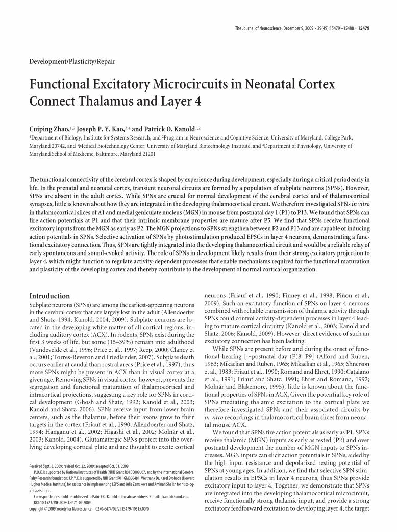
Figure 1. Intrinsic properties of SPNs in auditory cortex. A, Thalamocortical slice under DIC at P10. MGN and ACX are indicated. Scale bar, 500 �m. B, Higher-power picture during a recording froma SPN at P9. Note the clear differentiation of the subplate zone (SP) and cortical plate (CP) under DIC. The subplate appears horizontally organized while layer 6 appears darker. Scale bar, 200 �m.V indicates ventricle. C, P5 thalamocortical slice after recording from multiple cells and subsequent biocytin staining. Stimulation site in MGN is indicated (“stim”). Note the stained cells in thesubplate in ACX (arrowhead). High-power pictures show three of the stained subplate neurons, illustrating diverse morphology. Scale bars: 500 �m (slice), 50 �m (cells). D, Subplate neurons andlayer 6 neurons show different morphologies. Shown are biocytin stained neurons in a 600 �m slice counterstained with cresyl violet. The subplate zone (SP) is readily distinguished from the corticalplate in cresyl stains as being located below the transition between radial oriented cells and horizontally oriented cell bodies (future layer 6B). D1 and D2 show subplate neuron. The cell body islocated in the subplate and axons project onto the cortical plate while dendrites are in subplate and layer 6. VZ indicates the densely stained ventricular zone. Scale bars: 100 �m (D1), 50 �m (D2).D3 and D4 show pyramidal cells in layers 5/6. Note the prominent apical dendrites (arrows) that reach into superficial cortical layers. Scale bars: 100 �m (D3), 50 �m (D4 ). Due to the thickness ofthe section drying is uneven thus shrinkage of layer 2/3 is more pronounced. E, Current injections cause action potentials at all ages studied. Shown are traces evoked with various current injectionsat 3 ages. Cells were held at �75 mV. F, Average numbers of action potentials during 250 ms current injections. Note that the numbers of action potentials increase between P1/4 and P5/9. G, H,Input resistance (Rm) and resting potential (Vrest) determined from linear fits to I–V curve below action potential threshold. Both Rm and Vrest decrease during the postnatal period. **p � 0.05.
15480 • J. Neurosci., December 9, 2009 • 29(49):15479 –15488 Zhao et al. • Subplate Neurons Connect Thalamus and Layer 4
an upper sublamina below layer 6 (future layer 6b) and deeper more scat-tered neurons in the future white matter. The upper sublamina is separatedfrom layer 6 by a cell sparse zone. Compared to other neurons, the deepersubplate neurons in young rodents stain relatively poorly for markers such asNeuN but are apparent under differential interference contrast (DIC) wheresubplate neurons intermingled with horizontally traveling fiber can bereadily seen due to their horizontal appearance (see Figs. 1A,B, 6).
Electrophysiology. Whole-cell recordings are performed with a patch-clamp amplifier (Multiclamp 700B, Molecular Devices) using pipetteswith input resistance of 4 – 8 M�. Data are acquired with a Digidata ADboard (Molecular Devices) under pClamp (v9 and v10) and analyzedoffline using MATLAB (MathWorks). Electrodes are filled with (in mM)110 K-gluconate, 4 KCl, 4 NaCl, 0.2 CaCl2, 10 HEPES, 1.1 EGTA, 2Mg-ATP, 1 MgCl2 and 5 glutathione (pH 7.2, 300 mOsm). Membranevoltages are corrected for an estimated liquid junction potential of 14mV. Some cells were recorded using a solution containing 115 mM ce-sium methanesulfonate (CsMeSO3) 5 NaF, 10 EGTA, 10 HEPES, 15CsCl, 3.5 MgATP, 3 QX-314 (pH 7.25, 300 mOsm). We found no obvi-ous differences in EPSC sizes with these two solutions. For LSPS all cellswere recorded with this solution. Biocytin or neurobiotin (0.5%) isadded to the electrode solution as needed. Electrical stimulation: We usea bipolar electrode (Microprobe, biphasic, 0.2 ms, at 0.03 Hz) coupled to
a stimulus isolator (Cygnus). Photostimula-tion: 0.5–1 mM caged glutamate (N-(6-nitro-7-coumarylmethyl)-L-glutamate; Ncm-Glu)(Kao, 2006) is added to the ACSF. Without UVlight, this compound has no effect on neuronalactivity (N � 40 neurons) (Kao, 2006). UV la-ser light (500 mW, 355 nm, 100 kHz repetitionrate, DPSS, 2–10 ms pulses) is split via a 33%beam splitter (CVI Melles Griot) and coupledvia a fiber optical coupler (Oz Optics) into anoptical fiber (25 �m core diameter, Oz Optics).The distal end of the fiber is held within a bentglass pipette to reduce beam elongation andapply focused light to a small area. We visual-ized the stimulated site for all cells by taking apicture of the light beam to ensure that stimu-lation was limited to the subplate. In prelimi-nary experiments we varied laser power anddetermined that this power provided reliabledirect activation of neurons recorded in whole-cell patch configuration. For laser-scanningphotostimulation (LSPS), the other fraction ofthe split beam is coupled into a microscope viascan mirrors (Cambridge Technology) and adichroic mirror (Shepherd et al., 2003). Thelaser beam in LSPS enters the slice axiallythrough the objective (Olympus 4�, 0.16 NA/air) and has a diameter of �15 �m. Laserpower at the sample is �20 mW. In prelimi-nary experiments we varied laser power anddetermined that this power provided reliableactivation of neurons. We typically imaged16 � 16 or 32 � 32 stimulation sites spaced20 –50 �m enabling us to image areas of1mmx1 mm. Stimuli are applied at 0.5–1 Hz.Data acquisition is performed by National In-struments AD boards and custom software(Ephus) (Shepherd et al., 2003). Ephus (availableonline at http://research.janelia.org/labs/display/ephus/Ephus) is written in MATLAB andadapted to our setup. Analysis was performedsimilar to methods described previously(Shepherd et al., 2003) by using custom soft-ware written in MATLAB. To detect monosyn-aptically evoked EPSCs we detected peak EPSCamplitudes in a �60 ms time window after thestimulation. Traces containing a short-latency(�15–20 ms “direct”) response were discarded
from the analysis (see Fig. 6, black patches) as were traces that containedlonger latency inward currents of long duration (�100 ms). (Fig. 6, graypatches) These currents could be sometimes seen in locations surrounding(�50 �m) areas that gave a direct response. Occasionally, some of the directresponses contained synaptically evoked responses that we did not separateout, leading to an underestimation of local short-range connections. Cellsthat only showed direct responses were excluded from the analysis.
Drugs. We use NBQX (50 �M) to block AMPA currents and TTX (1�M) to block action potentials. All chemicals and drugs were obtainedfrom Sigma.
Statistics. Results are plotted as means � SD, compared with a Stu-dents t test, and deemed significant if p � 0.05.
ResultsWe investigated the physiological characteristics of subplate neu-rons in mouse auditory cortex over three age ranges (P1– 4, P5–P9, and P10 –P13), approximately corresponding to distinctfunctional states of the auditory system. Hair cells do not becomefunctional before P5 and thus only spontaneous evoked activity ispresent in the auditory system. After onset of hair cell function
P2-P4
P5-P9
P10-P13
P2-P4
P5-P9
P10-P13
P2-P4
P5-P9
P10-P13
P2-P4
P5-P9
P10-P13
minimal (pA) maximal (pA)
EPSC amplitude (AMPA)EPSC amplitude (pA)
A
C D
B
ratio (max/min)
stim re. max
0
0
50P8
P8
P13
P13
P4
P4
0
20
200
0 0.2 0.4 0.6 0.8 1
latency (ms)
0
25
threshold
maximum
saturation
**
**400
0 0
50
0
25
200p
A50
pA
50ms
50ms
50ms
20pA
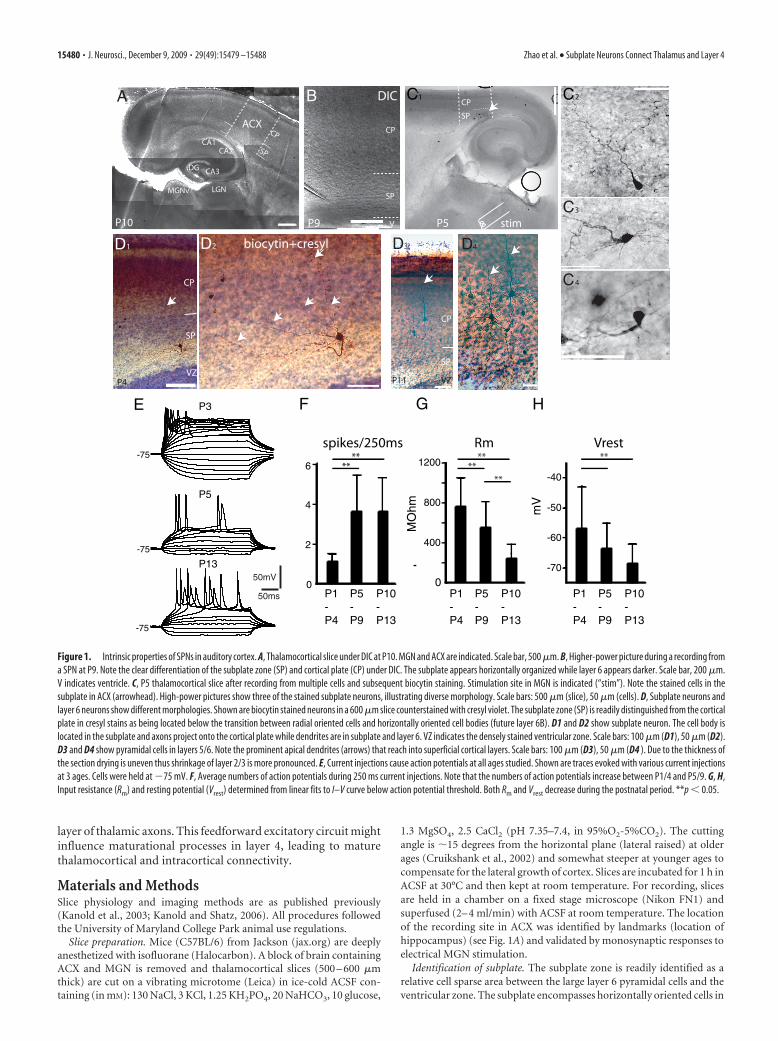
Figure 2. Excitatory MGN inputs to SPNs throughout the postnatal period. A, MGN evoked EPSCs with increasing stimulationcurrents at 3 ages. Traces are shown on the left. Peak EPSC amplitudes as functions of stimulus strength (IO-curve) are shown onright. For comparison, stimulus strength was normalized to the maximum stimulus strength used in a particular cell. Note thelimited dynamic range of EPSC amplitudes at young ages. Dashed lines indicate where saturation was reached. Note the occasionaldisynaptic responses (red traces). B, Graph shows a superposition of the IO curves in A normalized to maximum EPSC and saturationstimulus level. Note the steeper increase in the IO curve from threshold to saturation at younger ages. C, Quantification of EPSCamplitudes. Box plots show EPSC amplitude at threshold (minimal reliably evoked EPSC), maximal evoked EPSC, and the ratio ofthese values. Box plots show median (line), mean (square), and the 25% and 75% quartiles. The threshold EPSC amplitude does notchange (P1/4: 14.6 � 7.2 pA N � 9; P5/9: 19.9 � 8 pA N � 12; P10/13: 17.45 � 11.8 pA N � 8), while maximal EPSC amplitude(P1/4: 42.4 � 18.1 pA; P5/9: 121 � 105 pA; P10/13: 184 � 149 pA) and the ratio (P1/4: 3.1 � 1.2; P5/9: 4.3 � 5.6; P10/13:10.1 � 6.5) increase from P2/4 to P5/9 and P10/13 (*p � 0.05). D, Latencies of the monosynaptic EPSC (time to peak). Box plotsshow median (line), mean (square) and the 25% and 75% quartiles. Peak latencies were similar at all ages (P1/4: 11.6 � 3.2 ms,P5/9: 13.66 � 5.76 ms, P10/13: 14.74 � 4.32 ms, all p � 0.1).
Zhao et al. • Subplate Neurons Connect Thalamus and Layer 4 J. Neurosci., December 9, 2009 • 29(49):15479 –15488 • 15481

but before the opening of the ear canal,spontaneous and high-threshold sound-evoked responses might occur. In mice ofthe C57BL/6 strain the ear canal opens atP9 (Shnerson and Pujol, 1983; Pujol et al.,1997; Ruebsamen and Lippe, 1997). Afterear opening, low-threshold hearing devel-ops, followed by an early critical period(�P11–13 in rat) (de Villers-Sidani et al.,2007).
The intrinsic properties of subplateneurons mature over thepostnatal periodWe recorded from SPNs (N � 140) inthalamocortical slices of A1 over the first 2postnatal weeks using whole-cell patch-clamp recordings (Fig. 1A–C). SPNs arelocated in the future white matter belowlayer 6 and have diverse morphology(Allendoerfer and Shatz, 1994; Luhmannet al., 2000) (Fig. 1B–D). In response tocurrent injections, SPNs at all ages wereable to fire action potentials (APs) (Fig.1E). However, in response to depolariza-tion, the pattern of APs and the number ofspikes evoked varied strongly with age. Atearly ages (P1/4) sustained current injec-tion resulted in only one AP in 11 of 13 SPneurons, while this firing pattern was rareat ages older than P5 (4/37 cells). Figure1F shows that maximal number of evokedaction potentials to a 250 ms depolariza-tion increased from P1/4 to P10/13 (P1/4:1.1 � 0.3, P5/9: 3.6 � 1.8, P10/13: 3.6 �1.6, both p � 0.01). However, action po-tential threshold remained constant overthis age range (�48.4 � 6 mV vs �50.9 � 4 mV, p � 0.1).
One feature of SPNs at P1/4 is a sustained depolarization afterthe AP (Fig. 1E), which might be due to low expression levels ofK-channels. To test the availability of K-channels, we measuredthe membrane resistance (Rm) of SPNs by linear fits to the I–Vcurve below spike threshold and found that Rm decreased be-tween P1/4 and P10/13 (P1/4: 757 � 284 M�, P5/9: 554 � 260M�, P10/13: 285 � 95 M�, both p � 0.05) (Fig. 1G). In addition,in the first 2 postnatal weeks, the resting membrane potentialshowed a steady hyperpolarizing trend (P1/4: �56.9 � 13.9 mV,P5/9: �63.7 � 8.4 mV, P10/13: �68.5 � 6.3 mV; P1/4 to P5/8p � 0.13; P10/13 p � 0.05) (Fig. 1H). These results also indicatethat SPNs at early ages require less depolarization (8.5 mV at P1/4vs 17.6 mV at P10/13) to trigger an action potential—that is,SPNs at early ages can more easily respond to synaptic inputs.
SPNs receive functionally strong thalamic inputsSPNs are thought to receive thalamic inputs, as has been demon-strated directly in the cat visual and rodent somatosensory cortexby CSD analysis and optical recordings (Friauf et al., 1990; Allen-doerfer and Shatz, 1994; Hanganu et al., 2002; Higashi et al., 2002;Molnar et al., 2003). We therefore investigated if SPNs in ACXreceive inputs from the auditory thalamus [medial geniculatenucleus (MGN)] and how these inputs change over the course ofdevelopment. MGN fibers reach the cortical plate before birth(Gurung and Fritzsch, 2004). Electrical stimulation of the MGN
as early as P2 resulted in short-latency EPSCs in SPNs (Fig. 2A),indicating that monosynaptic transmission from thalamus tocortex is already present before the onset of hearing. Occasion-ally, we observed larger, long-latency EPSCs, which might reflectdisynaptic inputs (Fig. 2A, red traces). We focused our analysison the monosynaptically evoked EPSC, which was taken as eitherthe only evoked EPSC, or the first (short-latency) EPSC if multi-ple EPSCs were observed.
Increasing stimulation amplitudes resulted in correspondingincreases in EPSC amplitude (Fig. 2A). In some cells, especially atyoung ages, these increases occurred in a stepwise manner (Fig.2A, top), suggesting the activation of separate synapses, whereasat older ages such steps were less clear (Fig. 2A, bottom, B). Theminimally evoked EPSC amplitude was similar at P1/4 and P10/13, while the maximally evoked EPSCs were larger at older ages(Fig. 2C). The ratio of maximal to minimal EPSC is an estimate ofthe number of thalamic fibers innervating SPNs, and the in-creased ratio (Fig. 2C) suggests that the thalamo-subplate synap-tic connection does not prune but increases its numbers ofconnections during the postnatal period, even though SPNs arestarting to disappear. EPSC latencies (time to peak) were similarat all ages (Fig. 2D).
However, we did not observe any MGN stimulation evokedoutward currents under our recording conditions at any age,which suggests that MGN inputs might not engage inhibitorycircuits synapsing on SPNs.
Figure 3. MGN selectively activates SPNs. A, Picture of a slice (600 �m thick) from a P5 animal in which both layer 6 and SPnswere recorded. Boxes show position of high-magnification pictures. Dashed line indicates subplate/layer 6 border. High-magnification pictures show differences in morphology between SPNs and layer 6 neuron. Scale bars: 100 �m, 50 �m. VZindicates ventricular zone and H hippocampus. B, Traces show response of layer 6 neuron and SPN in A to thalamocorticalstimulation at same stimulation site (equal intensity). Note that the SPN but not the layer 6 neuron showed synaptic inputs.C, Picture of a thalamocortical slice (600 �m thick) from a P13 animal in which both layer 6 and SPns were recorded. Boxes showposition of high-magnification pictures. Dashed line indicates subplate/layer 6 border. V indicates ventricle and H hippocampus.High-magnification pictures show differences in morphology between SPNs and layer 6 neuron. Arrows show apical dendrite oflayer 6 neurons traveling to superficial layers. Scale bars: 1 mm, 50 �m. D, Traces from SPN and layer 6 neuron in C to electricalstimulation of MGN (multiple traces superimposed). Note the reliable short latency EPSC in the SPN while the layer 6 neuronshowed long latency EPSCs on some trials that started when SPNs ceased to receive inputs.
15482 • J. Neurosci., December 9, 2009 • 29(49):15479 –15488 Zhao et al. • Subplate Neurons Connect Thalamus and Layer 4
Since layer 6 neurons are located close to SPNs, we investi-gated if layer 6 neurons receive MGN inputs at these ages. Werecorded from layer 6 neurons (N � 8) (Figs. 1D, 3A,C) in thesame slice in which SPN showed MGN inputs (Fig. 3B,D). Inthese slices and by stimulating at the same stimulation site, layer6 cells did not show short latency inputs (Fig. 3B,D). However,2/8 layer 6 neurons showed long latency inputs (Fig. 3D). Com-parison of the layer 6 responses with SPN responses to stimula-tion at the same site (Fig. 3D) shows the large differences inlatencies. These data might also indicate that SPNs receive MGNinputs and drive cortical circuits that give rise to layer 6 inputs.
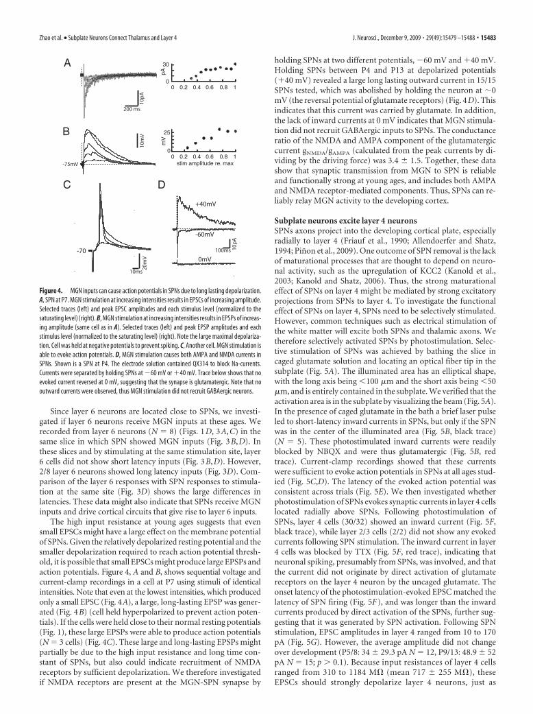
The high input resistance at young ages suggests that evensmall EPSCs might have a large effect on the membrane potentialof SPNs. Given the relatively depolarized resting potential and thesmaller depolarization required to reach action potential thresh-old, it is possible that small EPSCs might produce large EPSPs andaction potentials. Figure 4, A and B, shows sequential voltage andcurrent-clamp recordings in a cell at P7 using stimuli of identicalintensities. Note that even at the lowest intensities, which producedonly a small EPSC (Fig. 4A), a large, long-lasting EPSP was gener-ated (Fig. 4B) (cell held hyperpolarized to prevent action poten-tials). If the cells were held close to their normal resting potentials(Fig. 1), these large EPSPs were able to produce action potentials(N � 3 cells) (Fig. 4C). These large and long-lasting EPSPs mightpartially be due to the high input resistance and long time con-stant of SPNs, but also could indicate recruitment of NMDAreceptors by sufficient depolarization. We therefore investigatedif NMDA receptors are present at the MGN-SPN synapse by
holding SPNs at two different potentials, �60 mV and 40 mV.Holding SPNs between P4 and P13 at depolarized potentials(40 mV) revealed a large long lasting outward current in 15/15SPNs tested, which was abolished by holding the neuron at �0mV (the reversal potential of glutamate receptors) (Fig. 4D). Thisindicates that this current was carried by glutamate. In addition,the lack of inward currents at 0 mV indicates that MGN stimula-tion did not recruit GABAergic inputs to SPNs. The conductanceratio of the NMDA and AMPA component of the glutamatergiccurrent gNMDA/gAMPA (calculated from the peak currents by di-viding by the driving force) was 3.4 � 1.5. Together, these datashow that synaptic transmission from MGN to SPN is reliableand functionally strong at young ages, and includes both AMPAand NMDA receptor-mediated components. Thus, SPNs can re-liably relay MGN activity to the developing cortex.
Subplate neurons excite layer 4 neuronsSPNs axons project into the developing cortical plate, especiallyradially to layer 4 (Friauf et al., 1990; Allendoerfer and Shatz,1994; Pinon et al., 2009). One outcome of SPN removal is the lackof maturational processes that are thought to depend on neuro-nal activity, such as the upregulation of KCC2 (Kanold et al.,2003; Kanold and Shatz, 2006). Thus, the strong maturationaleffect of SPNs on layer 4 might be mediated by strong excitatoryprojections from SPNs to layer 4. To investigate the functionaleffect of SPNs on layer 4, SPNs need to be selectively stimulated.However, common techniques such as electrical stimulation ofthe white matter will excite both SPNs and thalamic axons. Wetherefore selectively activated SPNs by photostimulation. Selec-tive stimulation of SPNs was achieved by bathing the slice incaged glutamate solution and locating an optical fiber tip in thesubplate (Fig. 5A). The illuminated area has an elliptical shape,with the long axis being �100 �m and the short axis being �50�m, and is entirely contained in the subplate. We verified that theactivation area is in the subplate by visualizing the beam (Fig. 5A).In the presence of caged glutamate in the bath a brief laser pulseled to short-latency inward currents in SPNs, but only if the SPNwas in the center of the illuminated area (Fig. 5B, black trace)(N � 5). These photostimulated inward currents were readilyblocked by NBQX and were thus glutamatergic (Fig. 5B, redtrace). Current-clamp recordings showed that these currentswere sufficient to evoke action potentials in SPNs at all ages stud-ied (Fig. 5C,D). The latency of the evoked action potential wasconsistent across trials (Fig. 5E). We then investigated whetherphotostimulation of SPNs evokes synaptic currents in layer 4 cellslocated radially above SPNs. Following photostimulation ofSPNs, layer 4 cells (30/32) showed an inward current (Fig. 5F,black trace), while layer 2/3 cells (2/2) did not show any evokedcurrents following SPN stimulation. The inward current in layer4 cells was blocked by TTX (Fig. 5F, red trace), indicating thatneuronal spiking, presumably from SPNs, was involved, and thatthe current did not originate by direct activation of glutamatereceptors on the layer 4 neuron by the uncaged glutamate. Theonset latency of the photostimulation-evoked EPSC matched thelatency of SPN firing (Fig. 5F), and was longer than the inwardcurrents produced by direct activation of the SPNs, further sug-gesting that it was generated by SPN activation. Following SPNstimulation, EPSC amplitudes in layer 4 ranged from 10 to 170pA (Fig. 5G). However, the average amplitude did not changeover development (P5/8: 34 � 29.3 pA N � 12, P9/13: 48.9 � 52pA N � 15; p � 0.1). Because input resistances of layer 4 cellsranged from 310 to 1184 M� (mean 717 � 255 M�), theseEPSCs should strongly depolarize layer 4 neurons, just as
A
B
C D
10ms
-70
20m
V10
pA10
mV
-75mV
0
30
pAm
V
200 ms
0
25
0.2 0.4 0.6 0.8
0.2 0.4 0.6 0.8stim amplitude re. max
100ms 10pA
+40mV
-60mV
0mV
Figure 4. MGN inputs can cause action potentials in SPNs due to long lasting depolarization.A, SPN at P7. MGN stimulation at increasing intensities results in EPSCs of increasing amplitude.Selected traces (left) and peak EPSC amplitudes and each stimulus level (normalized to thesaturating level) (right). B, MGN stimulation at increasing intensities results in EPSPs of increas-ing amplitude (same cell as in A). Selected traces (left) and peak EPSP amplitudes and eachstimulus level (normalized to the saturating level) (right). Note the large maximal depolariza-tion. Cell was held at negative potentials to prevent spiking. C, Another cell. MGN stimulation isable to evoke action potentials. D, MGN stimulation causes both AMPA and NMDA currents inSPNs. Shown is a SPN at P4. The electrode solution contained QX314 to block Na-currents.Currents were separated by holding SPNs at �60 mV or 40 mV. Trace below shows that noevoked current reversed at 0 mV, suggesting that the synapse is glutamatergic. Note that nooutward currents were observed, thus MGN stimulation did not recruit GABAergic neurons.
Zhao et al. • Subplate Neurons Connect Thalamus and Layer 4 J. Neurosci., December 9, 2009 • 29(49):15479 –15488 • 15483

MGN inputs depolarize SPNs. Indeed,current-clamp recordings (N � 3 cells)revealed large EPSPs in layer 4 followingphotostimulation of SPNs (Fig. 5H ), in-dicating a strong functional impact ofSPNs on layer 4 neurons. Together, thesedata indicate a strong functional excita-tory connection between SPNs and layer 4neurons.
We next investigated the spatial pat-tern of SPN inputs to layer 4 by usinglaser-scanning photostimulation (Shepherdet al., 2003). The laser beam was targetedon a 16 � 16 to 32 � 32 pixel array cover-ing an area of the slice (Fig. 6A). Cell-attached patch recordings from SPNs(N � 3) showed that laser pulses were ableto trigger short latency action potentials inSPNs (Fig. 6B,C). Plotting the spatial lo-cation of stimulation sites that cause SPNspiking showed that photostimulationhad to be performed within �100 �m ofthe SPN soma (Fig. 6B,C). Plotting thelatency to the first spike (Fig. 6C) showsthat activation close to the soma causedspikes with the shortest latency. Thus,only photostimulation of the soma orproximal dendrites is sufficient to activateSPNs.
We next investigated the origin of syn-aptic inputs to layer 4 by whole-cell re-cordings from layer 4 neurons duringLSPS (N � 21). Photostimulation causedlarge amplitude short latency inward cur-rents (labeled direct in Fig. 6D1), whichreflect the direct activation of the cell un-der study (Shepherd et al., 2003; Barbourand Callaway, 2008). We also observedsmaller amplitude long latency inwardcurrents (labeled “evoked” in Fig. 6D1).These currents likely reflect EPSCs evokedby photostimulation of presynaptic cells.Plotting the amplitude of the pEPSC asfunction of stimulation location showedthat direct responses (Fig. 6D2, pinkboxes) were clustered around the cellbody (Fig. 6D2, red circle). Evoked re-sponses were also clustered but in a spa-tially distinct region (Fig. 6D2, yellowboxes). To identify the laminar source ofthe evoked response, we plotted the am-plitude of the responses and overlayed thisgraph with the DIC image of the slice (Fig.6D3). We find that the source of the exci-tatory inputs was located the subplate butnot layer 6 (Fig. 6D3). The source regionfor excitatory inputs in the subplate is relatively small (�200 �200 �m). Comparing the activation region in subplate (Fig.6B,C) with projection regions in layer 4 (Fig. 6D3) suggests thateach layer 4 neuron receives input from 2 to 3 SPNs. The latencyof the pEPSCs (Fig. 6D3, bottom) is similar to those of SPNspiking (Fig. 6C). The results of LSPS of a layer 4 neurons at P13are shown in Figure 6E. In addition to subplate inputs (13/21
cells), layer 4 neurons receive inputs locally from layer 4, fromlayer 2/3, and in some cases projections from layer 6 (9/21 cells)(Fig. 6, compare D, E).
In the adult layer 6, neurons can send some projections intolayer 4, even though this projection might be relatively weak inauditory cortex (Barbour and Callaway, 2008). Since layer 6 neu-rons can send dendrites into the subplate it is possible that our
50ms
hνNBQX
P4 P11
P11
D E
Layer 4 recording, stimulation in subplate
Subplate recording, stimulation in subplate
5pA
50 ms-58
10 mVhν hν
100ms
-65 20 600
75spike latency
% s
pike
s
A
B C
F
F
SP
L4
1
50 ms
HG pEPSC
ms
5pA
TTXhv
-70
10mV
100ms
hv
pA
# ce
lls
recording
?
UV light
cagedglutamate
subplate
layer 4
100 ms
20 pA
hv
20 40 600
6
-40 -20 00
3
pA
amplitude latency
count
ms
0 1800
6
Figure 5. SPNs excite layer 4 neurons. A, Left, Schematic of experimental setup. Recordings are obtained from layer 4 neurons whileSPNs are excited by photostimulation. Right, Actual setup of photostimulation experiment. Image shows part of the thalamocortical slice atP4. Layer 1 is to the right (1) and white matter (WM) is to the left. Recording of layer 4 neuron through whole-cell pipette (P) whilephotostimulation fiber (F) and light spot (white) are located in subplate. Note that the elliptical uncaging spot does not extend intooverlying cortical plate. The shape of the spot depends on the size of the fiber, its angle and distance to the slice. Imaging artifactssurrounding the fiber are due to DIC illumination. Scale bar, 100 �m. B–D, Recordings from SPNs. B, Photostimulation of SPN at P11.Followingalaserpulse(h�),arapidinwardcurrentdevelops(blacktrace)that isblockedbyNBQX(50�M;redtrace). C, D,Photostimulationof SPNs in current clamp at P4 (C) and P11 (D) reliably evokes action potentials. Occasionally 2 action potentials were observed. E, Spikelatency distribution in cell from D. Spike jitter is low: 75% of spikes occurred within 3 ms of each other during photostimulation. Absolutelatency was higher in this cell due to hyperpolarized holding potential. F–H, Recordings in layer 4 cells. F, Left, pEPSCs in layer 4 neuronselicitedbySPNphotostimulationatP11.Center,theaverageof15pEPSCs(blacktrace);thepEPSCswerecompletelyblockedbyTTX(0.5�M;red trace). Note prolonged delay after laser pulse (h�) before onset of inward current. Right, Histograms of pEPSC latency and amplitude.pEPSC latencies match those of SPN spiking (E). Amplitude histogram shows 2 peaks at �20 pA and at �40 pA possibly indicating thesummation of 2 presynaptic inputs. G, Distribution of pEPSC amplitudes in 27 layer 4 neurons. Mean amplitude was 40.6 � 41.2 pA.H, pEPSP in current clamp in P6 layer 4 neuron (spiking was blocked with the Na channel antagonist, QX314, in the pipette).
15484 • J. Neurosci., December 9, 2009 • 29(49):15479 –15488 Zhao et al. • Subplate Neurons Connect Thalamus and Layer 4

stimulation activates layer 6 neurons. To investigate if layer 6neuron activation can contribute to the observed EPSCs in layer 4neurons, we mapped directly the activation of layer 6 neurons(N � 2) and found that layer 6 neurons were readily excited bystimulation in a large area surrounding their soma but also in anarea extending radially toward the pia (Fig. 6F). The latter arealikely coincides with the prominent apical dendrite of layer 6pyramidal cells (Fig. 1D). This large activation area is similar to
those reported in layer 2/3 and layer 5 pyramidal cells (Shepherdet al., 2003). Thus, layer 6 neurons can be activated from a muchlarger area than SPNs. The large activation area of layer 6 neuronsis larger than the size of the source of excitatory connections tolayer 4 neurons. In particular, the region that gives rise to EPSCsin layer 4 (Fig. 6D,E) does not include the large activation regionsof layer 6 cells (Fig. 6F), suggesting that there is a excitatoryconnection between SPNs and layer 4 neurons.
Figure 6. SPN excitation to layer 4 is focal. A, LSPS stimulation grid (32�32 spots, 50 �m spacing) superimposed on infrared image. Red dot indicates cell body of recorded neuron, in this casea layer 4 neuron. B, Cell-attached recording from SPN at P13. Traces show action potentials that are evoked in SPN during LSPS. To the right is a DIC image showing the recording pipette. The layer6/subplate border is identified by large pyramidal cells in layer 6 and horizontal SPNs in upper subplate. The stimulation map is superimposed on the DIC image. Areas that caused action potentialsare labeled black. Note that photostimulation can evoke multiple spikes and that region of activation is very small. C, Cell attached recording from a SPN at P9. Image in C1 shows areas that causean action potential in black. The layer 6/subplate border is identified by large pyramidal cells in layer 6 and horizontal SPNs in upper subplate. Note that this are as confined to subplate. Image in C2shows, for the same cell as in C1, the latency to first spike of cell in C1 encoded in pseudocolor. Areas closer to the cell body show shorter latencies. D, Whole-cell patch recordings of layer neuron (P7)during LSPS. D1, Photostimulation (blue line) evoked short latency and long latency EPSCs labeled direct and evoked. D2, Traces show poststimulation currents (100 ms) at stimulus locations for thiscell. Areas shaded pink gave direct responses and are plotted on a reduced scale (2 nA scale). All other traces are plotted on a 100 pA scale. Areas shaded yellow gave rise to evoked pEPSCs (see D1).Note the large gap between these two areas. pEPSCs originating from superficial locations partially overlap direct response. D3, pEPSC amplitude (top) and latency (bottom) as function of stimulusxy-position and superimposed on DIC image of slice. Values are encoded in pseudocolor. Locations that led to short latency direct responses are marked in black. Red circle indicates location of cellbody. White box indicates stimulated area. E, LSPS of a layer 4 neuron at P13. E1 shows amplitude map. In addition to SPN input, this neuron also received intracortical inputs from layers 2/3 and4. E2, Traces show poststimulation currents during stimulation of areas overlying layer 6 and subplate (white box in E1). Note discontinuity of stimulation sites in layer 6 that gave responses fromthose in subplate that gave responses suggesting that these stimulation sites activate distinct populations of cells. F, Cell attached recording from layer 6 neuron. Left image shows areas that causean action potential. Note that this area is large and includes a wide area in layer 6 and a narrow area in layer 4. Right image shows latency to first spike encoded in pseudocolor. Note the short latencyto first spike for somatic stimulation. All scale bars, 200 �m.
Zhao et al. • Subplate Neurons Connect Thalamus and Layer 4 J. Neurosci., December 9, 2009 • 29(49):15479 –15488 • 15485

DiscussionOur data show that SPNs are a strong excitatory relay of thalamicinformation to the developing cortical layer 4 before and after theonset of hearing. We also show that SPNs provide strong excita-tory input to layer 4.
We report that SPNs receive MGN inputs and that these in-puts are strong enough to trigger action potentials in SPNs. Thisstrong effect of relatively weak synaptic inputs may be mediatedby the high input resistances of SPNs. Thus, in effect, the “imma-ture” intrinsic characteristics of SPNs (high Rm) might compen-sate for the immature synaptic inputs (small amplitudes). Highinput resistances in early development are not unique to SPNs:for example, layer 2/3 cells show a similar progression �10 d later(Oswald and Reyes, 2008). The decrease in input resistance andresting potential of SPNs over development are consistent withthe increased expression of K-channels during the postnatalperiod. Importantly, the functional compensation of weak im-mature synapses by coincident development of intrinsic mecha-nisms points to a general mechanism whereby neural circuits canbe functional even though their components have not matured.One feature of immature SPNs is that the action potential latencyis very short, allowing SPN firing to preserve the timing informa-tion present in the thalamic inputs. Such preservation of timinginformation has been shown to be important in computationalmodels of the role of SPNs in cortical development (Kanold andShatz, 2006).
While we observed a strengthening of the maximal MGNEPSC over development, we also observed a large increase invariance in EPSC amplitudes (Fig. 2C). We interpret this variabil-ity as originating from our preparation, since growth over devel-opment prevents us from preserving the entire thalamocorticalpathway in a slice preparation at older ages. It seems likely thatthe maximal observed EPSC at older ages represents a lower es-timate of the true total MGN input onto SPNs.
Thalamic input has been implicated in the survival of SPNs(Price and Lotto, 1996) in that removal of thalamic innervationfrom SPNs after their ingrowth and strengthening in layer 4would cause the death of SPNs by depriving them of their excita-tory inputs. However, we observe an increase rather than a de-crease in the number of MGN inputs to SPNs during a periodwhen thalamic inputs mature. Thus, retraction of thalamic in-puts is unlikely to be the “death signal” for SPN neurons. Imagingexperiments show that in S1 thalamic activation of subplateseems to decline by P10 (Higashi et al., 2005). This is consistentwith our observation of a decrease in input resistance and thusexcitability, which can counteract increases in synapse number.Thus, SPN death could be caused by reduced functional activa-tion of SPNs.
In some areas of the brain synaptic circuits refine by initialexuberant growth and later pruning, while in other areas thenumbers of synaptic inputs increase without evidence of hyper-innervation (LeVay et al., 1978; Bureau et al., 2004; Chen andRegehr, 2000; Friedlander and Martin, 1989; Price and Blakemore,1985). We here find that thalamic synapses onto SPNs strengthenover development. However, compared to other areas of thebrain there are distinct differences in SPN circuits, in that themajority of SPNs die. Thus, it might be that remaining SPNs“inherit” MGN inputs from SPNs targeted for programmed celldeath and that SPNs at older ages showing smaller MGN inputare the ones targeted for “cell death.” Such a mixed population ofSPNs could also contribute to the large variability of maximalEPSCs seen in our data (Fig. 2C).
Following MGN stimulation, we occasionally observed disyn-aptic EPSCs in SPNs, especially with higher stimulation intensi-ties. Thus, MGN stimulation likely activates excitatory SPNs,which appear to synapse on other SPNs (Fig. 7). Electrical stim-ulation of the white matter (which activates SPNs) activatesGABAergic inputs on SPNs, thus it is likely that GABAergic SPNsinnervate other SPNs (Voigt et al., 2001; Hanganu et al., 2002).However, we did not observe any IPSCs after MGN stimulation,even when holding SPNs at depolarized potentials. Thus,GABAergic SPNs might not be strongly driven by MGN inputs,but might be driven by input from other sources (Fig. 7).
SPN axons project to and ramify in cortical layer 4 (Friauf etal., 1990; Allendoerfer and Shatz, 1994; Pinon et al., 2009), but afunctional connection between SPNs and layer 4 had not beendirectly demonstrated. Since the large majority of SPN thatproject to layer 4 are found to be glutamatergic (Finney et al.,1998), an excitatory connection had been hypothesized. Our re-sults here show directly that SPNs provide strong excitatory in-puts to layer 4 neurons throughout the postnatal period. While inmany cortical regions in the adult layer 6 neurons project to layer4 (Douglas and Martin, 2004), our data suggests that such pro-jections might be weak or sparse at early ages in the auditorycortex. This is consistent with data in ferret visual cortex showinga late development of the axonal projection from layer 6 to layer4 (Callaway and Lieber, 1996). In addition, recent results suggestthat in auditory cortex the strength of layer 6 to layer 4 projec-tions might be low (Barbour and Callaway, 2008), consistent withour findings. Our observation of projections from layer 2/3 tolayer 4 in ACX confirms recent observation in older animals us-ing similar techniques (Barbour and Callaway, 2008).
In the adult, remaining subplate neurons (layer 6b) provideinput to cortical layer 6 (Torres-Reveron and Friedlander, 2007).This is consistent with our data showing that at older ages sub-plate activity might drive cortical circuits that excite layer 6 neu-rons (Fig. 3). Together, our data indicates that the developingcortical circuit operates in a feedforward and not recurrent man-ner. This is consistent with results in the developing barrel cortex(Bureau et al., 2004) showing early columnar pattern of connec-tions. The presence of long latency activity in layer 6 followingMGN stimulation is also consistent with the view that SPNs act asa generator or amplifier of cortical oscillatory activity (Dupont etal., 2006; Hanganu et al., 2009).
Our LSPS data show that layer 4 neurons receive SPN inputfrom locations in the superficial subplate. Many of these su-perficial SPNs survive and form layer 6B (Torres-Reveron andFriedlander, 2007) while many of the deeper SPNs disappear,
layer 4
subplate
MGNGABAergic
glutamatergic
?
?
Figure 7. Model circuit of SPNs in auditory cortex. Excitatory SPNs receive input from MGN,while inhibitory SPNs do not receive inputs from MGN but from other unknown sources (“?”).Excitatory SPNs project to layer 4 and to other SPNs, while the projection targets of inhibitorySPNs are unknown.
15486 • J. Neurosci., December 9, 2009 • 29(49):15479 –15488 Zhao et al. • Subplate Neurons Connect Thalamus and Layer 4

except for some sometimes termed “white matter neurons”(Torres-Reveron and Friedlander, 2007). Thus, the subplatemay contain multiple circuits of which one projects to layer 4.Recent advances in molecularly typing SPNs (Hoerder-Suabedissen et al., 2009; Osheroff and Hatten, 2009) might be atool to allow the functional dissection of these different circuits.
Cell culture studies had suggested that GABAergic SPNs play arole in organizing cortical activity (Voigt et al., 2001), but we didnot find evidence of GABAergic inputs from SPNs to layer 4 inour recordings. Thus, GABAergic SPNs might locally organizethe activity of excitatory SPNs, or project outside of layer 4. Wedid not observe MGN-driven inhibition in SPNs, suggesting thatMGN inputs do not drive GABAergic SPNs. Since we did notidentify the transmitter phenotype of the recorded SPNs it ispossible that some recorded SPNs are GABAergic. In fact weoccasionally recorded SPNs that did not receive MGN input.While the lack of MGN input could be due to slicing artifacts, it isalso possible that we recorded from GABAergic SPN that did notreceive MGN inputs. However, together with the absence ofMGN-driven inhibition in SPNs, it can be proposed that there aredistinct parallel circuits within the subplate, one providing feed-forward excitatory connections as studied here, and another con-taining GABAergic neurons.
The excitatory projection from SPNs to layer 4 neurons formsa substrate whereby SPNs can relay or influence early spontane-ous activity in cortex (Dupont et al., 2006). While SPNs can alsobe coupled to other SPNs via electrical synapses (Dupont et al.,2006), we did not see evidence of electrical coupling to layer 4neurons in experiments in which spiking was blocked by TTX(Fig. 5F). Gap junction coupling would have been revealed byshort-latency, TTX-resistant depolarization or inward current,which we did not observe. However, it is possible that electricalsynapses between SPNs and other cortical neurons outside oflayer 4 exist. It might also be the case that gap junction couplingmight exert a local effect on dendritic processing that we cannotobserve via somatic whole-cell recordings from layer 4 neuronsdue to insufficient voltage clamp. Figure 7 summarizes our cur-rent view of the SPN associated circuit in A1.
Together, results here show directly that SPNs provide strongexcitatory inputs to layer 4 neurons throughout the postnatalperiod. This excitatory projection can interact with learning rulespresent at layer 4 synapses to enable strengthening of thalamo-cortical synapses and plasticity during the critical period (Kanoldet al., 2003; Kanold and Shatz, 2006). For example, by depolariz-ing layer 4 neurons, SPNs can enhance mechanisms of heterosyn-aptic LTP at the thalamocortical synapse or increase correlationsbetween thalamic and cortical activity so that the thalamocorticalsynapse can be strengthened via spike-timing-dependent plastic-ity mechanisms (Kanold and Shatz, 2006).
Our results show that SPNs are tightly integrated in the devel-oping thalamocortical circuit. SPNs receive and relay thalamicinformation to the developing cortical layer 4. Via these feedfor-ward excitatory connections, SPNs may control activity levelsand patterns in layer 4 that ultimately lead to the functional mat-uration of layer 4. Loss of this excitatory feedforward circuitcaused by injury in development (McQuillen and Ferriero, 2005)could prevent activity-dependent maturational processes andleave layer 4 in an immature state, as has been observed experi-mentally following SPN ablation (Kanold et al., 2003; Kanold andShatz, 2006). Thus, SPNs form a key circuit during cortical de-velopment, tightly linking the thalamus to the developing corticallayer 4 and enabling thalamocortical development.
ReferencesAlford BR, Ruben RJ (1963) Physiological, behavioral and anatomical cor-
relates of the development of hearing in the mouse. Ann Otol RhinolLaryngol 72:237–247.
Allendoerfer KL, Shatz CJ (1994) The subplate, a transient neocorticalstructure: its role in the development of connections between thalamusand cortex. Annu Rev Neurosci 17:185–218.
Barbour DL, Callaway EM (2008) Excitatory local connections of superficialneurons in rat auditory cortex. J Neurosci 28:11174 –11185.
Bureau I, Shepherd GM, Svoboda K (2004) Precise development of func-tional and anatomical columns in the neocortex. Neuron 42:789 – 801.
Callaway EM, Lieber JL (1996) Development of axonal arbors of layer 6pyramidal neurons in ferret primary visual cortex. J Comp Neurol376:295–305.
Catalano SM, Robertson RT, Killackey HP (1991) Early ingrowth ofthalamocortical afferents to the neocortex of the prenatal rat. Proc NatlAcad Sci U S A 88:2999 –3003.
Chen C, Regehr WG (2000) Developmental remodeling of the retino-geniculate synapse. Neuron 28:955–966.
Clancy B, Silva-Filho M, Friedlander MJ (2001) Structure and projections ofwhite matter neurons in the postnatal rat visual cortex. J Comp Neurol434:233–252.
Cruikshank SJ, Rose HJ, Metherate R (2002) Auditory thalamocortical syn-aptic transmission in vitro. J Neurophysiol 87:361–384.
de Villers-Sidani E, Chang EF, Bao S, Merzenich MM (2007) Critical periodwindow for spectral tuning defined in the primary auditory cortex (A1) inthe rat. J Neurosci 27:180 –189.
Douglas RJ, Martin KA (2004) Neuronal circuits of the neocortex. AnnuRev Neurosci 27:419 – 451.
Dupont E, Hanganu IL, Kilb W, Hirsch S, Luhmann HJ (2006) Rapid devel-opmental switch in the mechanisms driving early cortical columnar net-works. Nature 439:79 – 83.
Ehret G, Romand R (1992) Development of tone response thresholds, laten-cies and tuning in the mouse inferior colliculus. Brain Res Dev Brain Res67:317–326.
Finney EM, Stone JR, Shatz CJ (1998) Major glutamatergic projection fromsubplate into visual cortex during development. J Comp Neurol398:105–118.
Friauf E, Shatz CJ (1991) Changing patterns of synaptic input to subplateand cortical plate during development of visual cortex. J Neurophysiol66:2059 –2071.
Friauf E, McConnell SK, Shatz CJ (1990) Functional synaptic circuits in thesubplate during fetal and early postnatal development of cat visual cortex.J Neurosci 10:2601–2613.
Friedlander MJ, Martin KA (1989) Development of Y-axon innervation ofcortical area 18 in the cat. J Physiol 416:183–213.
Ghosh A, Shatz CJ (1992) Involvement of subplate neurons in the forma-tion of ocular dominance columns. Science 255:1441–1443.
Gurung B, Fritzsch B (2004) Time course of embryonic midbrain and tha-lamic auditory connection development in mice as revealed by carbocya-nine dye tracing. J Comp Neurol 479:309 –327.
Hanganu IL, Kilb W, Luhmann HJ (2002) Functional synaptic projectionsonto subplate neurons in neonatal rat somatosensory cortex. J Neurosci22:7165–7176.
Hanganu IL, Okabe A, Lessmann V, Luhmann HJ (2009) Cellular mecha-nisms of subplate-driven and cholinergic input-dependent network ac-tivity in the neonatal rat somatosensory cortex. Cereb Cortex 19:89 –105.
Higashi S, Molnar Z, Kurotani T, Toyama K (2002) Prenatal developmentof neural excitation in rat thalamocortical projections studied by opticalrecording. Neuroscience 115:1231–1246.
Higashi S, Hioki K, Kurotani T, Kasim N, Molnar Z (2005) Functionalthalamocortical synapse reorganization from subplate to layer IV dur-ing postnatal development in the reeler-like mutant rat (shaking ratKawasaki). J Neurosci 25:1395–1406.
Hoerder-Suabedissen A, Wang WZ, Lee S, Davies KE, Goffinet AM, Rakic S,Parnavelas J, Reim K, Nicolic M, Paulsen O, Molnar Z (2009) Novelmarkers reveal subpopulations of subplate neurons in the murine cerebralcortex. Cereb Cortex 19:1738 –1750.
Kanold PO (2004) Transient microcircuits formed by subplate neurons andtheir role in functional development of thalamocortical connections.Neuroreport 15:2149 –2153.
Zhao et al. • Subplate Neurons Connect Thalamus and Layer 4 J. Neurosci., December 9, 2009 • 29(49):15479 –15488 • 15487

Kanold PO (2009) Subplate neurons: crucial regulators of cortical develop-ment and plasticity. Front Neuroanat 3:16.
Kanold PO, Shatz CJ (2006) Subplate neurons regulate maturation of cor-tical inhibition and outcome of ocular dominance plasticity. Neuron51:627– 638.
Kanold PO, Kara P, Reid RC, Shatz CJ (2003) Role of subplate neurons infunctional maturation of visual cortical columns. Science 301:521–525.
Kao JPY (2006) Caged molecules: principles and practical considerations.In: Current protocols in neuroscience (Gerfen C, Holmes A, Rogawski M,Sibley D, Skolnick P, Wray S, eds), pp 6.20.1–6.20.21. Hoboken, NJ: Wiley.
LeVay S, Stryker MP, Shatz CJ (1978) Ocular dominance columns and theirdevelopment in layer IV of the cat’s visual cortex: a quantitative study.J Comp Neurol 179:223–244.
Luhmann HJ, Reiprich RA, Hanganu I, Kilb W (2000) Cellular physiologyof the neonatal rat cerebral cortex: intrinsic membrane properties, so-dium and calcium currents. J Neurosci Res 62:574 –584.
McQuillen PS, Ferriero DM (2005) Perinatal subplate neuron injury: impli-cations for cortical development and plasticity. Brain Pathol 15:250 –260.
Mikaelian D, Ruben RJ (1965) Development of hearing in the normal Cba-Jmouse: correlation of physiological observations with behavioral re-sponses and with cochlear anatomy. Acta Otolaryngol 59:451– 461.
Mikaelian D, Alford BR, Ruben RJ (1965) Cochlear potentials and 8 nerveaction potentials in normal and genetically deaf mice. Ann Otol RhinolLaryngol 74:146 –157.
Molnar Z, Blakemore C (1995) Guidance of thalamocortical innervation.Ciba Found Symp 193:127–149; discussion 192–199.
Molnar Z, Kurotani T, Higashi S, Yamamoto N, Toyama K (2003) Devel-opment of functional thalamocortical synapses studied with currentsource-density analysis in whole forebrain slices in the rat. Brain Res Bull60:355–371.
Osheroff H, Hatten ME (2009) Gene expression profiling of preplate neu-rons destined for the subplate: genes involved in transcription, axon ex-tension, neurotransmitter regulation, steroid hormone signaling, andneuronal survival. Cereb Cortex 19 [Suppl 1]:i126 –i134.
Oswald AM, Reyes AD (2008) Maturation of intrinsic and synaptic proper-ties of layer 2/3 pyramidal neurons in mouse auditory cortex. J Neuro-physiol 99:2998 –3008.
Pinon MC, Jethwa A, Jacobs E, Campagnoni A, Molnar Z (2009) Dynamic
integration of subplate neurons into the cortical barrel field circuitryduring postnatal development in the Golli-tau-eGFP (GTE) mouse.J Physiol 587:1903–1915.
Price DJ, Blakemore C (1985) Regressive events in the postnatal develop-ment of association projections in the visual cortex. Nature 316:721–724.
Price DJ, Lotto RB (1996) Influences of the thalamus on the survival ofsubplate and cortical plate cells in cultured embryonic mouse brain.J Neurosci 16:3247–3255.
Price DJ, Aslam S, Tasker L, Gillies K (1997) Fates of the earliest generatedcells in the developing murine neocortex. J Comp Neurol 377:414 – 422.
Pujol R, Lavigne-Rebillard M, Lenoir M (1997) Development of sensoryand neural structures in the mammalian cochlea. In: Development of theauditory system (Rubel E, Popper A, Fay R, eds), pp 146 –192. Berlin:Spinger.
Reep RL (2000) Cortical layer VII and persistent subplate cells in mamma-lian brains. Brain Behav Evol 56:212–234.
Romand R, Ehret G (1990) Development of tonotopy in the inferior collicu-lus. I. Electrophysiological mapping in house mice. Brain Res Dev BrainRes 54:221–234.
Ruebsamen R, Lippe W (1997) The development of cochlear function. In:Development of the auditory system (Rubel E, Popper A, Fay R, eds), pp193–270. Berlin: Springer.
Shepherd GM, Pologruto TA, Svoboda K (2003) Circuit analysis ofexperience-dependent plasticity in the developing rat barrel cortex.Neuron 38:277–289.
Shnerson A, Pujol R (1983) Development: anatomy electrophysiology andbehavior. In: The auditory psychobiology of the mouse (Willot JF, ed), pp395– 425. Springfield IL: Charles C Thomas.
Shnerson A, Lenoir M, van de Water TR, Pujol R (1983) The pattern ofsensorineural degeneration in the cochlea of the deaf shaker-1 mouse:ultrastructural observations. Brain Res 285:305–315.
Torres-Reveron J, Friedlander MJ (2007) Properties of persistent postnatalcortical subplate neurons. J Neurosci 27:9962–9974.
Vandevelde IL, Duckworth E, Reep RL (1996) Layer VII and the gray mattertrajectories of corticocortical axons in rats. Anat Embryol 194:581–593.
Voigt T, Opitz T, de Lima AD (2001) Synchronous oscillatory activity inimmature cortical network is driven by GABAergic preplate neurons.J Neurosci 21:8895– 8905.
15488 • J. Neurosci., December 9, 2009 • 29(49):15479 –15488 Zhao et al. • Subplate Neurons Connect Thalamus and Layer 4