development of spinal cord & vertebral column dr. ahmed fathalla ibrahim & dr. zeenat zaidi
TRANSCRIPT

Development of Spinal Cord&
Vertebral Column
Dr. Ahmed Dr. Ahmed Fathalla Ibrahim Fathalla Ibrahim
&&Dr. Zeenat ZaidiDr. Zeenat Zaidi

OBJECTIVESOBJECTIVES
At the end of the lecture, students should be able At the end of the lecture, students should be able to:to:
List the layers layers of the spinal cord and its contents. List subdivisionssubdivisions of mantle & marginal zones. List meningeal layers meningeal layers and describe positional positional
changes of spinal cord.changes of spinal cord. Describe development development of vertebral column from
sclerotomic portion of paraxial mesoderm. Describe chondrification chondrification & ossificationossification stages in
vertebral development. Describe spina bifida spina bifida and its types.
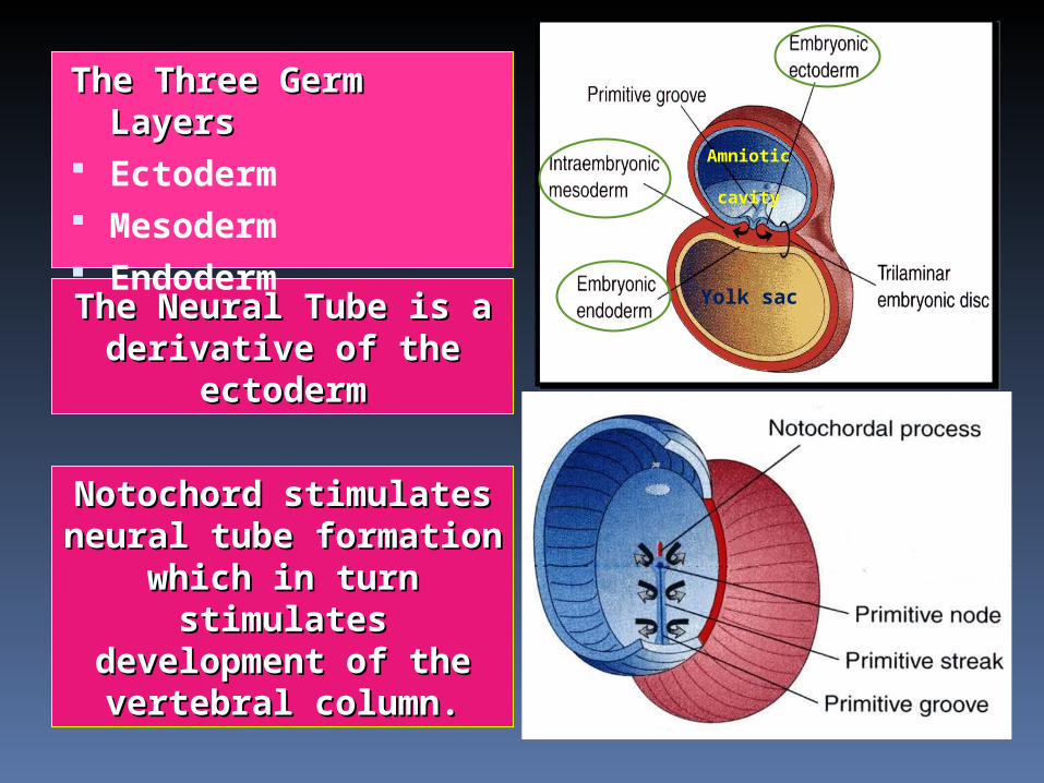
Yolk sac
Amniotic
cavity
Notochord stimulates Notochord stimulates neural tube formation neural tube formation
which in turn which in turn stimulates stimulates
development of the development of the vertebral column.vertebral column.
The Neural Tube is a The Neural Tube is a derivative of the derivative of the
ectodermectoderm
The Three Germ The Three Germ LayersLayers
Ectoderm Mesoderm Endoderm

Development of Neural TubeDevelopment of Neural Tube
Ectodermal cells dorsal to notochord thicken to form the the neural plateneural plate.
A longitudinal groove, neural neural groove,groove, develops in the neural plate ().().
The margins of the neural plate ((neural foldsneural folds) ) approach to each other and fuse to form the neural neural tubetube.

The spinal cord develops from the caudal 2/3 of the neural tube
Development of the Spinal CordDevelopment of the Spinal Cord

The cells of the neural tube form three layers:
An inner ventricular ventricular zone zone of undifferentiated cells
A middle mantle zonemantle zone of cell bodies of neurons (future grey matter)(future grey matter)
An outer marginal zone marginal zone of nerve fibers or axons of neurons (future white (future white matter)matter)
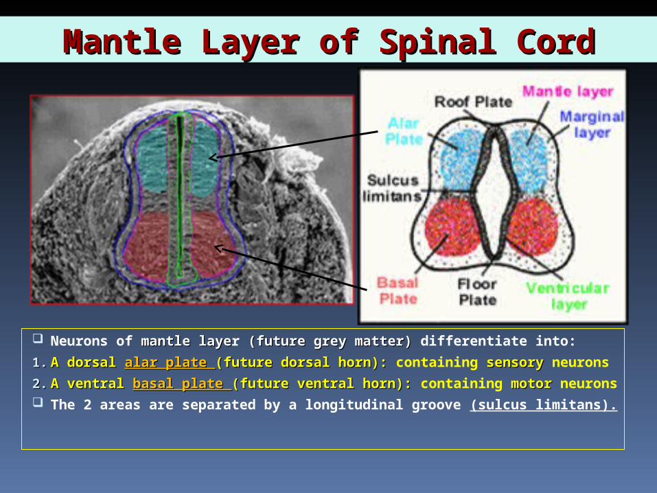
Mantle Layer of Spinal CordMantle Layer of Spinal Cord
Neurons of mantle layemantle layer (future grey matter) (future grey matter) differentiate into:
1.1. A dorsal A dorsal alar plate alar plate (future dorsal horn): (future dorsal horn): containing sensorysensory neurons
2.2. A ventral A ventral basal plate basal plate (future ventral horn): (future ventral horn): containing motor motor neurons The 2 areas are separated by a longitudinal groove (sulcus limitans).

Proliferation and bulging of both alar & basal plates cause: Formation of longitudinal dorsal & ventral median septadorsal & ventral median septa
Narrowing of the lumen to form a small central canalcentral canal
Central canal
Dorsal median septum
Ventral median fissure

Marginal Layer of Spinal cordMarginal Layer of Spinal cord
Marginal layer (future white matter) increases increases in size in size due to addition of ascending, descending & intersegmental nerve fibers.
Marginal layer is divided into: dorsaldorsal, , lateral lateral and and ventralventral funiculi funiculi
MyelinationMyelination of nerve fibers starts at 44thth month month & continues during the 1during the 1stst postnatal year. postnatal year. Motor fibers myelinate before sensory fibers.Motor fibers myelinate before sensory fibers.
Dorsalfuniculus
Lateral funiculus
Ventral funiculus

MeningesMeninges
These are 3 membranes covering the neural tube:
Outer thick dura dura mater: mesodermalmesodermal in origin
Middle arachnoidarachnoid mater: ectodermalectodermal in origin
Inner thin pia pia mater: ectodermal ectodermal in origin
A cavity appears between the arachnoid & the pia mater ((subarachnoid spacesubarachnoid space) ) & becomes filled with cerebrospinal fluid (CSF)cerebrospinal fluid (CSF).

Positional Changes of Spinal Positional Changes of Spinal CordCord
Initially, the spinal cord occupies the whole length of the vertebral canal.
As a result a a faster growth of faster growth of vertebral vertebral columncolumn, the caudal end of spinal cord ((conus conus medullarismedullaris)) shifts gradually shifts gradually to a higher to a higher level.level.

The vertebral column develops from the ventromedial parts (sclerotomes) of the somites
The somites develop from the para-axial mesoderm.
Development of the Vertebral Development of the Vertebral ColumnColumn

Intraembryonic MesodermIntraembryonic Mesoderm Located between Ectoderm & Endoderm between Ectoderm & Endoderm
EXCEPTEXCEPT in the central axis of embryo where NOTOCHORDNOTOCHORD is found.
Differentiates into 3 parts:
1.1. Paraxial mesodermParaxial mesoderm
2.2. Intermediate mesodermIntermediate mesoderm
3.3. Lateral mesodermLateral mesoderm Paraxial mesoderm Paraxial mesoderm divides into segments
called ‘somites’.somites’. Each somite divides into 3 parts:Each somite divides into 3 parts:
1.1. DermatomeDermatome
2.2. MyotomeMyotome
3.3. SclerotomeSclerotome
1
23
GutGut
NTNT
NN
somitessomites

At At 44thth week week, e, each sclerotome becomes subvidided into two parts: an anterior part, consisting of
loosely arranged cells a posterior part, of more
condensed tissue.
The posterior part of each somite fuses with the anterior part of the consecutive somite, around the notochord to form the bodyof the vertebra,called the centrum.
Formation of Body of VertebraFormation of Body of Vertebra
Each centrum develops from 2 Each centrum develops from 2 adjacent sclerotomesadjacent sclerotomes

The fused The fused sclerotomes grow sclerotomes grow dorsally around dorsally around the neural tube the neural tube and form the and form the vertebral (neural) vertebral (neural) arch.arch.
Ventrolaterally, Ventrolaterally, costal processes costal processes develop that give develop that give rise to ribs in rise to ribs in thoracic region.thoracic region.

Vertebral DevelopmentVertebral Development
All centers unite around 25 years25 years

Curvatures of Vertebral ColumnCurvatures of Vertebral Column
Primary curvatures: Primary curvatures: develop prenatallyprenatally1.1. ThoracicThoracic 2.2. Pelvic or SacralPelvic or Sacral
Secondary Secondary curvatures curvatures : : develop postnatallypostnatally1.1. CervicalCervical: : as a
result of lifting the head
2.2. LumbarLumbar:: as a result of walking

Fate of NotochordFate of Notochord In the region of the bodies In the region of the bodies
of vertebraeof vertebrae: : It degenerates Between bodies of Between bodies of
vertebraevertebrae: : It forms the central part, ’nucleus pulposus’ of the intervertebral discs
Annulus fibrosus Annulus fibrosus part part of the intervertebral of the intervertebral discs discs is formed by the mesoderm mesoderm surrounding the surrounding the notochord.notochord.

Spina BifidaSpina Bifida
Cause:Cause: Failure of fusion of the halves of vertebral arches
Incidence:Incidence: 0.04-0.15%
Sex:Sex: more frequent in femalesfemales
Types:Types:
1.1. Spina bifida Spina bifida occulta occulta (20%)
2.2. Spin bifida Spin bifida cystica cystica (80%)

Spina Bifida OccultaSpina Bifida Occulta
The closed type Only one
vertebra is affected
No clinical No clinical symptomssymptoms
Skin overlying it is intact
Sometimes covered by a tuft of hair
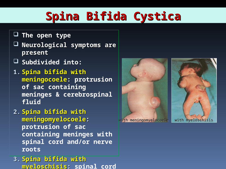
Spina Bifida CysticaSpina Bifida Cystica
The open type Neurological symptoms Neurological symptoms
are presentare present Subdivided into:
1.1. Spina bifida with Spina bifida with meningocoelemeningocoele: : protrusion of sac containing meninges & cerebrospinal fluid
2.2. Spina bifida with Spina bifida with meningomyelocoelemeningomyelocoele: : protrusion of sac containing meninges with spinal cord and/or nerve roots
3.3. Spina bifida with Spina bifida with myeloschisismyeloschisis: : spinal cord is open due to failure of neural folds
with meningomyelocoelewith meningomyelocoele with myeloschisiswith myeloschisis

Spina bifida with meningomyelocoele
Spina bifida occulta Spina bifida with meningocoele
Spina bifida with myeloschisis
