bilaminar & trilaminar embryonic disc dr. zeenat zaidi
TRANSCRIPT


Bilaminar & Bilaminar & Trilaminar Trilaminar
Embryonic DiscEmbryonic Disc
Dr. Zeenat ZaidiDr. Zeenat Zaidi

Bilaminar Embryonic Bilaminar Embryonic DiscDisc
The Second WeekThe Second Week

Formation of Amniotic CavityFormation of Amniotic Cavity As implantation of the As implantation of the
blastocyst progresses, blastocyst progresses, changes appear in the changes appear in the inner cell massinner cell mass (embryoblast)(embryoblast)
A cavity, A cavity, amniotic cavityamniotic cavity appears separating appears separating embryoblast from the embryoblast from the trophoblast, which soon trophoblast, which soon becomes lined by becomes lined by amnioblastsamnioblasts derived from derived from inner cell massinner cell mass
The cavity gradually The cavity gradually increases in size and is increases in size and is filled with filled with amniotic fluidamniotic fluid
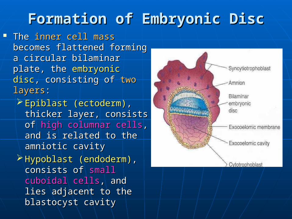
Formation of Embryonic DiscFormation of Embryonic Disc The The inner cell massinner cell mass
becomes flattened forming becomes flattened forming a circular bilaminar plate, a circular bilaminar plate, the the embryonic discembryonic disc, , consisting of consisting of two layerstwo layers:: Epiblast (ectoderm)Epiblast (ectoderm), ,
thicker layer, consists of thicker layer, consists of high columnar cellshigh columnar cells, and , and is related to the is related to the amniotic cavityamniotic cavity
Hypoblast (endoderm)Hypoblast (endoderm),, consists of consists of small small cuboidal cellscuboidal cells, and lies , and lies adjacent to the adjacent to the blastocyst cavityblastocyst cavity

Formation of Primitive Yolk SacFormation of Primitive Yolk Sac
The blastocyst cavity The blastocyst cavity becomes lined with becomes lined with exocelomic membrane exocelomic membrane and is called and is called exocelomic cavityexocelomic cavity
The The hypoblastic cellshypoblastic cells soon replace the soon replace the exocelomic membrane exocelomic membrane and the cavity is then and the cavity is then named as the named as the primitive (primary) primitive (primary) yolk sacyolk sac

At this stage the embryonic disc is:At this stage the embryonic disc is:
A circular bilaminar A circular bilaminar disc, that lies between disc, that lies between the the amniotic cavityamniotic cavity and the and the primitive yolk primitive yolk sacsac
TheThe epiblast forms the epiblast forms the floorfloor of the amniotic of the amniotic cavitycavity & the & the hypoblast hypoblast lies in the lies in the roofroof of the of the primitive yolk sacprimitive yolk sac

Formation of Extraembryonic Formation of Extraembryonic MesodermMesoderm
Endoderm of the Endoderm of the yolk sac gives rise yolk sac gives rise to a layer of loosely to a layer of loosely arranged arranged connective tissue, connective tissue, extraembryonic extraembryonic mesoderm (EEM)mesoderm (EEM), , which surrounds the which surrounds the amniotic cavity and amniotic cavity and the yolk sac.the yolk sac.

Formation of Extraembryonic CelomeFormation of Extraembryonic Celome
Isolated spaces Isolated spaces appear in the EEMappear in the EEM
These spaces rapidly These spaces rapidly fuse to form a large fuse to form a large fluid filledfluid filled, C-shaped , C-shaped cavity, the cavity, the extraembyonic extraembyonic celomecelome surrounding surrounding the amniotic cavity the amniotic cavity and the yolk sacand the yolk sac

Formation of Connecting StalkFormation of Connecting Stalk The region where no The region where no
cavity has appeared, cavity has appeared, forms the forms the connecting connecting stalk,stalk, that connects the that connects the amniotic cavity, yolk amniotic cavity, yolk sac and the embryonic sac and the embryonic disc to the outer walldisc to the outer wall
The site of the The site of the connecting stalk connecting stalk determines the determines the caudal caudal polepole of the embryonic of the embryonic discdisc

With the formation of With the formation of extraembryonic extraembryonic celomecelome::
The The extraembryonicextraembryonic mesoderm mesoderm splits into splits into two layers: two layers: • an outer an outer
extraembryonic extraembryonic parietal (somatic)parietal (somatic) mesodermmesoderm
• an inner an inner extraembryonicextraembryonic visceral (splanchnicvisceral (splanchnic) ) mesodermmesoderm

The primary yolk sac The primary yolk sac decreases in size and decreases in size and becomes the becomes the secondary (definitive) secondary (definitive) yolk sacyolk sac
Wall of the yolk sac, Wall of the yolk sac, amnion & chorionamnion & chorion are are formedformed

AmnionAmnion: Two layers:: Two layers:• AmnioblastsAmnioblasts• Extraembronic Extraembronic
splanchnic mesodermsplanchnic mesoderm Wall of the yolk sacWall of the yolk sac: :
Two layers:Two layers:• EndodermEndoderm• Extraembronic Extraembronic
splanchnic mesodermsplanchnic mesoderm ChorionChorion: Three layers:: Three layers:
• Extraembryonic somatic Extraembryonic somatic mesodermmesoderm
• CytotrophoblastCytotrophoblast• SyncytiotrophoblastSyncytiotrophoblast

chorion
amnion
bilaminar disc
wall of the yolk sac
Connecting stalk EEC
Extraembryonic celome is now called the
CHORIONIC CAVITY

Trilaminar Embryonic Trilaminar Embryonic DiscDisc
The Third WeekThe Third Week
The significant event of third The significant event of third week is week is GastrulationGastrulation

GastrulationGastrulation
The process by which the bilaminar The process by which the bilaminar disc is converted into a disc is converted into a trilaminar trilaminar discdisc
It is the beginning of It is the beginning of morphogenesismorphogenesis (formation of body form)(formation of body form)
Consists of formation of the Consists of formation of the primitive primitive streakstreak, the , the threethree germ layersgerm layers & the & the notochordnotochord
Embryo is referred to as a Embryo is referred to as a GastrulaGastrula

Primitive StreakPrimitive Streak The primitive streak The primitive streak results results
from proliferation of the from proliferation of the epiblastic cellsepiblastic cells in the in the median plane,median plane, in the in the caudal half caudal half of the of the epiblastepiblast, , and lies along the cranio-and lies along the cranio-caudal axis. caudal axis.
Its Its cranial cranial end forms end forms primitive nodeprimitive node
A groove, A groove, primitive grooveprimitive groove, , appears in the primitive appears in the primitive streak, which continues streak, which continues with a small depression, with a small depression, primitive pitprimitive pit, in the , in the primitive node.primitive node.

A circular thickening A circular thickening appears in the appears in the hypoblast hypoblast near the near the cranial endcranial end, in the , in the midlinemidline, to form the , to form the prechordal plate,prechordal plate, that that marks the future site marks the future site of mouthof mouth
A circular thickening A circular thickening appears in the appears in the hypoblasthypoblast caudal to caudal to primitive streak in primitive streak in the midline to form the midline to form the the cloacal cloacal membranemembrane, the future , the future site of the anussite of the anus

By this stage of By this stage of development, it is development, it is possible to identify possible to identify the embryo’s:the embryo’s:craniocaudal axiscraniocaudal axiscranial and caudal cranial and caudal
endsendsdorsal and ventral dorsal and ventral
surfacessurfacesright and left right and left
sides.sides.Connecting stalk

Formation of Intraembryonic Formation of Intraembryonic MesodermMesoderm
The epiblastic cells from The epiblastic cells from the the primitive streak primitive streak (groove)(groove) proliferate to proliferate to form form mesenchymalmesenchymal tissue tissue
The newly formed cells The newly formed cells invaginateinvaginate, , migratemigrate ventrally, laterally & ventrally, laterally & cranially between the cranially between the epiblast and hypoblast epiblast and hypoblast & & organizeorganize to form the to form the intraembryonic intraembryonic mesodermmesoderm

Formation of Intraembryonic Formation of Intraembryonic MesodermMesoderm cont’d cont’d
Intraembryonic mesoderm Intraembryonic mesoderm merges with the extra-merges with the extra-embryonic mesoderm at embryonic mesoderm at the periphery of the the periphery of the embryonic discembryonic disc
By the end ofBy the end of 3 3rdrd week, week, mesoderm lies between mesoderm lies between embryonic ectoderm and embryonic ectoderm and endoderm everywhereendoderm everywhere except in the region ofexcept in the region of prechordal plate prechordal plate andand cloacal membrane, cloacal membrane, as the as the embryonic ectoderm & embryonic ectoderm & endoderm are fused at endoderm are fused at these regionsthese regions

Formation of Intraembryonic Formation of Intraembryonic MesodermMesoderm cont’d cont’d
Some mesenchymal Some mesenchymal cells displace the cells displace the hypoblasts forming hypoblasts forming the the embryonic embryonic endodermendoderm
Cells remaining in Cells remaining in the epiblast form the the epiblast form the embryonic ectodermembryonic ectoderm

Each of the three germ layers gives Each of the three germ layers gives rise to specific tissues and organsrise to specific tissues and organs
Thus the Thus the EPIBLASTEPIBLAST gives rise to gives rise to all all three germ layers,three germ layers, EctodermEctoderm, , MesodermMesoderm, , EndodermEndoderm in the in the
embryoembryo

Fate of Primitive StreakFate of Primitive Streak Actively forms mesoderm until the Actively forms mesoderm until the
early part of early part of 44thth week week Then it starts regressing and becomes Then it starts regressing and becomes
an insignificant structure in the an insignificant structure in the sacrocooccygeal regionssacrocooccygeal regions
Normally it degenerates and Normally it degenerates and disappears by the disappears by the end of 4end of 4thth week week
Remnants may persist and give rise to Remnants may persist and give rise to a large tumor called a large tumor called Sacrococcygeal Sacrococcygeal TeratomasTeratomas

NotochordNotochord
A rod of A rod of mesenchymal cells mesenchymal cells located located craniallycranially, in , in the the midlinemidline, , extending between extending between the primitive node the primitive node and the prechordal and the prechordal plateplate

Formation of NotochordFormation of Notochord Mesenchymal cells Mesenchymal cells
migrate cranially from migrate cranially from primitive pitprimitive pit toward the toward the prechordal plate, and prechordal plate, and form a rod like form a rod like notochordal processnotochordal process
The notochordal The notochordal process becomes process becomes canalized forming a canalized forming a hollow tube, the hollow tube, the notochordal canalnotochordal canal, , communicating with communicating with the primitive pit. the primitive pit.

Formation of NotochordFormation of Notochord cont’d cont’d The floor of the The floor of the
tube and the tube and the underlying underlying endoderm break endoderm break down, forming a down, forming a notochordal plate notochordal plate
The notochordal The notochordal plate becomes plate becomes continuous with the continuous with the endodermal layer.endodermal layer.

Formation of NotochordFormation of Notochord cont’d cont’d
A temporary A temporary communication communication is established is established between the between the amniotic cavity amniotic cavity and the yolk sac, and the yolk sac, termed the termed the neurenteric neurenteric canalcanal. .

Notochordal plateNotochordal plate folds to form the folds to form the notochordnotochord. .

Functions of NotochordFunctions of Notochord Defines primordial axis of the embryoDefines primordial axis of the embryo Provides rigidity to the embryoProvides rigidity to the embryo Serves as a basis for the development Serves as a basis for the development
of the axial skeletonof the axial skeleton Indicates the future site of the vertebral Indicates the future site of the vertebral
bodies/columnbodies/column Regulates differentiation of surrounding Regulates differentiation of surrounding
structures including the overlying structures including the overlying ectoderm (neural plate) and mesoderm ectoderm (neural plate) and mesoderm (somites).(somites).

Fate of NotochordFate of Notochord
Degenerates and disappears as the Degenerates and disappears as the bodies of the vertebrae develop, but bodies of the vertebrae develop, but it persists as the it persists as the nucleus pulposusnucleus pulposus of of each intervertebral disceach intervertebral disc
Remnants of notochordal tissue give Remnants of notochordal tissue give rise to tumors called rise to tumors called ChordomasChordomas
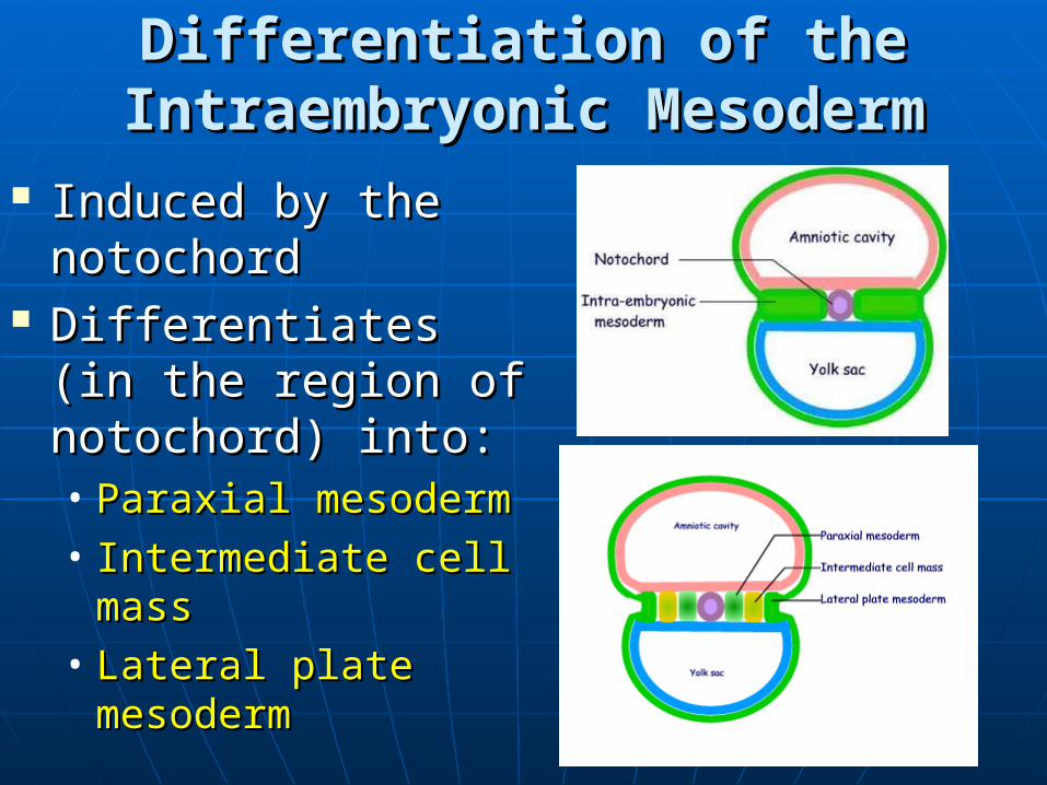
Differentiation of the Differentiation of the Intraembryonic MesodermIntraembryonic Mesoderm
Induced by the Induced by the notochordnotochord
Differentiates (in the Differentiates (in the region of notochord) region of notochord) into:into:• Paraxial mesodermParaxial mesoderm• Intermediate cell Intermediate cell
massmass• Lateral plate Lateral plate
mesodermmesoderm
