development of antimicrobial protective food...
TRANSCRIPT
DEVELOPMENT OF ANTIMICROBIAL PROTECTIVE FOOD COATING MATERIALS
FROM EDIBLE ALGINATE FILMS
A Thesis Submitted to the Graduate School of Engineering and Science of
Izmir Institute of Technology in Partial Fulfilment of Requirements for the Degree of
MASTER OF SCIENCE
in Biotechnology
by Fatih Yalçın Güne� YENER
July 2007 �ZM�R

ii
We approve the thesis of Fatih Yalçın Güne� YENER
Date of Signature
....................................... 13 July 2007 Assist. Prof. Dr. Figen KOREL Supervisor Department of Food Engineering Izmir Institute of Technology
....................................... 13 July 2007 Assoc. Prof. Dr. Ahmet YEMEN�C�O�LU Co-Supervisor Department of Food Engineering Izmir Institute of Technology
....................................... 13 July 2007 Assist. Prof. Dr. Alper ARSLANO�LU Co-Supervisor Department of Biology Izmir Institute of Technology
....................................... 13 July 2007 Assist. Prof. Dr. Canan TARI Department of Food Engineering Izmir Institute of Technology
....................................... 13 July 2007 Assoc. Prof. Dr. Sacide ALSOY ALTINKAYA Department of Chemical Engineering Izmir Institute of Technology
....................................... 13 July 2007 Assist. Prof. Dr. Gül�ah �ANLI Department of Chemistry Izmir Institute of Technology
....................................... 13 July 2007 Prof. Dr. Semra ÜLKÜ Head of Department Izmir Institute of Technology
…………..................................... Prof. Dr. M. Barı� ÖZERDEM
Head of the Graduate School
iii
ACKNOWLEDGMENTS
I would like to thank my thesis advisor Assist. Prof. Figen Korel and my co-
advisor Assoc. Prof. Ahmet Yemenicio�lu for their directions and unending patience
during the whole year. Without their guidance in this thesis, it would not have been
completed. I would also like to thank my other co-advisor Assist. Prof. Alper
Arslano�lu for his aid with suggestions on my studies.
The supportive and friendly demeanor of everyone in Food Engineering
Department made the three years enjoyable. Special thanks to the people who devoted
their time and effort during the course of my research, namely, �lke Uysal, Nihan
Gö�ü�, and F. I�ık Üstok. I also wish to thank to give my special thanks to A. Emrah
Çetin for his valuable assistance and recommendations at the beginning of my thesis. I
would also like to thank the very knowledgeble and cooperative departmental staff that
is always available, Burcu Okuklu. In addition, I would also thank to my office mates,
�lke Uysal, Levent Yurdaer Aydemir and Dilhun Keriman Arserim for their sincere
helps and the nice days we lived together.
I would also thank my grandparents, my elder aunt, and my sister for their love
and support, and most importantly, I would like to thank my mother Mahinur Yener
who is a catalyst behind all that I do.

iv
ABSTRACT
DEVELOPMENT OF ANTIMICROBIAL PROTECTIVE FOOD
COATING MATERIALS FROM EDIBLE ALGINATE FILMS
Consumer interests in high quality, healthy, convenient and safe food continue
to increase, presenting food processors with new challenges to which functional edible
coating and film concepts offer potential solutions. The interest in the research of edible
film which has many advantages and applications has increased during last decade.
There is a particular interest in the use of antimicrobial biopreservatives in edible films
and to increase food safety without application of chemical preservatives. In this study,
we have developed antimicrobial or protective edible films by incorporation of
antimicrobial enzyme lactoperoxidase or protective cultures (Lactobacillus delbrueckii
subsp. lactis and Lactobacillus plantarum) into alginate films, respectively. The main
objective of this research was to increase food safety by using lactoperoxidase or lactic
acid bacteria incorporated into alginate films. The results obtained in the study showed
that in reaction mixtures, the lactoperoxidase system has antimicrobial activity against
E. coli, L. innocua, and P. fluorescens. The developed lactoperoxidase incorporated
antimicrobial films also reduced the total microbial load of a selected seafood during
cold storage. The lactic acid bacteria, used in edible films for the first time, also
successfully incorporated into alginate films. The bacteria showed sufficient stability in
alginate films and at surface of red meat during cold storage. The results of this study
clearly showed the good potential of using lactoperoxidase and lactic acid bacteria
incorporated alginate films in food packaging. The developed films can be used in
antimicrobial packaging or protective packaging. However, further studies are needed to
show the beneficial effects of developed films on different food systems.
v
ÖZET
YEN�LEB�L�R ALG�NAT F�LMLER KULLANILARAK
ANT�M�KROB�YAL-KORUYUCU ETK�S� OLAN GIDA KAPLAMA
MATERYALLER�N�N GEL��T�R�LMES�
Tüketicilerin kaliteli, sa�lıklı, kolay hazırlanabilir ve güvenli gıdalara
gösterdikleri yüksek talep, gıda üreticilerini yeni çözüm arayı�larına yöneltmektedir.
Fonksiyonel yenebilir fimler ve kaplamalar kullanılarak gıdaların kalite ve güvenli�inin
arttırılması konsepti üreticilerin bu arayı�larına potansiyel çözümler getirmekte ve
birçok avantajlar sa�lanmasına ve uygulamalara olanak sa�lamaktadır. Örne�in,
üzerinde en yo�un olarak çalı�ılan konulardan birisi de antimikrobiyal etkiye sahip
biyoprezervatiflerin yenebilir filmlerle birlikte gıdalarda kullanılması ve gıda
güvenli�inin kimyasal koruyucular uygulanmadan arttırılmasıdır. ��te bu çalı�mada
laktoperoksidaz ve koruyucu kültürler kullanılarak (Lactobacillus delbrueckii subsp.
lactis ve Lactobacillus plantarum) sırasıyla antimikrobiyal ve koruyucu etkisi olan
alginat filmler üretilmi�tir. Bu ara�tırmanın temel amacı belirtilen biyoprezervatifler
kullanılarak gıda güvenli�inin arttırılmasıdır. Elde edilen sonuçlar üretilmi� olan
laktoperoksidaz içeren alginate filmlerin olu�turulan deneysel reaksiyon karı�ımlarında
E. coli, L. innocua, and P. fluorescens bakterilerine kar�ı antimikrobiyal etkiye sahip
oldu�unu göstermi�tir. Geli�tirilmi� olan laktoperoksidaz içeren alginat filmler seçilmi�
bir deniz ürününde de uygulanmı� ve bu ürünün depolanması sırasında toplam canlı
bakteri sayısında kayda de�er bir azalma sa�lanmı�tır. Di�er yandan, literatürde ilk kez
gerçekle�tirilen laktik asit bakterilerinin alginat filmlere ilave edilmesi çalı�ması da
ba�arıyla gerçekle�tirilmi�tir. Kullanılmı� olan laktik asit bakterileri gerek filmler
içerisinde gerekse uygulandıkları seçilmi� gıda olan kırmızı et yüzeyinde yeterli
stabiliteyi göstermektedirler. Bu çalı�mada elde edilmi� olan sonuçlar laktoperoksidaz
ve laktik asit bakterisi içeren alginat filmlerin gıda paketleme uygulamalarında
kullanılabilece�ini göstermi�tir. Geli�tirilmi� olan filmler antimikrobiyal paketleme
veya koruyucu paketleme amacıyla kullanılabileceklerdir. Ancak, geli�tirilen filmlerin
çe�itli gıdalarda denenmesi amacıyla ilave çalı�malar gerçekle�tirilmesi gerekmektedir.
vi
TABLE OF CONTENTS
LIST OF FIGURES ......................................................................................................... xi
LIST OF TABLES........................................................................................................ xiii
CHAPTER 1. INTRODUCTION .................................................................................. 1
CHAPTER 2. PACKAGING ......................................................................................... 4
2.1. Packaging............................................................................................. 4
2.1.1. Antimicrobial Food Packaging....................................................... 4
2.1.1.1. Types of Antimicrobial Packaging ........................................ 5
2.1.1.1.1. Addition of Sachets/Pads Containing Volatile
Antimicrobial Agents into Packages......................... 5
2.1.1.1.2. Incorporation of Volatile and Non-Volatile
Antimicrobial Agents Directly into Polymers .......... 6
2.1.1.1.3. Coating or Adsorbing Antimicrobials to
Polymer Surfaces ...................................................... 7
2.1.1.1.4. Immobilization of Antimicrobials to Polymers
by Ion or Covalent Linkages..................................... 8
2.1.1.1.5. Use of Polymers that are Inherently
Antimicrobial ............................................................ 8
2.1.1.2. Antimicrobial Packaging Systems......................................... 9
2.1.1.3. Important Factors Considered in the Manufacturing of
Antimicrobial Films............................................................. 10
2.1.1.3.1. Process Conditions and Residual
Antimicrobial Activity............................................ 10
2.1.1.3.2. Characteristics of Antimicrobial Substances
and Foods ............................................................... 10
2.1.1.3.3. Chemical Interaction of Additives with Film
Matrix...................................................................... 11
2.1.1.3.3. Storage Temperature............................................... 11
2.1.1.4. Testing the Effectiveness of Antimicrobial Packaging ....... 11

vii
2.1.1.4.1. Minimum Inhibitory Concentrations (MIC)
Method.................................................................... 12
2.1.1.4.2. Dynamic Shake Flask Test ..................................... 12
2.1.1.4.3. Agar Plate Test........................................................ 12
CHAPTER 3. EDIBLE FILMS AND COATINGS..................................................... 13
3.1. Edible Films and Coatings ................................................................. 13
3.2. Functionality of Edible Films and Coatings ...................................... 14
3.3. Film Components............................................................................... 16
3.3.1. Hydrocolloid Films ...................................................................... 16
3.3.1.1. Carbohydrate Films ............................................................. 16
3.3.1.1.1. Starch ...................................................................... 17
3.3.1.1.2. Alginate .................................................................. 17
3.3.1.1.3. Carrageenan ........................................................... 20
3.3.1.1.4. Cellulose Derivatives.............................................. 20
3.3.1.1.5. Pectin Films ............................................................ 20
3.3.1.1.6. Chitosan .................................................................. 21
3.3.1.2. Protein Films........................................................................ 21
3.3.1.2.1. Zein ......................................................................... 21
3.3.1.2.2. Whey Protein Isolates (WPI) .................................. 22
3.3.1.2.3. Collagen Casings .................................................... 22
3.3.2. Lipid Films ................................................................................... 23
3.3.2.1. Waxes .................................................................................. 23
3.3.2.2. Glycerides ............................................................................ 23
3.3.3. Composite Films .......................................................................... 24
3.4. Film Formation .................................................................................. 24
3.4.1. Coacervation................................................................................. 24
3.4.2. Solvent Removal .......................................................................... 24
3.4.3. Solidification of Melt ................................................................... 25
3.5. Film Additives ................................................................................... 25
CHAPTER 4. BIOPRESERVATION.......................................................................... 28
4.1. Definition of Biopreservation ............................................................ 28
4.1.1. Protective Culture......................................................................... 28
viii
4.1.1.1. Organic Acids ...................................................................... 29
4.1.1.2. Diacetyl................................................................................ 30
4.1.1.3. Hydrogen Peroxide .............................................................. 31
4.1.1.4. Fatty Acids........................................................................... 31
4.1.1.5. Phenyllactic Acid................................................................. 31
4.1.1.6. Cyclic Dipeptides ............................................................... 32
4.1.1.7. Bacteriocins ......................................................................... 32
4.1.1.8. Reutericyclin........................................................................ 32
4.1.1.9. 2-Pyrrolidone-5-Carboxylic Acid........................................ 33
4.1.2. Factors Affecting Protective Culture Performance ...................... 33
4.1.2.1. Temperature Effect .............................................................. 33
4.1.2.2. Protective culture and food poisoning-spoilage organisms ........ 33
4.1.2.3. Inoculum Effect .................................................................. 35
4.1.2.4. Food Effect .......................................................................... 36
4.1.3. Potential Benefits of PCs.............................................................. 37
4.1.4. Application of PCs ....................................................................... 37
4.1.5. Antimicrobial Enzymes ................................................................ 39
4.1.5.1. Lactoperoxidase................................................................... 39
4.1.5.2. Lysozyme............................................................................. 44
4.1.5.3. Glucose Oxidase .................................................................. 44
4.1.5.4. Chitinase ............................................................................. 45
4.1.6. Bioactive Proteins and Peptides ................................................... 45
4.1.6.1. Lactoferrin .......................................................................... 45
4.1.6.2. Phosvitin ............................................................................. 46
CHAPTER 5. MATERIALS AND METHODS.......................................................... 47
5.1. Materials ............................................................................................ 47
5.2. Methods ............................................................................................. 47
5.2.1. Alginate Films Incorporating Lactoperoxidase............................ 47
5.2.1.1. Preparation of Lactoperoxidase from Bovine Milk............. 47
5.2.1.2. Determination of Lyophilized Lactoperoxidase Activity........... 48
5.2.1.3. Preparation of Alginate Films ............................................. 49
5.2.1.4. Determination of Lactoperoxidase Activity Released
from the Alginate Film ........................................................ 49

ix
5.2.1.5. Determination of Immobilized Lactoperoxidase
Activity in the Alginate Film............................................... 49
5.2.1.6. Antimicrobial Activity of Alginate Films ........................... 50
5.2.1.6.1. Bacterial Strains...................................................... 50
5.2.1.6.2. Determination of Antimicrobial Activity of
Alginate Films Incorporating Lactoperoxidase ...... 50
5.2.1.7. Application of Lactoperoxidase Incorporated Alginate
Films to Calamari ................................................................ 52
5.2.2. Alginate Films Incorporating Lactic Acid Bacteria ..................... 53
5.2.2.1. Bacterial Strains................................................................... 53
5.2.2.2. Test of the Ability of LAB to Produce Hydrogen
Peroxide ............................................................................... 53
5.2.2.3. Test for the Ability of L.delbrueckii subsp. lactis and
L.plantarum to Produce H2O2 and Lactic Acid at
Different Storage Temperatures .......................................... 54
5.2.2.4. Preparation of Lyophilized Culture ..................................... 54
5.2.2.5. Preparation of Alginate Films Incorporating Lactic
Acid Bacteria ....................................................................... 55
5.2.2.5.1. Determination of the Number of Free and
Immobilized Lactic Acid Bacteria in Alginate
Films ....................................................................... 55
5.2.2.5.2. Determination of the Stability of the Lactic
Acid Bacteria Incorporated into the Alginate
Film Forming Solution............................................ 56
5.2.2.5.3. Determination of the Stability of the Lactic
Acid Bacteria in Powdered Alginic Acid ............... 56
5.2.3. Applications of Alginate Films Incorporating LAB to Beef
Fresh Cubes .................................................................................. 56
CHAPTER 6. RESULTS AND DISCUSSIONS......................................................... 58
6.1. Incorporation of Lactoperoxidase into Alginate Films ..................... 58
6.1.1. Determination of the LP Activity Released from the
Alginate Films .............................................................................. 58

x
6.1.2. Determination of the LP Activity Immobilized in the
Alginate Films .............................................................................. 59
6.1.3. Antimicrobial Activity of LPS-H2O2-Thiocyanate System ........ 60
6.1.3.1. Effects of LP System in Reaction Mixtures Containing
Alginate Films Incorporating LP against E.coli .............. 60
6.1.3.2. Effects of LP System in Reaction Mixtures Containing
Alginate Films Incorporating LP against L. innocua ......... 66
6.1.3.3. Effects of LP System in Reaction Mixtures Containing
Alginate Films Incorporating LP against P.fluorescens ..... 72
6.1.4. Test of Developed LP Incorporated Alginate Films on
Refrigerated Fresh Calamari ....................................................... 78
6.2. Alginate Films Incorporated with Lactic Acid Bacteria .................... 80
6.2.1. Comparison of L.casei and L.delbrueckii subsp. lactis for
Hydrogen Peroxide Production at 37oC ....................................... 80
6.2.2. H2O2 and Lactic Acid Production of L. delbrueckii subsp.
lactis and L. plantarum at Different Storage Temperatures ........ 82
6.2.3. Incorporation of Lactic Acid Bacteria into Alginate Films.......... 86
6.2.4. Determination of the Number of Free and Immobilized
Lactic Acid Bacteria in Alginate Films........................................ 86
6.2.5. Determination of the Stability of the Lactic Acid Bacteria
Incorporated into the Alginate Film Forming Solution................ 87
6.2.6. Determination of the Stability of the Lactic Acid Bacteria in
Alginic Powder .............................................................................. 88
6.2.7. Test of Developed Lactic Acid Bacteria Incorporated
Alginate Films on Refrigerated Fresh Beef Cubes....................... 89
CHAPTER 7. CONCLUSIONS................................................................................... 91
REFERENCES ............................................................................................................... 92
APPENDICES
APPENDIX A. E. coli .................................................................................................... 99
APPENDIX B. L. innocua ............................................................................................ 100
APPENDIX C. P. fluorescens ...................................................................................... 101
xi
LIST OF FIGURES
Figure Page
Figure 2.1. The migration of antimicrobial agent from the coating material................. 7
Figure 2.2. Immobilization antimicrobial agents onto food packaging
materials....................................................................................................... 9
Figure 2.3. Packaging/Food systems.............................................................................. 9
Figure 2.4. Package/Headspace/Food Systems ............................................................ 10
Figure 3.1. Selective functions of edible films and coatings ....................................... 15
Figure 3.2. Structure of alginates consisting of (a) M block, (b) G block,
and (c) alternating M and G blocks ........................................................... 18
Figure 3.3. Film Components....................................................................................... 27
Figure 4.1. Summary of the main antimicrobial compounds produced by
LAB ........................................................................................................... 29
Figure 4.2. Oxidation of protein (enzyme) sulphydryls by lactoperoxidase
(LP) catalysed reactions, mediated by products of SCN- ........................ 41
Figure 6.1. The immobilized lactoperoxidase activity in different H2O2
concentrations ............................................................................................ 59
Figure 6.2. Change of microbial counts (log10 cfu/mL) of E. coli in
reaction mixtures containing 4000 �M KSCN, 1188 U
lactoperoxidase/disc and different concentrations of H2O2 ....................... 62
Figure 6.3. Change of microbial counts (log10 cfu/mL) of E. coli in
reaction mixtures containing 200 �M H2O2, 1188 U
lactoperoxidase/disc and different concentrations of KSCN..................... 65
Figure 6.4. Change in the microbial counts (log10 cfu/ml) of L. innocua in
reaction mixtures with different concentrations of H2O2 and
having 660 �g (A: 1st trial) and 453 �g (B: 2nd trial) of
lactoperoxidase in discs in 24 hr................................................................ 68
Figure 6.5. Change in the microbial counts (log10 cfu/ml) of L. innocua in
reaction mixtures with different concentrations of KSCN and
having 479 �g (A: 1st trial) and 453 �g (B: 2nd trial) of
lactoperoxidase in discs in 24 hr................................................................ 71
xii
Figure 6.6. Change in the microbial counts (log10 cfu/ml) of P. fluorescens
in reaction mixtures with different concentrations of H2O2 and
having 398 �g (A: 1st trial) and 344 �g (B: 2nd trial) of
lactoperoxidase in discs in 24 hr................................................................ 74
Figure 6.7. Change in the microbial counts (log10 cfu/ml) of P. fluorescens
in reaction mixtures with different concentrations of KSCN and
having 545 �g (A: 1st trial) and 398 �g (B: 2nd trial) of
lactoperoxidase in discs in 24hr ................................................................ 77
Figure 6.8. The change in the total viable counts of the calamari rings
coated with alginate films incorporating LP during storage...................... 80
Figure 6.9. The microbial growth of L. casei (A) and L. delbrueckii subsp.
lactis (B) at 37oC........................................................................................ 82
Figure 6.10. The growth curve of L.delbrueckii subsp. lactis at 4°C (A) and
23°C (B)..................................................................................................... 83
Figure 6.11. The growth curve of L. plantarum at 4 °C (A) and 23 °C (B) ................... 85
Figure 6.12. The change in the counts of the different lactic acid bacteria
incorporated alginate films during storage ................................................ 90
Figure A.1. The growth curve of E. coli ....................................................................... 99
Figure B.1. The growth curve of L. innocua............................................................... 100
Figure C.1. The growth curve of P. fluorescens ......................................................... 101

xiii
LIST OF TABLES
Table Page
Table 2.1. Antimicrobials covalently/ionically immobilized in polymer
supports. ....................................................................................................... 8
Table 4.1. Application of Protective Cultures to Non-Refrigerated Foods ............... 34
Table 4.2. Factors Effecting Protective Cultures Performance in Non-
Fermented Foods....................................................................................... 36
Table 4.3. Physico-chemical characteristics of LP ..................................................... 40
Table 6.1. E. coli counts in reaction mixtures having different
concentrations of H2O2 during 24 hr of incubation at 37°C ...................... 61
Table 6.2. The change of H2O2 concentration in reaction mixtures during
24 hr of incubation at 37°C........................................................................ 61
Table 6.3. E. coli counts in reaction mixtures having different
concentrations of KSCN during 24 hr of incubation at 37°C.................... 64
Table 6.4. The change of H2O2 concentration in reaction mixtures during
24 hr of incubation at 37°C........................................................................ 64
Table 6.5. L. innocua counts in reaction mixtures having different
concentrations of H2O2 during 24 hr of incubation at 37°C ...................... 67
Table 6.6. The change of H2O2 concentration in reaction mixtures during
24 hr of incubation at 37°C........................................................................ 67
Table 6.7. L. innocua counts in reaction mixtures having different
concentrations of KSCN during 24 hr of incubation at 37°C.................... 70
Table 6.8. The change of H2O2 concentration in reaction mixtures during
24 hr of incubation at 37°C........................................................................ 70
Table 6.9. P. fluorescens counts in reaction mixtures having different
concentrations of H2O2 during 24 hr of incubation at 26°C ...................... 73
Table 6.10. The change of H2O2 concentration in reaction mixtures during
24 hr of incubation at 26°C........................................................................ 73
Table 6.11. P. fluorescens counts in reaction mixtures having different
concentrations of thiocyanate and H2O2 during 24 hr incubation
at 26°C ....................................................................................................... 76
xiv
Table 6.12. The change of H2O2 concentration in reaction mixtures during
24 hr incubation at 26°C ............................................................................ 76
Table 6.13. Effect of LP incorporated alginate films on total viable counts
of coated calamari rings during refrigerated storage ................................. 79
Table 6.14. The amount of hydrogen peroxide and change in the optical
density of L.casei and L.delbrueckii subsp. lactis at 37 °C
during 24 hr incubation ............................................................................ 81
Table 6.15. The H2O2 and lactic acid production ability of L. delbrueckii
subsp. lactis and pH change at different storage temperatures ................. 84
Table 6.16. The H2O2 and lactic acid production ability of L. plantarum
and pH change at different storage temperatures....................................... 86
Table 6.17. The free and immobilized lactic acid bacteria counts in alginate
films incorporating different amounts of L. delbrueckii subsp.
lactis........................................................................................................... 87
Table 6.18. The free and immobilized lactic acid bacteria counts in the
alginate films incorporating different amounts of L. plantarum ............... 87
Table 6.19. The free and immobilized lactic acid bacteria counts (L.
delbrueckii subsp. lactis) in the stored alginate film
incorporating lactic acid bacteria ............................................................... 88
Table 6.20. The free and immobilized lactic acid bacteria counts (L.
plantarum) in the stored alginate film incorporating lactic acid
bacteria....................................................................................................... 88
Table 6.21. Lactic acid bacteria counts of cold stored alginate powders
containing different amounts of L. delbrueckii subsp. lactis ..................... 89
Table 6.22. Lactic acid bacteria counts of cold stored alginate powders
containing different amounts of L. plantarum ........................................... 89
1
CHAPTER 1
INTRODUCTION
Foods may undergo physical, chemical and microbiological deterioration during
storage and distribution. Their stability is dependent on a function of changes in their
components (proteins, lipids, carbohydrates, and water) owing to environmental and
processing factors (Cha and Chinnan 2004). An adequate selection of food packaging
and edible coatings could prevent food quality loss by providing barrier and protective
properties (Labuza 1996). Various kinds of antimicrobial substances can also be
incorporated to improve its functionality since these substances could limit or prevent
microbial growth on food surface (Han 2000).
Due to the occurrence of foodborne outbreaks, researchers have been focused on
antimicrobial packaging technologies to inhibit microbial growth in the foods while
maintaining quality, freshness, and safety. The antimicrobial chemicals incorporated
into packaging materials contain organic or inorganic acids, metals, alcohols,
ammonium compounds or amines (Appendini and Hotchkiss 2002, Suppakul et al.
2003). However, consumers expect to have natural, more stable and safe products
(Hugas et al. 1998, Lemay et al. 2002, Leray et al. 2002). This can be achieved by mild
processes without using chemical preservatives. Due to consumers’ concerns about
chemicals, there is a particular interest in food industry to use natural biopreservatives
such as antimicrobial enzymes and bacteriocins for antimicrobial packaging instead of
chemical agents (Devlieghere et al. 2004, Padgett et al. 1998). Biopreservatives could
be incorporated into biodegradable films since the incorporation of these agents into
plastic films is not suitable in terms of denaturating effects of processing methods. The
biodegradable film formation is materialized under mild conditions and they are
consumable (Suppakul et al. 2003, Appendini and Hotchkiss 2002, Han 2000,
Quantavalla and Vicini 2002). They are generally made from proteins, polysaccharides
and lipids used alone or together (Khwaldia et al. 2004). Various biopreservatives could
be incorporated into edible films to broaden their antimicrobial properties. Park et al.
(2004) incorporated lysozyme into chitosan in order to increase its antimicrobial
activity. Min et al. (2005) investigated the antimicrobial effects of lactoferrin, lysozyme
and lactoperoxidase system together with edible whey protein films. Natrajan et al.
2
(2000) examined the use of protein and polysaccharide based films containing nisin
against Salmonellae on fresh broiler skin. Siragusa and Dickson (1992) added organic
acids to calcium alginate gels to develop a potential for raw meat decontamination.
In this study, lactoperoxidase (LP), one of the most important enzymes used in
the food packaging as an antimicrobial agent, was used as a biopreservative in alginate
films. It catalyzes the oxidation of thiocyanate (SCN-) by hydrogen peroxide (H2O2).
This is called lactoperoxidase system (LP system) and this system has a broad
antimicrobial activity. The effect of the LP system is either bactericidal or bacteriostatic,
depending on the bacterial species, but it has also virucidal, fungicidal, and tumoricidal
activity in vitro. This antimicrobial activity is caused by some oxidizing agents such as
hypothiocyanite anion (OSCN-), hypothiocyanous acid (HOSCN) and other short-lived
antimicrobial products. These agents can oxidize essential sulphydryl groups (-SH) in
metabolic enzymes, thereby inhibiting bacterial growth (Kamau et al. 1990, de Wit et al.
1996, and Jacob et al. 2000). The addition of thiocyanate and/or hydrogen peroxide to
milk to activate naturally occurring LP system is used to improve microbial quality of
milk and cheese (Seifu et al. 2000a, Jacob et al. 2000, Pakkanen and Aalto 1997, Seifu
et al. 2000b). The addition of LP and other components of this antimicrobial system to
thermally processed skim milk, meat and vegetable products in order to prevent the
development of pathogenic bacteria has also been studied (Elliot et al. 2004, Touch et
al. 2004, Kennedy et al. 2000). Recently, the LP was incorporated into edible whey
protein films to determine the antimicrobial effect of these films on different
microorganisms and also on smoked salmon samples. The results revealed that edible
whey protein films incorporating LP had good potential for using as antimicrobial
packaging (Min and Krochta 2005, Min et al. 2005a, Min et al. 2005b).
Another biopreservative used in this study was the protective cultures. Protective
cultures are considered as food grade bacteria (Rodgers 2001), which are also termed as
antagonistic cultures (Devlieghere et al. 2004). Lactic acid bacteria constitute the
majority of protective cultures (Rodgers 2001a and 2001b). The aim of their
antagonistic activity is to inhibit other microorganisms through competition for
nutrients and/or production of primary or secondary metabolites (Vermeiren et al.2004,
Devlieghere et al. 2004). Some of the compounds released by the bacteria have low
molecular weight, such as lactic acid, H2O2, CO2, alcohols, phenyllactic acids, cyclic
dipeptides and short or medium chain fatty acids (Rodgers 2003, Vereecken and Van
Impe 2002, Schnürer and Magnusson 2005) and some have high molecular weight,

3
amongst which are polysaccharides and bacteriocins (Vereecken and Van Impe 2002).
Bradholt et al. (1999) showed that lactic acid bacteria might be used as protective
cultures to inhibit growth of L. monocytogenes and E. coli in cooked meat products.
Budde et al. (2003) investigated lactic acid bacteria in vacuum-packed meat products
and evaluated their potential as protective cultures in vacuum-packed meat products.
Rodgers et al. (2004) also studied the growth of non-proteolytic C. botulinum and
protective cultures, toxins and bacteriocin production in a liquid medium at refrigeration
temperatures.
In this study, we have developed protective antimicrobial edible alginate films
incorporating antimicrobial enzyme lactoperoxidase or protective cultures,
Lactobacillus delbrueckii subsp. lactis and L. plantarum. The main objective of this
research was to increase food safety by the use of lactoperoxidase or lactic acid bacteria
incorporated alginate films. The formation of lactoperoxidase mechanism in alginate
films and use of these films to increase food safety was achieved. It was also observed
that the protective cultures incorporated into edible films could be utilized as an
alternative method for food preservation.

4
CHAPTER 2
PACKAGING
2.1. Packaging
Packaging is today indispensible vehicle to maintain the quality of foods during
storage, transport and handling. It is necessary to protect food products from outside
influences and damage, to hold the food products together, to provide consumers with
ingredient and nutritional information. In addition, it helps minimizing the use of
chemical additives and reducing the impact of packaging waste on the environment
(Marsh and Bugusu 2007, Schou et al. 2004).
Active packaging is a kind of packaging aiming at changing the condition of
packed food in order to extend its shelf-life, improve safety or enhance sensory
properties (Quantavalla and Vicini 2002). Major active packaging techniques are
concerned with a variety of chemical (chelators, antioxidants, flavors, essential oils,
etc.) or antimicrobial compounds (bacteriocins, organic acids, lysozyme, etc.); gas (i.e.
ethylene, carbon dioxide, oxygen, nitrogen, etc.) scavengers or emitters; humidity
absorbers or controllers; aroma absorbers or emitters; or active enzyme systems.
2.1.1. Antimicrobial Food Packaging
The presence and growth of pathogenic or spoilage organisms spoil fresh foods
easily. This is also a reason of increase in the risk of foodborne illnesses. There are
some traditional methods of preserving foods such as thermal processing, drying,
freezing, refrigeration, irradiation, modified atmosphere packaging, and adding
antimicrobial agents or salts. Unfortunately, some of these techniques cannot be applied
to some food products, such as fresh meats and ready-to-eat products (Quantavalla and
Vicini 2002).
Considering the fact that microbial contamination of these foods occurs
primarily at the surface, due to post-processing handling, attempts have been made to
improve safety and to delay spoilage by use of antibacterial sprays or dips. Different
5
chemicals such as organic or inorganic acids, metals, alcohols, ammonium compounds
or amines can be incorporated into the packaging materials as antimicrobials. On the
other hand, direct surface application of antibacterial substances onto foods have limited
benefits. The active agents are neutralized on contact or diffuse rapidly from the surface
into the bulk of food. This may result in partial inactivation of the active substances by
product constituents such as lipids and proteins (Hoffman et al. 2001, Pranoto et al.
2005, Quantavalla and Vicini 2002). Therefore, it is clear that this type of protection has
limited effect on the surface microflora.
The increased demand for safe and minimally processed fresh produce has
intensified the researches on antimicrobial packaging. It is a promising form of active
food packaging. The use of packaging containing antimicrobial agents could be more
efficient. Slow migration of the agents from the packaging material to the surface of the
product localizes the functional effect at the food surface. Moreover, they remain at
high concentrations for extended periods of time (Hoffman et al. 2001, Pranoto et al.
2005, Quantavalla and Vicini 2002).
2.1.1.1. Types of Antimicrobial Packaging
The antimicrobial food packaging acts to reduce, inhibit or retard the growth of
microorganisms that may be present in the packaged food or packaging material itself. It
can take several forms as discussed below.
2.1.1.1.1. Addition of Sachets/Pads Containing Volatile Antimicrobial
Agents into Packages
The most successful commercial application of antimicrobial packaging has
been sachets. They can be enclosed loose or attached to the interior of a package. There
are three forms: oxygen absorbers, moisture absorbers, and ethanol vapor generators
(Appendini and Hotchkiss 2002).
Oxygen Absorbers: These system consumes oxygen from the package
headspace and oxygen that enters through the package wall. Oxygen scavenging is an
effective way to prevent growth of aerobic bacteria and molds in dairy and bakery
products (Suppakul et al.2003). Oxygen absorbers cause oxidation of either iron or

6
ascorbic acid. The former is more common. By using iron powder, it is possible to
reduce the oxygen concentration in the headspace to less than 0.01%. One drawback of
iron-based scavengers is that they normally cannot pass the metal detectors. As an
alternative to the iron-based absorbers, ascorbic acid, ascorbate salts or catechol can be
utilized. However, their uses are not widespread (Robertson 2006).
Moisture Absorbers: Liquid water can be accumulated in packages as a result
of respiration in horticulture produce, melting of ice, temperature fluctuations in food
packs with a high equilibrium relative humidity, or drip of tissue fluid from cut meats
and produce. The purpose of these scavenging system is to lower water activity of the
product. Thereby, it suppresses the growth of microorganisms on the foodstuff. Pouches
containing NaCl and desiccants have been successfully used for moisture control in a
wide range of foods, such as tomato, cheeses, meats, chips, nuts, popcorn, candies,
gums and spices (Suppakul 2003).
Ethanol Vapor Generators: In ethanol generating system, sachets containing
encapsulated ethanol release its vapor into the packaging headspace. Therefore, it
maintains the preservative effect on the product (Suppakul 2003). It is only effective in
products with reduced water activity (aw< 0.92), and retards molds in bakery and dried
fish products (Appendini and Hotchkiss 2002).
2.1.1.1.2. Incorporation of Volatile and Non-Volatile Antimicrobial
Agents Directly into Polymers
Antimicrobials may be incorporated into polymers in two ways: (1)
incorporation of the agents when the polymer is in the melt form, (2) incorporation of
antimicrobial substances into solvents containing the polymer (solvent compounding).
Thermal polymer processing methods, extrusion and injection molding, may be utilized
with thermally stable antimicrobials. Silver substituted zeolites, for example, can resist
very high temperatures (up to 800oC). For heat sensitive antimicrobials like enzymes
and volatile compounds, solvent compounding may be more appropriate method for
their incorporation into polymers (Appendini and Hotchkiss 2002).
The antimicrobial substances incorporated into packaging material may be
volatile or non-volatile. Antimicrobial packaging materials must contact the food
surface if they are non-volatile. Hence, antimicrobial agents can diffuse to the surface.

7
At this point, surface characteristics and diffusion kinetics become crucial. The
diffusion of antimicrobial from the film should occur at a suitable rate (in other words
controlled release is expected which means release in a slow manner, not a rapid
release) because the surface concentration can be maintained at minimum inhibitory
concentration at the food surface.
Packaging systems that release volatile antimicrobials have also been developed.
These are chlorine dioxide, sulfur dioxide, carbon dioxide and allylisothiocyanate. They
can penetrate the bulk matrix of the food. In this case, the polymer does not need to
contact the product (Appendini and Hotchkiss 2002).
2.1.1.1.3. Coating or Adsorbing Antimicrobials to Polymer Surfaces
There are some antimicrobials which cannot tolerate the temperatures. They
have also been used in polymer processing (extrusion and injection molding). For this
reason, heat sensitive antimicrobials are often coated onto the material after forming or
are added to cast films. Cast edible films, for example, have been utilized as carriers for
antimicrobials and applied as coating onto packaging materials and/or foods. An
example includes nisin incorporated zein coatings for poultry (Appendini and Hotchkiss
2002). Another example is the LDPE films coated with a mixture of polyamide resin in
propanol and a bacteriocin solution (Suppakul et al. 2003). The most important point is
that the location of coating may influence the migration of antimicrobial substance (Fig.
2.1)
Figure 2.1. The migration of antimicrobial agent from the coating material. (Source: Han 2000)
8
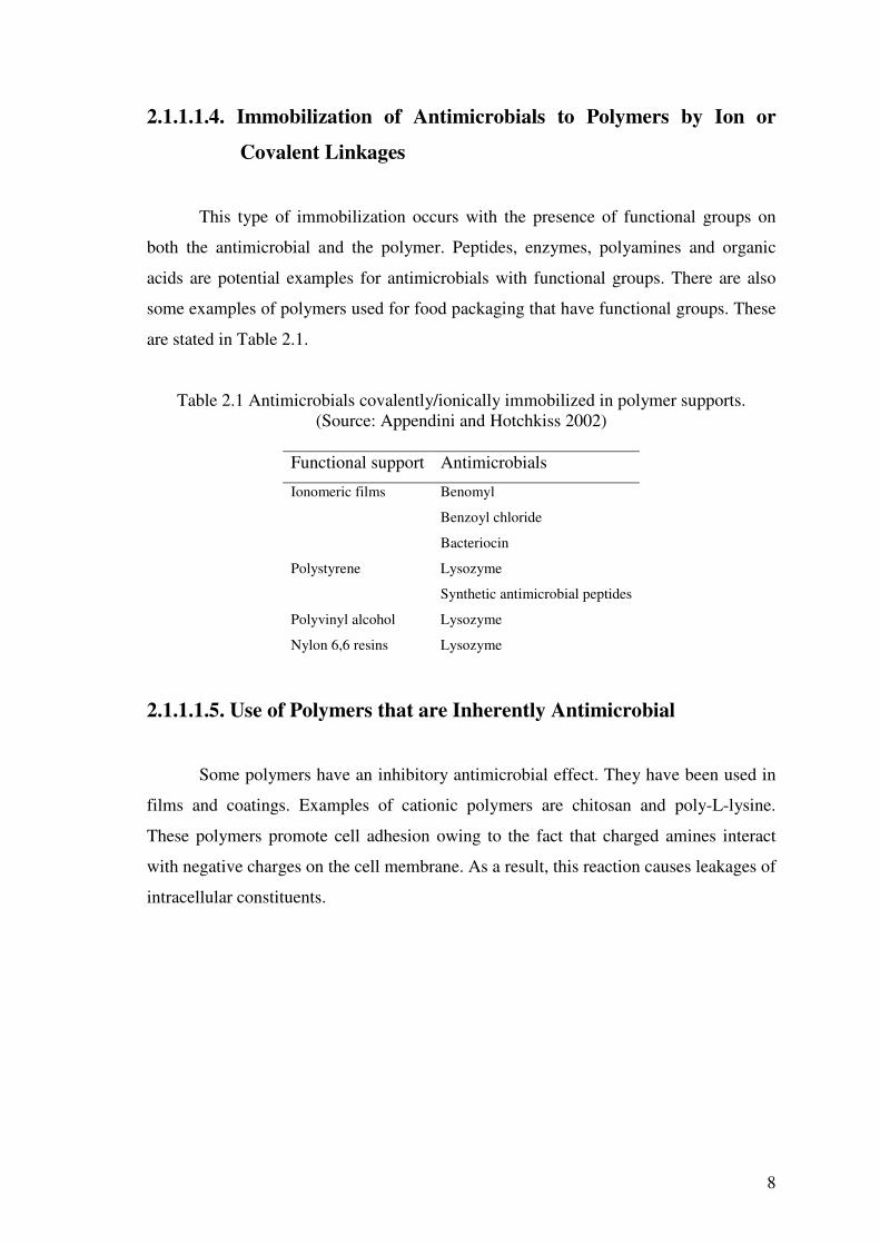
2.1.1.1.4. Immobilization of Antimicrobials to Polymers by Ion or
Covalent Linkages
This type of immobilization occurs with the presence of functional groups on
both the antimicrobial and the polymer. Peptides, enzymes, polyamines and organic
acids are potential examples for antimicrobials with functional groups. There are also
some examples of polymers used for food packaging that have functional groups. These
are stated in Table 2.1.
Table 2.1 Antimicrobials covalently/ionically immobilized in polymer supports. (Source: Appendini and Hotchkiss 2002)
Functional support Antimicrobials
Ionomeric films Benomyl
Benzoyl chloride
Bacteriocin
Polystyrene Lysozyme
Synthetic antimicrobial peptides
Polyvinyl alcohol Lysozyme
Nylon 6,6 resins Lysozyme
2.1.1.1.5. Use of Polymers that are Inherently Antimicrobial
Some polymers have an inhibitory antimicrobial effect. They have been used in
films and coatings. Examples of cationic polymers are chitosan and poly-L-lysine.
These polymers promote cell adhesion owing to the fact that charged amines interact
with negative charges on the cell membrane. As a result, this reaction causes leakages of
intracellular constituents.
9
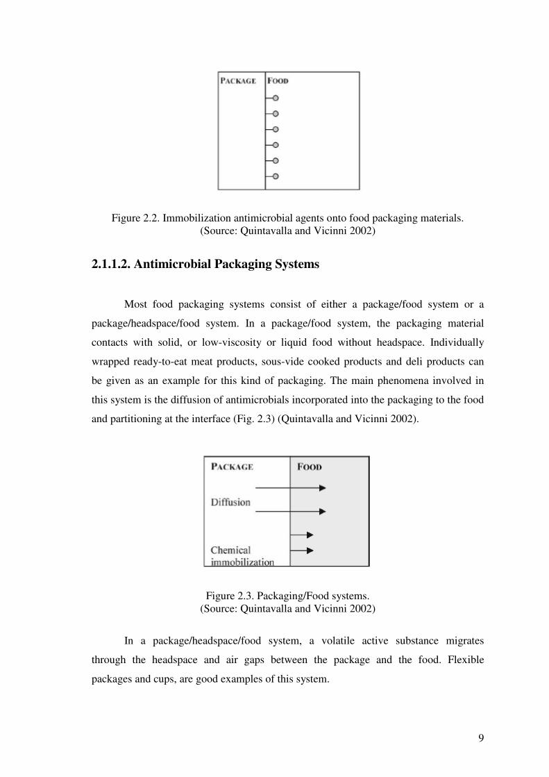
Figure 2.2. Immobilization antimicrobial agents onto food packaging materials. (Source: Quintavalla and Vicinni 2002)
2.1.1.2. Antimicrobial Packaging Systems
Most food packaging systems consist of either a package/food system or a
package/headspace/food system. In a package/food system, the packaging material
contacts with solid, or low-viscosity or liquid food without headspace. Individually
wrapped ready-to-eat meat products, sous-vide cooked products and deli products can
be given as an example for this kind of packaging. The main phenomena involved in
this system is the diffusion of antimicrobials incorporated into the packaging to the food
and partitioning at the interface (Fig. 2.3) (Quintavalla and Vicinni 2002).
Figure 2.3. Packaging/Food systems. (Source: Quintavalla and Vicinni 2002)
In a package/headspace/food system, a volatile active substance migrates
through the headspace and air gaps between the package and the food. Flexible
packages and cups, are good examples of this system.

10
Figure 2.4. Package/Headspace/Food Systems. (Source: Quintavalla and Vicinni 2002)
2.1.1.3. Important Factors Considered in the Manufacturing of
Antimicrobial Films
There are several factors to be considered in the design or modelling of the
antimicrobial film or package since an antimicrobial agent may affect the inherent
physico-mechanical properties of the package.
2.1.1.3.1. Process Conditions and Residual Antimicrobial Activity
The effectiveness of an antimicrobial agent used for the packaging may be
changed during film fabrication, distribution, and storage. The chemical stability of an
incorporated antimicrobial substance is likely to be influenced by the extrusion
conditions (high temperatures, shearing forces and pressure). Moreover, some processes
(laminating, printing, and drying) may affect the antimicrobial compounds which could
be lost during storage. All these parameters should be considered in evaluating the
effectiveness of an antimicrobial agent (Quintavalla and Vicinni 2002, Suppakul 2003).
2.1.1.3.2. Characteristics of Antimicrobial Substances and Foods
Food components significantly influence the effectiveness of the antimicrobial
substances and their release. Physico-chemical characteristics of food , including water
activity, pH and acidity, could change the activity of antimicrobial substances. For
example, the pH of food alters the ionisation of most active chemicals, and could

11
influence the antimicrobial activity of organic acids and their salts. The food water
activity could change the microflora, antimicrobial activity, and chemical stability of the
incorporated active ingredients (Quintavalla and Vicinni 2002). Moreover, aerobic
microorganisms can use headspace oxygen. Therefore, oxygen present in the headspace
may influence the microbial growth (Suppakul 2003).
2.1.1.3.3. Chemical Interaction of Additives with Film Matrix
Polarity and molecular weight of an additive are of great significance during
incorporation of additives into a polymer. Antimicrobials with low polarity are
compatible with non-polar polymeric materials. Furthermore, the molecular weight,
ionic charge and solubility of different additives influence their diffusion rates in the
polymer. For example, amongst ascorbic acid, potassium sorbate and sodium ascorbate
in calcium-alginate films at 8,15 and 20oC, the most diffused agent was the ascorbic
acid. These was due to the ionic states of the additives (Suppakul 2003).
2.1.1.3.3. Storage Temperature
Storage temperature can influence the antimicrobial activity of chemical
preservatives. Generally, the migration of the active agents in the film/coating layers
can be accelerated by the increased storage temperature. In contrast, refrigeration slows
down the diffusion rate. The temperature conditions during production and distribution
have to be taken into consideration in order to determine the residual antimicrobial
activity. (Suppakul 2003, Han 2000).
2.1.1.4. Testing the Effectiveness of Antimicrobial Packaging
There are several methods to assess the antimicrobial effectiveness of films on
microorganisms. The most common techniques are discussed in the following sections
(Appendini and Hotchkiss 2002).

12
2.1.1.4.1. Minimum Inhibitory Concentrations (MIC) Method
The minimum inhibitory concentration is the lowest concentration of an
antimicrobial in a polymer that still inhibits the growth of a particular microorganism.
This can be determined using tube dilution procedures. In this method, films containing
different dosage of antimicrobials are incorporated into tubes containing growth media
inoculated with the target microorganisim. The growth of microorganism is monitored
as a change in turbidity. By this way, the break point of the antimicrobial in the polymer
that prevents growth of the microorganism in-vitro can be determined.
2.1.1.4.2. Dynamic Shake Flask Test
This method provides more detailed confirmation on antimicrobial kinetics.
Liquid media (buffer, growth media, or foods) are seeded with the target
microorganisms and the antimicrobial polymer. The flasks are incubated with mild
agitation at appropriate temperatures. Samples are taken over time and enumerated in
order to observe the reduction in microbial growth rate.
2.1.1.4.3. Agar Plate Test
An antimicrobial containing film is placed on a solid agar medium containing
the test microorganism. The agar plates are incubated until growth is visible. A clear
zone surrounding the film is explained as antimicrobial diffusion from the film and this
diffusion of the antimicrobial from the film causes an inhibition of growth of target
microorgansim. Lack of growth under a film may show inhibition. However, suitable
controls must be included. This may be because of simple restriction to oxygen. The
agar plate tests method simulates wrapping of foods. It may suggest what can happen
when films contact contaminated surfaces. To make this method quantitative the
diameter of clear zones can be measured. Another possible uses of this method is to
inoculate the target microorgansim on an antimicrobial containing film. As known,
post-processing surface contamination is a major issue for food industry. This method
may suggest what can happen when microbial contamination occurs on films or
coatings contacting foods (Min and Krochta 2005).

13
CHAPTER 3
EDIBLE FILMS AND COATINGS
3.1. Edible Films and Coatings
Edible films and coatings have become popular in the food industry, because
they produce less waste, are cost effective, and offer protection after the package has
been opened (Cha and Chinnan 2004). They are defined as continuous matrices
prepared from proteins, polysaccharides, and lipids (Ça�rı et al. 2003). These are thin
layers of edible materials and are formed on the surface of a food as a coating or
between food components. They are natural and biodegradable substances, can be
consumed along with the food, can provide additional nutrients, can enhance sensory
characteristics and may include antimicrobials (Ryu et al. 2002).
During 12th and 13th century, in China, oranges and lemons were dipped in wax
in order to slow water loss (Hardenberg 1967). During 15th century, Yuba, the first-
standing edible film, was developed from soymilk in Japan (Debeaufort et al.1998). In
the 16th century, “larding”, coating food products with fat, was used to prevent moisture
loss in foods in England, for example, the edible protective coating was applied on
meats to prevent shrinkage (Krochta et al. 1994, Debeaufort et al. 1998). In the 19th
century, nuts, almonds, and hazelnuts were coated with sucrose to prevent oxidation and
rancidness during storage (Debeaufort et al.1998). The more important application of
edible films and coatings until now is the use of an emulsion made of waxes and oil in
water that was spread on fruits to improve their appearance, such as their shininess,
color, softening, onset of mealiness, carriage of fungicides, and to better control their
ripening and to retard the water loss.
The main purposes of the films applied on foods are: (Multon 1996, Krochta et
al. 1994)
• To provide protection against humidity and/or oxygen
• To limit surface desiccation
• To delay microbial spoilage of the surface
• To maintain water activity in heterogeneous foodstuff

14
• To control transfer of solutes, pigments, aromas,... etc. in heterogeneous
foodstuff
• To prevent absorption of brine, osmotic dehydration of syrup, or frying oil by
the foodstuff
• To improve the mechanical properties to facilitate handling on the
manufacturing line or during storage and to reduce spoilage and provide structural
integrity of a food product
• To protect individual items on foodstuffs
• To fulfill a surface which does not stick or which is not oily
• To prevent the sticking together of small portions
• To improve or modify the color, aroma, or flavor of foodstuffs
• To trap flavor during manufacture and storage.
Foods are described as: “ All raw, partially treated or treated substances used for
human nutrition and feeding” by Debeaufort et al. (1998). From this point of view,
edible films and coatings can be classified as foods. On the other hand, edible
packagings do not provide a significant nutritional value to the coated food. Therefore,
we should consider them more like an additive than an ingredient. It is clear that it all
depends on the application of the edible packaging. It can also be employed to improve
the nutritional quality of the food, and so be qualified as a food ingredient. As food
components, edible films and coatings usually have to be as tasteless as possible in
order not to be detected during the consumption of the edible-packaged food product.
Consequently, if we focus on them as a packaging and a food component, edible films
and coatings have to fullfill some requirements: they should have good sensory
qualities, high barrier and mechanical efficiencies, enough biochemical, physico-
chemical and microbial stability, free of toxics, and safe for health, simple technology,
low cost of raw materials and process, and be nonpolluting.
3.2. Functionality of Edible Films and Coatings
Edible packagings have functional properties; which are selective and active
properties. Selective properties of edible films and coatings are illustrated in Figure 3.1.
In most cases, they can be used as water barrier efficiency in order to retard the surface

15
dehydration of fresh (meat, fruits, and vegetables) or frozen products. For example,
coatings could delay the water absorption inducing the caking in food powder or the
loss of crispness in dried cakes. The control of gas exchanges ( O2, CO2, ethylene ), in
particular oxygen, allows better control of the ripening of fruits or to significantly
reduce the oxidation of oxygen-sensitive foods and the rancidity of polyunsaturated fats.
Organic vapor transfers can be prevented, which enables aroma compounds retain in the
product during storage. Moreover, by the prevention of the transfers, solvent penetration
is not allowed in foods, which can involve toxicity or off-flavors. The penetration of oil
during frying and of sucrose or sodium chloride during osmotic dehydration can be
limited by an edible film. For example, one of the more interesting applications of
edible films and coatings is the reduction of water migration in a pie because of their
use inside a composite food to control mass transfers between the different
compartments of the product.
Figure 3.1.Selective functions of edible films and coatings. (Source: Debeaufort et al. 1998)
As seen from the figure above, the film can also reduce the effect of light and
UV light that concerns radical reactions in foods. This property can be improved by
incorporating pigments or light absorbers. Therefore, they are active when films are
carriers or used for encapsulation of food additives or ingredients. Edible packagings
can improve mechanical properties of food to facilitate handling and carriage. Sensorial
characteristics such as color, shininess, transparency, roughness or sticking can be

16
improved. The films and coatings enable to protect or separate small pieces or food
portion for individual consumption.
3.3. Film Components
Components of edible films and coatings can be classified into three groups:
hydrocolloids, lipids, and composites. Below each type of edible films were discussed
in the following sections.
3.3.1. Hydrocolloid Films
Hydrocolloid films can be used in applications where barrier to oxyen, carbon
dioxide, and lipids are required (Krochta et al. 1994), but they are sensitive to moisture
and show poor water vapor barrier properties. They have suitable mechanical and
optical properties (Ryu et al. 2002), and for this reason it makes them useful for
strengthening the structure of the foodstuff. Hydrocolloids can be grouped into two
classes as carbohydrates or proteins according to their composition (Krochta et al.
1994).
3.3.1.1. Carbohydrate Films
Polysaccharides are nontoxic and widely available. They also have selective
permeablity to CO2 and O2, and hence retard the respiration and ripening of many fruits
and vegetables by limiting the availability of O2. Polysaccharide-based films have a
hydrophilic nature. For this reason, they are a poor barrier to water vapor. The poor
water vapor barrier property allows for the movement of water vapor across the film,
thus, preventing water condensation that can be a potential source of microbial spoilage
in horticulture commodities (Cha and Chinnan 2004). Namely, the coating which has a
fairly high water content acts by losing water before the product.

17
3.3.1.1.1. Starch
Starch is very biodegradable and cost effective, but is also very hydrophilic.
Their mechanical properties are generally inferior to synthetic polymer films. When a
plasticizer such as water is incorporated, starch exhibit thermoplastic behavior (Cha and
Chinnan 2004). Starch is composed of amylose and amylopectin. It is primarily derived
from cereal grains, potatoes, tapioca, or arrowroot. Amylose is responsible for the film-
forming capacity of starches. Amylose, the linear fraction of starch, is known to form a
coherent and relatively strong, freestanding film, in contrast to amylopectin films,
which are brittle and noncontinuous. Starch-based coatings containing potassium
sorbate have been applied to extend the storage life of strawberries. The formulations
with potassium sorbate have not only reduced the microbial count, but also extended the
storage life from 14 to 28 days in coated starwberries. In addition, since the water
activity (aw) is critical for microbial, chemical, and enzymatic activities, studies have
demonstrated that films can resist the migration of moisture into the meat or poultry
during the storage (Cha and Chinnan 2004).
3.3.1.1.2. Alginate
Alginates are salts of alginic acid. They are linear copolymers of D-mannuronic
acid and L-guluronic acid and extracted from seaweeds, major structural
polysaccahrides of brown seaweeds known as Phaeophyceae (Hui 1991). There are
some seaweeds used as commercial production, including Macrocystis pyrifera,
Laminaria hyberborea, Laminaria digitata, and Ascophyllum nodosum.
Alginic acid is a linear ( 1->4 ) linked polyuronic acid containing three types of
block structures:
� poly-�-D-mannopyranosyluronic acid ( M ) blocks
� poly-�-L-gulopyranosyluronic acid ( G )blocks
� MG blocks containing both polyuronic acids
18
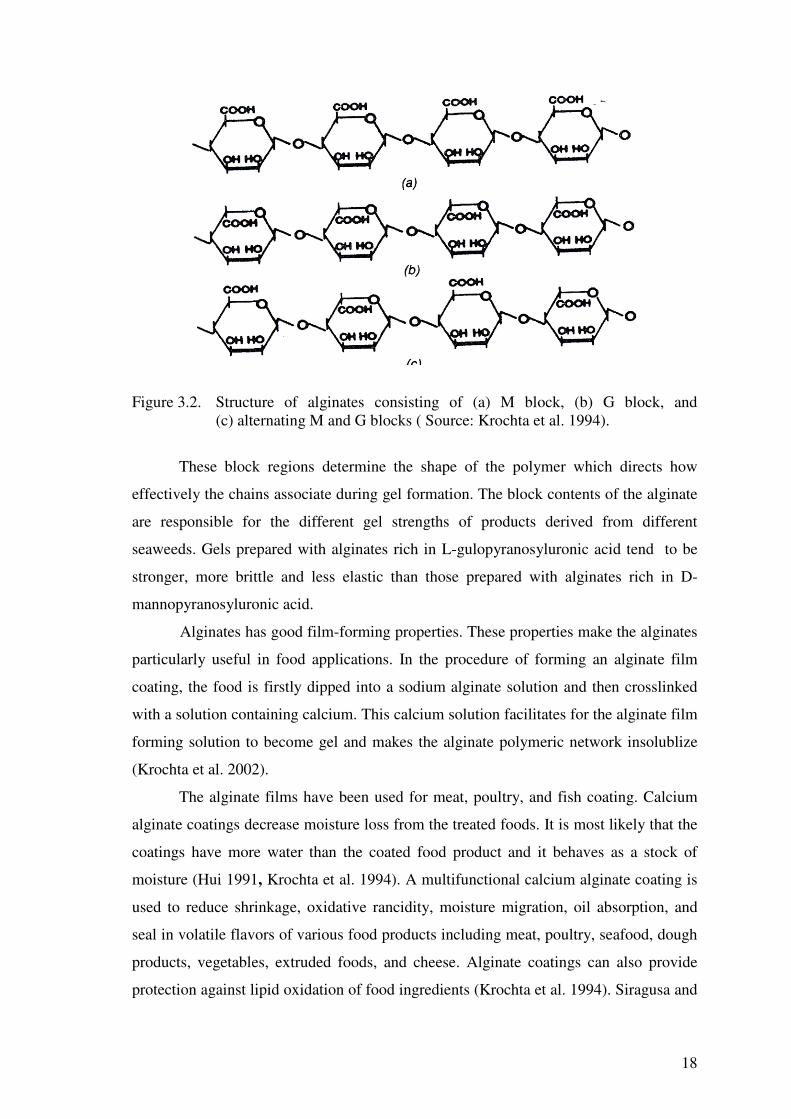
Figure 3.2. Structure of alginates consisting of (a) M block, (b) G block, and
(c) alternating M and G blocks ( Source: Krochta et al. 1994).
These block regions determine the shape of the polymer which directs how
effectively the chains associate during gel formation. The block contents of the alginate
are responsible for the different gel strengths of products derived from different
seaweeds. Gels prepared with alginates rich in L-gulopyranosyluronic acid tend to be
stronger, more brittle and less elastic than those prepared with alginates rich in D-
mannopyranosyluronic acid.
Alginates has good film-forming properties. These properties make the alginates
particularly useful in food applications. In the procedure of forming an alginate film
coating, the food is firstly dipped into a sodium alginate solution and then crosslinked
with a solution containing calcium. This calcium solution facilitates for the alginate film
forming solution to become gel and makes the alginate polymeric network insolublize
(Krochta et al. 2002).
The alginate films have been used for meat, poultry, and fish coating. Calcium
alginate coatings decrease moisture loss from the treated foods. It is most likely that the
coatings have more water than the coated food product and it behaves as a stock of
moisture (Hui 1991, Krochta et al. 1994). A multifunctional calcium alginate coating is
used to reduce shrinkage, oxidative rancidity, moisture migration, oil absorption, and
seal in volatile flavors of various food products including meat, poultry, seafood, dough
products, vegetables, extruded foods, and cheese. Alginate coatings can also provide
protection against lipid oxidation of food ingredients (Krochta et al. 1994). Siragusa and
19
Dickson (1992) tested the use of organic acids immobilized on the surface of inoculated
beef tissue to inhibit microbial growth. The researchers showed that the lactic and acetic
acids used in immobilized calcium alginate gels inhibited Listeria monocytogenes on a
lean beef tissue. These acids-alginate gels indicated a promising method for sanitizing
and preserving raw meat. Natrajan and Sheldon (2000) examined the use of calcium
alginate film including nisin against Salmonella typhimurium. They studied the effect of
alginate film use for coating broiler drumstick skin samples contaminated with S.
typhimurium and then coated with the film. The results indicated that the level of
reduction in microbial growth was affected by film type and gel concentration, exposure
time, and nisin concentration. Pranoto et al.(2005) reported the antibacterial effect of
alginate-based edible film incorporated with garlic oil against Escherichia coli,
Salmonella typhimurium, Staphylococcus aureus and Bacillus cereus. The researchers
also examined the concentration of garlic oil in the film and concluded that
incorporation of garlic oil into alginate film at levels more than 0.2 % led to a
significant inhibitory effect on S.aureus and B.cereus. Cha et al.(2002) studied Na-
alginate based antimicrobial films including lysozyme, nisin, grape fruit seed extract
and EDTA. The film incorporated with grape fruit seed extract-EDTA showed the
strongest inhibitory effect on Listeria innocua, Escherichia coli, Salmonella enteritidis,
Staphylococcus aureus, and Microccocus luteus. Oussalal et al. (2006) evaluated the
concentration of the pretreatment used (2% or 20 % CaCl2) and the antimicrobial effect
of cinnamon, oregano, and savory essential oils in alginate based films against E. coli
O157:H7 and S. typhimurium on beef muscle. They obtained that after 5 days of
storage, S.typhimurium was mostly affected by the films containing oregano or
cinnamon essential oils regardless of the concentration of the cross-linking agent. In
addition, oregano incorporated films were effective against E.coli together with the
pretreatment of 2% CaCl2. Ogunbanwo and Okanlawon (2006) immobilized bacteriocin
produced by L. brevis in edible alginate film (1% w/v). In this study, they inoculated the
chicken samples with S .kentucky AT1 and then examined the shelf-life of refrigerated
chicken treated with bacteriocin containing film. They indicated that the immobilized
bacteriocin into alginate film could extend shelf-life of samples up to 14 days at
refrigerated storage.

20
3.3.1.1.3. Carrageenan
Carrageenan is a complex mixture of several polysaccharides. As for alginates,
the films obtained are transparent, odorless, and very lightly salted in the case of the
addition of calcium salts. Their mechanical and protective properties are average, and
their applications are restricted to improving the appearance, preventing sticking, and
protecting against rancidity of dried products such as dried fruits and protecting frozen
meats and fish from surface drying. Some authors searched the reduction of
Staphylococcus aureus on the surface of an intermediate moisture food by using a
coating composed of a carrageenan agarose gel to obtain a surface pH of 0.5 units less
than the pH of the rest of the foodstuff (Bureau and Multon 1996).
3.3.1.1.4. Cellulose Derivatives
The modified cellulose ethers, including methyl cellulose, hydroxypropyl
cellulose, hydroxypropyl methyl cellulose, and ethyl cellulose, are the basis of many
edible film coatings. The methyl celluloses have hydrophilic nature. Therefore, it makes
them suitable for components of nonpolar lipid films having moisture barrier properties
(Hui 1991). Cha et al (2002) studied the antimicrobial effect of lysozyme incorporating
carragenan films. The results showed that these films were not superior than lysozyme
incorporated alginate films under the same conditions.
3.3.1.1.5. Pectin Films
Pectins are a group of plant-derived polysaccharides. They work well with low-
moisture foods, but they are poor moisture barriers. Pectins are composed of methyl
esters of linear chains of 1,4-�-D-galacturonic acid units. During the processing methyl
esters may be hydrolyzed, producing low methoxy pectin. The low methoxy pectin have
a divalent cation gelling mechanism similar to that of the alginates and carrageenan, and
the use of low methoxy pectins as edible coatings is similar to that of the alginates. The
food to be coated is first dipped into a solution of low methoxy pectin and then into a
solution of low methoxy pectin and then into a solution of calcium chloride. The
thickness of the coating can be controlled by the concentration of the pectin or the

21
viscosity of the pectin solution. These gels have been used as coatings for almonds,
candied fruit, and dried fruit (Hui 1991).
3.3.1.1.6. Chitosan
Chitosan is an edible and biodegradable polymer. It is derived from chitin.
Chitosan is also an abundant natural polymer available. It forms films without the
addition of additives, exhibits good oxygen and carbon dioxide permeability beside
excellent mechanical properties. This material not only acts as a chelator in biological
system, but also exhibits antimicrobial activity against bacteria, yeasts and molds.
However, one disadvantage with chitosan is its high sensitivity to moisture. Quattara et
al.(2000) investigated the inhibition effect of chitosan films containing cinnamaldehyde
against S.liquefaciens and Enterobacteriaceae on bologna. They reported that growth of
the microorganisms were delayed or completely inhibited as a result of film application.
3.3.1.2. Protein Films
Proteins may be derived from corn, wheat, soybeans, peanut, milk, or gelatin.
They are suitable for coating fruits and vegetables. Protein-based coatings have good
barriers to O2 and CO2, but not to water. Several protein based edible film discussed in
the following section.
3.3.1.2.1. Zein
Zein is a protein extracted from maize or corn. It is only soluble in the mixture
of water and alcohol because it is protein. It has good humidity barrier property. This
type of protein based edible films has been used commercially in coating formulations
of shelled nuts, candy, and pharmaceutical tablets (Bureau and Multon 1996, Hui 1991,
Cha and Chinnan 2004). These films are also suitable coatings for dried fruits and
intermediate moisture foodstuffs (Bureau and Multon 1996). Mecito�lu et al.(2005)
reported that corn zein films incorporated with partially purified lysozyme showed
antimicrobial effect on Bacillus subtilis and Lactobacillus plantarum. Hoffman et
al.(2000) studied the effect of corn zein films impregnated with nisin, lauric acid, and
22
ethylene diamine tetracetic acid (EDTA) on L.monocytogenes and Salmonella
enteritidis. They found that L.monocytogenes could not be detected after 24h exposure
to the film combination including lauric acid. The film with EDTA and lauric acid and
EDTA, lauric acid, nisin had bacteriostatic effect when the initial inoculum of S.
Enteritidis was 104 CFU/ml was used.
3.3.1.2.2. Whey Protein Isolates (WPI)
Whey proteins account for 20% of total milk proteins. They are characterized by
their solubility at pH 4.6. Liquid whey is a by-product of cheese manufacture and is
produced in large quantities, but much of this whey is not utilized. Hence, it causes
serious waste disposal problems. Whey proteins are water soluble and form hydrophilic
edible films (Krochta et al.2002). WPI can form transparent, flexible, colorless, and
odorless films that provide excellent oxygen, aroma, and oil barrier properties (Min and
Krochta 2005). Ça�rı et al.(2001) developed an edible film (pH5.2) from whey protein
isolate containing p-aminobenzoic acid (PABA) or sorbic acid (SA). The researchers
showed that incorporating 0.5% to 1.5% of SA or PABA into WPI films led to
inhibition of L.monocytogenes, E.coli O157:H7, and S. typhimurium DT104 on
trypticase soy agar + 0.6% yeast extract.
3.3.1.2.3. Collagen Casings
Collagen is a fibrous protein. The film-forming ability of collagen has been
traditionally utilized in the meat industry for production of edible sausage casings.
Collagen is manufactured from corium layer of bovine hide which is more than 90%
collagen on a dry basis. To obtain casings, the hide coriums is delcalcified and grinded
into small pieces. Then, ground collagenous material with acid is mixed to produce a
swollen slurry (4-5% solids). The slurry is homogenized and extruded into tubular
casings( 8-10%). Finally, tha casings free of salts are washed, treated with plasticizer
and cross-linking agents and dried (Min and Krochta 2005).

23
3.3.2. Lipid Films
Lipid compounds contain neutral lipids of glycerides. Glycerids are esters of
glycerol and fatty acids and waxes, which are esters of long-chain monohydric alcohols
and fatty acids. From this group, acetylated monoglycerides, natural waxes, and
surfactants are commonly utilized in edible coatings. Lipids are commonly added to
food coatings to impart hydrophobicity. Lipids are mainly used for their efficiency as a
water-vapor barrier in edible films. The structure, degree of saturation, chain length,
physical state, shape and dimension of crystal, and distribution of lipids into the film
influence the functional properties of the film (Cha and Chinnan 2004). Wax and
glycerides are examples of lipid based films and described below.
3.3.2.1. Waxes
Waxes belong to the non-polar lipid class. They are insoluble in bulk water and
do not spread to form a monolayer on the surface. Their hydrophobicity is high. There
are differences in permeability of wax films. These differences is owing to their
chemical composition and crystal type. The waxy skin on fresh fruit and vegetables is
applied to reduce dehydration and control the exchange of gases to prolong preservation
period (Cha and Chinnan 2004). There are some examples of waxes used for coating,
including paraffin wax, carnauba wax, beeswax, candelilla wax, polyethylene wax
(Krochta et al. 2002).
3.3.2.2. Glycerides
Monoglycerides are used in edible films as emulsifiers, especially for stabilizing
emulsified film and increasing adhesion between two components with different
hydrophobicity. Triglycerides are insoluble in bulk water, but will spread at the
interface to form a stable monolayer. Water affinity or hydrophobicity of triglyceride
depends on its structure. By adding palmitic, stearic, lauric acids, and stearyl alcohols to
edible films, the moisture barrier properties are greatly enhanced (Cha and Chinnan
2004).

24
3.3.3. Composite Films
Composite films can be designed by combining lipid and hydrocolloid elements.
By this way, it can decrease the disadvantages of each film. When a barrier to water
vapor is desired, the lipid component can serve this function while hydrocolloid
component provides the necessary durability. Composite films consisting of a
conglomerate of casein and acetylated monoglycerides have been studied by Krochta et
al.(1990). These films can be used as coatings for processed fruit and vegetables.
3.4. Film Formation
There are several techniques developed to form edible films. These techniques
include coacervation, solvent removal and solidification of melt and are given below.
3.4.1. Coacervation
The principle of coacervation is to separate a polymeric coating material from a
solution by heating, altering pH, adding solvents, or changing the charge on the polymer
concerned (Krochta et al. 1994). In the simple coacervation a hydrocolloid is dispersed
in aqueous solution and precipitated or gelified by the removal of the solvent, by the
addition of a non-electrolyte solute in which the polymer is not soluble, by the addition
of an electrolyte substance inducing a “salting out” effect or by the modification of pH
of the solution. Complex coacervation, where two hydrocolloid solutions with opposite
charges are combined, inducing interactions and the precipitation of the polymer
mixture (Debeaufort et al. 1998).
3.4.2. Solvent Removal
It is generally applied for the production of hydrocolloid edible films. In this
process, film-forming material is dispersed in a solvent such as water, ethanol, or acetic
acid. These solvents contain several additives including plasticizers, cross-linking
agents, solutes,...etc. The film-forming solution is then cast in a thin layer, dried and

25
peeled from the surface (Ça�rı et al. 2003). Solvent removal is of great importance to
get solid film formation (Krochta et al. 1994).
3.4.3. Solidification of Melt
Melting followed by solidification is one of the commonly used techniques to
produce lipid films. In this method the rate of solvent removal and the cooling rate play
an impotant role in the overall properties of the resulting film (Krochta et al.1994).
3.5. Film Additives
Additives may be added to edible films to improve their mechanical, protective,
sensory, and nutritional properties. These are plasticizers, antimicrobial agents,
antioxidizing agents, colorants,...etc. (Bureau and Multon 1996).
A plasticizer is defined as “a substantially nonvolatile, high boiling,
nonseparating substance, which when added to another material changes the physical
and/or mechanical properties of that material”(McHugh and Krochta 1994). A
plasticizing agent must be compatible with the film-forming polymer. Moreover, its
permanence within solvent-polymer system is also of prime importance since this
influences the physical and mechanical stability of the film. In addition, the polymer
and the plasticizer must also have similar solubility in the solvent used. The plasticizer
should not be volatile (or only very slightly volatile) and its degree of retention by the
film should be high. Furthermore, other properties, such as its chemical stability,
hygroscopocity, color, flavor, and so on, are also more or less important depending on
the type of film under discussion. Finally, the concentration of a plasticizer necessarily
varies from 10-60% (dry base) according to the nature and type of film and the method
of application. The most often used plasticizers in edible films and coatings are mono-,
di-, and oligosaccharides (generally glucose syrups or glucose-fructose honey, polyos
(principally glycerol and its derivatives, polyethylene glycols, sorbitol), and lipids and
its derivatives (fatty acids, monoglycerides and their esters, acetoglycerides,
phospholipids, and other emulsifiers) (Bureau and Multon 1996). McHugh and Krochta
(1994) used sorbitol as a plasticizer in whey protein isolate solution and reported that
sorbitol was more effective than glycerol in those films of equal tensile strength,

26
elongation, and elastic modulus. However, the workers determined that the sorbitol used
in the film had lower oxygen permeabilities. Ghanbarzadeh et al.(2006) studied the
effects of sugar plasticizers on the viscoelastic and thermal properties of zein resins. The
researchers found that the films containing galactose had better tensile strength and
strain at break than films containing fructose and glucose.
Edible films and coatings may be used as means for preservatives
(antibacteriological or antioxidizing agents) in order to maintain an effective
concentration of these substances on the surface of the foodstuff. The use of edible films
as a carrier for preservatives makes it possible to combat the deterioration which occur
during the onset of storage (e.g., oxidation of dried fruits or development of
microorganisms such as molds, yeasts, or osmophilic bacteria on numerous foodstuffs
of medium humidity). The addition of preservatives can be used without destruction of
the food. In addition, it also uses low amounts of additives in relation to the weight of
the product owing to the fact that a sufficient concentration of the additive is maintained
on the surface (Bureau and Multon 1996). Park et al.(2005) incorporated potassium
sorbate into chitosan coating solution to enhance preservative effects on the storability
of strawberries during storage. Theivendran et al.(2006) evaluated the inhibitory effect
of grape seed extract (GSE) and green tea extract (GTE) against Listeria
monocytogenes. This research demonstrated that the use of soy protein edible film
coating containing both nisin and natural extracts was a promising means of controlling
the growth and recontamination of L.monocytogenes on ready-to-eat meat products.
Seydim and Sarikus (2006) determined the inhibitory effects of whey protein based
edible films incorporated with essential oils against E.coli O157:H7, S.aureus,
S.enteritidis, L.monocytogenes, and L.plantarum. The researchers observed that the film
containing oregano essential oil was the most effective film against these bacteria at 2%
level.
Incorporation of antioxidants into packaging materials has also become very
popular since oxidation is one of the problems influencing the food quality (Güçbilmez
et al. 2007). An antioxidant agent can prevent enzymatic (lipogenase) and non-
enzymatic peroxidation of essential fatty acids (Hortmann and Meisel 2007). In the
same way, films and coatings may serve to maintain high concentration of colorants,
flavors, spices, herbs, acids, sugars, salts,...etc. This may give the foodstuff a strong
surface taste and nutritional enrichment without destruction of the food (Bureau and
Multon 1996).
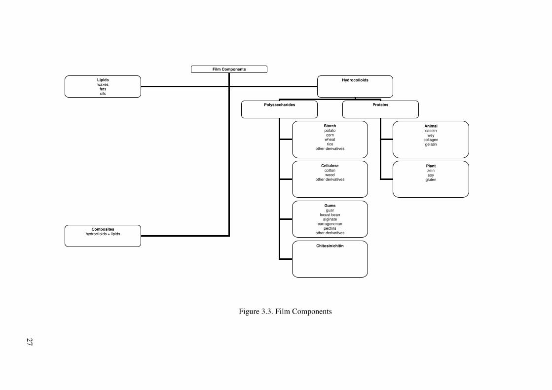
27
Figure 3.3. Film Components
Film Components
Lipids waxes
fats oils
Hydrocolloids
Composites hydroclloids + lipids
Polysaccharides Proteins
Starch potato corn
wheat rice
other derivatives
Cellulose cotton wood
other derivatives
Gums guar
locust bean alginate
carragenenan pectins
other derivatives
Chitosin/chitin
Animal casein
wey collagen gelatin
Plant zein soy
gluten
27

28
CHAPTER 4
BIOPRESERVATION
4.1. Definition of Biopreservation
Consumers expect to have natural, more stable and safe products (Hugas et al. 1998,
Lemay et al. 2002, Leroy et al. 2002). This can be achieved by the use of mild processes
without utilizing chemical additives. It is clear that these additives damage the product’s
preservative-free status. They, therefore, are not agreeable with the fresh image (Rodgers
2003, Rodgers 2004, Devlieghere et al. 2004, Schnürer and Magnusson 2005, Osmano�lu
2005). Recently, the food industry and food research have intensified on natural
antimicrobial compounds owing to the consumers’ ban against chemical preservatives
(Devlieghere et al. 2004).
In the early days it was noticed that fermented foods had delicate and refreshing
tastes, with an extended shelf life. They also decreased the outbreak caused by
foodborne illnesses (Ray 2004). Nowadays, novel technologies such as
‘biopreservation’ have attracted great attention as means of naturally controlling the
growth of pathogenic and spoilage organisms (Mataragas et al. 2003). As it is known,
that food additives are widely employed so as to hamper undesired microorganisms and
to enhance shelf life. However, biopreservation is a useful device to prevent an overuse
of food additives (Schewenninger and Meille 2004). A definition of the biopreservation
is as follows: “Biopreservation is a way to prolong storage life and enhance safety by
the use of natural or controlled microflora and/or their antimicrobial products.”(Rodgers
2003, Hugas 1998, Devlieghere et al. 2004).
As a conclusion, biopreservation is provided by the use of protective cultures
and their antmicrobial products or some natural compounds isolated from food. These
biopreservatives are briefly explained in the following section.
4.1.1. Protective Culture
Protective cultures are food grade bacteria (Rodgers 2001), which are also
termed as antagonistic cultures (Devlieghere et al. 2004). Lactic acid bacteria are

29
considered as the major group of the antagonistic cultures (Rodgers 2001a, Rodgers
2001b). The objective of their antagonistic activity is to inhibit other microorganisms
through competition for nutrients and/or production of primary or secondary
metabolites (Vermeiren et al.2004, Devlieghere et al. 2004). Some of the compounds
released by the bacteria have low molecular weight, such as lactic acid, H2O2, CO2,
alcohols, phenyllactic acids, cyclic dipeptides and short or medium chain fatty acids
(Rodgers 2003, Vereecken and Van Impe 2002, and Schnürer and Magnusson 2005)
and some have high molecular weight, amongst which are polysaccharides and
bacteriocins (Vereecken and Van Impe 2002).
Alcohols reutericyclin Caproic acid Phenyllactic acid 3-hydroxy fatty acids H2O2 Cyclic dipeptides Reuterin Organic acids diacetyl proteinaous compounds reutericyclin CO2 2-pyrrolidone-5-carboxylic acid
Figure 4.1. Summary of the main antimicrobial compounds produced by LAB
(Source: Salmien et al. 2004)
4.1.1.1. Organic Acids
Acetic acid is commercially produced by Acetobacter aceti. In general, when the
level of acetic acid is 0.2%, it shows bacteriostatic activity, however, bacteriocidal above
0.3%. It is more effective against Gram-negative bacteria. Actually, this effect depends on
pH and the bactericidal effect is more definite at low pH (below pH 4.5). It is used as an
antimicrobial agent to salad dressings, mayonnaise, and carcass wash (Ray 2004).
Lactic acid is manufactured by some Lactobacillus spp. which are able to
produce L-lactate (or DL-lactate).The antibacterial effect is pronounced when utilized at
1 to 2 % levels (�pH 5). At low pH 5, lactic acid can have a bactericidal activity
particularly against Gram-negative bacteria (Ray 2004). It is capable of inhibiting the

30
growth of various food spoilage bacteria, such as Gram-negative species of the families
Enterobacteriaceae and Pseudomonadaceae (Alakomi et al. 2000). However, it may not
have an antimicrobial activity on fungi in the food environment (It is usually employed
on carcasses) (Ray 2004).
Propionic acid by Propionibacterium spp. has fungistic effect in the food
environment and also capable of inhibiting the growth of Gram-negative and Gram-
positive bacteria. At pH 5 or below, Gram-negative bacteria are more sensitive than
Gram-positive ones as used 0.1 - 0.2 % levels,. It is applied to control molds in cheeses,
butter, and bakery products and to hamper growth of bacteria and yeasts in syrup,
applesauce, and some fresh fruits (Ray 2004).
The antimicrobial property of these organic acids is due to the fact that they are
present in the form of undissociated molecules. Their dissociation constants ( pKa ) for
acetic, propionic, lactic acid are 4.8, 4.9 and 3.8, respectively (Ray 2004). They enter
the cytoplasmic membrane and result in decrease in intracellular pH and disruption of
the transmembrane proton motive force (Alakami et al.2000), eventually cause
denaturation of proteins and viability loss (Ray 2004). On the other hand, some studies
reported that these weak acids provide antimicrobial action with the combined effects of
undissociated molecules and dissociation ions (Ray 2004).
4.1.1.2 Diacetyl
Diacetyl is produced by some lactic acid bacteria during citrate fermentation (Ray
2004, Schnürer and Magnusson 2005). The more diacetyl concentration makes a change in
the taste and aroma of the product (Schnürer and Magnusson 2005). Diacetyl (2,3-
butanedione) has antimicrobial effects at low pH (Schnürer and Magnusson 2005). Gram-
negative bacteria is especially susceptible at pH 5.0 or below. When it is utilized with the
combination of heat, it is more bactericidal than when it is used alone (Ray 2004).
Under reduced conditions, acetoin is formed by the conversion of diacetyl. This
conversion leads to decrease in antibacterial activity. The antimicrobial effect is likely
due to its deactivating property on some important enzymes. The dicarbonyl group (-
CO-CO-) makes a reaction with arginine in the enzymes and eventual modification in
their catalytic sites (Ray 2004).
31
4.1.1.3. Hydrogen Peroxide
Hydrogen peroxide (H2O2) produced by most lactic acid bacteria is widespread
under aerobic conditions (Ray 2004). LAB have flavoprotein oxidases which enable
them to produce H2O2 in the presence of oxygen (Schnürer and Magnusson 2005).
However, they don’t have cellular catalase, pseudocatalase, or peroxidase, and therefore
the culture release the agent into the environment to avoid its antimicrobial action (Ray
2004). The production of H2O2 by LAB can be useful for food preservation and
prevention of growth of foodborne spoilage and pathogenic bacteria and pathogens
(Villegas and Gilliland 1998).
The synthesis of H2O2 by LAB should enable them to prolong the shelf life of
some refrigerated foods without altering the acidity of the food (Zálan et al. 2005).
H2O2 has a strong oxidizing effect on the bacterial cell and on the destruction of basic
molecular structures of cellular proteins (Schnürer and Magnusson 2005, Zálan et al.
2005). Bacteriostatic action was shown at 6 to 8 �g/ml H2O2 levels, 30 to 40 �g/ml
induce bacteriocidal effect. It can also show antimicrobial action against fungi and
viruses (also bacteriophages) (Ray 2004).
4.1.1.4. Fatty Acids
Antimicrobial fatty acids can be produced by lipolytic LAB. These acids also
make a contribution to the sensory quality of fermented foods. The antimicrobial
property can change according to the chain length. For example, caprylic (C8) acid and
longer fatty acids are the most effective, exception of undecanoic, (C11). The researchers
found that the growth of yeast cells treated with 10 mM of the fatty acids and
monoglycerids was inhibited due to the capric and lauric acid (Schnürer and Magnusson
2005).
4.1.1.5. Phenyllactic Acid
Phenyllactic acid has antimicrobial action only on yeasts and molds at a given
concentrations. This metabolite is active in combination with other compounds
produced by LAB. In sourdough bread the use of L.plantarum delayed the growth of the

32
mold Aspergillus niger, compared to the ose of Lactobacillus brevis that did not
produce phenyllactic acid (Schnürer and Magnusson 2005).
4.1.1.6. Cyclic Dipeptides
The hydroxylated fatty acids are stronger in terms of antifungal activity than the
cyclic dipeptides. These peptides show antifungal activity at given concentrations.
(Schnürer and Magnusson 2005).
4.1.1.7. Bacteriocins
Bacteriocins are ribosomally-produced, cationic, and amphiphathic pose �-
helical and/or �-sheet structures and can also have thioethers, disulfide bridges, or free
thiol groups (Ray 2004). These agents synthesized by LAB have antimicrobial action on
closely related species of the bacteria and other Gram-positive spoilage and pathogenic
bacteria (Hugas 1998, Alakomi et al. 2000, Osmano�lu 2003, Devlieghere et al. 2004,
Ray 2004,, and Zálan et al. 2005).
The principle of their antimicrobial action is to bind to the surface of the cell
wall or the membrane of target organisms, and then form pores in the membrane in
order to reduce transmembrane potential and/or pH gradient, namely, its functional
destabilization, and provide leakage of cellular materials. Eventually, this causes the
death of the cell (Ray 2004, Zálan et al., 2005).
4.1.1.8. Reutericyclin
It is also produced by L.reuteri. It is negatively-charged and has highly
hydrophobic character. The inhibitory action is rised at higher salt concentration (2%)
and low pH. The minimal inhibitory concentration was found to be approximately 0.05-
1 mg/L for Gram-negative bacteria and yeasts. It behaves as a proton ionophore, in
contrast to other antimicrobial agents which forms pores in the membrane of the target
cells. It separates the cytoplasmic membrane, owing to its hydrophobicity, eventually,
disrupts the transmembrane pH.

33
4.1.1.9. 2-Pyrrolidone-5-Carboxylic Acid
Pyroglutamic acid, or PCA, is produced by Lactobacillus casei ssp. casei,
L.casei ssp. pseudoplantarum and Streptococcus bovis. It is also present in fruit,
vegetables and grasses. It has an inhibitory activity to Bacillus subtilis, Enterobacter
cloacae, Pseudomonas putida, and Pseudomonas fluorescens. It is heat stable (20 min at
121 oC). It is affected by the decrease in pH. Its mechanism of action resembles to that
of organic acids (Salmien et al. 2004).
4.1.2. Factors Affecting Protective Culture Performance
The inhibitory effectiveness of a protective culture could vary according to some
factors stated below:
4.1.2.1. Temperature Effect
Temperature can affect growth rate, and bacteriocin production of the
microorgansims. The protective culture should maintain its viablity in products under
refrigerated temperatures. In addition, it should be able to grow and start an antagonistic
action at the desired temperatures. Its growth rate should be faster than the food
poisoning and spoilage organisms’growth rates. Finally, if the inoculated product is
intended for cooking, the heat resistance of the protective culture should be considered
in order not to leave the product unprotected after heating. Production rate of
bacteriocins and their ability depend on incubation temperatures. Based on temperature,
a food poisoning and spoilage organisms can gain sensitiveness to inhibition. For
example, psychrotrophic strains of LAB isolated from commercial fresh salads
hampered the growth of different pathogens at different temperatures (Rodgers 2001).
4.1.2.2. Protective Culture and Food Poisoning-Spoilage Organisms
A protective culture can be effective against one or several food poisoning-spoilage
organisms.The inhibitory substance production rate by a PC and the sensitivity of a FPSO
to inhibition depends on the identity of the interactive cultures (Rodgers 2001).

34
Table 4.1. Application of Protective Cultures to Non-Refrigerated Foods. (Source: Rodgers 2001)
PC Temperature
( Co ) Inoculum ( cfu/g)
FSPO Food Reference
Raw meat,milk, and eggs L.delbrueckii subsp. bulgaricus
5 5x10⁸ Pseudomonas spp. Ground beef Gilland and Speck (1975)
P.cerevisiae 3 109 Pseudomonas spp. Raw poultry Raccach,Baker, Regenstein,and Mulnox (1979)
P.cerevisiae Lc. lactis 7 5x10⁷ E.coli o157:H7 Raw chicken
meat Brashers,Reilly, and Gilliland (1998)
L.delbrueckii subsp. bulgaricus
5,7 108 P.fragi Psychrotrophs
Milk Fresh crab meat
Gilliland and Speck (1975)
P.cerevisiae 3 109 Pseudomonas spp.
Pasteurised liquid whole egg
Raccach and Baker (1979)
L.plantarum S.typhimurium Ready-to-eat meals
E.faecium 3 104, 107 L.monocytogenes C.botulinum C.perfringens B.thermosphacta
Sous vide fish Smith (1975)
Lc. lactis 30 106 C.botulinum Chicken a la king Saleh and Ordal (1955)
L.delbrueckii subsp. bulgaricus
L.plantarum ATCC 8014
5,15,25 106 C.botulinum type A,B and E
Refrigerated can pea soup
Skinner et al. (1999)
L.bavaricus 4,10 103,105 L.monocytogenes Sous-vide beef Winkowski et al. (1999 )
L.plantarum 4 4x10⁶ C.botulinum Cured meat Tanaka,Meske,Doyle, Traisman,Thayer, and Johnson (1985)
P.acidilactici 7 L.alimentarius 4 1010 Micrococci Cooked ham Kotzekidou and
Bloukas (1996) S.xylous Staphylococci
Brochothix thermosphacta Aerobic bacteria Pseudomonas
L.casei 4,8 108 L.monocytogenes Ready-to-eat vegetables
Vescovo et al. (1996)
L.plantarum S.typhimurium
35
Table 4.1. Application of Protective Cultures to Non-Refrigerated Foods. (cont.) (Source:Rodgers 2001)
Pediococcus spp.
S.aureus Aeromonas hydrophila
P.acidilactici L.delbrueckii subsp. bulgaricus
6 4x10⁹ Background microflora
Tuna and Potato salad
Raccach et al. (1979)
Seafood Leuconostoc spp
5 106, 107 L.monocytogenes Shrimp extracts Jeppesen and Huss (1993)
L.plantarum Yersinia enterocolitica
Carnobacterium piscicola
4 108 H2S-producing bacteria
Smoked salmon Leroi et al. (1996 )
L.plantarum Yeasts and molds Lc.lactis subsp. lactis ATCC 11454
10 3x10⁶ L.monocytogenes Cold-smoked salmon
Wessels and Huss (1996)
Carnobacterium piscicola
L.plantarum Others L.plantarum 15 108 Salmonella Model food slurries Ernsaw et al.
(1993) L.mesenteroides
L.monocytogenes
P.pentosaceus Lc. lactis 10 106 C.botulinum 17B TPGY broth Rybka-
Rodgers (1989)
P.pentosaceus 107 L.plantarum 15 108-109 S.typhimurium
Staphylococcus aureus
Buttered brain heard Infusion Broth
Raccach et al. (1979)
Pediococcus spp.
S.aureus Aeromonas hydrophila
P.acidilactici 10-35 106 C.botulinum Chicken salad Hutton et al. (1991)
L.delbrueckii subsp. bulgaricus
6 4x10⁹ Background microflora
Tuna and Potato salad
Raccach et al. (1979)
PC: protective culture FSPO: food poisoning-spoilage organsims
4.1.2.3. Inoculum Effect
Inoculum size affects the production rate and speed of the antimicrobial
substances, the sensory quality of the product, and the cost-effectiveness of the method.
As seen from the Table 4.1., generally high inoculums (106-109 CFU/g) are required to
show inhibitory activity. The inoculation levels of PC also vary according to the growth

36
medium, the initial load of other organisms and the identity of the PC used. Excessive
microbial load may have a limitation on the growth of the protective culture (Rodgers
2001).
4.1.2.4. Food Effect
Food composition and structure can support or inhibit the protective cultures.
For examples, salts, acids, spices, added preservatives have a synergetic effect with
PCs. Food can also contain some ingredients, which provide the growth of PCs, such as
glucose, yeast extract, biotin,...etc. Indigenous microflora can manufacture its own
inhibitory agents on the pathogen involved. The low pH of a product favors bacteriocin
production and increase their activity. Growth rates are usually slower in structured
food than in liquid environment (Rodgers 2001).
Table 4.2. Factors Effecting Protective Cultures Performance in Non-Fermented Foods. (Source: Rodgers 2001)
Factors Possible impacts
Increase in PC inoculation level
� Reduces the time for production of sufficient quantity of inhibitory substance
� Excessively high inoculums can prevent culture growth and bacteriocin production
Increase in incubation temperature
� Increases the growth rate of PC and FPSO � Can reduce bacteriocin-producing capacity and
bacteriocin activity � Can reduce FPSO sensitivity
Choice of a PC � FPSO target range � Rate of inhibiting substance production � Minimum growth temperature � Possible alteration of redox potential � Ability to spoil the product � Survival during processing � Possible health benefits/hazards
Type of food � Growth and inhibitory substances diffusion rate � Indigenous microflora can produce its own inhibitory
effect � Antimicrobial substances can have a synergetic effect
with the antibiosis � Growth and bacteriocin-production promoting substances
can enhance the antagonistic effect

37
4.1.3. Potential Benefits of PCs
There are some potential benefits of protective cultures (Rodgers 2003, Rodgers
2001). These are as follows:
� Enhancing food safety without imparting the parameters in relation to the
process
� Reducing the severity of processing
� Prolonging the shelf life of product, therefore decreasing waste and increasing
convenience
� Additive-free preservation
� Natural image
� Temperature-responsive inhibition ( against temperature abuse situations )
� Decreasing the energy costs
4.1.4. Application of PCs
Rodgers et al. (2003) reported that bacteriocinogenic lactic acid bacteria could
be employed as protective cultures. They found that the inhibitory activity of the
selective protective cultures on the different Clostridium botulinum strains tested at
lower temperatures were high. These protective cultures were Lactobacillus,
Lactococcus, Streptoccocus, and Pediococcus species. Rodgers et al. (2004) conducted
a research on the inhibition of nonproteolytic Clostridium botulinum with lactic acid
bacteria and their bacteriocins at refrigeration temperatures. They found that there was
no change in the populations of the organisms during incubation at 5oC. On the other
hand, the PCs showed inhibitory effect on that pathogen at 10oC , and signified that it
was related with the nisin level produced by L.lactis. Unfortunately, the sufficient
amount of nisin produced by the culture to avert toxin formation by the pathogen was
lately accumulated in the media.
Millette et al. (2004) immobilized living cells of lactic acid bacteria in calcium
alginate beads and tested for their ability to produce bacteriocins and to inhibit the
growth of undesirable organisms. Minor-Pérez et al. (2003) studied that the changes in
long-chain fatty acids as well as pH and microbial growth ( lactic acid bacteria and

38
enterobacteria ) have an inhibitory effect in pork inoculated with two biopreservative
strains, Lactobacillus alimentarius and Staphylococcus carnosus. Both produce lactic
acid, but S.carnosus was more efficient in decreasing enterobacteria populations at
20oC. On the other hand, at that temperature, there was a rapid increase of free fatty
acids which causes inedibility of the food. In contrast, there was no significant increase
in long-chain fatty acid concentration in the samples at 4oC.
Buncic et al. (1997) indicated that on vacuum packaged raw beef (pH 5.3-5.4)
and on vacuum packaged emulsion-type sausages (pH 6.4) the amounts of bacteriocin
produced in situ by low initial numbers (103/g) of the protective strains were inadequate
to show listericidal activity at 4oC. In addition, they also explained that high initial
numbers of lactic acid bacteria are not desired for the sensory qualities of the products.
Vescova et al.(2006) studied that the growth of Listeria innocua could be controlled or
reduced by the help of antimicrobial-producing lactic acid cultures in vacuum packed
cold-smoked salmon. They reported that it could be achieved by the addition of these
cultures alone or in combination. Budde et al. (2003) incorporated Leuconostoc
carnosum 4010 into a vacuum-packaged meat sausage and found that the protective
culture decreased the viable number of Listeria monocytogenes cells to a level below
the detection limit and there was no increase of that pathogenic bacterium during
storage at 5oC for 21 days. They suggested that Leuconostoc carnosum was useful as a
new protective culture for cold –stored, cooked, sliced, and vacuum-packed meat
products. Luukkonen et al. (2005) achieved to inhibit the growth of Listeria in Edam
cheeses from organic milk by the help of a protective culture containing Lactobacillus
rhamnosus LC705. The protective culture decreased the numbers of Listeria, by 0.5 log
counts, after 70 days of storage in cheeses. Amézquita and Brashears (2002) suggested
that LAB cells during refrigerated storage would prevent the growth of
L.monocytogenes to infective levels, hence ensuring the safety of the products. They
used a bacteriocin producer, P.acidilactici, and organic acid producers, Lactobacillus
casei and Lactobacillus paracasei. Their method indicated a potential antilisterial
intervention in RTE meats, because it inhibited the growth of the pathogen at
refrigeration temperatures without causing sensory changes.
Muthukumarasamy et al. (2003) performed a study to determine the
bactericidal effects of the two naturally occurring antimicrobial agents, Allyl
isothiocyanate (AITC) and Lactobacillus reuteri, on Escherichia coli O157:H7 in
refrigerated ground beef. The results indicated that the use of AITC with L.reuteri was
39
not effective, but when they are applied alone, the antimicrobial effects were apparent.
Saad et al.(2001) conducted a research about the influence of lactic acid bacteria on
survival of E.coli O157:H7 in inoculated Minas cheese during storage at 8.5oC. They
stated that this could be an additional safeguard to well-established good
manufacturing practices and hazard analysis and critical control point programs in
control of its growth in the cheese.
Kotzekidou and Bloukas (1998) reported that the shelf-life of sliced vacuum-
packed frankfurter-type sausage, known as pariza, was prolonged by the inoculation of
L.alimentarius. However, while suppressing the saprophytic microflora of the samples,
L.alimentarius could not avert S.enteritidis growing with lower growth rates. So, the
biopreservation system tested here could not guarantee the safety of sliced vacuum-
packed pariza when severe Salmonella contamination occurs in this product.
Leisner et al. (1996) controlled the growth of the lactic acid bacteria,
Lactobacillus sake, which is capable of spoilage of vacuum-packaged meat, by the help
of bacteriocinogenic, nonspoiling lactic acid bacterium, Leuconostoc gelidum during
anaerobic storage at 2oC. It delayed the spoilage by L.sake for up to 8 weeks of storage,
but chill stored, vacuum-packaged beef inoculated with sulfide-producing L.sake
developed a distinct sulfide odor within 3 weeks of storage at 2oC.
4.1.5. Antimicrobial Enzymes
Some enzymes that have antimicrobial activity are also used in food industry.
These are discussed in the following section.
4.1.5.1. Lactoperoxidase
Research on natural antimicrobials used to enhance in food safety has intensified
in recent years. All these studies indicate that consumers will pay more attention to
natural antimicrobials than in the past. Lactoperoxidase is one of these agents attracted
special interest because of its antimicrobial properties (Yoshida and Ye-Xluyun 1990).
The enzyme lactoperoxidase is a glycoprotein found in mammalian milk, saliva,
tears (Jacob 2000). Bovine LP is composed of a single polypeptide chain. It has a
molecular mass of 78.431 kDa. LP is a basic protein having an iso-electric point of 9.6
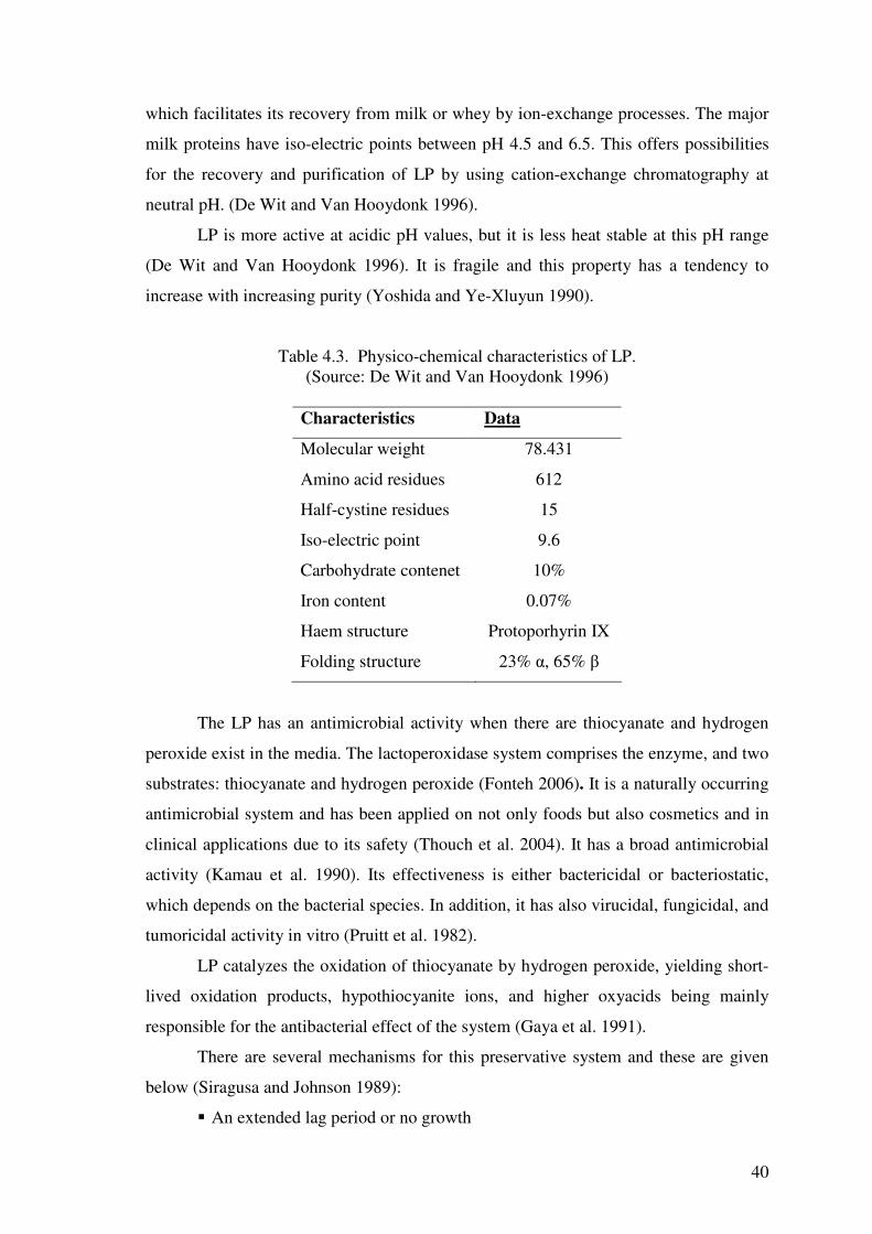
40
which facilitates its recovery from milk or whey by ion-exchange processes. The major
milk proteins have iso-electric points between pH 4.5 and 6.5. This offers possibilities
for the recovery and purification of LP by using cation-exchange chromatography at
neutral pH. (De Wit and Van Hooydonk 1996).
LP is more active at acidic pH values, but it is less heat stable at this pH range
(De Wit and Van Hooydonk 1996). It is fragile and this property has a tendency to
increase with increasing purity (Yoshida and Ye-Xluyun 1990).
Table 4.3. Physico-chemical characteristics of LP. (Source: De Wit and Van Hooydonk 1996)
Characteristics Data
Molecular weight
Amino acid residues
Half-cystine residues
Iso-electric point
Carbohydrate contenet
Iron content
Haem structure
Folding structure
78.431
612
15
9.6
10%
0.07%
Protoporhyrin IX
23% �, 65% �
The LP has an antimicrobial activity when there are thiocyanate and hydrogen
peroxide exist in the media. The lactoperoxidase system comprises the enzyme, and two
substrates: thiocyanate and hydrogen peroxide (Fonteh 2006). It is a naturally occurring
antimicrobial system and has been applied on not only foods but also cosmetics and in
clinical applications due to its safety (Thouch et al. 2004). It has a broad antimicrobial
activity (Kamau et al. 1990). Its effectiveness is either bactericidal or bacteriostatic,
which depends on the bacterial species. In addition, it has also virucidal, fungicidal, and
tumoricidal activity in vitro (Pruitt et al. 1982).
LP catalyzes the oxidation of thiocyanate by hydrogen peroxide, yielding short-
lived oxidation products, hypothiocyanite ions, and higher oxyacids being mainly
responsible for the antibacterial effect of the system (Gaya et al. 1991).
There are several mechanisms for this preservative system and these are given
below (Siragusa and Johnson 1989):
� An extended lag period or no growth

41
� Reduced O2 uptake
� Reduced lactate production by fermentative organisms
� Inhibition of key metabolic enzymes such as hexokinase, glyceraldehyde-3P-
dehydrogenase, and D-lactate dehydrogenase
� Inhibition of glucose uptake
� Cytoplasmic membrane damage with leakage of ions and UV-absorbing
material
� Inhibition of nucleic acid and protein synthesis.
The oxidizing products generated by LP system inhibit microorganisms by the
oxidation of sulphydryl (SH) groups of microbial enzymes and other proteins. So, it
damages the microbial cytoplasmic membrane through the oxidation of SH groups.
Finally, this leads to leakage of potassium ions, amino acids, and peptides from
microbial cells and eventually causes the death of the cells (Min et al. 2005).
The oxidation of thiol groups (-SH) of cytoplasmic membrane and damage to
other cellular elements such as the outer membrane, cell wall or cytoplasmic membrane,
transport sytems, glycolytic enzymes and nucleic acids are the results of the LP-
catalysed reactions (Touch et al. 2004).
LPO
2SCN- + H2O2 ( SCN )2 + 2e-
( SCN )2 + H2O HOSCN + H+ + SCN-
HOSCN ( pK = 5.3 ) OSCN- + H+
OSCN- + protein - SH protein - S - SCN + OH-
R-S-SCN + R-SCN R-S-S-R + SCN- + H+
Figure 4.2. Oxidation of protein (enzyme) sulphydryls by lactoperoxidase (LP)
catalysed reactions, mediated by products of SCN- (Source: De Wit and Van Hooydonk 1996).
As mentioned above, this system yields a variety of oxidizing products. The
reaction can continue by direct oxidation of SCN- to OSCN- or by oxidation of SCN- via
(SCN)2 (thiocyanogen). Among these products, hypothiocyanite ion (OSCN-) is the
major inhibitory product at physiologic pH (Siragusa and Johnson 1989, Pruitt et al.
1982).

42
OSCN- is in equilibrium with HOSCN (pK 5.3). This indicates that at pH 5.3 (at
the pH of maximal LP activity) half is in the form of HOSCN and half is in the form of
OSCN- (De Wit and Van Hooydonk 1996). At low pH, antibacterial action is greater,
owing to the ability of the uncharged HOSCN to penetrate microbial membranes and
thus to attack functional groups of essential intracellular enzymes (Thomas et al. 1983).
It is also reported that the inhibitory effect caused by resulting OSCN- ions is
dependent on the holding temperature, pH, and microbial load. However, the LP system
inhibition can be reversed by incorporating sulfhydryl-containing compounds, such as
cysteine, dithiothreitol, and glutathione, as well as by high levels of catalase or by
heating milk at pasteurization temperatures (Siragusa and Johnson 1989).
There are several studies performed on LP system. Fonteh et al. (2006) studied
that the shelf life of raw milk could be effectively extended with small amounts of
thiocyanate (20 ppm) and peroxide (20 ppm) under Cameroonian conditions by
approximately 9 h without refrigeration. LP system-activated milk can be stored for as
long as 21 h, allowing sufficient time for its suitable disposal. Jacob et al. (2000)
observed that the exploitation of LP-thiocyanate-H2O2 system was an effective system
against many disease causing organisms in plants and animals.
Gaya et al. (1991) found that LP system activation was shown to be a feasible
procedure for controlling the development of L.monocytogenes in refrigerated raw milk.
The bactericidal activity of LP system in raw milk prevented the growth and also
reduced significantly the L.monocytogenes load during refrigerated storage for 5 days.
According to the researchers, the LP system exhibited a bactericidal activity at 4 and
8oC; the activity was dependent on temperature, incubation time, and strain of
L.monocytogenes. Kamau et al.(1990) determined that the effect of the LP system on
thermal resistance of bacteria suspended in milk. They reported that the thermal
destruction of L.monocytogenes and Staphylococcus aureus when exposed to the LP
system following the heating process was increased. By this way, LP system processes
at low temperatures does not affect the nutritional value and the quality of foods. Garcia
et al.(2000) investigated the combined action of the LP system and high pressure
treatment on four strains of E.coli and L.innocua inoculated in milk. The aim of this
research was to find a potential mild food preservation method for producing
microbiologically safe foods. They concluded that the LP system immediately after high
pressure treatment could be effective against L.innocua, but high pressure and the LP
system could not exhibit a synergetic interaction on the inactivation of E.coli. However,

43
Garcia-Graells et al. (2000) found that at low cell concentration of E.coli used in the
study were inactivated within the early hr by the help of the combined pressure-LP
system treatment. They showed that the pressure resistant strain of E.coli needed a
higher pressure to become sensitized to the LP system.
Touch et al.(2004) investigated the use of the LP system against S.enteritidis in
tomato juice, carrot juice, milk, liquid whole egg, and chicken skin extract under various
conditions. They found that the system was more effective against the organism in
vegetable juices than in animal products, at low pH than at neutral pH, and at higher
temperatures than at lower temperatures. They concluded that combination of the system
with other preservatives or treatments would be needed to effectively inhibit growth and
survival of Salmonella in animal products.
Björck et al. (1975) incorporated glucose and glucose oxidase into raw milk
containing 0.085 mM SCN-. They observed that P. fluorescens EF 1998 could not grow
in the presence of 3% glucose and 0.1U of glucose oxidase, and 0.085mM SCN- up to
10h.
Microbial growth on food surfaces is a main reason for food spoilage (Pranoto
et al. 2004). Due to the fact that microbial growth in foods takes place at the surface,
some methods have been developed to to reduce the growth on the surface by using
antimicrobial sprays or dips (Pranoto et al. 2004).On the other hand, direct surface
application of antibacterial substances has limited effect on surface flora because of the
rapid diffusion of the active agent within the bulk of food or neutralization of the
substances by contacting product constituents (Pranoto et al. 2004, Quattara et al. 2000).
In the indirect application, the antimicrobial agent localizes the functional
efficiancy at the food surface. By this way, the agents migrate slowly to the surface and
so they remain at high concentrations where they are necessary. This can be provided by
edible films or coatings, which are means to carry antimicrobials (Pranoto et al. 2004).
Min and Krochta (2005) conducted a study with Penicillium commune, which causes
the deterioration of foods, as well as produce a mycotoxin. They found that
lactoperoxidase system incorporated into whey protein isolate films at a level of 59 mg
LP/g film inhibited growth of P.commune. In addition, Min et al.(2005) also
demonstrated the effect of LP system on the inhibition of L.monocytogenes and the
effect of LP system -WPI films and/or coating on smoked salmon. The WPI coatings
incorporating LP system prevented the growth of L.monocytogenes in smoked salmon at
4 and 10oC for 35 and 14d, respectively (Min et al 2005). Furthermore, they also studied
44
the efficiancy of WPI films with LP system on the inhibition of Salmonella enterica and
Escherichia coli O157:H7. LP system -WPI films (0.15g/g) completely prevented the
growth of these pathogens, (4 log CFU/cm2) (Min et al 2005).
4.1.5.2. Lysozyme
Lysozyme (EC 3.2.1.17) is a commercially produced enzyme. It is used in food
technology in order to prevent late gas blowing in cheese, and reduce sterilizing
temperature for food canning (Jiang et al. 2001). In Japan, it has been used for
preservation of oyster, shrimp, other seafood, sushi, sake, kimchi (pickled cabbage),
Chinese noodles, potato salad, and custard (Davidson and Branen 1993).
This enzyme is a natural component of tears, plant tissues, milk, and eggs. It
specifically degrades the peptidoglycan portion of the rigid bacterial cell wall (Jiang et al.
2001). It has an antimicrobial property against common food spoilage and food-borne
disease-causing bacteria, including B.cereus, B.stearothermophilus, Campylobacter jejuni,
C.botulinum types A,B, and E, Clostridium butyricum, Clostridium perfringens,
Clostridium sporogenes, Clostridium thermosaccharolyticum, Clostridium tyrobutyricum,
E.coli O157:H7, Klebsiella pneumoniae, L.monocyotogenes, S.typhimurium, S.aureus,
Vibrio cholerae, and Yersinia enterocolitica (Davidson and Branen 1993). Furthermore,
some strains of C.botulinum, C.thermosaccharolyticum, C.tyrobutyricum,
B.stearothermophilus, and L.monocytogenes were effectively inhibited or lysed by
lysozyme (Davidson and Branen 1993). The presence of other chemicals (e.g., EDTA),
temperature, growth phase, and growth media may affect the activity of lysozyme.
Kandemir et al.(2005) found that the combinational addition of lysozyme and
EDTA.2H2O2 in pullulan film gave antimicrobial property against Escherichia coli. This
may be used to obtain antimicrobial films to increase the safety of foods.
4.1.5.3. Glucose Oxidase
Glucose oxidase (�-D-glucose; 1-oxido-reductase) is one of the important
enzymes used in the food industry. It is utilized as a stabilizing agent in wine, beer,
carbonated soft drinks, mayonnaise and sauce salad dressings. In addition, the enzyme
is applied in the manufacturing process of liquid egg before drying to avoid Maillard

45
reactions (Massa et al. 2001). Consequently, it has been used in the industry for
desugaring, and as an antioxidant (Jeong et al. 1992). It is produced by molds, including
Penicillium notatum and Aspergillus niger (Jeong et al. 1992).
The enzyme is an oxidoreductase. It catalyzes the oxidation of glucose to gluconic
acid and hydrogen peroxide by using molecular oxygen as an electron acceptor (Jeong et
al. 1992, Kana et al. 2001, Massa et al. 2001). Gluconic acid produced by this enzymatic
reaction reduces the pH. It is thought that the growth of spoilage microorganisms present
on food is inhibited in the more acidic environment (Jeong et al. 1992). In order to
understand whether the inhibition of enterotoxic E.coli PM 015 and Salmonella derby BP
177 was caused by gluconic acid or hydrogen peroxide, Massa et al. (2001) added
catalase to glucose oxidase-glucose system. Since catalase decomposes hydrogen
peroxide to water and oxygen, they attributed the antibacterial effect to the result of a pH
decrease by the formation of gluconic acid.
4.1.5.4. Chitinase
Chitinase (EC 3.2.1.14) catalyzes the hydrolysis of chitin to its monomer N-
acetyl-D-glucosamine. Chitin is one of the major components of fungal cell wall.
Therefore, the antimicrobial effect of this enzyme is against fungi. In biological
research, chitinases are used to generate fungal protoplasts. The hydrolytic property of
chitinases gains interest as an environmentally safe biocontrol agent. Chitinase
generating organisms have been utilized to inhibit the growth of phytopathogens.
Paenibacillus spp. is found to be a high chitinase producer (Kao et al. 2007).
4.1.6. Bioactive Proteins and Peptides
There are several bioactive proteins and peptides present in foods. These agents
are also called naturally occurring antimicrobial agents.
4.1.6.1. Lactoferrin
Lactoferrin is a molecule of 80.000 D molecular weight. It is found in
colostrum, milk and other exocrine fluids such as tears. It is active at neutral pH and in
46
the presence of bicarbonate ions (Pakkanen and Aalto 1997). It is highest in bovine
colostrum. Its role in milk may also involve iron binding. It may bind the iron so tightly
that it is no longer available for microbial growth (Davidson and Branen 1993). The
protein has been shown to inhibit the growth of some organisms, including E.coli,
S.typhimurium, S.dysenteria, L.monocytogenes, S.mutans, B.stearothermophilus and B.
subtilis (Pakkanen and Aalto 1997). The antimicrobial activity of lactoferrin can be
decreased or totally reversed in the presence of trysin, ferrous sulfate, magnessium
sulfate, hematin, and citrate. It is very stable to heat (90oC for 60 min) (Davidson and
Branen 1993).
Lactoferricin is a peptide derived from pepsin digestion of bovine lactoferrin. It
has a low-molecular-weight, single-chain peptide of 25 amino acid residues (Davidson
and Branen 1993). It also has antimicrobial activities against various bacteria and
Candida albicans (Pakkanen and Aalto 1997).
4.1.6.2. Phosvitin
Phosvitin is an egg yolk protein. It has a molecular weight between 36.000 and
40.000 D (Lee et al. 2002). This egg yolk phosphoglycoprotein is a highly
phosphorylated serine-rich glycoprotein. It contains approximately 10 % phosphorus
and 6.5 % carbohydrates (Khan et al. 1998).
Phosvitin has an antimicrobial spectrum because it is an amphiphilic protein
with chelating ability. The protein is a good candidate for damaging the outer
membrane of Gram-negative bacteria. It binds and limits iron which is necessary for
bacterial growth (Khan et al. 2000). Kahan et al.(2000) demonstrated that phosvitin
damaged the outer membrane and killed E.coli under thermal stress. In addition, to
suppress the lipid oxidation reactions in food is of great importance to the food industry.
The aim is to prevent undesirable changes to food products that result in loss of
nutritional value, formation of off-flavors, and production of potential toxins. Phosvitin
also has an antioxidant potential (Nakamura et al. 1998). Lee et al. (2002) investigated
the antioxidant property of phosvitin in muscle food. The researchers suggested that due
to its insufficient antioxidant activity, it should be utilized together with other
antioxidants.
47
CHAPTER 5
MATERIALS AND METHODS
5.1. Materials
Whole milk used to produce LP enzyme was obtained from a local farm in �zmir
in every two months (2005-2006). Potassium thiocyanate was purchased from Sigma-
Aldrich (Stenheim, Germany). Toyopearl sulphopropyl (SP) cation-exchanger (SP-
550C, fast flow, size: 100 �m) was purchased from Supelco (Bellefonte, PA, USA).
Dialysis tubes (cut off: 12000 MW), dextran (from Leuconostoc mesenteroides, 73.200
MW), ABTS (2,2-Azino-bis-(3-ethylbenz-thiazoline-6-sulfonic acid)), and the sodium
salt of alginic acid (from Macrocystis pyrifera, viscosity of 2% solution at 25 °C is 3500
cpc) were purchased from Sigma Chem. Co. (St. Louis, MO, USA). Rennet was
obtained from ICN Biomedicals Inc. (Aurora, Ohio, USA). For microbiological
analyses, nutrient agar (NA), DeMan, Rogosa and Sharpe (MRS) agar, plate count agar
(PCA), violet red bile agar (VRBA), peptone water, and tween 80 were purchased from
Fluka (Spain), nutrient broth (NB) and MRS broth were obtained from Merck
(Germany).
5.2. Methods
5.2.1. Alginate Films Incorporating Lactoperoxidase
5.2.1.1. Preparation of Lactoperoxidase from Bovine Milk
The LP was produced from bovine whey by using the method of Mecito�lu and
Yemenicio�lu (2007). Skim milk was prepared by centrifugation at 5000g for 20 min at
30 °C. It was filtered through a cheese-cloth, and then warmed to 37 °C. After the
temperature reached to 37 °C, 90 mg rennet was added to 900 mL of skim milk and kept
at 37 °C for 1 hr. The rennet whey was separated by filtration through a cheese-cloth

48
and centrifuged at 10000g for 25 min at 4 °C for removing the remaining fat and
insoluble proteins.
Chromatography was carried out using cation exchange chromatography
coloumn, a 11.5 cm x 2.8 cm Toyopearl SP column previously equilibrated with 0.05 M
Na-phosphate buffer at pH 6.5. Rennet whey (400 mL) was passed through the column
and then washed with 500 mL of 0.05 M sodium phosphate buffer (pH 6.5). Adsorbed
proteins were eluted with a linear concentration gradient of 0 – 0.55 M NaCl in 300 mL
of 0.05 M sodium phosphate buffer (pH 6.5). Elute fractions of 10 mL were collected in
tubes. The absorbance of each tube was measured at 280 nm and LP activity was
determined. LP activity test was performed based on a color change. The aliquot (0.2
mL) from each tube was reacted with 1 mL of 0.65 mM ABTS and 0.5 mL of 0.1 mM
H2O2. The tubes having dark or close to dark blue colors were collected. Finally, the
solution was dialysed using distilled water for 24 hr at 4 °C. After dialysis, 250-300 mg
of dextran as a supporting agent was mixed with the dialyzed enzyme and it was
lyophilized in a Labconco freeze-dryer (FreeZone 61, Kansas City, MO, USA), working
approximately at -47 °C collector temperature and 50-100 x 10-3 mBar vacuum. The
lyophilized enzyme was stored at -18 °C prior to analyses.
5.2.1.2. Determination of Lyophilized Lactoperoxidase Activity
LP activity was spectrophotometrically determined in triplicate by monitoring
the oxidation of ABTS using a (Shimadzu, model 2450, Tokyo, Japan)
spectrophotometer together with a constant temperature cell holder running at 30 °C
(Chávarri et al. 1998). Briefly, 0.1 mL of enzyme solution was mixed with 2.3 mL, 0.65
mM ABTS (prepared in Na-phosphate buffer) in a cuvette. The reaction was initiated by
adding 0.1 mL H2O2 (0.2 mM) to the mixture. The reaction was monitored for 5 min at
412 nm. The enzyme activity was estimated from the slope of the initial linear portion
of absorbance vs. time curve. Finally, the results were expressed as Unit (0.001
absorbance change in 1 min).

49
5.2.1.3. Preparation of Alginate Films
The alginate films were produced as described in Mecito�lu and Yemenicio�lu
(2007). To prepare films, lyophilized LP preparation (0.26-0.597 mg) was dissolved per
gram of 2% (w/v) alginic acid solution by mixing slowly with a magnetic stirrer. 10 g
portions of this solution were then spread onto the glass Petri dishes (9.5 cm in
diameter). The Petri dishes were dried at room temperature for three days and 0.8 mL of
0.3 M CaCl2 was pipetted onto the films for cross-linking, but before using the films
they were washed with 10 mL sterile deionized water for 15 sec in order to remove
excessive CaCl2.
5.2.1.4. Determination of Lactoperoxidase Activity Released from the
Alginate Film
The release tests were performed in a refrigerated incubator at 4 °C. The alginate
film incorporated with 900 U/cm2 LP was placed in a glass Petri dish containing 50 mL
distilled water (4 °C). The Petri dish was then covered with a parafilm in order to
prevent moisture loss and was incubated at 4 °C for 24 hr with stirring at 200 rpm using
a magnetic stirrer (2 cm long Teflon coated rod). The LP activity in release test solution
was monitored by taking 0.2 mL aliquots in triplicate from the release test solution at
different time intervals. The LP activity measurements were conducted by mixing 0.2
mL of the aliquot with 2.2 mL of 0.65 mM ABTS prepared in 0.1 M Na-phosphate
buffer at pH 6.0 and 0.1 mL of 0.4 mM H2O2. The absorbance of the reaction mixtures
was measured at 412 nm using a spectrophotometer (Shimadzu, model 2450, Tokyo,
Japan).
5.2.1.5. Determination of Immobilized Lactoperoxidase Activity in
the Alginate Film
The immobilized LP activity in the alginate film was determined by slightly
modifying the method of Mecito�lu and Yemenicio�lu (2007). The dried and cross-
linked alginate film incorporating 900 U/cm2 LP was halved with a clean razor and one
of the halves was placed into a glass Petri dish containing 23 mL of 0.65 mM ABTS

50
solution (prepared in 0.1M Na-phosphate buffer at pH 6.0) and 2 mL of 200, 400, or
800 µM H2O2 solution which was brought to 30 °C prior to mixing. The Petri dishes
were incubated at 30 °C and stirred at 200 rpm with a magnetic stirrer. The LP activity
was monitored by measuring the reaction mixture absorbance at 412 nm at different
time intervals and it was determined from the slope of the initial linear portion of
absorbance vs. time curve. The measurements were repeated for the remaining halves of
the film and average of two measurements was considered in all measurements. The
activity was expressed as U (0.001 absorbance change in one minute) per cm2 of the
film.
5.2.1.6. Antimicrobial Activity of Alginate Films
5.2.1.6.1. Bacterial Strains
The bacterial strains used in this study were Listeria innocua (NRRL-33314),
Escherichia coli (NRRL-B-3008), Pseudomonas fluorescens (NRRL B-253). These
strains were supplied from the United States Department of Agriculture, Microbial
Genomics and Bioprocessing Research Unit, Peoria, Illinois. The frozen stock cultures
were maintained in nutrient broth containing 15% glycerol at - 80 °C prior to analyses.
5.2.1.6.2. Determination of Antimicrobial Activity of Alginate Films
Incorporating Lactoperoxidase
Alginate films with or without LP were prepared as it was described in section
5.2.1.3. The films were washed with 25 mL sterile deionized water for 15 sec and the
discs (1.3 cm in diameter) were cut aseptically using a cork borer. The discs having 900
U/cm2 LP were placed into the test tubes containing 3 mL sterile Nutrient broth, 0.5 mL
culture, 0.1 mL potassium thiocyanate (final concentration in reaction mixture: 4000
µM), and 0.1 mL H2O2 (final concentration in tubes: 200, 400, or 800 µM). E. coli, L.
innocua or P. fluorescens were used as test cultures in the reaction mixtures. E. coli and
L. innocua were incubated using nutrient broth at 37 °C for 16-18 hr and P. fluorescens
was incubated using nutrient broth at 26°C for 16-18 hr.

51
In order to determine the effectiveness of LP system at different concentrations
of H2O2 and constant thiocyanate concentration, six different reaction mixtures were
prepared. Reaction mixture #1 was the control sample having only the nutrient broth
(NB), sterile water, and culture; #2 contained NB, sterile water, culture, and disc; #3
contained NB, sterile water, culture, disc, and thiocyanate; #4 contained NB, culture,
disc, thiocyanate, and 200 µM H2O2; #5 contained the same constituents as in #4 except
the concentration of H2O2 which was 400 µM instead of 200 µM; #6 was the same as #4
except the concentration of H2O2 was 800 µM instead of 200 µM. The test tubes having
E. coli and L. innocua were incubated at 37 °C for 24 hr and the tubes inoculated with
P. fluorescens were incubated at 26 °C for 24 hr. E. coli, L. innocua or P. fluorescens
were enumerated on nutrient agar by taking samples from reaction mixtures at 0, 6 and
24 hr of incubation. Pour plate method was performed and the duplicate plates for each
dilution were incubated at 37°C/24 hr for E. coli, at 37 °C/48 hr for L. innocua, and at
26 °C/48 hr for P. fluorescens. The microbial counts were expressed as log10 cfu/mL.
The residual H2O2 in the reaction mixtures were determined using semi-quantitative
H2O2 test papers (detection limit 1 mg/ L, Quantofix Peroxide 100, Macherey-Nagel,
Düren, Germany) at 0, 6 and 24 hr of incubation.
To determine the effectiveness of LP system at different concentrations of
thiocyanate (1000 µM or 2000 µM in the reaction mixture) and constant H2O2
concentration (200 µM), six different reaction mixtures were prepared. Reaction
mixture #1 was the control sample containing only NB, sterile water, and culture; #2
contained NB, sterile water, culture, and disc; #3 had NB, sterile water, culture, disc,
and 1000 µM thiocyanate; #4 contained NB, culture, disc, 1000 µM thiocyanate and
200 µM H2O2; #5 contained NB, sterile water, culture, disc, and 2000 µM thiocyanate;
#6 contained broth, culture, disc, 2000 µM thiocyanate and 200 µM H2O2.
The test tubes having E. coli and L. innocua were incubated at 37 °C for 24 hr
and the tubes inoculated with P. fluorescens were incubated at 26 °C for 24 hr. E. coli,
L. innocua or P. fluorescens were enumerated on nutrient agar by taking samples from
reaction mixtures at 0, 6 and 24 hr of incubation. Pour plate method was performed and
the duplicate plates for each dilution were incubated at 37 °C/24 hr for E. coli, at 37
°C/48 hr for L. innocua, and at 26 °C/48 hr for P. fluorescens. The microbial counts
were expressed as log10 cfu/mL. The residual H2O2 in the reaction mixtures were

52
determined using semi-quantitative H2O2 test papers (detection limit 1 mg/L,
Quantofix Peroxide 100, Macherey-Nagel, Düren, Germany) at 0, 6 and 24 hr of
incubation. All the experiments were performed in duplicate using LP produced from
different batches of milk samples.
Data was analyzed by one-way analysis of variance (ANOVA) using Minitab
version 13 (Minitab, Inc., State College, PA, U.S.A.). Means with a significant
difference (p<0.05) were compared using the Tukey’s test.
5.2.1.7. Application of Lactoperoxidase Incorporated Alginate Films
to Calamari
Fresh and cleaned calamari (5 kg) was purchased from a local seafood market in
�zmir. It was transferred to the laboratory in ice in the same day. In the laboratory,
calamari was washed in tab water and cut into rings. The calamari rings were randomly
separated into four batches. The treatments applied to each batch were as follows:
1) Untreated samples were placed in a flask containing 45 mL sterile deionized
water;
2) Samples coated with alginate film solution without having lactoperoxidase,
cross-linked with 0.3 M CaCl2, and placed in flasks containing 45 mL sterile deionized
water;
3) Samples coated with alginate film solution incorporating lactoperoxidase (22
mg/10 g film solution) and 4000 µM KSCN, cross-linked with 0.3 M CaCl2, and placed
in flasks containing 45 mL 4000 µM H2O2;
4) Samples coated with alginate film solution incorporating lactoperoxidase (22
mg/10 g film solution) and 4000 µM KSCN, cross-linked with 0.3 M CaCl2, and placed
in flasks containing 45 mL 8000 µM H2O2.
The untreated and treated calamari samples were stored at 4 °C for 7 days. Total
viable count was performed at days 0, 1, 2, 3, 5, and 7. The calamari samples (10 g)
were transferred aseptically into stomacher bags containing 90 mL of sterile peptone
water (0.1%) and homogenized in a stomacher (Interscience, France) at medium speed
for 60 sec. For each sample, appropriate serial decimal dilutions were prepared using
sterile peptone water (0.1%) and plated in duplicate on plate count agar. The plates were

53
incubated at 30 °C for 48 hr and microbial counts were determined. The counts were
expressed as log10 cfu/g.
The experiment was carried out in triplicate. Data was analyzed by one-way
analysis of variance (ANOVA) using Minitab version 13 (Minitab, Inc., State College,
PA, U.S.A.). Means with a significant difference (p<0.05) were compared using the
Tukey’s test.
5.2.2. Alginate Films Incorporating Lactic Acid Bacteria
5.2.2.1. Bacterial Strains
Lactobacillus delbrueckii subsp. lactis (NRRL B-4525), Lactobacillus casei
(NRRL B-441) were obtained from the United States Department of Agriculture,
Microbial Genomics and Bioprocessing Research Unit, Peoria, Illinois. The frozen
stock cultures of Lactobacillus casei ( NRRL B-441), Lactobacillus delbrueckii subsp.
lactis (NRRL B-4525) and Lactobacillus plantarum (DSM NR 1954) were maintained
in MRS broth containing 15% glycerol at -80 °C prior to the analysis.
5.2.2.2. Test of the Ability of Lactic Acid Bacteria to Produce
Hydrogen Peroxide
Two loops from the frozen stock cultures of L. casei and L . delbrueckii subsp.
lactis were inoculated with 25 mL MRS broth and incubated anaerobically (in an
incubator 5% CO2 and 50% humidity) at 37 °C for 24 hr. Then, the culture broth (1 mL)
was again inoculated with 90 mL MRS broth and incubated anaerobically at 37 °C for
24 hr. During the second incubation, samples were taken for bacterial enumeration at 0,
4, 6, 9, and 24 hr. They were serially diluted in 0.1% peptone water and pour plate
method was applied using MRS agar. The plates were incubated at 37oC for 48 hr. At 0,
4, 6, 9, and 24 hr samples were tested for optical density (OD) and H2O2 concentration.
OD measurements were done at 650 nm using a spectrophotometer (Shimadzu, model
2450, Tokyo, Japan), and H2O2 concentration (mg/L) was determined by using semi-
quantitative test papers (detection limit 1 mg/L, Quantofix Peroxide 100, Macherey-
54
Nagel, Düren, Germany). The concentrations of H2O2 produced during the growth of
these two lactic acid bacteria were determined.
5.2.2.3. Test for the Ability of L. delbrueckii subsp. lactis and L.
plantarum to Produce H2O2 and Lactic Acid at Different
Storage Temperatures
Two loops from the frozen stock cultures of L. delbrueckii subsp. lactis and L.
plantarum were inoculated into 25 mL MRS broth and incubated anaerobically at 37 °C
(in the CO2 incubator) for 16 hr. Then, the culture broth (1 mL) was aseptically
inoculated into 99 mL MRS broth and incubated at 4 and 23 °C for 144 hr (7 days). The
pH values, lactic acid bacteria count, H2O2 concentration, and lactic acid content (D and
L form respectively) were determined at predetermined time intervals of incubation.
The pH was monitored using a pH-meter (Microprocessor pH meter, Hanna
Instruments, Romania). Lactic acid bacteria counts were enumerated using MRS agar
by pour plate method. The duplicated plates were incubated anaerobically at 37 °C for
48 hr. The H2O2 concentration was determined using semi-quantitative test papers
(detection limit 1 mg/ L, Quantofix Peroxide 100, Macherey-Nagel, Düren, Germany)
and the lactic acid content was determined by test kits (Boehringer Mannheim,
Germany).
5.2.2.4. Preparation of Lyophilized Culture
MRS broth (10 mL) was inoculated with a loop of frozen stock cultures of L.
delbrueckii subsp. lactis, and L. plantarum. The broth was incubated anaerobically at 37
°C for 24 hr (in an incubator 5% CO2 and 50% humidity). Then, 10 mL incubated broth
was inoculated with 240 mL sterile MRS broth and incubated anaerobically at 37 °C
for 16 hr (in an incubator 5% CO2 and 50% humidity). The culture was washed in
sterile saline solution (0.9 % NaCl w/v) after two consecutive centrifugations (5000g) at
4 °C for 15 min. The pellets were suspended in reconstituted skim milk containing 7%
(w/v) sucrose, and the solution was stored at -20 °C before lyophilization. The frozen
solution was lyophilized in a Labconco freeze-dryer (FreeZone 61, Kansas City, Mo,

55
USA), working approximately at - 47 °C collector temperature and 50-100 x 10-3 mBar
vacuum.
5.2.2.5. Preparation of Alginate Films Incorporating Lactic Acid
Bacteria
To prepare films, 2-12 mg of lyophilized lactic acid bacteria (L. delbrueckii
subsp. lactis, and L. plantarum) was dissolved in 2% (w/v) alginic acid solution by
mixing slowly with a magnetic stirrer. 10 g portions of this solution were then spread
onto glass Petri dishes (9.5 cm in diameter). The Petri dishes were dried at room
temperature for 1 hr and 5 mL, 0.3 M CaCl2 was pipetted into them to cross-link the
dried films.
5.2.2.5.1. Determination of the Number of Free and Immobilized
Lactic Acid Bacteria in Alginate Films
The free lactic acid bacteria in the alginate films were determined by using a
shaking incubator (Barnstead International, LabLine 4000-BCE, Iowa, USA) at 25 °C.
The cross-linked film was weighed and then placed in a Petri dish containing 25 mL,
0.1% steril peptone water. The Petri dish was incubated for 5 min with continuous
stirring at 90 rpm. The amounts of lactic acid bacteria were determined by taking 1mL
aliquots from the solution in the Petri dish and serially diluted with 0.1 % peptone
water. Pour plate method using MRS agar was employed by using duplicate plates. The
plates were incubated anaerobically at 37 °C (5 % CO2, 50 % humidity) for 48 hr and
counts were expressed as log10 cfu/g film.
After 5 min of incubation, the alginate film incorporated with lactic acid bacteria
was homogenized using a homogenizator (Waring Commercial Blendor, New Hartford,
CT, 06057, USA) to determine the immobilized lactic acid bacteria in the film. The
serial dilutions were made using 0.1% peptone water and MRS agar was used to
enumerate lactic acid bacteria. The Petri dishes were incubated at 37 °C (5 % CO2, 50
% humidity) for 48 hr. The counts were expressed as log10 cfu/g film.

56
5.2.2.5.2. Determination of the Stability of the Lactic Acid Bacteria
Incorporated into the Alginate Film Forming Solution
To prepare films, 60 mg of lyophilized lactic acid bacteria (L. delbrueckii subsp.
lactis, and L. plantarum) was added in 2 % alginic acid solution. The film solutions
were stored at 4 °C for 7 days in firmly-sealed Petri dishes. At different time periods,
the film forming solutions were taken and cross-linked by pipetting 5 mL 0.3 M CaCl2
into the Petri dishes. The number of lactic acid bacteria in the film was enumerated at
days 0, 1, 3, and 7. Then, the free and immobilized lactic acid bacteria counts were
determined to evaluate the stability of lactic acid bacteria. The experiments were
performed as stated in section 5.2.2.5.1.
5.2.2.5.3. Determination of the Stability of the Lactic Acid Bacteria in
Powdered Alginic Acid
The alginic acid was mixed with 60 and 120 mg lactic acid bacteria (L.
delbrueckii subsp. lactis, and L. plantarum) and stored at 4oC. To determine the lactic
acid bacteria counts, 50 mg alginic acid containing lactic acid bacteria was taken and
dissolved in 10 mL sterile peptone water (0.1 %). The number of lactic acid bacteria
was determined by serially-diluting using sterile peptone water and pour plating with
MRS agar. The Petri dishes were incubated anaerobically at 37 °C (5 % CO2, 50 %
humidity) for 48 hr. The results were expressed as log10 cfu/g.
5.2.3.4. Applications of Alginate Films Incorporating Lactic Acid
Bacteria to Fresh Beef Cubes
The vacuum packaged beef loaf (approximately 4.5 kg) was obtained from Pınar
Et A.�. (�zmir, Turkey). The outer surfaces of the meat were removed to avoid possible
contamination before cutting into cubes. The beef cubes were separated into four
batches and treated as follows: 1) untreated; 2) treated with alginate film solution and
cross-linked with 0.3 M CaCl2; 3) treated with 90 mg L. debrueckii subsp. lactis per 10
g of alginate solution and cross-linked with 0.3 M CaCl2; 4) treated with 90 mg L.
plantarum per 10 g of alginate solution and cross-linked with 0.3 M CaCl2. The cubes
57
first coated with the alginate film solution and then immersed into 0.3 M CaCl2 solution
for 2-3 sec to cross-link. The uncoated and coated samples were placed in sterile Petri
dishes, tightly wrapped with a stretch film and stored at 4oC for 14 days. At days 0, 1, 2,
3, 7, and 14, approximately 10 g samples were taken from each treatment, and they
were homogenized using a stomacher (Interscience, France). Each homogenized sample
was serially diluted in 0.1% sterile peptone water and plated in duplicate on MRS agar.
The plates were incubated anaerobically at 37 °C (5 % CO2, 50 % humidity) for 48 hr
and lactic acid bacteria counts were determined. The counts were expressed as log10
cfu/g.
58
CHAPTER 6
RESULTS AND DISCUSSIONS
In the present study, the lyophilized lactoperoxidase and lactic acid bacteria
were incorporated into the edible alginate films. Lactoperoxidase (LP) forms the
naturally occurring antimicrobial system milk. Thus, one of the aims of this study is the
exploitation of the naturally occurring antimicrobial mechanism by adapting it in
alginate films used frequently for coating of meat, poultry and fish (Lindstrom et al.,
1992). The lactic acid bacteria also exist naturally in meat, poultry and fish. In fact,
enrichment of food by lactic acid bacteria is currently applied extensively to create an
additional safeguard for temperature abuse conditions occurred during storage of
refrigerated meat and meat products. The rapid development of lactic acid bacteria
during temperature abuse conditions prevents growth of pathogenic bacteria and food is
spoiled by nontoxic lactic acid bacteria by causing easily detectable acidity. This
prevents poisoning by pathogens which mostly cause no detectable change in food. The
second aim of this work is to incorporate lactic acid bacteria into alginate films. By this
way, the application of bacterial cultures to meat and meat products will become a very
practical process which needs no dealing with liquid culture and complicated aseptic
spraying equipment.
6.1. Incorporation of Lactoproxidase into Alginate Films
6.1.1. Determination of the LP Activity Released from the Alginate
Films
Release tests were performed at 4 °C in distilled water (50 mL, stirring rate 200
rpm) for 24 hr. No LP activity release was determined from the alginate films
incorporated with 900U/cm2 LP. The alginate films are consisted of linear copolymers
of D-mannuronic acid and L-guluronic acid which are cross-linked by the CaCl2. The
enzyme has a high isoelectric point (pI 9.6). Thus, it may bind to films by the negatively
charged carboxylic acid groups on polymeric chains of alginate. In this research, the
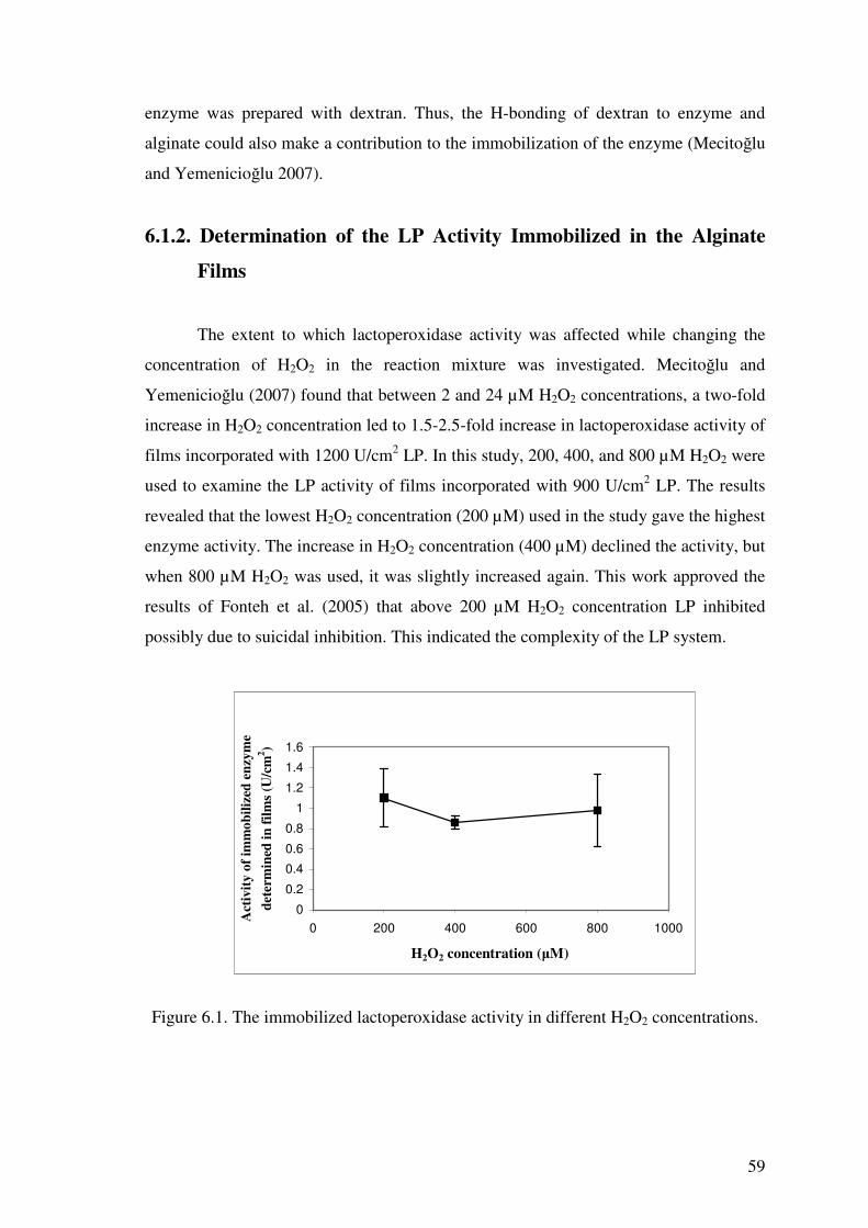
59
enzyme was prepared with dextran. Thus, the H-bonding of dextran to enzyme and
alginate could also make a contribution to the immobilization of the enzyme (Mecito�lu
and Yemenicio�lu 2007).
6.1.2. Determination of the LP Activity Immobilized in the Alginate
Films
The extent to which lactoperoxidase activity was affected while changing the
concentration of H2O2 in the reaction mixture was investigated. Mecito�lu and
Yemenicio�lu (2007) found that between 2 and 24 µM H2O2 concentrations, a two-fold
increase in H2O2 concentration led to 1.5-2.5-fold increase in lactoperoxidase activity of
films incorporated with 1200 U/cm2 LP. In this study, 200, 400, and 800 µM H2O2 were
used to examine the LP activity of films incorporated with 900 U/cm2 LP. The results
revealed that the lowest H2O2 concentration (200 µM) used in the study gave the highest
enzyme activity. The increase in H2O2 concentration (400 µM) declined the activity, but
when 800 µM H2O2 was used, it was slightly increased again. This work approved the
results of Fonteh et al. (2005) that above 200 µM H2O2 concentration LP inhibited
possibly due to suicidal inhibition. This indicated the complexity of the LP system.
0
0.2
0.4
0.6
0.8
1
1.2
1.4
1.6
0 200 400 600 800 1000
H2O2 concentration (�M)
Act
ivity
of i
mm
obili
zed
enzy
me
dete
rmin
ed in
film
s (U
/cm
2 )
Figure 6.1. The immobilized lactoperoxidase activity in different H2O2 concentrations.
60
6.1.3. Antimicrobial Activity of LP-H2O2-Thiocyanate System
The antimicrobial activity of LP-H2O2-thiocyanete system on Escherichia coli
(NRRL B-3008), Listeria innocua (NRRL B-33314), and Pseudomonas fluorescens
(NRRL B-253) was investigated by using 1.3 cm in diameter discs prepared from
alginate films incorporated with 900 U/cm2 LP in the presence of 0, 200, 400, or 800
�M H2O2 and 4000 �M KSCN in one set of experiments and in the other set of
experiments the antimicrobial activity of LP system on the previously mentioned
microorgansims by using the alginate discs incorporating 900 U/cm2 LP in the presence
of 0 or 200 µM H2O2 and 0, 1000, and 2000 µM KSCN .
6.1.3.1. Effects of LP System in Reaction Mixtures Containing
Alginate Films Incorporating LP against E.coli
The results of the effect of LP system against the growth of E. coli were
summarized in Table 6.1. According to these results, the LP system had an inhibitory
effect on the growth of E. coli in the presence of 200, 400, and 800 �M H2O2.
Incubation time had a significat effect on each treatment (p<0.05). The growth of the
microorganism was delayed up to the 6th hr (Figure 6.2). It was observed that the
bacteria were inhibited in the presence of high H2O2 especially in the 2nd trial (Figure
6.2). Thus, findings showed that the enzymes used in the 1st and 2nd trials had different
kinetic properties. The different H2O2 consumption rates also supported this hypothesis.
Although the enzyme activity used in the films was at the same level (900 U/cm2), the
H2O2 was used more rapidly in the 2nd trial than in the 1st one (Table 6.3). On the other
hand, the bacteria began to grow immediately between 6 and 24 hr at the same levels as
the control regardless of the treatments with the system. It could be explained by the
absence of H2O2 to be used for the inhibitory reaction. However, these films could be
applied for minimally-processed foods stored at 0-4 °C. Under these conditions, the
enzyme would work more slowly than at 37 °C, and therefore, the antimicrobial
effectiveness would be prolonged.

61
Table 6.1. E. coli counts in reaction mixtures having different concentrations of H2O2 during 24 hr incubation at 37 °C.
No
Total LP activity
in discs
(U/cm2)1
Thiocyanate
conc.
(�M)
H2O2
conc.
(�M)
E. coli counts
(log10 cfu/mL)
Incubation time at 37 oC (hr)
0 6 24
12 - - - 3.8c 7.5b 9.3a
23 - - - 4.3c 7.8b 9.0a
3 900 (500)4 4000 - 5.7c 8.0b 9.0a
4 900 (500) 4000 200 5.7b 5.8b 8.8a
5 900 (500) 4000 400 5.0c 5.8b 9.2a
1st T
rial
6 900 (500) 4000 800 5.6c 6.2b 8.8a,
12 - - - 4.2c 8.7b 9.2a
23 - - - 4.2c 8.7b 9.2a
3 900 (597) 4000 - 4.3c 8.6b 9.3a
4 900 (597) 4000 200 4.3c 6.7b 9.1a
5 900 (597) 4000 400 4.2b 3.0c 8.8a
2nd T
rial
6 900 (597) 4000 800 4.2b 2.0c 8.6a
1 LP activity of films used for 1st trial: 1188 U/disc (660 �g/disc) and for 2nd trial: 1188 U/disc (788 �g/disc) 2 Reaction mixture contains only nutrient broth and E. coli 3 Reaction mixture contains nutrient broth, E. coli and discs without lactoperoxidase enzyme 4 Lactoperoxidase enzyme content as �g per disc a-c Row means having a different letter are significantly different (P<0.05).
Table 6.2. The change of H2O2 concentration in reaction mixtures during 24 hr incubation at 37 °C.
H2O2 concentration (µM)
Incubation time at 37 °C (hr) No
Initial H2O2
concentration
(µM) 0 6 24
1 200 88-200 - -
2 400 294 0-29 -
1st T
rial
3 800 294-800 29-88 -
1 200 88-200 - -
2 400 88-294 - -
2nd T
rial
3 800 294-800 29-88 -

62
0
1
2
3
4
5
6
7
8
9
10
0 5 10 15 20 25 30Time (hr)
C
hang
e in
mic
ro lo
ad (
log
cfu/
ml)
1
2
3
4
5
6
-4
-2
0
2
4
6
8
10
0 5 10 15 20 25 30
Time (hr)
C
hang
e in
mic
ro lo
ad (l
og c
fu/m
l)
1
2
3
4
5
6
Figure 6.2. Change of microbial counts (log10 cfu/mL) of E. coli in reaction mixtures
containing 4000 �M KSCN, 1188 U lactoperoxidase/disc (this activity was reached by using 660 �g/disc (A: 1st trial) or 788 �g/disc (B: 2nd trial) enzyme for different LP preparations) and different concentrations of H2O2. Composition of reaction mixtures; 1: Nutrient broth (NB)+E. Coli (E) + sterile deionized water (W); 2: NB+E+Alginate disc (A)+W; 3: NB+E+Lactoperoxidase incorporated alginate disc (L) + Potassium thiocyanate (T)+W; 4: NB+E+L+T+H1 (200 �M); 5: NB+E+L+T+H1 (400 �M); 6: NB+E+L+T+H1 (800 �M).
A
B
63
The effect of LP system in presence of different concentrations of KSCN (1000
or 2000 µM) and H2O2 (0, 200 µM) against E. coli was also investigated. The results
were given in Table 6.3. In this case, bacteriostatic effect, instead of inhibitory action,
occurred when 1000 or 2000 �M KSCN, and 200 �M H2O2 were used. The obtained
antimicrobial effect was also lasted up to 6 hr. Between 6 and 24 hr, the microorganism
started to develop readily. The present work showed that the antimicrobial effect did not
improve when the thiocyanate concentration was increased from 1000 to 2000 �M in
the presence of 200 �M H2O2. In fact, the increase in KSCN concentration caused a
decline in the effectiveness of LP system (Figure 6.3.). As seen from Table 6.4, the
H2O2 concentration was declined to an undetectable level within the 6th hr. Thus, it is
hard to compare the H2O2 consumption rates of different reaction mixtures. However,
higher antimicrobial activity of 1000 �M KSCN than 2000 �M KSCN in presence of
200 �M H2O2 suggested that the limiting factor for the effectiveness of LP system is
H2O2. This was expected since the short lived antimicrobial products of KSCN
oxidation should have been formed and then degraded more rapidly in presence of
higher concentraiton of KSCN.

64
Table 6.3. E. coli counts in reaction mixtures having different concentrations of KSCN during 24 hr incubation at 37 °C.
E. coli counts
(log10 cfu/mL) No
Incubation time at 37 °C (hr)
Total LP activity
in discs
(U/cm2)1
Thiocyanate
conc.
(µM)
H2O2
conc.
(µM) 0 6 24
12 - - - 3.0c 7.4b 9.2a
23 - - - 3.0c 7.5b 9.3a
3 900 (344)4 1000 - 4.1c 7.8b 9.2a
4 900 (344) 1000 200 4.9c 6.3b 9.1a
5 900 (344) 2000 - 4.3c 7.8b 9.1a
1st T
rial
6 900 (344) 2000 200 4.3c 6.2b 9.1a
12 - - - 3.2c 8.1b 9.2a
23 - - - 3.2c 8.3b 9.3a
3 900 (597)4 1000 - 3.4c 8.2b 9.2a
4 900 (597) 1000 200 3.3b 3.5b 9.0a
5 900 (597) 2000 - 3.5c 8.4b 9.3a
2nd T
rial
6 900 (597) 2000 200 3.2c 5.8b 9.2a
1 LPS activity of films used for 1st trial: 1188 U/disc (454 �g/disc) and for 2nd trial: 1188 U/disc (788 �g/disc)
2 Solution contains only nutrient broth and E. coli 3 Solution contains nutrient broth, E. coli and discs without lactoperoxidase enzyme 4 Lactoperoxidase enzyme amount in �g in each disc a-c Row means having a different letter are significantly different (P<0.05).
Table 6.4. The change of H2O2 concentration in reaction mixtures during 24 hr incubation at 37 °C.
H2O2 concentration (µM)
Incubation time at 37 °C (hr) No
Thiocyanate
conc.
(µM)
H2O2
conc.
(µM) 0 6 24
1 1000 200 88-200 - -
1st T
rial
2 2000 200 88-200 - -
1 1000 200 88-200 - -
2nd T
rial
2 2000 200 88-200 - -

65
0
1
2
3
4
5
6
7
0 5 10 15 20 25 30Time (hr)
C
hang
e in
mic
ro lo
ads
(log
cfu
/ml)
1
2
3
4
5
6
0
1
2
3
4
5
6
7
0 5 10 15 20 25 30Time (hr)
C
hang
e in
mic
ro lo
ad (l
og c
fu/m
l)
1
2
3
4
5
6
Figure 6.3. Change of microbial counts (log10 cfu/mL) of E. coli in reaction mixtures
containing 200 �M H2O2, 1188 U lactoperoxidase/disc (this activity was reached by using 454 �g/disc (A: 1st trial) or 788 �g/disc (B: 2nd trial) enzyme for different LP preparations) and different concentrations of KSCN. Composition of reaction mixtures; 1: Nutrient broth (NB) + E. Coli (E) + sterile deionized water (W); 2: NB + E + Alginate disc (A) + W; 3: NB + E + Lactoperoxidase incorporated alginate disc (L) + Potassium thiocyanate (T) + W; 4: NB + E + L + T(1000 �M) + H (200 �M); 5: NB + E + L + T (2000 �M) + H; 6: NB + E + L + T + H.
A
B

66
6.1.3.2. Effects of LP System in Reaction Mixtures Containing Alginate
Films Incorporating LP against L. innocua
Alginate films incorporating LP were also evaluated for their ability to inhibit L.
innocua. As seen in Table 6.5, incubation for 6 hr caused an inhibitory effect against L.
innocua in the presence of lactoperoxidase-H2O2-thiocyanate system containing 200 and
400 �M H2O2. The inhibitory effect increased further by increasing H2O2 to 800 �M
(Figure 6.4). In contrast, rapid growth of L.innocua was observed in these reaction
mixtures between 6 and 24 hr of incubation, with the exception of the 2nd trial reaction
mixture containing 800 �M H2O2. In the 2nd trial conducted with a different batch of LP,
the inhibition in the presence of 800 �M H2O2 lasted longer and continued even at the
end of 24 hr. Thus, the number of L. innocua in this reaction mixture reduced to the
lowest level reached in this study. As given in Table 6.6, the H2O2 residue was still
present in this reaction mixture at the end of 24 hr incubation period. It seems that the
remaining H2O2 contributed to the long lasting antimicrobial effect of reaction mixture
6 of the 2nd trial.

67
Table 6.5. L. innocua counts in reaction mixtures having different concentrations of H2O2 during 24 hr incubation at 37 °C.
L. innocua counts
(log10 cfu/mL) No
Total LP activity
in discs
(U/cm2)1
Thiocyanate
conc.
(µM)
H2O2
conc.
(µM) Incubation time at 37 °C (hr)
0 6 24
12 - - - 4.1c 5.6b 9.2a
23 - - - 4.2c 5.8b 8.9a
3 900(459)4 4000 - 5.3c 7.3b 8.9a
4 900(459) 4000 200 5.3b 5.2b 8.8a
5 900(459) 4000 400 5.0c 5.3b 9.0a
1st T
rial
6 900(459) 4000 800 5.4b 4.9c 8.7a
12 - - - 4.0c 6.5b 9.0a
23 - - - 4.3c 7.1b 9.1a
3 900(343)4 4000 - 4.1c 7.2b 9.3a
4 900(343) 4000 200 4.6c 5.9b 9.2a
5 900(343) 4000 400 4.1b 3.9b 9.2a
2nd T
rial
6 900(343) 4000 800 4.1a 3.1c 3.6b
1 LPS activity of films used for 1st trial: 1188 U/disc (660 �g/disc) and for 2nd trial: 1188 U/disc (453�g/disc)
2 Solution contains only nutrient broth and L.innocua 3 Solution contains nutrient broth, L. innocua and discs without lactoperoxidase enzyme 4 Lactoperoxidase enzyme amount in �g in each disc a-c Row means having a different letter are significantly different (P<0.05).
Table 6.6. The change of H2O2 concentration in reaction mixtures during 24 hr
incubation at 37 °C.
H2O2 concentration (µM)
Incubation time at 37 °C (hr) No H2O2 concentration
(µM) 0 6 24
1 200 88 - -
2 400 294 29-88 -
1st T
rial
3 800 294-800 88-294 -
1 200 88-200 - -
2 400 294 29-88 -
2nd T
rial
3 800 294-800 294 29

68
-2
0
2
4
6
8
10
0 5 10 15 20 25 30
Time (hr)
C
hang
e in
mic
ro lo
ad (l
og c
fu/m
l)
1
2
3
4
5
6
-2
0
2
4
6
8
10
0 5 10 15 20 25 30
Time (hr)
C
hang
e in
mic
ro lo
ad (l
og c
fu/m
l)
1
2
3
4
5
6
Figure 6.4. Change in the microbial counts (log10 cfu/ml) of L. innocua in reaction
mixtures with different concentrations of H2O2 and having 660 �g (A: 1st trial) and 453 �g (B: 2nd trial) of lactoperoxidase in discs in 24 hr (the legends 1, 2, 3, 4, 5, and 6 were the same as those in the Figure 6.2).
A
B

69
In further studies, 200 �M H2O2 and 1000 or 2000 �M KSCN concentrations
were used for the LP system. As summarized in Table 6.7, the antimicrobial effect
lasted up to the 6th hr of incubation. In this time period, the LP system showed a
bacteriostatic effect on L. innocua. In contrast, between 6th and 24th hrs of incubation
there was almost no antimicrobial effect on the bacteria and this caused a rapid
microbial growth (Figure 6.5.). The results of this study clearly showed the higher
susceptibility of L. innocua to LP system than E. coli. The LP system is particularly
effective on L. innocua, in the first 6th hr of incubation. In both cases the antimicrobial
effect of the LP system is mostly short lived and rarely exceeded 6 hr. However, it
should be noted that the reaction mixtures contain significant number of bacteria which
is hard to find in real food applications. In fact, in a food application, the expectation
from LP antimicrobial system would be the inhibition of limited number of pathogenic
bacteria originated from contamination. The duration of antimicrobial effect of
lactoperoxidase system depends on kinetic properties of the enzyme, initial
concentrations of hydrogen peroxide and thiocyanate, and catalase activity of the food
product. Thus, it is essential to use suitable concentrations of each ingredient during
food applications and choose food with low catalase activity at the surface.

70
Table 6.7. L. innocua counts in reaction mixtures having different concentrations of KSCN during 24 hr incubation at 37 °C.
No Total LP activity
in discs
(U/cm2)1
Thiocyanate
conc.
(µM)
H2O2
conc.
(µM)
L. innocua counts
(log10 cfu/mL)
Incubation time at 37 °C (hr)
0 6 24
12 - - - 3.0c 5.4b 8.8a
23 - - - 3.0c 5.5b 8.8a
3 900 (363)4 1000 - 3.8c 6.3b 8.9a
4 900 (363) 1000 200 3.5c 4.8b 8.9a
5 900 (363) 2000 - 3.5c 6.2b 9.0a
1st T
rial
6 900 (363) 2000 200 3.5c 4.5b 8.9a
12 - - - 3.1c 5.5b 8.8a
23 - - - 3.1c 5.7b 8.6a
3 900 (343)4 1000 - 3.2c 5.9b 9.3a
4 900 (343) 1000 200 3.8c 4.7b 9.1a
5 900 (343) 2000 - 3.3c 6.2b 9.1a
2nd T
rial
6 900 (343) 2000 200 3.4b 3.9b 9.1a
1 LPS activity of films used for 1st trial:1188 U/disc (479 �g/disc) and for 2nd trial: 900 U/cm2 (453 �g/disc) 2 Solution contains only nutrient broth and L. innocua 3 Solution contains nutrient broth, L. innocua and discs without lactoperoxidase enzyme 4 Lactoperoxidase enzyme amount in �g in each disc a-c Row means having a different letter are significantly different (P<0.05).
Table 6.8. The change of H2O2 concentration in reaction mixtures during 24 hr incubation at 37 °C.
H2O2 concentration (µM)
Incubation time at 37 °C (hr) No Thiocyanate
conc. (µM)
H2O2
conc.
(µM) 0 6 24
1 1000 200 88-200 29 -
1st T
rial
2 2000 200 88-200 0-29 -
1 1000 200 88-200 29-88 -
2nd T
rial
2 2000 200 88-200 29-88 -

71
0
1
2
3
4
5
6
7
0 5 10 15 20 25 30Time (hr)
C
hang
e in
mic
ro lo
ad (
log
cfu/
ml)
1
2
3
4
5
6
0
1
2
3
4
5
6
7
0 5 10 15 20 25 30Time (hr)
C
hang
e in
mic
ro lo
ad (l
og c
fu/m
l)
1
2
3
4
5
6
Figure 6.5. Change in the microbial counts (log10 cfu/ml) of L. innocua in reaction
mixtures with different concentrations of KSCN and having 479 �g (A: 1st trial) and 453 �g (B: 2nd trial) of lactoperoxidase in discs in 24 hr (the legends 1, 2, 3, 4, 5, and 6 were the same as those in the Figure 6.3).
A
B

72
6.1.3.3. Effects of LP System in Reaction Mixtures Containing
Alginate Films Incorporating LP against P. fluorescens
Effect of the LP incorporated alginate films on inhibition of P. fluorescens was
tested under the same conditions used for the other cultures. The antibacterial effect of
the alginate film incorporated with 900 U/cm2 LP on P. fluorescens was given in Table
6.9. In the 1st trial, the results showed that all tubes containing 4000 µM thiocyanate and
200, 400, or 800 µM H2O2 were effective against the inhibiton of P. fluorescens.
Particularly at 800 µM H2O2, the LP incorporated alginate films showed a considerable
inhibition on P. fluorescens up to 24th hr of incubation. In the 2nd trial conducted with a
different batch of LP, the desired antibacterial effect was seen in the tubes containing
400 and 800 µM H2O2 at 6th hr of incubation, but at 24th hr the antibacterial effect was
diminished. The H2O2 consumption in reaction mixtures of 1st and 2nd trials were
different (Table 6.10). Thus, it seems that there are differences between the kinetic
properties of different batches of LPs. The differences (almost 1 log) in the initial
microbial numbers of reaction mixtures may also contribute to variations in
antimicrobial effect of LPs since the amount of organic matter may affect the half-life
of reactive antimicrobial products formed by the LP system.
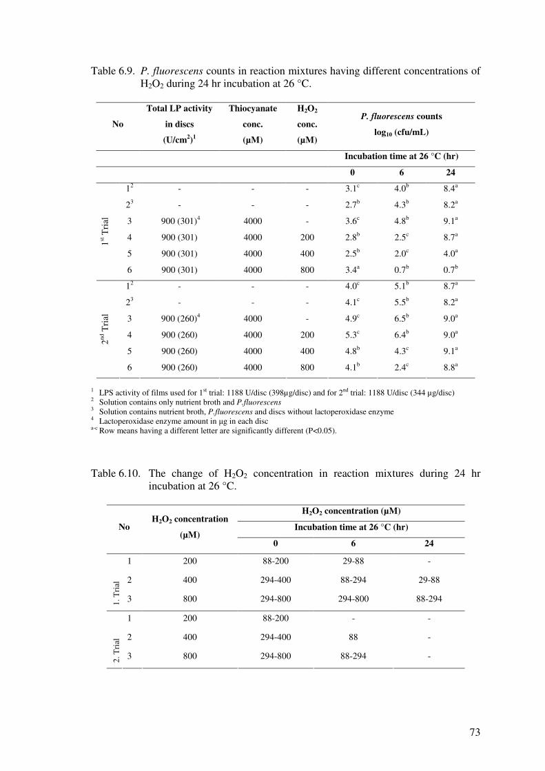
73
Table 6.9. P. fluorescens counts in reaction mixtures having different concentrations of H2O2 during 24 hr incubation at 26 °C.
No
Total LP activity
in discs
(U/cm2)1
Thiocyanate
conc.
(µM)
H2O2
conc.
(µM)
P. fluorescens counts
log10 (cfu/mL)
Incubation time at 26 °C (hr)
0 6 24
12 - - - 3.1c 4.0b 8.4a
23 - - - 2.7b 4.3b 8.2a
3 900 (301)4 4000 - 3.6c 4.8b 9.1a
4 900 (301) 4000 200 2.8b 2.5c 8.7a
5 900 (301) 4000 400 2.5b 2.0c 4.0a
1st T
rial
6 900 (301) 4000 800 3.4a 0.7b 0.7b
12 - - - 4.0c 5.1b 8.7a
23 - - - 4.1c 5.5b 8.2a
3 900 (260)4 4000 - 4.9c 6.5b 9.0a
4 900 (260) 4000 200 5.3c 6.4b 9.0a
5 900 (260) 4000 400 4.8b 4.3c 9.1a
2nd T
rial
6 900 (260) 4000 800 4.1b 2.4c 8.8a
1 LPS activity of films used for 1st trial: 1188 U/disc (398µg/disc) and for 2nd trial: 1188 U/disc (344 µg/disc) 2 Solution contains only nutrient broth and P.fluorescens 3 Solution contains nutrient broth, P.fluorescens and discs without lactoperoxidase enzyme 4 Lactoperoxidase enzyme amount in �g in each disc a-c Row means having a different letter are significantly different (P<0.05).
Table 6.10. The change of H2O2 concentration in reaction mixtures during 24 hr
incubation at 26 °C.
H2O2 concentration (µM)
Incubation time at 26 °C (hr) No H2O2 concentration
(µM) 0 6 24
1 200 88-200 29-88 -
2 400 294-400 88-294 29-88
1. T
rial
3 800 294-800 294-800 88-294
1 200 88-200 - -
2 400 294-400 88 -
2. T
rial
3 800 294-800 88-294 -
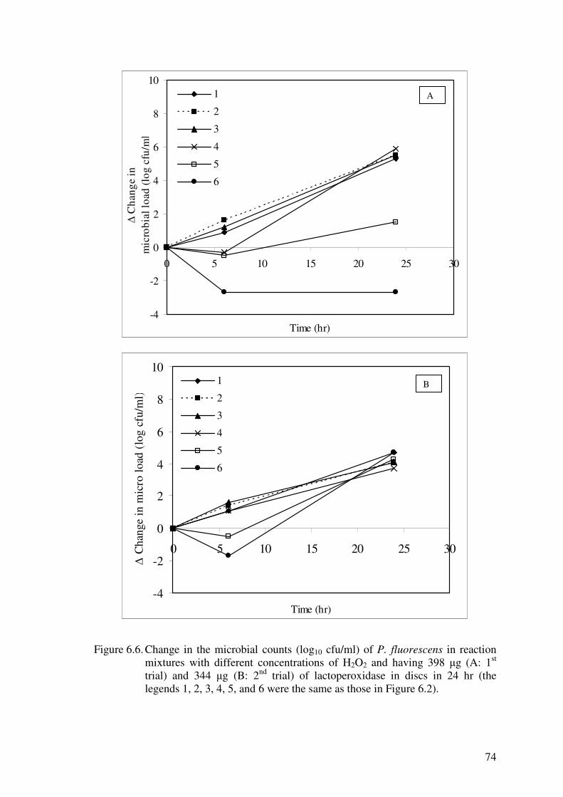
74
-4
-2
0
2
4
6
8
10
0 5 10 15 20 25 30
Time (hr)
C
hang
e in
m
icro
bial
load
(log
cfu
/ml)
1
2
3
4
5
6
-4
-2
0
2
4
6
8
10
0 5 10 15 20 25 30
Time (hr)
C
hang
e in
mic
ro lo
ad (
log
cfu/
ml)
1
2
3
4
5
6
Figure 6.6. Change in the microbial counts (log10 cfu/ml) of P. fluorescens in reaction
mixtures with different concentrations of H2O2 and having 398 �g (A: 1st trial) and 344 �g (B: 2nd trial) of lactoperoxidase in discs in 24 hr (the legends 1, 2, 3, 4, 5, and 6 were the same as those in Figure 6.2).
A
B

75
The antimicrobial effect of LP system on P. fluorescens was also tested at 1000
and 2000 µM thiocyanate and 200 µM H2O2 (Table 6.11). As seen in Figure 6.7 the
reaction mixture containing LP incorporated alginate films and KSCN and H2O2
showed considerable antimicrobial effect on P. fluorescens in the first 6th hr of
incubations. Then the antimicrobial effect was diminished and rapid growth occurred
between 6th and 24th hrs of incubation. In reaction mixtures containing KSCN but
lacking H2O2, the microbial growth occurred more rapidly than the other reaction
mixtures, including the controls. This result suggested the activatory activity of KSCN,
but it occurred only with P. fluorescens.

76
Table 6.11. P. fluorescens counts in reaction mixtures having different concentrations
of thiocyanate and H2O2 during 24 hr incubation at 26 °C.
No Total LP activity
in discs
(U/cm2)1
Thiocyanate
conc.
(µM)
H2O2
conc.
(µM)
P. fluorescens counts
(log10 cfu/mL)
Incubation time at 26 °C (hr)
0 6 24
12 - - - 2.5c 3.2b 8.4a
23 - - - 2.6b 2.9b 8.2a
3 900 (413)4 1000 - 3.3c 5.3b 8.3a
4 900 (413) 1000 200 2.7b 1.5b 5.6a
5 900 (413) 2000 - 3.0c 5.5b 8.9a
1st T
rial
6 900 (413) 2000 200 2.7c 1.4b 7.1a
12 - - - 3.6b 3.6b 7.5a
23 - - - 3.6c 4.7b 7.8a
3 900 (260)4 1000 - 3.8c 4.8b 8.9a
4 900 (260) 1000 200 3.7b 3.4b 8.6a
5 900 (260) 2000 - 3.7c 4.7b 8.9a
2nd T
rial
6 900 (260) 2000 200 3.7b 3.1c 8.5a
1 LPS activity of films used for 1st trial: 1188 U/disc (545 �g/disc) and for 2nd trial: 1188 U/disc (398 �g/disc)
2 Solution contains only nutrient broth and P. fluorescens 3 Solution contains nutrient broth, P. fluorescens and discs without lactoperoxidase enzyme 4 Lactoperoxidase enzyme amount in �g in each disc a-c Row means having a different letter are significantly different (P<0.05).
Table 6.12. The change of H2O2 concentration in reaction mixtures during 24 hr incubation at 26 °C.
H2O2 concentration (µM)
Incubation time at 26 °C (hr) No Thiocyanate
conc. (µM)
H2O2
conc.
(µM) 0 6 24
1 1000 200 88-200 - -
1st T
rial
2 2000 200 88-200 0-29 -
1 1000 200 88-200 88 -
2nd T
rial
2 2000 200 88-200 88 -

77
-2
-1
0
1
2
3
4
5
6
7
0 5 10 15 20 25 30
Time (hr)
C
hang
e in
mic
ro lo
ad (l
og c
fu/m
l)
1
2
3
4
5
6
-1
0
1
2
3
4
5
6
7
0 5 10 15 20 25 30
Time (hr)
C
hang
e in
mic
ro lo
ad (l
og c
fu/m
l)
1
2
3
4
5
6
Figure 6.7. Change in the microbial counts (log10 cfu/ml) of P. fluorescens in reaction
mixtures with different concentrations of KSCN and having 545 �g (A: 1st trial) and 398 �g (B: 2nd trial) of lactoperoxidase in discs in 24 hr (the legends 1, 2, 3, 4, 5, and 6 were the same as those in Figure 6.3).
A
B
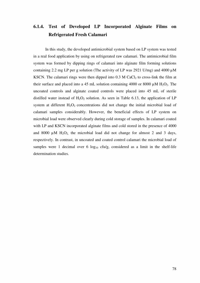
78
6.1.4. Test of Developed LP Incorporated Alginate Films on
Refrigerated Fresh Calamari
In this study, the developed antimicrobial system based on LP system was tested
in a real food application by using on refrigerated raw calamari. The antimicrobial film
system was formed by dipping rings of calamari into alginate film forming solutions
containing 2.2 mg LP per g solution (The activity of LP was 2921 U/mg) and 4000 µM
KSCN. The calamari rings were then dipped into 0.3 M CaCl2 to cross-link the film at
their surface and placed into a 45 mL solution containing 4000 or 8000 µM H2O2. The
uncoated controls and alginate coated controls were placed into 45 mL of sterile
distilled water instead of H2O2 solution. As seen in Table 6.13, the application of LP
system at different H2O2 concentrations did not change the initial microbial load of
calamari samples considerably. However, the beneficial effects of LP system on
microbial load were observed clearly during cold storage of samples. In calamari coated
with LP and KSCN incorporated alginate films and cold stored in the presence of 4000
and 8000 µM H2O2, the microbial load did not change for almost 2 and 3 days,
respectively. In contrast, in uncoated and coated control calamari the microbial load of
samples were 1 decimal over 6 log10 cfu/g, considered as a limit in the shelf-life
determination studies.

79
Table 6.13. Effect of LP incorporated alginate films on total viable counts of coated calamari rings during refrigerated storage.
Total viable count (log cfu/g)
Storage period (days)
Tre
atm
ents
0 1 2 3 5 7
Uncoated calamari
(stored in water)
5.12±0.04e,x 7.16±0.13d,x 7.57±0.07c,x 8.09±0.27b,x 8.90±0.11a,x 8.81±0.19a,x
Control calamari coated with alginate film
(stored in water)
5.08±0.15d,x 7.30±0.08c,x 7.49±0.23c,x 8.32±0.16b,x 8.90±0.14a,x 9.10±0.04a,x
Calamari coated with alginate film incorporated with LP and KSCN
(stored in 4000 µM H2O2 solution)
4.91±0.14c,xy 5.04±0.23c,y 5.00±0.65c,y 6.55±0.34b,y 7.81±1.05ab,xy 8.57±0.34a,x
Calamari coated with alginate film incorporated with LP and KSCN
(stored in 8000 µM H2O2 solution)
4.68±0.15 b,y 4.57±0.22 b,z 4.30±0.40 b,y 4.58±1.36b,z 6.39±1.04 ab,y 6.75±1.12a,y
a-e Row means having a different letter are significantly different (P<0.05). x-z Column means having a different letter are significantly different (P<0.05).
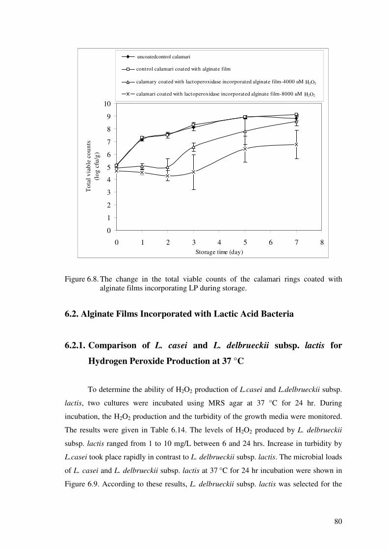
80
calamari
0
1
2
3
4
5
6
7
8
9
10
0 1 2 3 4 5 6 7 8Storage time (day)
Tot
al v
iabl
e co
unts
(l
og c
fu/g
)
untreated control calamari
control calamari coated with alginate film
calamary coated with lactoperoxidase incorporated alginate film-4000 uM H2O2
calamari coated with lactoperoxidase incorporated alginate film-8000 uM H2O2
Figure 6.8. The change in the total viable counts of the calamari rings coated with
alginate films incorporating LP during storage.
6.2. Alginate Films Incorporated with Lactic Acid Bacteria
6.2.1. Comparison of L. casei and L. delbrueckii subsp. lactis for
Hydrogen Peroxide Production at 37 °C
To determine the ability of H2O2 production of L.casei and L.delbrueckii subsp.
lactis, two cultures were incubated using MRS agar at 37 °C for 24 hr. During
incubation, the H2O2 production and the turbidity of the growth media were monitored.
The results were given in Table 6.14. The levels of H2O2 produced by L. delbrueckii
subsp. lactis ranged from 1 to 10 mg/L between 6 and 24 hrs. Increase in turbidity by
L.casei took place rapidly in contrast to L. delbrueckii subsp. lactis. The microbial loads
of L. casei and L. delbrueckii subsp. lactis at 37 °C for 24 hr incubation were shown in
Figure 6.9. According to these results, L. delbrueckii subsp. lactis was selected for the
uncoatedcontrol calamari
H2O2
H2O2

81
next studies due to its ability of H2O2 production. The maximum amount of H2O2 was
produced at the end of the exponential growth.
Table 6.14. The amount of hydrogen peroxide and change in the optical density by L.casei and L.delbrueckii subsp. lactis at 37 °C during 24 hr incubation.
L. casei L. delbrueckii subsp. lactis
Time
(hour) H2O2
(mg/L) OD 650 nm
H2O2
(mg/L) OD 650 nm
0 0 0.0426 0 -0.0012
3 0 0.1647 0 0.0093
4 0 0.3339 0 -
5 0 0.5056 0 0.0157
6 0 0.7957 1 0.0365
7 0 1.1127 3 0.0608
8 0 1.4875 3 0.0981
9 0 1.9528 10 0.1546
24 0 2.5127 10 1.491
82
L. casei (A)
0
2
4
6
8
10
0 4 8 12 16 20 24 28
Time (hr)
Mic
robi
al c
ount
s(l
og10
cfu
/mL
)
L. delbrueckii subsp. lactis (B)
0
2
4
6
8
0 4 8 12 16 20 24 28
Time (hr)
Mic
robi
al c
ount
s(lo
g 10 c
fu/m
L)
Figure 6.9. The microbial growth of L. casei (A) and L. delbrueckii subsp. lactis (B) at 37 °C.
6.2.2. H2O2 and Lactic Acid Production of L. delbrueckii subsp. lactis
and L. plantarum at Different Storage Temperatures
Lactic acid bacteria could be used as protective cultures because they become
dominant on microflora during storage and also they produce antimicrobial products
such as lactic acid, H2O2, bacteriocin, etc. to inhibit the growth of other
microorganisms. One of the most effective mechanisms that the bacteria utilize to form
the inhibitory action is lactic acid production. L.delbrueckii subsp.lactis bacterium can
produce lactic acid as well as H2O2 at storage temperatures (Zalan et al. 2005). Thus, it

83
may have a potential for incorporation into edible films as protective bacteria. Another
advantage of L. delbrueckii subsp. lactis is the production of lactic acid in lower
amounts and especially at the stationary phase. This is of great significance in terms of
not altering the products’ sensorial quality at the early stages of storage.
L. delbrueckii subsp. lactis (A)
0
2
4
6
8
10
0 50 100 150 200
Time (hr)
Mic
robi
al c
ount
s(l
og10
cfu
/mL
)
L. delbrueckii subsp. lactis (B)
0
1
2
3
4
5
6
7
8
9
0 50 100 150 200
Time (hr)
Mic
robi
al c
ount
s(l
og10
cfu
/mL
)
Figure 6.10. The growth curve of L.delbrueckii subsp. lactis at 4°C (A) and 23°C (B).

84
The growth curves at 4 °C and 23 °C as well as lactic acid and H2O2
concentrations and pH values of growth medium during incubation were given in Figure
6.10 and Table 6.15, respectively. According to the obtained results, it was clear that at
4°C L. delbrueckii subsp. lactis cannot multiply and produce lactic acid and H2O2. On
the other hand, at 23 °C, there was an increase in the microbial load, and bacteria began
producing H2O2 and lactic acid after 24 h and 144 h, respectively. The bacteria were
unable to produce L-lactic acid and it formed only D-lactic acid under the studied
conditions. On the other hand, the required time at 23 °C to reach 10 mg H2O2/L was
almost 5-fold longer than that necessary to obtain the same concentration of H2O2 at 37
°C (see Table 6.14). Thus, it is clear that the increase in temperature reduced the time
period to reach the stationary phase at which most of the antimicrobial products are
produced by the bacteria. The results of this study showed that the L.delbrueckii subsp.
lactis is not an extensive producer of H2O2 and lactic acid at the studied conditions.
However, the production of both of these antimicrobial products may be effective on
growth of pathogenic bacteria when L. delbrueckii subsp. lactis was incorporated into
alginate films for antimicrobial packaging. It is also possible that this bacteria show
protective effect by dominating over other bacteria by occupying of food surface and
minimizing nutritive factors.
Table 6.15. The H2O2 and lactic acid production ability of L. delbrueckii subsp. lactis and pH change at different storage temperatures.
Incubation Temperature
4 °C 23 °C Time
(hr) pH
H2O2
(mg/L)
D-Lactic acid
(mg/L)
L-Lactic
acid (mg/L) pH
H2O2
(mg/L)
D-Lactic acid
(mg/L)
L-Lactic acid
(mg/L)
0 6.59 0 0 0 6.57 0 0 0
8 6.60 0 0 0 6.58 0 0 0
24 6.61 0 0 0 6.45 3 0 0
48 6.60 0 0 0 6.14 10 0 0
72 6.60 0 0 0 5.93 10 0 0
144 6.64 0 0 0 5.72 3-10 50 0
The growth curves of L. plantarum at 4° and 23 °C were given in Figure 6.11.
The bacteria cannot grow at 4 °C, but it grows at 23 °C and produces both D- and L-
forms of lactic acid (Table 6.16.). At both 4° and 23 °C, the bacteria could not produce

85
any H2O2. However, the significant amount of lactic acid production by the bacteria
showed that it could be used as a protective bacterium particularly at temperature abuse
conditions.
L. plantarum (A)
0
1
2
3
4
5
6
7
0 20 40 60 80 100 120 140 160
Time (hr)
Mic
robi
al c
ount
s (l
og c
fu/m
L)
L. plantarum (B)
0
2
4
6
8
10
0 20 40 60 80 100 120 140 160
Time (hr)
Mic
robi
al c
ount
s (l
og c
fu/m
L)
Figure 6.11. The growth curve of L. plantarum at 4 °C (A) and 23 °C (B).

86
Table 6.16. The H2O2 and lactic acid production ability of L. plantarum and pH change at different storage temperatures.
Incubation Temperature
4ºC 23ºC Time
(hour) pH
H2O2
(mg/L)
D-Lactic acid
(mg/L)
L-Lactic
acid (mg/L) pH
H2O2
(mg/L)
D-Lactic
acid (g/L)
L-Lactic
acid (g/L)
0 6.78 0 0 0 6.78 0 0 0
8 6.80 0 0 0 6.71 0 0 0
24 6.82 0 0 0 6.33 0 0.048 0.307
48 6.83 0 0 0 5.80 0 0.495 0.663
72 6.86 0 0 0 5.46 0 0.735 0.953
144 6.86 0 0 0 4.87 0 1.695 2.569
6.2.3. Incorporation of Lactic Acid Bacteria into Alginate Films
The lactic acid bacteria in the films may dominate the potential pathogenic
contaminants at the food surface and act as a protective mechanism in case of
temperature abuse conditions. Therefore, L. plantarum and L. delbrueckii subsp. lactis
tested for some of their properties which were stated above were incorporated into
alginate films following preparation and lyophilization by the methods described in the
materials and methods section (Chapter 5, section 5.2.2.4).
6.2.4. Determination of the Number of Free and Immobilized Lactic
Acid Bacteria in Alginate Films
This work was conducted to determine both the number of free and entrapped
immobilized lactic acid bacteria in the films. For this purpose, the films were kept in
peptone water for some time to release free bacteria. The number of immobilized
bacteria, on the other hand, was determined by homogenization of the films kept in
peptone water. As shown in Table 6.17 and 6.18, the number of lactic acid bacteria
immobilized in the film was at least 1000-fold higher than that of the free bacteria
released from the film. However, there were also considerable numbers of free lactic
acid bacteria in the films and these bacteria can diffuse into food surface. The increase
of incorporated lactic acid bacteria increased the number of free and immobilized lactic
acid bacteria in the films. However, to obtain 1 decimal increase in the number of free

87
and immobilized bacteria, the incorporated lactic acid bacteria content should be
increased 6 fold (from 2 to 12 mg/g film forming solution).
Table 6.17. The free and immobilized lactic acid bacteria counts in alginate films incorporating different amounts of L. delbrueckii subsp. Lactis.
Amount of lactic acid bacteria
incorporated into the film (mg/g
film forming solution)a
Lactic acid bacteria counts
(free in the film)
(log10 cfu/g film)
Lactic acid bacteria counts
(immobilized in the film)
(log10 cfu/g film)
2 2.98 6.16
4 3.61 6.84
6 3.74 6.76
8 3.18 7.10
10 3.76 7.04
12 3.98 7.25
athe count of L.debruekii subsp. lactis in the preparate was 3.0 x 10⁹ cfu/g.
Table 6.18. The free and immobilized lactic acid bacteria counts in the alginate films incorporating different amounts of L. plantarum.
Amount of lactic acid bacteria
incorporated into the film (mg/g
film forming solution)a
Lactic acid bacteria counts
(free in the film)
(log10 cfu/g film)
Lactic acid bacteria counts
(immobilized in the film)
(log10 cfu/g film)
2 3.61 7.77
4 4.78 7.62
6 4.77 7.78
8 4.60 8.11
10 4.98 8.29
12 4.85 8.91
*the count of L.plantarum in the preparate was 3.27 x 10¹⁰ cfu/g.
6.2.5. Determination of the Stability of the Lactic Acid Bacteria
Incorporated into the Alginate Film Forming Solution
In this part of the study, the lyophilized lactic acid bacteria were suspended in
the alginate film forming solutions, and then the solutions were cast into Petri dishes
and stored at 4 °C for 7 days. At different time periods, the film forming solutions were
taken, cross-linked with CaCl2 and enumerated for their lactic acid bacteria counts.
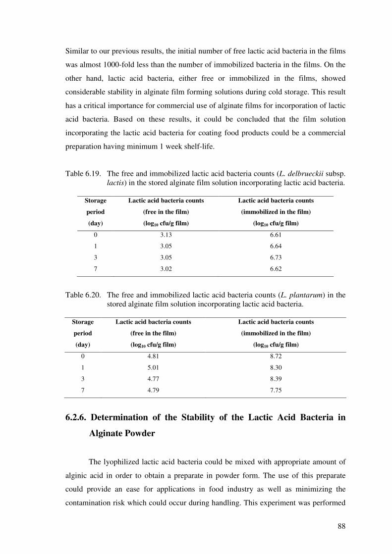
88
Similar to our previous results, the initial number of free lactic acid bacteria in the films
was almost 1000-fold less than the number of immobilized bacteria in the films. On the
other hand, lactic acid bacteria, either free or immobilized in the films, showed
considerable stability in alginate film forming solutions during cold storage. This result
has a critical importance for commercial use of alginate films for incorporation of lactic
acid bacteria. Based on these results, it could be concluded that the film solution
incorporating the lactic acid bacteria for coating food products could be a commercial
preparation having minimum 1 week shelf-life.
Table 6.19. The free and immobilized lactic acid bacteria counts (L. delbrueckii subsp. lactis) in the stored alginate film solution incorporating lactic acid bacteria.
Storage
period
(day)
Lactic acid bacteria counts
(free in the film)
(log10 cfu/g film)
Lactic acid bacteria counts
(immobilized in the film)
(log10 cfu/g film)
0 3.13 6.61
1 3.05 6.64
3 3.05 6.73
7 3.02 6.62
Table 6.20. The free and immobilized lactic acid bacteria counts (L. plantarum) in the stored alginate film solution incorporating lactic acid bacteria.
Storage
period
(day)
Lactic acid bacteria counts
(free in the film)
(log10 cfu/g film)
Lactic acid bacteria counts
(immobilized in the film)
(log10 cfu/g film)
0 4.81 8.72
1 5.01 8.30
3 4.77 8.39
7 4.79 7.75
6.2.6. Determination of the Stability of the Lactic Acid Bacteria in
Alginate Powder
The lyophilized lactic acid bacteria could be mixed with appropriate amount of
alginic acid in order to obtain a preparate in powder form. The use of this preparate
could provide an ease for applications in food industry as well as minimizing the
contamination risk which could occur during handling. This experiment was performed
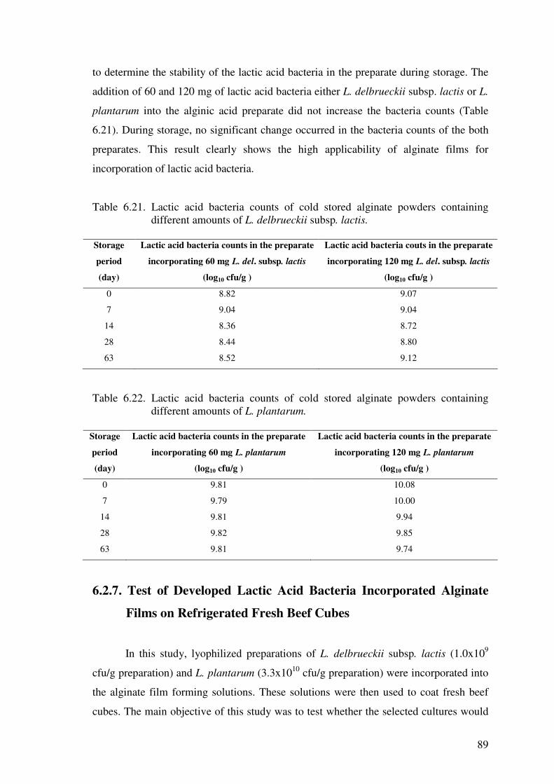
89
to determine the stability of the lactic acid bacteria in the preparate during storage. The
addition of 60 and 120 mg of lactic acid bacteria either L. delbrueckii subsp. lactis or L.
plantarum into the alginic acid preparate did not increase the bacteria counts (Table
6.21). During storage, no significant change occurred in the bacteria counts of the both
preparates. This result clearly shows the high applicability of alginate films for
incorporation of lactic acid bacteria.
Table 6.21. Lactic acid bacteria counts of cold stored alginate powders containing different amounts of L. delbrueckii subsp. lactis.
Storage
period
(day)
Lactic acid bacteria counts in the preparate
incorporating 60 mg L. del. subsp. lactis
(log10 cfu/g )
Lactic acid bacteria couts in the preparate
incorporating 120 mg L. del. subsp. lactis
(log10 cfu/g )
0 8.82 9.07
7 9.04 9.04
14 8.36 8.72
28 8.44 8.80
63 8.52 9.12
Table 6.22. Lactic acid bacteria counts of cold stored alginate powders containing
different amounts of L. plantarum.
Storage
period
(day)
Lactic acid bacteria counts in the preparate
incorporating 60 mg L. plantarum
(log10 cfu/g )
Lactic acid bacteria counts in the preparate
incorporating 120 mg L. plantarum
(log10 cfu/g )
0 9.81 10.08
7 9.79 10.00
14 9.81 9.94
28 9.82 9.85
63 9.81 9.74
6.2.7. Test of Developed Lactic Acid Bacteria Incorporated Alginate
Films on Refrigerated Fresh Beef Cubes
In this study, lyophilized preparations of L. delbrueckii subsp. lactis (1.0x10⁹
cfu/g preparation) and L. plantarum (3.3x10¹⁰ cfu/g preparation) were incorporated into
the alginate film forming solutions. These solutions were then used to coat fresh beef
cubes. The main objective of this study was to test whether the selected cultures would

90
increase lactic acid bacteria load of raw beef. It is also intended to test the viability of
incorporated bacteria in the films on beef surface during refrigerated storage. As seen in
Figure 6.10, the lactic acid bacteria counts of the samples coated with protective culture
incorporated films increased 4 to 5 log, compared to uncoated samples. During 2 weeks
of storage period, there was an inconsiderable slight decrease in the microbial load of
beef cubes coated with lactic acid bacteria incorporated alginate films. Thus, it is clear
that the L. delbrueckii subsp. lactis and L. plantarum incorporated into alginate films
are quite stable at the beef surface. On the other hand, the initial microbial counts of
uncoated beef cubes were almost 2 decimal higher than that of uncoated beef cubes. It
appeared that the coating of beef cubes with alginate has some protective effect on
naturally occurring lactic acid bacteria. However, this effect lasted only for several days
and lactic acid counts of coated and uncoated beef cubes reached to the same level
during cold storage.
0
1
2
3
4
5
6
7
8
0 5 10 15time (days)
Mic
robi
al c
ount
s lo
g C
FU/g
untreatedtreated with alginate solutiontreated with L.delbrueckii subsp. lactis incorporated alginate solutiontreated with L.plantarun incorporated alginate solution
Figure 6.12. The change in the counts of the different lactic acid bacteria incorporated
alginate films during storage.

91
CHAPTER 7
CONCLUSIONS
• The antimicrobial system formed by lactoperoxidase incorporated alginate
films, thiocyanate and hydrogen peroxide show antimicrobial activity on different Gram
(+) and Gram (-) bacteria. The system also showed sufficient antimicrobial activity in
food application conducted on refrigerated fresh calamari.
• The duration of antimicrobial effect of lactoperoxidase system depends on
kinetic properties of the enzyme, initial concentrations of hydrogen peroxide and
thiocyanate, and catalase activity of the food product. Thus, it is essential to use suitable
concentrations of each ingredient during food applications and choose food with low
catalase activity at the surface.
• For food applications, dry powders of thiocyanate and lactoperoxidase
preparation can be mixed with alginate powder. Once this mixture is solubilized in
water, the foods can be coated with alginate and further treated with CaCl2 to cross-link
the film. The hydrogen peroxide can be employed before transportation or storage.
• The lactic acid bacteria can also be incorporated into alginate films. The
bacteria showed sufficient stability in the films and on surface of refrigerated beef
cubes.
• In industry, the lactic acid bacteria incorporated films can be used in coating of
refrigerated fresh and processed meat products to provide a protective effect in case of
temperature abuse.
• For food applications, dry powders of lactic acid bacteria can be mixed with
alginate powder. Once this mixture is solubilized in water the foods can be coated with
alginate and further treated with CaCl2 to cross-link the film. The films can be
employed before transportation to market.
• Further studies are needed to test the effectiveness of the developed films on
different foods during refrigerated storage and temperature abuse.

92
REFERENCES
Alakomi, H.L., Skyttä, E., Saarela, M., Mattila-Sandholm, T., Latva-Kala, K.,and Helander, I.M., 2000. “Lactic Acid Permeabilizies Gram-Negative Bacteria by Disrupting the Outer Membrane”. Applied and Environmental Microbiology, 66 ( 5 ), 2001-2005.
Amézquita, A. and Brashears, M.M., 2002. “Competitive Inhibition of Listeria
monocytogenes in Ready-to-Eat Meat Products by Lactic Acid Bacteria”. Journal of Food Protection. 65 ( 2 ), 316-325.
Appendini, P. and Hotchkiss, J.H., 2002. “Review of Antimicrobial Food Packaging”,
Innovative Food Science & Emerging Technologies,Vol.3, pp., 113-126. Bredholt, S., Nesbakken, T., and Holck, A., 1999. “Protective Cultures Inhibit Growth
of Listeria monocytogenes and Escherichia coli O157:H7 in Cooked, Sliced, Vacuum- and Gas- Packaged Meat”, International Journal of Food Microbiology, Vol.(53),pp.43-52.
Budde, B.B., Hornbeak, T., Jacobsen, T., Barkholt V., and Koch, A.G., 2003.
“Leuconostoc carnosum 4010 has the Potential for use as a Protective Culture for Vacuum-Packed Meats: Culture Isolation, Bacteriocin Identification, and Meat Application Experiments”. International Journal of Food Microbiology. 83,171-184.
Buncic, S., Avery, S.M., and Moorhead, S.M., 1997. “Insufficient Antilisterial Capacity
of Low Inoculum Lactobacillus Cultures on Long-Term Stored Meats at 4oC”. International Journal of Food Microbiology. 34,157-170.
Bureau, G., and Multon, J.L., 1996. Food Packaging Technology, (VCH press). Volume
1. Ça�rı, A., Üstünol, Z., and Ryser, E.T., 2001. “Antimicrobial, Mechanical, and
Moisture Barrier Properties of Low pH Whey Protein-Based Edible Films Containing p-Aminobenzoic or Sorbic Acids”, Journal of Food Science,Vol.66, No:6, pp.865-870.
Ça�rı, A., Üstünol, Z., and Ryser, E.T., 2004. “Antimicrobial Edible Films and
Coatings”, Journal of Food Protection, 67(4),833-848. Cha, D.S., Choi, J.H., Chinnan, M.S., and Park, H.J., 2002. “Antimicrobial Films Based
on Na-Alginate and k-Carrageenan”, Lebensm.-Wiss.u-Technol.,35,715-719. Cha, D.S., and Chinnan, M.S. 2004. “Biopolymer-Based Antimicrobial Packaging: A
Review”, Food Science and Nutrition, Vol.(44), pp.223-237. Chávarri, F., Santisbean, A., Virto, M., de Renobales, M. “Alkaline Phosphatase. Acid
Phosphates, Lactoperoxidase, and Lipoprotein Lipase Activities in Industrial Ewe’s Milk and Cheese”, J. Agric. Food Chem. Vol. 46, pp. 2926-2932.

93
Coma, V., Sebti, L., Pardon, P., Deschamps, A., and Pichavant, F.H., 2001.
“Antimicrobial Edible Packaging Based on Cellulosic Ethers, Fatty Acids, and Nisin Incorporation to Inhibit Listeria innocua and Staphylococcus aureus”. Journal of Food Protection, 64(4),470-475.
Davidson, P.M., and Branen, A.L., 1993. “Antimicrobials in Foods”. Second Edition,
pressed by Marcel Dekker, pp. 452-454. Debeaufort, F., Quezada-Gallo, J., and Voilley, A., 1998. “Edible Films and Coatings:
Tomorrow’s Packagings”, Critical Reviews in Food Science, 38(4), 299-313. De Roever, C., 1998. “Microbiological Safety Evaluations and Recommendations on
Fresh Produce”, Food Control, Vol.(9), pp.321-347. Devlieghere, F., Vermeiren, L., and Debevere, J., 2004. New Preservation
Technologies: Possibilities and Limitations. International Dairy Journal. 14,273-285.
Elliot, R.M., McLay, J.C., Kennedy, M.J., and Simmonds, R.S., 2004. “Inhibition of
Foodborne Bacteria by the Lactoperoxidase System in a Beef Cube System”, Int. J. Food Microbiol., Vol.(91), pp.73-81.
Fonteh, F.A., Grandison, A.S., and Lewis, M.J., 2005. “Factors Affecting
Lactoperoxidase Activity”, International Journal of Dairy Technology, Vol.(58), pp. 233-236.
Ghanbarzadeh, B., Oromiehie, A.R., Musavi, M., D-Jomeh, Z.E., Rad, E.R., and Milani,
J., 2006. “Effect of Platicizing Sugars on Rheological and Thermal Properties of Zein Resins and Mechanical Properties of Zein Films”, Food Research International, Vol.70, Nr.39, pp. 882-890.
Gmez, R., Mu�oz, M., de Ancos, B., and Cano, M.P., 2002. New Procedure for The
Detection of Lactic Acid Bacteria in Vegetables Producing Antibacterial Substances. Lebensm.-Wiss.u-Technol..35,284-288.
Güçbilmez, Ç.M., Yemenicioglu, A., and Arslano�lu, A., 2007. “Antimicrobial and
Antioxidant Activity of Edible Zein Films Incorporated with Lysozyme, Albumin Proteins and Disodium EDTA”, Food Research International, 40,80-91.
Han, J.H., 2000. “Antimicrobial Food Packaging”, Food Technology, Vol.54(3), pp.56-
65. Hartmann, R. and Meisel, H., 2007. “Food-Derived Peptides with Biological Activity:
from Research to Food Applications”, Current Opinion in Biotechnology,18,163-169.

94
Hugas, M., 1998. Bacteriocinogenic Lactic Acid Bacteria for the Biopreservation of Meat and Meat Products. Meat Science. 49 ( 1 ), 139-150.
Hui, Y.H., 1991. Encyclopedia of Food Science and Technology, volume 2. pp. 659-
663. (A Wiley-Interscience Publication). Jeong, D.K., Harrison, M.A., Frank, J.F., and Wicker, L., 1992. “Trials on the
Antibacterial Effect of Glucose Oxidase on Chicken Breast Skin and Muscle”, Journal of Food Safety, Vol.(13), pp. 43-49.
Kana, R.P., Qureshi, N., and Pai, J.S., 2001. “Production of Glucose Oxidase Using
Aspergillus niger and Corn Steep Liquar”, Bioresource Technology, Vol.(78), pp. 123-126.
Kandemir, N., Yemenicio�lu, A., Mecito�lu, Ç., Elmacı, Z.S., Arslano�lu, A.,
Göksungur, Y., and Baysal, T., 2005. “Production of Antimicrobial Films by Incorporation of Partially Purified Lysozyme into Biodegradable Films of Crude Exopolysaccharides Obtained from Aureobasidium pullulans Fermentation”, Food Technol. Biotechnol., Vol.43(4), pp. 343-350.
Kennedy, M., O’Rourke, A.L., McLay, J., and Simmonds, R., 2000. “ Use of Ground
Beef Model to Assess the Effect of the Lactoperoxidase System on the Growth of Escherichia coli O157:H7, Listeria monocytogenes and Staphylococcus aureus in Red Meat”, Int. J. Food Microbiol., Vol.(57),pp. 147-158.
Kao, P., Chen, C., Huang, S., Chang, Y., Tsai, P., and Liu, Y., 2007. “Effects of Shear
Stress and Mass Transfer on Chitinase Production by Paenibacillus sp. CHE-N1”, Biochemical Engineering Journal, Vol.(34),pp.172-178.
Khan, M.A.S., Babiker, E.E., Azakami, H., and Kato, A., 1998. “Effect of Protease
Digestion and Dephosphorylation on High Emulsifying Properties of Hen Egg Yolk Phosvitin”, J. Agric. Chem., Vol.(46), pp.4977-4981.
Khan, M.A.S., Nakamura, S., Ogawa, M., Akita, E., Azakami, H., and Kato, A., 2000.
“Bactericidal Action of Egg Yolk Phosvitin Against E.coli Under Thermal Stress”, J. Agric. Chem., Vol.(48), pp.1503-1506.
Kotzekidou, P. and Bloukas, J.G., 1998. “Microbial And Sensory Changes in Vacuum-
Packed Frankfurter-Type Sausage By Lactobacillus alimentarius and Fate of Inoculated Salmonella enteritidis”. Food Microbiology.15,101-111.
Khwaldia, K., Banon, S., Desorby, S., and Hardy, J., 2004. “Mechanical and Barrier
Properties of Sodium Caseinate-Anhydrous Milk Fat Edible Films”, International Journal of Food Science and Technology, Vol.(39), pp. 403-411.
Krochta, J.M., Baldwin, E.A., Nisperos-Carriedo, M., 1994. Edible Coating and Films
to Improve Food Quality. (CRC Pres). Labuza, T.P., 1996. “An Introduction to Achieve Packaging for Foods”. Food
Technol.Vol(50), pp. 68-71.

95
Lee, S.K., Han, J.H., and Decker, E.A., 2002. “Antioxidant Activity of Phosvitin in Phosphatidylchline Liposomes and Meat Model Systems”, Food Chemistry and Toxicology, Vol.(67), Nr.1, pp.37-41.
Leisner, J.J., Greer, G.G., and Stiles, M.E., 1996. “Control of Beef Spoilage by a
Sulfide-Producing Lactobacillus sake Strain with Bacteriocinogenic Leuconostoc gelidum UAL 187 during Anaerobic Storage At 2oC”. Applied and Environmental Microbiology.Vol. 62(7), pp.2610-2614.
Lindstrom, T.R., Morimoto, K. and Cante, C.J., 1992. Edible Films and Coatings, in:
Y.H. Hui Editor, Encyelopedia of Food Science and Technology Vol.2, John Wiley and Sons. Inc, New York (1992), pp.39-663.
Luukkonen, J., Kemppinen, A., Kärki, M., Laitinen, H., Mäki, M., Sivelä, S., Taimista,
A.M., and Ryhänen, E.L., 2005. “The Effect of A Protective Culture and Exclusion of Nitrate on the Survival of Enterohemorrhagic E.coli and Listeria in Edam Cheese made from Finnish Organic Milk”,International Dairy Journal. Vol. 15, pp.449-457.
Massa, S., Petruccioli, M., Brocchi, G.F., Atieri, C., Sinigaglia, M., and Spano, G.,
2001. “Growth Inhibition by Glucose Oxidase System of Enterotoxic Escherichia coli and Salmonella derby; in vitro Studies”, World Journal of Microbiology & Biotechnology, Vol.(17), pp.287-291.
Mataragas, M., Drosinos, E.H., Metaxopoulas, J., 2003. “Antagonistic Activity Of
Lactic Acid Bacteria Against Listeria monocytogenes in Sliced Cooked Cured Pork Shoulder Stored under Vacuum or Modified Atmosphere At 4±2oC”. Food Microbiology. Vol. 20, pp.259-265.
McHugh, T.H., and Krochta, J.M., 1994. “Sorbital-vs Glycerol-Plasticized Whey
Protein Edible Films: Integrated Oxygen Permeability and Tensile Property Evaluation”, Journal of Agricultural and Food Chemistry, Vol.42, No.42, pp 841-845.
Mecito�lu, Ç., and Yemenicio�lu, A., 2007. “Partial Purification and Preparation of
Bovine Lactoperoxidase and Charaterization of Kinetic Properties of its Immobilized Form Incorporated into Cross-Linked Alginate films”, Food Chemistry. Vol. 104, pp. 726-733.
Millette, M., Smoragiewicz, W., and Lacroix, M., 2004. “Antimicrobial Potential of
Immobilized Lactococcus lactis subsp. lactis ATCC 11454 against Selected Bacteria”. Journal of Food Protection. Vol.67(6), pp.1184-1189.
Minor-Pérez, H., Ponce-Alquicira, E., Macias-Bravo, S., and Guerrero-Legarreta, I.,
2004. “Changes In Fatty Acids and Microbial Popolations of Pork Inoculated with Two Biopreservative Strains”. Meat Science. Vol. 66, pp.793-800.
Muthukumarasamy, P., Han, J.H., and Holley, R.A., 2003. “Bactericidal Effects of
Lactobacillus reuteri and Allyl Isothiocyanate on Escherichia coli O157:H7 in Refrigerated Ground Beef”. Journal of Food Protection. Vol.66(11), pp.2038-2044.

96
Nakamura, S., Ogawa, M., Nakai, S., Kato, A., and Kitts, D.D., 1998. “Antioxidant Activity of a Maillard-Type Phosvitin-Galactomannan Conjugate with Emulsifying Properties and Heat Stability”, J. Agric. Food Chem., Vol.(46), pp. 3958-3963.
Natrajan, N., and Sheldon, B.W., 2000. “Inhibition of Salmonella on Poultry Skin Using
Protein- and Polysaccharide-Based Films Containing a Nisin Formulation”, Journal of Food Protection, Vol.63(9), pp.1268-1272.
Ogunbanwo, S.T. and Okanlawon, B.M., 2006. “Microbial and Sensory Changes
During the Cold Storage of Chicken Meat Treated with Bacteriocin from L.brevis OG1”, Pakistan Journal of Nutrition, Vol.5(6), pp.601-605.
Oussalah, M., Caillet, S., Salmieri, S., Saucier L., and Lacroix, M., 2006.
“Antimicrobial Effects of Alginate-Based Film Containing Essential Oils for The Preservation of Whole Beef Muscle”, Journal of Food Protection, Vol.69(10), pp.2364-2369.
Pakkanen, R., and Aalto, J., 1997. “Growth Factors and Antimicrobial Factors”,
International Dairy Journal, Vol.(7), pp.285-297. Park, S., Stan, S.d., Daeschel, M.A., and Zhao, Y., 2005. “Antifungal Coatings on Fresh
Strawberries (Fragaria x ananassa) to Control Mold Growth During Cold Storage”, Journal of Food Science, Vol.70(4), pp.202-207.
Pranoto, Y., Salokhe, V.M., and Rakshit, S.K., 2005. “Physical and Antibacterial
Properties of Alginate-Based Edible Film Incorporated with Garlic Oil”, Food Research International, Vol.38, pp.267-272.
Quintavalla, S., and Vicini, L. 2002. “Antimicrobial Food Packaging in Meat Industry”,
Meat Science, Vol.62, pp.373-380. Ray, B., 2004. “Fundamental Food Microbiology”. (CRC Press, Third Edition). Robertson, G. L., 2006. “ Food Packaging Principles and Practice”, (CRC press,
second edition) pp. 292-293. Rodgers, S., 2001. “Preserving Non-Fermented Refrigerated Foods with Microbial
Cultures-a review”. Food Science and Technology. Vol.12, pp.276-284. Rodgers, S., 2003. “Potential Applications of Protective Cultures in Cook-Chill
Catering”. Food Control. Vol.14, pp.35-42. Rodgers, S., Kailasapathy, K., Cox, J., and Peiris, P., 2004. “Co- incubation of
Clostridium botulinum with Protective Cultures”, Food Research International. Rodgers, S., Peiris, P., and Casadei, G., 2003. “Inhibition of Nonproteolytic Clostridium
botulinum with Lactic Acid Bacteria and Their Bacteriocins at Refrigeration Temperatures”. Journal of Food Protection, Vol.66 ( 4 ), pp.674-678.

97
Rodgers, S., Kailasapathy, K., Cox, J., and Peiris, P., 2004. “Coincubation of Clostridium botulinum with protective cultures. Food Research International”, article in press
Rybka-Rodgers, S., 2001. “Improvement of Food Safety Design of Cook-chill Foods”.
Food Research International. Vol.34, pp.449-455. Ryu, S.Y., Rhim, H.J., and Kim, S.S., 2002. “Preparation and Physical Properties of
Zein-Coated High-Amylose Corn Starch Film”, Lebensm.-Wiss.u-Technol., Vol.35, pp.680-686.
Saad, S.M.I., Vanzin, C., Oliveira, M.N., and Franco, B.D.G., 2001. “Influence of
Lactic Acid Bacteria on Survival of Escherichia coli O157:H7 in Inoculated Minas Cheese During Storage at 8.5oC”. Journal of Food Protection. Vol.64(8 ), pp.1151-1155.
Salmien, S., Von Wright, A., and Ouwehand, A., 2004. “Lactic Acid Bacteria,
Microbiological and Functional Aspects”, (Third Edition, Revised and Expanded), p 379.
Schnürer, J., and Magnusson, J., 2005. “Antifungal Lactic Acid Bacteria as
Biopreservatives”. Food Science and Technology. Article in press 1-9.Review. Schwenninger, S.M. and Meille, L., 2004. “A Mixed Culture of Propionibacterium
jensenii and Lactobacillus paracasei subsp. paracasei Inhibits Food Spoilage Yeasts System”. Appl. Microbiol. Vol.27, pp.229-237.
Seifu, E., Buys, E.M., and Donkin, E.F., 2000. “Significance of the Lactoproxidase
System in the Dairy Industry and Its Potential Applications: A Review”, Trends Food Sci. Tech., Vol.(16), pp.1-18.
Seifu, E., Buys, E.M., and Donkin, E.F., 2004. “Quality Aspects of Gouda Cheese Made
from Goat Milk Preserved by the lactoperoxidase System”, International Dairy Journal, Vol.(14), pp.581-589.
Seydim, A.C., and Sarikus, G., 2006. “Antimicrobial Activity of Whey Protein Based
Edible Films Incorporated with Oregano, Rosemary and Garlic Essential Oils”, Food Research International. Vol.39, pp.639-644.
Siragusa, G.R., and Dickson, J.S., 1992. “Inhibition of Listeria monocytogenes on Beef
Tissue by Application of Organic Acids Immobilized in a Calcium Alginate Gel”, Journal of Food Science, Vol.57(2), pp.293-296.
Suppakul, P., Miltz, J.,Sonneveld, K., and Bigger, S.W., 2003. “Active Packaging
Technologies with an Emphasis on Antimicrobial Packaging and its Applications”, Journal of Food Science, Vol.68, Nr.2, pp. 408-420.

98
Theivendran, S., Hettiarachchy, N.S., and Johnson, M.G., 2006. “Inhibition of Listeria monocytogenes by Nisin Combined with Grape Seed Extract or Green Tea Extract in Soy Protein Film Coated on Turkey Frankfurters”, Journal of Food Science. Vol.71(2), pp.39-44.
Touch, V., Hayakawa, S., Yamada, S., and Kaneko, S.,2004. “Effects Of A
Lactoperoxidase-Thiocyanate-Hydrogen Peroxide System on Salmonella enteritidis in Animal or Vegetable Foods”, International Journal of Food Microbiology, Vol.93, pp.175-183.
Vereecken, K.M., and Van Impe, J.F., 2002. “Analysis and Practical Implementation of
a Model for Combined Growth and Metabolic Production of Lactic Acid Bacteria”. International of Food Microbiology.Vol.73, pp.239-250.
Vermeiren, L., Devlieghere, F., and Debevere, J., 2004. “Evaluation of Meat Born
Lactic Acid Bacteria as Protective Cultures for the Biopreservation of Cooked Meat Products”. International Journal of Food Microbiology. Vol.96, pp.149-164.
Vescovo, M., Scolari, G., and Zacconi, C.,2006. “Inhibition of Listeria innocua Growth
by Antimicribial-Producing Lactic Acid Cultures in Vacuum-Packed Cold-Smoked Salmon”. Food Microbiology. Vol.23, pp.689-693.
Villegas, E. and Gilliland, S.E.,1998. “Hydrogen Peroxide Production by Lactobacillus
delbrueckii subsp.lactis I at 5oC”. Journal of Food Science.Vol.63(6), pp.1070-1074.
Zalán, Z., Nèmeth, E., Baráth, Á., and Halász, A., 2005. “Influence of Growth Medium
on Hydrogen Peroxide and Bacteriocin Production of Lactobacillus Strains”. Food Technol.Biotechnol. Vol.43(3), pp.219-225.
99
APPENDICES
APPENDIX A
E. coli
0
2
4
6
8
10
0 4 8 12 16 20 24
Time (h)
log
CFU
/mL
Figure A.1. The growth curve of E. coli
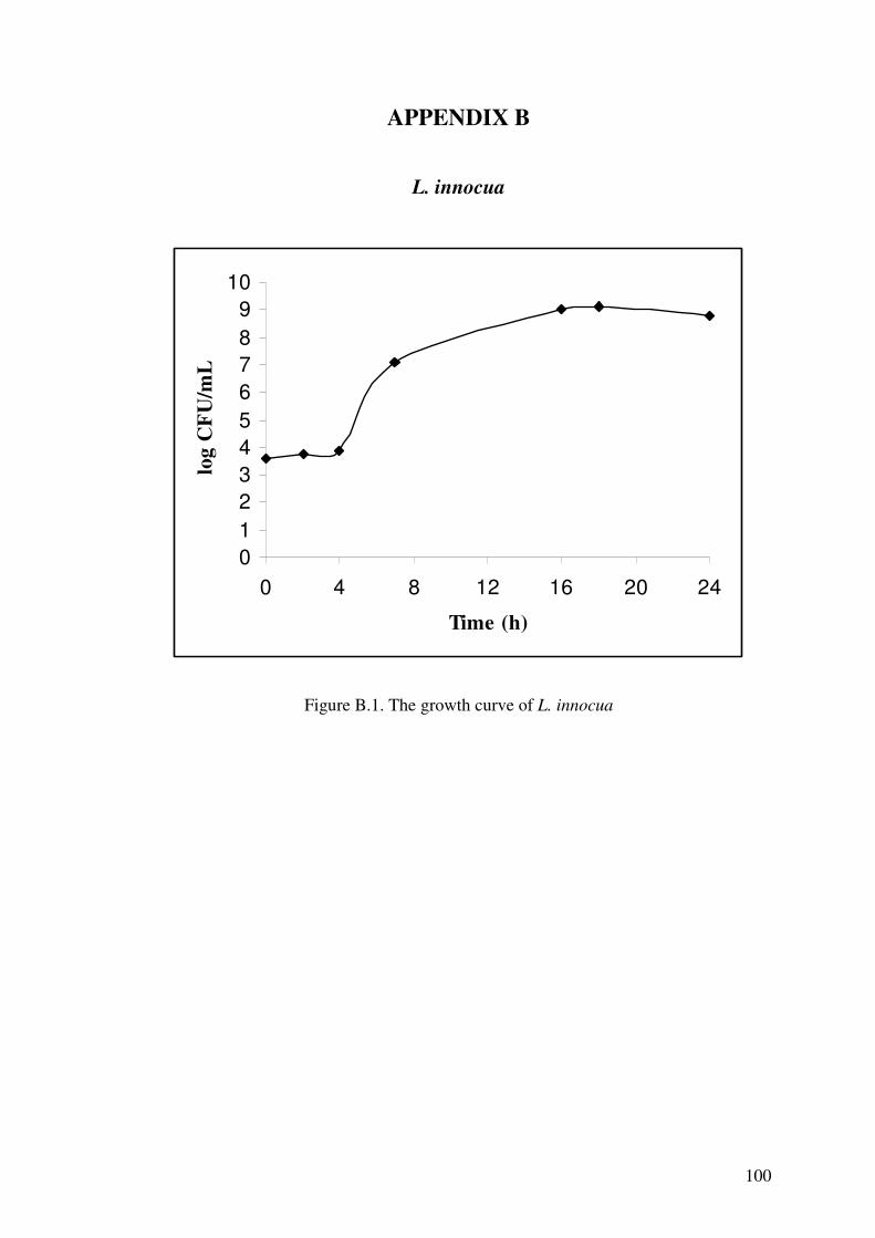
100
APPENDIX B
L. innocua
0123456789
10
0 4 8 12 16 20 24
Time (h)
log
CFU
/mL
Figure B.1. The growth curve of L. innocua

101
APPENDIX C
P. fluorescens
0123456789
10
0 4 8 12 16 20 24
Time (h)
log
CFU
/mL
Figure C.1. The growth curve of P. fluorescens