determination of the core of a minimal bacterial gene set† · 180 housekeeping genes that were...
TRANSCRIPT

MICROBIOLOGY AND MOLECULAR BIOLOGY REVIEWS, Sept. 2004, p. 518–537 Vol. 68, No. 31092-2172/04/$08.00�0 DOI: 10.1128/MMBR.68.3.518–537.2004Copyright © 2004, American Society for Microbiology. All Rights Reserved.
Determination of the Core of a Minimal Bacterial Gene Set†Rosario Gil,1,2* Francisco J. Silva,1,2 Juli Pereto,1,3 and Andres Moya1,2
Institut Cavanilles de Biodiversitat i Biologia Evolutiva, Universitat de Valencia, Valencia,1 andDepartament de Genetica2 and Departament de Bioquımica i Biologia Molecular,3
Universitat de Valencia, Burjassot (Valencia), Spain
INTRODUCTION .......................................................................................................................................................518MAIN FEATURES OF THE MINIMAL SET ........................................................................................................520
Information Storage and Processing....................................................................................................................520DNA metabolism. (i) Basic replication machinery ........................................................................................520
(ii) DNA repair, restriction, and modification ...........................................................................................524RNA metabolism .................................................................................................................................................524
(i) Basic transcription machinery ................................................................................................................524(ii) Translation................................................................................................................................................525(iii) RNA degradation ....................................................................................................................................526
Protein Processing, Folding, and Secretion ........................................................................................................527Cell Structure and Cellular Processes.................................................................................................................527
Cell wall................................................................................................................................................................527Cell shape and division......................................................................................................................................527Substrate transport ............................................................................................................................................528
Energetic and Intermediary Metabolism.............................................................................................................528Glycolysis, gluconeogenesis, pyruvate metabolism, and the TCA cycle ......................................................529Electron transport chain and proton motive force generation.....................................................................530Pentose phosphate pathway...............................................................................................................................530Biosynthesis of amino acids ..............................................................................................................................531Biosynthesis of lipids..........................................................................................................................................531Biosynthesis of nucleotides................................................................................................................................531Biosynthesis of cofactors....................................................................................................................................533
Poorly Characterized Genes ..................................................................................................................................534CONCLUSIONS .........................................................................................................................................................534ACKNOWLEDGMENTS ...........................................................................................................................................535REFERENCES ............................................................................................................................................................535
INTRODUCTION
Complete genome sequences are becoming available for alarge number of diverse bacterial species. Comparative geno-mics shows that most bacterial proteins are highly conserved inevolution, allowing predictions to be made about the functionsof most products of uncharacterized genomes based on modelorganisms, such as Escherichia coli and Bacillus subtilis (gram-positive and gram-negative bacteria, respectively), for whichabundant high-quality genetic and biochemical informationhas been obtained in the past.
One important question raised by the availability of com-plete genomic sequences is how many genes are essential forcellular life. Although bacterial genomes differ vastly in theirsizes and gene repertoires, no matter how small, they mustcontain all the information to allow the cell to perform manyessential (housekeeping) functions that give the cell the abilityto maintain metabolic homeostasis, reproduce, and evolve, thethree main properties of living cells (53). Cells usually can
import metabolites but not functional proteins; therefore, theyhave to rely on their own gene products to perform suchessential functions.
The determination of the minimal set of protein-codinggenes necessary to maintain a living cell is becoming an in-creasingly appealing issue, considering that such minimal geneset should include “the smallest possible group of genes thatwould be sufficient to sustain a functioning cellular life formunder the most favorable conditions imaginable, that is, in thepresence of a full complement of essential nutrients and in theabsence of environmental stress” (48). Reconstruction of theminimal gene set can take advantage of the increasing knowl-edge of completely characterized genomes. In recent years,several research groups have tried to define the essential setof survival protein-encoding genes in bacteria by different ex-perimental and computational methods (reviewed in refer-ences 22, 24, 38, and 79). Three different experimental ap-proaches have been used to identify genes that are essentialunder particular growth conditions: massive transposon mu-tagenesis strategies (the most widely used approach), the useof antisense RNA to inhibit gene expression (22, 37), andthe systematic inactivation of each individual gene presentin a genome (31, 45, 64; http://www.genome.wisc.edu/functional/tnmutagenesis.htm). However, all these approaches have lim-itations. Transposon mutagenesis might overestimate the set
* Corresponding author. Mailing address: Institut Cavanilles deBiodiversitat i Biologia Evolutiva, Universitat de Valencia, ApartatOficial 2085, 46071 Valencia, Spain. Phone: 34 96 354 36 29. Fax: 34 96354 36 70. E-mail: [email protected].
† Supplemental material for this article may be found at http://mmbr.asm.org.
518
on February 16, 2019 by guest
http://mm
br.asm.org/
Dow
nloaded from

by misclassification of nonessential genes that slow growthwithout arresting it but can also miss essential genes that tol-erate transposon insertions. The use of antisense RNA is lim-ited to the genes for which an adequate expression of theinhibitory RNA can be obtained in the organism under study.Finally, inactivation of single genes does not detect essentialfunctions encoded by redundant genes, and the essential geneset is not the same as the minimal genome, since it is clear thatgenes that are individually dispensable may not be simulta-neously dispensable.
Computational analysis has also been extensively used to tryto get closer to the minimal gene set (28, 48, 65, 81). For thispurpose, the smallest bacterial genomes fully sequenced, frombacterial parasites or endosymbionts, have been very useful,since they must retain all genes involved in housekeeping func-tions and a minimum amount of metabolic transactions forcellular survival and replication in their given niche. The small-est complete bacterial genome reported thus far correspondsto the human pathogen Mycoplasma genitalium, with only 480protein-coding genes in a 540-kb genome (23), which can beconsidered an upper-limit estimate for the minimal bacterialgene set. However, smaller genomes have been described forother bacteria, such as some strains of Buchnera aphidicola, theendosymbiotic bacteria of aphids, whose genomes have anestimated size of around 450 kb, which can contain about 400protein-coding genes (27).
The two first bacterial genomes completely sequenced werethose from the parasitic bacteria Haemophilus influenzae (21)and M. genitalium (23), gram-negative and gram-positive bac-teria, respectively, with reduced genomes (1.83 Mb and 0.58Mb respectively). Soon after that, Mushegian and Koonin per-formed a computational comparison between them, assumingthat genes conserved across the large phylogenetic distancesthat exist between these two species are likely to be essential(65). The authors suggested a list of 256 conserved genes as aclose estimate of the minimal gene set. However, some of thegenes considered essential by this approach can be disruptedby transposon insertion (34), and it is reasonable to considerthat enlargening the number of genomes used in the compar-ison will significatively reduce the number of genes consideredto be essential.
In the last few years, complete genomes of five bacterialendosymbionts of insects have been described (2, 28, 77, 80,83). These bacteria have a permanent host-associated life-style, and therefore they have relaxed selection on the main-tenance of genes that are not required for survival in theirprotected environments. Based on the above-mentioned as-sumption that the genes shared by multiple genomes are likelyto be essential and to be good candidates for inclusion in theminimal gene set, we performed a comparative analysis of allfive endosymbiont genomes, which revealed that they shareonly 277 orthologous protein-coding genes (28). The obtainedminimal endosymbiont gene set was then compared with thegenome of M. genitalium; this resulted in the identification of180 housekeeping genes that were shared by all six genomes.The number of shared genes among endosymbiotic and para-sitic bacteria was further reduced to 156 when the parasitesRickettsia prowazekii and Chlamydia trachomatis were added tothe comparison (44). However, computational analysis has alsolimitations, since it is likely to underestimate the minimal set
because it takes into account only the genes that have re-mained similar enough during the course of evolution to berecognized as true orthologues. Therefore, it will not includegenes with a high rate of evolution, which may not show theirrelationship in comparisons of distant taxons. In the above-mentioned study, the displacement of unrelated but function-ally analogous protein-coding genes was not considered, thuspresenting only a core of the possible minimum gene set nec-essary to sustain life.
In an attempt to get closer to the minimal bacterial genome,in this paper we present an enhanced review of all previouslyused strategies for addressing this issue. Taking as a start ourprevious computational comparison of gene orthologues forthe five completely sequenced endosymbionts, we added to thecomparison functional equivalent genes that do not presentsequence similarity. The obtained gene set was compared withthe B. subtilis essential genes determined by systematic inacti-vation (45) and with the proposed essential genes for E. colibased on the genome-scale genetic footprinting performed byGerdes et al. (25), the information found at the PEC (for“Profiling E. coli Chromosome”) website (http://www.shigen.nig.ac.jp/ecoli/pec), and the results obtained by F.R. Blatt-ner and coworkers (http://www.genome.wisc.edu/functional/tnmutagenesis.htm). It should be noticed that the PEC andBlattner’s E. coli sequencing-project databases are incomplete,and in some cases there are discrepancies about the essentialityof E. coli genes depending on the source. Some differencesmight be due to the different growth conditions used in theexperiments, while others might be reflecting the inability of amutant with a severe decrease in fitness to maintain an enoughvigorous growth to be isolated in the footprinting experiments(25). We also took into account the computationally derivedminimal gene set proposed for M. genitalium (65) and theresults of the global transposon mutagenesis for mycoplasmas(34) in order to detect genes that appear to be dispensable.Genes that are present in all five endosymbionts, and for whichno transposon insertion could be detected in mycoplasmas,were considered essential in reduced genomes, even if theyappear to be nonessential in bacteria with larger genomes. Wehave also included in our minimal gene set some of the genesthat were disrupted by transposon insertion in mycoplasmasbut were proven to be essential in other bacteria and are pres-ent in all five endosymbionts under study. We also comparedour set with the list of essential genes identified in Staphylo-coccus aureus by antisense RNA experiments (22, 37) and therecently sequenced genome of Phytoplasma asteris (70). Fi-nally, the resulting gene list was reanalyzed to detect gaps inmetabolic pathways that could be considered essential to main-tain a reasonable metabolic homeostasis for any living cell.
The following gene and protein servers and databases wereused to identify the corresponding orthologous genes and pro-tein functions, as well as to reconstruct the metabolic pathways:BRENDA (for “Braunschweig Enzyme Database”) (http://www.brenda.uni-koeln.de/index.php4), COG (for “Clusters of Or-thologous Groups of Proteins”) (http://www.ncbi.nlm.nih.gov/COG/), ExPASy (for “Expert Protein Analysis System”) mo-lecular biology server (http://us.expasy.org), KEGG (for “KyotoEncyclopedia of Genes and Genomes”) (http://www.genome.ad.jp/kegg2.html), MBGD (for “Microbial Genome Databasefor Comparative Analysis”) (http://mbgd.genome.ad.jp), and
VOL. 68, 2004 MINIMAL BACTERIAL GENE SET 519
on February 16, 2019 by guest
http://mm
br.asm.org/
Dow
nloaded from

Pfam (for “Protein Families Database of Alignments andHMMs”) (http://www.sanger.ac.uk/Software/Pfam).
The results of this analysis are summarized in Table 1 and,with more detail, in Table S2 presented in the supplementalmaterial. The functional classification of the genes is based onthe categories used in the sequencing work on Aquifex aeolicus(19). The number of genes included in each category is indicat-ed parenthetically in the tables. The gene name column gen-erally indicates the most common synonymous for the corre-sponding gene in gram-negative bacteria (except when no genewith such function has been identified in E. coli), although insome cases it does not correspond with the given name for thegram-positive bacteria orthologous gene. Table S2 in the sup-plemental material also includes the homologous genes pres-ent in the six completely sequenced reduced genomes (M. geni-talium, B. aphidicola strains BAp, BSg and BBp, CandidatusBlochmannia floridanus, and Wigglesworthia glossinidia) andthe two model bacteria used in this analysis (E. coli and B. sub-tilis). The B. subtilis gene numbers are those given by the Jap-anese and European Consortium involved in its sequencing(http://bacillus.genome.ad.jp/ and http://genolist.pasteur.fr/SubtiList/) (50) and can also be found in the KEGG database.In the cases of nonorthologous or nonhomologous genes en-coding proteins with the same function, we have included inTable S2 (in the supplemental material) the gene that waspresent in more analyzed organisms.
The notion of a minimal cell cannot be sharply defined, sincedifferent essential functions can be defined depending on theenvironmental conditions, and numerous versions of minimalgene sets can be conceived to fulfill such functions even for thesame set of conditions (49). Nevertheless, it is possible to try todefine which functions should be performed in any living celland to list the genes that would be necessary to maintain suchfunctions. The number of these genes, but not the number offunctions, could show some minor variation associated withputative gene fusions. In this work, we present the hypotheticalcore of one of the possible minimal protein-coding gene setsable to sustain a functional bacterial cell under ideal condi-tions, and we examine the main features of the proposed min-imal gene set. The rationale followed to decide which genesshould be included in it is described in detail.
MAIN FEATURES OF THE MINIMAL SET
Information Storage and Processing
DNA metabolism. (i) Basic replication machinery. One ofthe most important housekeeping functions in all living cells isDNA replication, a multistage reaction with four basic steps.First, a replication origin sequence within the genome is rec-ognized by protein components. Second, initiation of replica-tion occurs through the recruitment of replisomal proteins atthe origin. Third, the general replication reaction duplicatesboth strands of DNA. Fourth, replication terminates and thetwo daughter chromosomes are separated.
The specific mechanism for replication initiation dependsboth on the structure of the replication origin and on thenature of the initiation protein (47). Several mechanisms havebeen described in bacteria (46), but the analysis of the genomeof B. floridanus, the endosymbiotic bacteria of carpenter ants,
revealed that it does not encode any of the proposed initiationproteins (DnaA, RecA, and PriA). Another recruitment pro-tein (DnaC in E. coli, DnaI in B. subtilis) is also required toform a complex with the helicase DnaB (EC 3.6.1.-) to preventthe indiscriminate binding of the helicase to single-strandedDNA, transferring it only in the correct DNA location which isnot coated by Single-Stranded DNA Binding protein. (SSB).However, DnaC is absent in B. floridanus and W. glossinidia(endosymbiont of tsetse flies), indicating that it is also dispens-able in these bacteria. Hence, it can be assumed that DNAreplication can take place without the need for these initiationand recruiting proteins under some conditions, which might bethe case for small genomes, or that there are other, as yetunidentified proteins that perform such functions in these re-duced genomes. The recruitment and loading of helicase at thereplication origin generally requires the presence of a histone-like protein for the destabilization of the duplex DNA near theorigin of replication. At least one histone-like protein is alsoencoded by all five analyzed endosymbiont genomes, althoughnone of these proteins is conserved in all of them. In M. geni-talium, MG353 appears to encode a nucleoid DNA bindingprotein similar to hupA. Since this gene is also present in allendosymbionts analyzed except B. floridanus, we decided to in-clude this histone-like protein-coding gene in our minimal set.
To begin DNA replication, the helicase DnaB (EC 3.6.1.-)attracts the primase DnaG (EC 2.7.7.-) to the replication fork,both of them present in all bacteria with reduced genomesanalyzed in this study. Once the DNA is melted and primed,the general DNA replication begins, with the action of DNApolymerase III (EC 2.7.7.7). Genes encoding the �, ε, � and �,�, �’, and � subunits of DNA polymerase III, all subunits thatare present both in gram-negative and gram-positive bacteria,are also conserved. It should be noticed that in gram-positivebacteria there are two genes encoding the � subunits of thisholoenzyme, both of them essential in B. subtilis, but one ofthem (dnaE) has been proven to be dispensable in M. geni-talium, while polC encodes both the � and ε subunits of DNApolymerase III. Two more proteins required at this stage, gy-rase (EC 5.99.1.2, encoded by gyrA and gyrB) and ligase (EC6.5.1.2, encoded by lig), are also conserved, thus allowing thecompletion of the third stage of DNA replication. Although ligwas previously annotated as a pseudogene in B. aphidicola BSgdue to the absence of the C-terminal protein end (80), it hasbeen reannotated as a functional gene encoding the catalyticdomain of the protein but with the absence of the BRCTdomain (28). On the other hand, the gene encoding the proteinnecessary to stop replication by inhibiting helicase transloca-tion (tus in E. coli, rtp in B. subtilis) cannot be found in thereduced genomes under study. Several topoisomerases that arepresent in M. genitalium and essential in B. subtilis and E. colito allow independent segregation of the two daughter chromo-somes (EC 5.99.1.2, encoded by topA, and EC 5.99.1.-, encodedby parC and parE) must also be considered dispensable, sinceparC and parE have been lost in all endosymbionts analyzed,while topA is only present in two strains of B. aphidicola. It ispossible that the above-mentioned GyrA/B, which is present inall these bacteria, performs all the topoisomerase functionsrequired for DNA replication and segregation, since it hasbeen suggested that type IA (TopA) and type II (GyrA/B andParD/E) topoisomerases may use comparable mechanisms to
520 GIL ET AL. MICROBIOL. MOL. BIOL. REV.
on February 16, 2019 by guest
http://mm
br.asm.org/
Dow
nloaded from

TABLE 1. Protein-coding genes in the hypothetical minimal cell
Category Subcategory Gene E.C. no. Protein function
DNA metabolism(16 genes)
Basic replication machinery (13 genes) dnaB 3.6.1.- Replicative DNA helicasednaE 2.7.7.7 DNA polymerase III, � subunitdnaG 2.7.7.- DNA primasednaN 2.7.7.7 DNA polymerase III, � subunitdnaQ DNA polymerase III, ε subunitdnaX 2.7.7.7 DNA polymerase III, � and � subunitsgyrA 5.99.1.3 DNA gyrase, A subunitgyrB 5.99.1.3 DNA gyrase, B subunitholA 2.7.7.7 DNA polymerase III, � subunitholB 2.7.7.7 DNA polymerase III, �� subunithupA DNA binding proteinlig 6.5.1.2 DNA ligase (NAD dependent)ssb SSB
DNA repair, restriction, and modification(3 genes)
nth 4.2.99.18 Endonuclease IIIpolA 3.1.11.- 5�-3� exonuclease domain of DNA polymerase Iung 3.2.2.- Uracil-DNA glycosylase
RNA metabolism(106 genes)
Basic transcription machinery (8 genes) deaD ATP-dependent RNA helicasegreA Transcription elongation factornusA Transcription-translation couplingnusG Transcription antitermination proteinrpoA 2.7.7.6 RNA polymerase, � subunitrpoB 2.7.7.6 RNA polymerase, � subunitrpoC 2.7.7.6 RNA polymerase, �� subunitrpoD RNA polymerase major � factor
Translation: aminoacyl-tRNA synthesis(21 genes)
alaS 6.1.1.7 Alanyl-tRNA synthaseargS 6.1.1.19 Arginyl-tRNA synthaseasnS 6.1.1.22 Asparaginyl-tRNA synthaseaspS 6.1.1.12 Aspartyl-tRNA synthasecysS 6.1.1.16 Cysteinyl-tRNA synthaseglnS 6.1.1.18 Glutaminyl-tRNA synthasegltX 6.1.1.17 Glutamyl-tRNA synthaseglyS 6.1.1.14 Glycyl-tRNA synthase, b subunithisS 6.1.1.21 Histidyl-tRNA synthaseileS 6.1.1.5 Isoleucyl-tRNA synthaseleuS 6.1.1.4 Leucyl-tRNA synthaselysS 6.1.1.6 Lysyl-tRNA synthasemetS 6.1.1.10 Methionyl-tRNA synthasepheS 6.1.1.20 Phenylalanyl-tRNA synthase, a subunitpheT 6.1.1.20 Phenylalanyl-tRNA synthase, b subunitproS 6.1.1.15 Prolyl-tRNA synthaseserS 6.1.1.11 Seryl-tRNA synthasethrS 6.1.1.3 Threonyl-tRNA synthasetrpS 6.1.1.2 Tryptophanyl-tRNA synthasetyrS 6.1.1.1 Tyrosyl-tRNA synthasevalS 6.1.1.9 Valyl-tRNA synthase
Translation: tRNA maturation andmodification (6 genes)
iscS 4.4.1- Cysteine desulfurase-NifS homologmnmAa 2.1.1.61 tRNA (5-methylaminomethyl-2-thiouridylate) methyl-
transferasemnmEb GTP binding protein involved in biosynthesis of
5-methilaminomethyl-2-thiouridinemnmGc Glucose-inhibited division protein A, involved in bio-
synthesis of 5-methylaminomethyl-2-thiouridinepth 3.1.1.29 Peptidyl-tRNA hydrolasernpA 3.1.26.5 Protein component of RNa P
Translation: ribosomal proteins (50 genes) rplA 50S ribosomal protein L1rplB 50S ribosomal protein L2rplC 50S ribosomal protein L3rplD 50S ribosomal protein L4rplE 50S ribosomal protein L5rplF 50S ribosomal protein L6rplI 50S ribosomal protein L9rplJ 50S ribosomal protein L10rplK 50S ribosomal protein L11rplL 50S ribosomal protein L12rplM 50S ribosomal protein L13rplN 50S ribosomal protein L14rplO 50S ribosomal protein L15rplP 50S ribosomal protein L16
Continued on following page
VOL. 68, 2004 MINIMAL BACTERIAL GENE SET 521
on February 16, 2019 by guest
http://mm
br.asm.org/
Dow
nloaded from

TABLE 1—Continued
Category Subcategory Gene E.C. no. Protein function
rplQ 50S ribosomal protein L17rplR 50S ribosomal protein L18rplS 50S ribosomal protein L19rplT 50S ribosomal protein L20rplU 50S ribosomal protein L21rplV 50S ribosomal protein L22rplW 50S ribosomal protein L23rplX 50S ribosomal protein L24rpmA 50S ribosomal protein L27rpmB 50S ribosomal protein L28rpmC 50S ribosomal protein L29rpmE 50S ribosomal protein L31rpmF 50S ribosomal protein L32rpmG 50S ribosomal protein L33rpmH 50S ribosomal protein L34rpmI 50S ribosomal protein L35rpmJ 50S ribosomal protein L36rpsB 30S ribosomal protein S2rpsC 30S ribosomal protein S3rpsD 30S ribosomal protein S4rpsE 30S ribosomal protein S5rpsF 30S ribosomal protein S6rpsG 30S ribosomal protein S7rpsH 30S ribosomal protein S8rpsI 30S ribosomal protein S9rpsJ 30S ribosomal protein S10rpsK 30S ribosomal protein S11rpsL 30S ribosomal protein S12rpsM 30S ribosomal protein S13rpsN 30S ribosomal protein S14rpsO 30S ribosomal protein S15rpsP 30S ribosomal protein S16rpsQ 30S ribosomal protein S17rpsR 30S ribosomal protein S18rpsS 30S ribosomal protein S19rpsT 30S ribosomal protein S20
Translation: ribosome function, maturationand modification (7 genes)
cspR 2.1.1.- Ribosomal methytransferaseengA GTP binding proteinera GTP binding proteinksgA 2.1.1.- Dimethyladenosine transferaseobg GTP binding proteinrbfA Ribosome binding factor AychF GTP binding protein
Translation factors (12 genes) efp Elongation factor PfusA 3.6.1.48 Elongation factor Gfrr Ribosome-recycling factorhemK 2.1.1.- N5-glutamine methyltransferase, modulation of
release factor activityinfA Initiation factor IF-1infB Initiation factor IF-2infC Initiation factor IF-3lepA GTP binding elongation factorprfA Peptide chain release factor 1 (RF1)smpB tmRNA binding proteintsf Elongation factor TstufA 3.6.5.3 Elongation factor Tu
RNA degradation (2 genes) pnp 2.7.7.8 Polyribonucleotide nucleotidyltransferasernc 3.1.26.3 Ribonuclease III
Protein processing,folding, and secre-tion (15 genes)
Protein posttranslational modification(2 genes)
map 3.4.11.18 Methionine aminopeptidasepepA 3.4.11.1 Aminopeptidase A/I
Protein folding (5 genes) dnaJ Hsp70 cochaperonednaK Chaperone Hsp70groEL Class I heat shock proteingroES Class 1 heat shock proteingrpE Hsp70 cochaperone
Protein translocation and secretion (5 genes) ffh Protein component of signal recognition particleftsY Signal recognition particle receptor
Continued on following page
522 GIL ET AL. MICROBIOL. MOL. BIOL. REV.
on February 16, 2019 by guest
http://mm
br.asm.org/
Dow
nloaded from
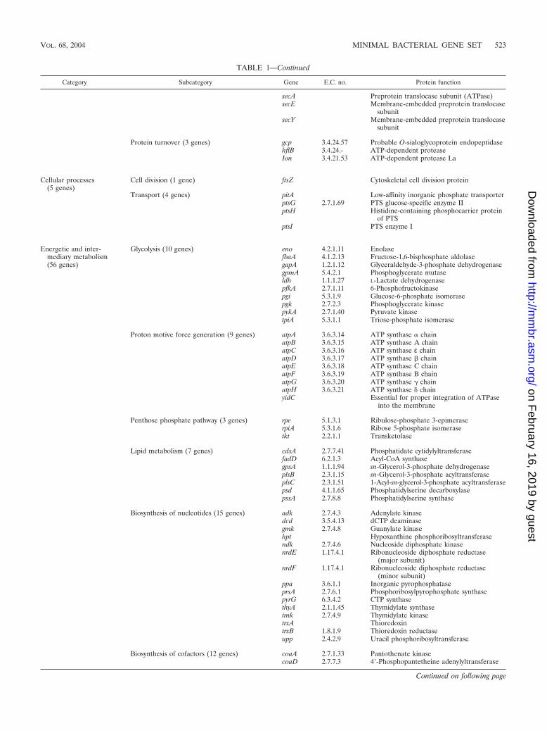
TABLE 1—Continued
Category Subcategory Gene E.C. no. Protein function
secA Preprotein translocase subunit (ATPase)secE Membrane-embedded preprotein translocase
subunitsecY Membrane-embedded preprotein translocase
subunit
Protein turnover (3 genes) gcp 3.4.24.57 Probable O-sialoglycoprotein endopeptidasehflB 3.4.24.- ATP-dependent proteaseIon 3.4.21.53 ATP-dependent protease La
Cellular processes(5 genes)
Cell division (1 gene) ftsZ Cytoskeletal cell division protein
Transport (4 genes) pitA Low-affinity inorganic phosphate transporterptsG 2.7.1.69 PTS glucose-specific enzyme IIptsH Histidine-containing phosphocarrier protein
of PTSptsI PTS enzyme I
Energetic and inter-mediary metabolism(56 genes)
Glycolysis (10 genes) eno 4.2.1.11 EnolasefbaA 4.1.2.13 Fructose-1,6-bisphosphate aldolasegapA 1.2.1.12 Glyceraldehyde-3-phosphate dehydrogenasegpmA 5.4.2.1 Phosphoglycerate mutaseldh 1.1.1.27 L-Lactate dehydrogenasepfkA 2.7.1.11 6-Phosphofructokinasepgi 5.3.1.9 Glucose-6-phosphate isomerasepgk 2.7.2.3 Phosphoglycerate kinasepykA 2.7.1.40 Pyruvate kinasetpiA 5.3.1.1 Triose-phosphate isomerase
Proton motive force generation (9 genes) atpA 3.6.3.14 ATP synthase � chainatpB 3.6.3.15 ATP synthase A chainatpC 3.6.3.16 ATP synthase ε chainatpD 3.6.3.17 ATP synthase � chainatpE 3.6.3.18 ATP synthase C chainatpF 3.6.3.19 ATP synthase B chainatpG 3.6.3.20 ATP synthase � chainatpH 3.6.3.21 ATP synthase � chainyidC Essential for proper integration of ATPase
into the membrane
Penthose phosphate pathway (3 genes) rpe 5.1.3.1 Ribulose-phosphate 3-epimeraserpiA 5.3.1.6 Ribose 5-phosphate isomerasetkt 2.2.1.1 Transketolase
Lipid metabolism (7 genes) cdsA 2.7.7.41 Phosphatidate cytidylyltransferasefadD 6.2.1.3 Acyl-CoA synthasegpsA 1.1.1.94 sn-Glycerol-3-phosphate dehydrogenaseplsB 2.3.1.15 sn-Glycerol-3-phosphate acyltransferaseplsC 2.3.1.51 1-Acyl-sn-glycerol-3-phosphate acyltransferasepsd 4.1.1.65 Phosphatidylserine decarboxylasepssA 2.7.8.8 Phosphatidylserine synthase
Biosynthesis of nucleotides (15 genes) adk 2.7.4.3 Adenylate kinasedcd 3.5.4.13 dCTP deaminasegmk 2.7.4.8 Guanylate kinasehpt Hypoxanthine phosphoribosyltransferasendk 2.7.4.6 Nucleoside diphosphate kinasenrdE 1.17.4.1 Ribonucleoside diphosphate reductase
(major subunit)nrdF 1.17.4.1 Ribonucleoside diphosphate reductase
(minor subunit)ppa 3.6.1.1 Inorganic pyrophosphataseprsA 2.7.6.1 Phosphoribosylpyrophosphate synthasepyrG 6.3.4.2 CTP synthasethyA 2.1.1.45 Thymidylate synthasetmk 2.7.4.9 Thymidylate kinasetrxA ThioredoxintrxB 1.8.1.9 Thioredoxin reductaseupp 2.4.2.9 Uracil phosphoribosyltransferase
Biosynthesis of cofactors (12 genes) coaA 2.7.1.33 Pantothenate kinasecoaD 2.7.7.3 4�-Phosphopantetheine adenylyltransferase
Continued on following page
VOL. 68, 2004 MINIMAL BACTERIAL GENE SET 523
on February 16, 2019 by guest
http://mm
br.asm.org/
Dow
nloaded from

bind and cleave DNA, based on similarities in several struc-tural elements (6). Other proteins involved in chromosomecondensation that are essential in B. subtilis (SMC, ScpA, andScpB) are not conserved either, and they have not been in-cluded in the minimal set. SMC proteins are conserved in most(but not all) prokaryotes, and E. coli mutants lacking the en-coding gene are viable (85).
(ii) DNA repair, restriction, and modification. A commonfeature of reduced genomes is the loss of most genes involvedin DNA recombination and repair, which has been extensivelystudied in endosymbiotic bacteria. It has been proposed thatthe loss of DNA repair mechanisms at the beginning of thesymbiotic relationship started a process of continuous degen-eration of these genomes (62) while the loss of genes involvedin homologous recombination has probably contributed to thechromosome stability of reduced genomes (78, 80). However, arudimentary system of DNA repair is still maintained in everyendosymbiont genome and in M. genitalium. As in B. aphidi-cola, M. genitalium has lost the DNA polymerase domain ofpolA (which encodes DNA polymerase I), and only the portionwith the 5�-3� exonuclease activity of the protein (EC 3.1.11.-)has been conserved, indicating that the truncated protein mustbe involved only in DNA repair mechanisms. Furthermore,endonuclease III (EC 4.2.99.18, encoded by nth), which repairsDNA at apurinic or apyrimidinic sites, is present in all endo-symbionts, while M. genitalium retains the nfo gene, whichencodes an endonuclease IV (EC 3.1.21.2) with similar activity.The exonuclease V (EC 3.1.11.5), encoded by the recBCDsystem in gram-negative bacteria, is present in all endosymbi-onts under study, but its gram-positive counterpart, encoded bythe addAB system, is not present in M. genitalium. Instead, M.genitalium retains the UvrABC system, although uvrA hasproven to be dispensable (34). None of them seems to beessential for B. subtilis and E. coli, probably due to the exis-tence of several overlapping DNA repair mechanisms in largegenomes or because they are dispensable in the absence of
selective pressure. The ung gene, encoding the DNA repairenzyme uracil-DNA glycosylase (EC 3.2.2.-), is also present inall reduced genomes that have been analyzed, although it is notconsidered essential in B. subtilis and E. coli. Taking everythinginto account, we propose that a minimal gene set should in-clude at least the genes that encode one endonuclease (nth ornfo), one exonuclease (encoded by a truncated version ofpolA), and ung. The presence of these genes would allow thecell to maintain the rate of mutation at tolerable levels.
RNA metabolism. RNA metabolism refers to all processesthat involve RNA, including transcription, processing, andmodification of transcripts; translation; and RNA degradationand its regulation. It is the central and most evolutionarilyconserved part of cell physiology. The genes involved in thesepathways represent more than 50% of the total number ofgenes included in our proposed minimal set (107 of 206 genes).
(i) Basic transcription machinery. Five genes that encodecomponents of the basic transcription machinery (rpoA, rpoB,rpoC, and rpoD, related to RNA polymerase function [EC2.7.7.6], and nusA, involved in the coupling between translationand termination of RNA synthesis) are essential in B. subtilisand are conserved in all genomes analyzed in this study. Theyhave all been included in the minimal gene set, although,surprisingly, rpoD (encoding the major � factor of RNA poly-merase; which promotes the transcription of a wide variety ofgenes) can be disrupted in M. pneumoniae. However, the au-thors recognize that there might exist some nonidentified prob-lems with the method of mutagenesis used, which result inleakage for some of the mutants (34). We have also included inthe minimal gene set four more genes involved in transcriptionwhich are present in all five endosymbionts and M. genitalium:deaD (ATP-dependent RNA helicase) and greA and nusG,encoding two transcription factors. Although none of them areessential in B. subtilis and some experiments indicate that theymight not be essential in E. coli either, all of them seem to beessential in M. genitalium, since none of them have been dis-
TABLE 1—Continued
Category Subcategory Gene E.C. no. Protein function
coaE 2.7.1.24 Dephospho-CoA kinasedfp 6.3.2.5 Phosphopantothenate cysteine ligase
4.1.1.36 4�-Phospho-pantothenyl-L-cysteine decarboxylasefolA 1.5.1.3 Dihydrofolate reductaseglyA 2.1.2.1 Glycine hydroxymethyltransferasemetK 2.5.1.6 Methionine adenosyltransferasenadR 2.7.7.1 AdenylyltransferasenadV Nicotinamide phosphoribosyltransferasepdxY 2.7.1.35 Pyridoxal kinaseribF 2.7.1.26 Riboflavin kinase
2.7.7.2 Flavin mononucleotide adenylyltransferaseyloS 2.7.6.2 Thiamine pyrophosphokinase
Poorly characterized(8 genes)
mesJ Conserved hypothetical proteinmraW 2.1.1.- MethyltrasferaseybeY Conserved hypothetical proteinycfF HIT familyycfH 3.1.21.- Putative deoxyribonuclease, tatD familyyoaE Conserved hypothetical proteinyqgF Conserved hypothetical proteinyraL Conserved hypothetical protein
a Old gene name: trmU.b Old gene name: thdF.c Old gene name: gidA.
524 GIL ET AL. MICROBIOL. MOL. BIOL. REV.
on February 16, 2019 by guest
http://mm
br.asm.org/
Dow
nloaded from

rupted in the massive transposon knockout experiment per-formed by Hutchison et al. (34). Furthermore, deaD is the onlygene encoding an RNA helicase that has been preserved in thereduced genomes analyzed.
Two more genes that are essential in B. subtilis, members ofa two-component system involved in regulation of RNA syn-thesis (yycF and yycG, EC 2.7.3.-), are nonessential in E. coliand are absent in the reduced genomes under study. We havenot included in the minimal gene set any transcription regula-tor, since it does not seem to be an essential function in bac-teria with reduced genomes.
(ii) Translation. The largest category of preserved genescorresponds to those involved in protein synthesis (78 genes),represented mainly by aminoacyl-tRNA synthases and ribo-somal proteins.
Aminoacyl-tRNA synthases for all amino acid present inproteins are represented in all genomes with the exception ofonly glutaminyl-tRNA synthase (glnS, EC 6.1.1.18), which isabsent in B. subtilis and in M. genitalium. These two gram-positive bacteria contain instead the genes that encode thedifferent subunits of the glutamyl-tRNA amidotransferase (EC6.3.5.-), gatA, gatB, and gatC, the last of which is missing inM. genitalium. It has been proposed that only the A and Bsubunits of the heterotrimeric protein are necessary for itsactivity, since only homologues of the gatA and gatB genes canbe found in all the archaea, gram-positive bacteria, and or-ganelles analyzed (18). In all genomes analyzed in this work,the glycyl-tRNA synthase is an heterotetramer composed oftwo � and two � subunits, except in M. genitalium, where it iscomposed only of two � subunits. Thus, it can be consideredthat the smallest set of genes necessary to encode all 20 ami-noacyl-tRNA synthases (EC 6.1.1.-) will contain the genes thatare present in all five endosymbionts, but considering that afunctional glycyl-tRNA synthase can be encoded only by theglyS gene, as in M. genitalium. The fmt gene, required for theformylation of methionyl tRNA (EC 2.1.2.9), is also preservedin all the genomes under study, is essential in B. subtilis, andE. coli strains lacking this gene have severely impaired growthrates (30). However, it was not identified in the recently se-quenced genome of Phytoplasma asteris (70), and formylationhas proven not to be essential for all bacteria, since it is dis-pensable in Pseudomonas aeruginosa (66), while several E. coli,S. aureus, and S. pneumoniae mutants simultaneously lackingfmt and def (encoding a peptide deformylase) have been ob-tained (13, 55, 56, 59). Therefore, it has not been included inthe minimal gene set.
Among the genes involved in tRNA modification that havebeen described as essential in B. subtilis, only rnpA (encodingthe protein component of RNase P, essential for tRNA pro-cessing; EC 3.1.26.5), pth (which encodes a peptidyl-tRNAhydrolase, EC 3.1.1.29), and trmU (encoding a tRNA methyl-transferase that participates in the hypermodification of tRNA,EC 2.1.1.61) seem to be essential in small genomes. rnpA alsoappears to be essential in E. coli (39), but published reportsdisagree about whether pth and trmU (renamed mnmA [52])are essential in E. coli (16, 25, 29, 40). mnmA can be disruptedin M. genitalium (34), but it is also essential in S. aureus (22).With respect to the other B. subtilis essential genes, cca (whichencodes a tRNA nucleotidyl transferase involved in tRNArepair, EC 2.7.7.25), which is also essential in E. coli, is absent
in M. genitalium. The loss of trmD (encoding a tRNA methyl-ase, EC 2.1.1.31) reduces the growth rate in E. coli (72), buttrmD appears as a pseudogene in W. glossinidia and is absent inM. genitalium. Three more genes in this category that are notconsidered essential in B. subtilis and E. coli have been con-served in all five analyzed endosymbionts and M. genitalium:gidA (glucose-inhibited division protein A), thdF (GTPase pro-tein involved in hypermodification of tRNA, renamed mnmE[52]), and truA (site-specific pseudouridine synthase, EC4.2.1.70), although the last of these can be disrupted in M. geni-talium (five different mutants were found in the global trans-poson mutagenesis experiment [34]) and therefore has notbeen included in our minimal set. The gidA gene was previ-ously described as a gene involved in cell division, but it ap-pears to be also involved in the hypermodification of tRNAsand has been renamed mnmG (7). MnmA, MnmE, and MnmGare involved in the first steps of the biosynthesis of the hyper-modified nucleoside 5-methylaminomethyl-2-thiouridine, whichis found in the wobble position of some tRNAs. Mutations inmnmE result in excessive frameshifting during protein synthe-sis. However, in E. coli some of the mutations can be toleratedin some genetic backgrounds, which proves that it can be non-essential depending on features of other elements involved intranslation (9). iscS, the gene that encodes a cysteine desul-furase (EC 4.4.1.-), which has also been involved is in vitro2-thiouridine biosynthesis in E. coli as a sulfur transferase (40),is also present in all analyzed reduced genomes. Since thesefour genes have been preserved in reduced genomes and sincethe 2-thiouridine modification of tRNAs stabilizes anticodonstructure, confers ribosome binding ability to tRNA, and im-proves open reading frame maintenance, we decided to includethem in the minimal set.
The largest number of conserved genes encode ribosomalproteins. Four of the ribosomal proteins present in all fiveendosymbionts (L25, L30, S1, and S21) are not encoded by thegenome of M. genitalium, and disruptions in the gene thatencodes L28 are viable in this microorganism, although theauthors consider that this finding does not prove that L28 is notessential (34). Therefore, we assume that the minimal numberof ribosomal proteins required for proper functioning of theribosome corresponds to the gene set present in M. genitalium,which includes 31 proteins for the large ribosomal subunit and19 proteins for the small one.
Among the genes involved in ribosome maturation and mod-ification, three genes encoding the GTP-binding proteinsEngA, Era, and Obg, whose function is not yet well under-stood, have been described as being essential in B. subtilis andE. coli. The GTPase superfamily of cellular regulators is wellrepresented in bacteria, and a small number of GTPases areuniversally conserved over the entire range of bacterial species,suggesting that they play important roles in bacterial cellularsystems (11). Although the function of most of the universallyconserved bacterial GTPases is poorly understood (with theexception of the factors necessary for protein synthesis andsecretion, which are described in the corresponding section),recent studies support the idea that GTPases of the Obg andEra groups regulate and coordinate ribosome function, cellcycle activity, and DNA partitioning and segregation. TwoObg-like GTPases, Obg and YchF, are present in all analyzedbacteria with reduced genomes. Furthermore, obg appears to
VOL. 68, 2004 MINIMAL BACTERIAL GENE SET 525
on February 16, 2019 by guest
http://mm
br.asm.org/
Dow
nloaded from

be essential in E. coli, B. subtilis, and S. aureus (22, 37), andychF is also essential in E. coli and S. aureus (22). Very little isknown about YchF, although it is also widely distributed. It hasbeen found that its coding gene is cotranscribed with pth inE. coli (15), which links this bacterial GTPase to regulation ofprotein synthesis and ribosome function. The orthologous genecan be disrupted in M. pneumoniae, which probably indicatesthat another GTPase can perform its function in this microor-ganism. Nevertheless, since no direct M. genitalium mutantwas obtained in the transposon insertion experiment (34),we decide to include this gene in our minimal set. Amongthe GTPases of the Era group, only ThdF (already describedin this section) and EngA are present in all reduced genomesanalyzed. EngA is a unique GTPase that contains two GTP-binding domains arranged in tandem and that has been in-volved in ribosome assembly or stability. It is essential in E. coliand S. aureus (22). The era gene is essential in E. coli and B. sub-tilis and was included in the minimal gene set proposed by Mu-shegian and Koonin (65). The orthologous gene can be foundin all three analyzed strains of B. aphidicola but not in B. flori-danus and W. glossinidia, where another conserved GTPasemight perform its function. Therefore, we have included bothera and engA in our minimal gene set.
The rRNA methyltransferase CspR (EC 2.1.1.-) is also es-sential in B. subtilis. This protein is also present in M. geni-talium and E. coli but not in the five analyzed endosymbionts.Instead, ftsJ (renamed rrmJ [10]), encoding a 23S rRNA meth-yltransferase, has no orthologous gene identifiable in theB. subtilis and M. genitalium genomes, but some studies indi-cate that it is essential in E. coli (69), and it is present in all fiveendosymbionts analyzed. It has been proposed that methyla-tion could modulate rRNA maturation, affect rRNA stability,or alter translation rates, although the real function is poorlyunderstood. At least one 23S rRNA methylase has been iden-tified in all reduced genomes analyzed, and therefore we de-cided to include cspR in our minimal set, since this gene can befound in both gram-positive and gram-negative bacteria. Fourmore genes in this category that are nonessential in B. subtilisare conserved in all five endosymbionts, and three of thesegenes also have an orthologue in M. genitalium (ksgA [EC2.1.1.-], rbfA, and rluD [EC 4.2.1.70]). KsgA and RbfA arerequired for efficient processing of the 16S rRNA. However,rluD, whose product is involved in 23S rRNA maturation,appears to be dispensable in M. genitalium (34) and has notbeen included in the minimal gene set.
All the essential translation factors in B. subtilis are presentin all five endosymbionts analyzed. infA, infB, and infC arerequired for translation initiation and are also essential inE. coli (17, 25) and S. aureus (22). The status of the infC genein B. aphidicola BSg, initially annotated as a pseudogene due tothe absence of a translation start codon (80), was recentlychanged (28) because, as in other enteric bacteria, it possessesan unusual AUU initiator codon. The genes tufA (EC 3.6.5.3),tsf, and fusA (EC 3.6.1.48), which encode elongation factors,are also present in all analyzed reduced genomes and areessential in S. aureus (22, 37). tufA has been proven to be dis-pensable in E. coli (88) and Salmonella enterica serovar Typhi-murium (33), because these microorganisms, like many othergram-negative bacteria, have a duplication (tufA and tufB).prfA and prfB encode the two codon-specific bacterial release
factors RF1 and RF2. prfB has been lost in M. genitalium, sinceit recognizes the UGA codon that, in this microorganism, hasbeen reassigned to the tryptophan codon during evolution.Since both the prfA and prfB genes are paralogous, since thereis only one release factor in eukaryotes, and since it has beenproven that a single mutation in RF2 can allow the new pro-tein encoded to recognize all three stop codons (36), it canbe assumed that a single ancestral RF existed for the directreading of the three stop codons, and therefore only one prfgene has been included in the minimal set. The last conservedB. subtilis essential gene, frr, is required for ribosome recycling.Two more elongation factors, efp and lepA, and a modulator ofthe release factors activity, hemK, which are nonessential inB. subtilis, are present in all reduced genomes under study,which probably reveals that they are essential in bacteria withsmall genomes. All of them have orthologous genes in B. sub-tilis and E. coli. Furthermore, efp and hemK are essential inE. coli, and lepA is essential in S. aureus (22, 37). The smpBgene is also present in all resident genomes analyzed. Althoughit can be disrupted in M. pneumoniae and is nonessential inB. subtilis and E. coli, it has been proven to be essential inS. aureus (22) and H. influenzae (1). smpB encodes the smallprotein B, essential for the activity of tmRNA in releasingstalled ribosomes from damaged mRNAs and targeting incom-pletely synthesized protein fragments for degradation. SincetmRNA has also been identified in all prokaryotic genomes se-quenced (41), we have included smpB in the minimal gene set.
(iii) RNA degradation. The catabolism of RNA molecules isknown to encompass a wide variety of reactions and to requirea large number of distinct RNases, although many of themappear to overlap functionally.
Exoribonucleases belonging to three different superfamilies(87) have been identified in the bacteria analyzed in this study,although there is no single conserved exoribonuclease-codinggene. The RNR family is composed of the RNase II (EC 3.1.13.1)and RNase R (EC 3.1.-.-) types. Although both types can nor-mally be found in �-proteobacteria, B. floridanus and W. glossin-idia lack both of them. M. genitalium contains only one RNaseR (encoded by MG104, homologous to vacB). RNase T (EC3.1.13.-) and oligoribonuclease (EC 3.1.-.-), members of theDEDD family, are exoribonucleases that are found in onlysome bacteria (including �-proteobacteria) and are represent-ed in all five endosymbionts analyzed. The PDX family includespolynucleotide phosphorylase (EC 2.7.7.8, encoded by the pnpgene), a highly conserved protein that has been found in everysequenced bacterial genome except those of mycoplasmas.
Some endoribonucleases are also present in small genomes.However, only rnc, an essential gene in B. subtilis that en-codes RNase III, is present in all analyzed reduced genomes.RNase III (EC 3.1.26.3) is a multifunctional endoribonucleaseinvolved in the processing of rRNA precursors and somemRNAs and in the maturation and decay of RNAs, and italso cleaves double-stranded RNA. M. genitalium also containsRNase HII (EC 3.1.26.4), which acts on RNA-DNA hybridsbut is absent in most endosymbionts analyzed. On the otherhand, RNase E, the major endoribonuclease participating inmRNA turnover, is present in all five endosymbionts but not inM. genitalium.
RNA degradation must be one of the essential functions inany living cell. However, only the endonuclease encoded by rnc
526 GIL ET AL. MICROBIOL. MOL. BIOL. REV.
on February 16, 2019 by guest
http://mm
br.asm.org/
Dow
nloaded from
is conserved in all bacteria with reduced genomes analyzed inthis study. Nevertheless, at least one exoribonuclease must bealso included in the minimal gene set. In fact, some speciesseem to need only one exoribonuclease (87). Therefore, wehave also included in our minimal set the widely conservedexoribonuclease encoded by pnp.
Protein Processing, Folding, and Secretion
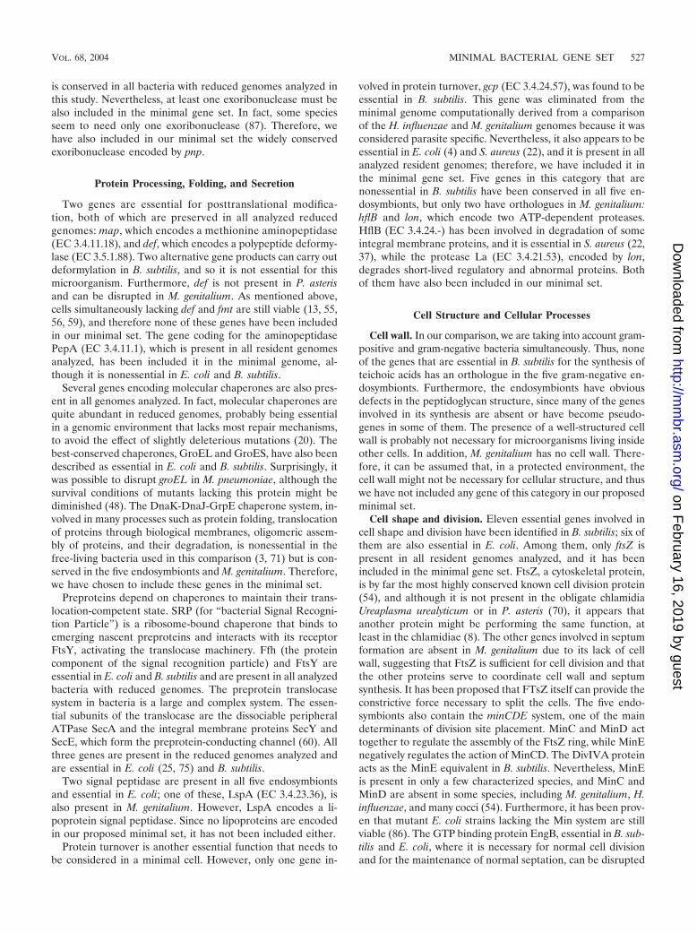
Two genes are essential for posttranslational modifica-tion, both of which are preserved in all analyzed reducedgenomes: map, which encodes a methionine aminopeptidase(EC 3.4.11.18), and def, which encodes a polypeptide deformy-lase (EC 3.5.1.88). Two alternative gene products can carry outdeformylation in B. subtilis, and so it is not essential for thismicroorganism. Furthermore, def is not present in P. asterisand can be disrupted in M. genitalium. As mentioned above,cells simultaneously lacking def and fmt are still viable (13, 55,56, 59), and therefore none of these genes have been includedin our minimal set. The gene coding for the aminopeptidasePepA (EC 3.4.11.1), which is present in all resident genomesanalyzed, has been included it in the minimal genome, al-though it is nonessential in E. coli and B. subtilis.
Several genes encoding molecular chaperones are also pres-ent in all genomes analyzed. In fact, molecular chaperones arequite abundant in reduced genomes, probably being essentialin a genomic environment that lacks most repair mechanisms,to avoid the effect of slightly deleterious mutations (20). Thebest-conserved chaperones, GroEL and GroES, have also beendescribed as essential in E. coli and B. subtilis. Surprisingly, itwas possible to disrupt groEL in M. pneumoniae, although thesurvival conditions of mutants lacking this protein might bediminished (48). The DnaK-DnaJ-GrpE chaperone system, in-volved in many processes such as protein folding, translocationof proteins through biological membranes, oligomeric assem-bly of proteins, and their degradation, is nonessential in thefree-living bacteria used in this comparison (3, 71) but is con-served in the five endosymbionts and M. genitalium. Therefore,we have chosen to include these genes in the minimal set.
Preproteins depend on chaperones to maintain their trans-location-competent state. SRP (for “bacterial Signal Recogni-tion Particle”) is a ribosome-bound chaperone that binds toemerging nascent preproteins and interacts with its receptorFtsY, activating the translocase machinery. Ffh (the proteincomponent of the signal recognition particle) and FtsY areessential in E. coli and B. subtilis and are present in all analyzedbacteria with reduced genomes. The preprotein translocasesystem in bacteria is a large and complex system. The essen-tial subunits of the translocase are the dissociable peripheralATPase SecA and the integral membrane proteins SecY andSecE, which form the preprotein-conducting channel (60). Allthree genes are present in the reduced genomes analyzed andare essential in E. coli (25, 75) and B. subtilis.
Two signal peptidase are present in all five endosymbiontsand essential in E. coli; one of these, LspA (EC 3.4.23.36), isalso present in M. genitalium. However, LspA encodes a li-poprotein signal peptidase. Since no lipoproteins are encodedin our proposed minimal set, it has not been included either.
Protein turnover is another essential function that needs tobe considered in a minimal cell. However, only one gene in-
volved in protein turnover, gcp (EC 3.4.24.57), was found to beessential in B. subtilis. This gene was eliminated from theminimal genome computationally derived from a comparisonof the H. influenzae and M. genitalium genomes because it wasconsidered parasite specific. Nevertheless, it also appears to beessential in E. coli (4) and S. aureus (22), and it is present in allanalyzed resident genomes; therefore, we have included it inthe minimal gene set. Five genes in this category that arenonessential in B. subtilis have been conserved in all five en-dosymbionts, but only two have orthologues in M. genitalium:hflB and lon, which encode two ATP-dependent proteases.HflB (EC 3.4.24.-) has been involved in degradation of someintegral membrane proteins, and it is essential in S. aureus (22,37), while the protease La (EC 3.4.21.53), encoded by lon,degrades short-lived regulatory and abnormal proteins. Bothof them have also been included in our minimal set.
Cell Structure and Cellular Processes
Cell wall. In our comparison, we are taking into account gram-positive and gram-negative bacteria simultaneously. Thus, noneof the genes that are essential in B. subtilis for the synthesis ofteichoic acids has an orthologue in the five gram-negative en-dosymbionts. Furthermore, the endosymbionts have obviousdefects in the peptidoglycan structure, since many of the genesinvolved in its synthesis are absent or have become pseudo-genes in some of them. The presence of a well-structured cellwall is probably not necessary for microorganisms living insideother cells. In addition, M. genitalium has no cell wall. There-fore, it can be assumed that, in a protected environment, thecell wall might not be necessary for cellular structure, and thuswe have not included any gene of this category in our proposedminimal set.
Cell shape and division. Eleven essential genes involved incell shape and division have been identified in B. subtilis; six ofthem are also essential in E. coli. Among them, only ftsZ ispresent in all resident genomes analyzed, and it has beenincluded in the minimal gene set. FtsZ, a cytoskeletal protein,is by far the most highly conserved known cell division protein(54), and although it is not present in the obligate chlamidiaUreaplasma urealyticum or in P. asteris (70), it appears thatanother protein might be performing the same function, atleast in the chlamidiae (8). The other genes involved in septumformation are absent in M. genitalium due to its lack of cellwall, suggesting that FtsZ is sufficient for cell division and thatthe other proteins serve to coordinate cell wall and septumsynthesis. It has been proposed that FTsZ itself can provide theconstrictive force necessary to split the cells. The five endo-symbionts also contain the minCDE system, one of the maindeterminants of division site placement. MinC and MinD acttogether to regulate the assembly of the FtsZ ring, while MinEnegatively regulates the action of MinCD. The DivIVA proteinacts as the MinE equivalent in B. subtilis. Nevertheless, MinEis present in only a few characterized species, and MinC andMinD are absent in some species, including M. genitalium, H.influenzae, and many cocci (54). Furthermore, it has been prov-en that mutant E. coli strains lacking the Min system are stillviable (86). The GTP binding protein EngB, essential in B. sub-tilis and E. coli, where it is necessary for normal cell divisionand for the maintenance of normal septation, can be disrupted
VOL. 68, 2004 MINIMAL BACTERIAL GENE SET 527
on February 16, 2019 by guest
http://mm
br.asm.org/
Dow
nloaded from

in M. genitalium and is absent in B. floridanus. The other es-sential B. subtilis genes necessary for determination of cellshape are not present in all endosymbionts either.
Substrate transport. Bacteria with small genomes are un-able to synthesize many of their essential metabolites, and theymust rely on the environment to obtain them. Thus, it could beexpected that many transport systems must be present in orderto obtain the necessary molecules, as it has been proven forP. asteris (70). In fact, the presence of many specific transport-ers in this bacterium can help to explain why many metabolicpathways for the synthesis of small molecules are missing inthis microorganism but have been preserved in its close rela-tive, M. genitalium, with a smaller genome. However, B. aphidi-cola has a reduced number of genes devoted to such function,a number that is slightly increased in the other endosymbioticbacteria analyzed and in M. genitalium. This fact might be re-lated to the presence of a less highly structured cell envelope,which might be more permeable to small metabolites. Further-more, the analyzed bacteria with small genomes differ in thetransport systems that have been preserved from the reduc-tive process suffered by their genomes, and they probably com-pensate for the reduced transporter spectrum by encoding trans-porters with broadened specificity. Several porins and low-affinityactive transporters, as well as a few ATP binding cassette (ABC)transporters have been identified in the five endosymbionts un-der study, while M. genitalium has a larger number of ABC trans-porters. ABC transporters are heterotrimeric transport systemsmade up of a specificity (ligand binding) subunit, a permease,and an ATP binding protein. Many ATP binding subunits ap-pear to be “orphan” proteins with unknown partners, which areapparently overrepresented compared to the other two subunitsin all genomes sequenced thus far (32), including the reducedgenomes under study. However, none of them has been pre-served in all analyzed reduced genomes simultaneously.
Phosphate transport is thought to be an essential function.However, only the low-affinity inorganic phosphate transporterencoded by pitA is present in all endosymbionts under study,although it is not present in the smallest described B. aphidi-cola genome, which is being sequenced in our laboratory(unpublished results). No orthologous gene has been foundin M. genitalium, which instead contains an ABC phosphatetransporter, although two of its three subunits (MG410 andMG411) can be disrupted. It appears that another yet uniden-tified phosphate transport system exists in mycoplasmas and inB. aphidicola BCce.
Several more or less specific transporters for mono- and di-valent cations have also been identified in every microorganismanalyzed in this study, but none of them is shared in all cases.
All the components of a phosphoenolpyruvate-dependentsugar phosphotransferase system (PTS), a major carbohydrateactive-transport system that catalyzes sugar phosphorylation andtransport across the cytoplasmic membrane, are present in allbacteria with reduced genomes analyzed except W. glossinidia,although the specific permease differs among them. The B. flori-danus genome encodes the mannose permease, which can trans-port a wide variety of sugar monomers, while B. aphidicolaencodes the genes for intake of mannitol and glucose and M.genitalium contains the corresponding glucose and fructose per-meases. It is not known if the presence of different permeasesis due to different metabolic requirements and/or environmen-
tal conditions or if such permeases are less specific in thesemicroorganisms and can thus be used to transport other sugars.
Based on the present analysis, it is clear that no singletransport system is shared by all analyzed bacteria. B. aphidi-cola appears to be close to the minimal “free diffusing cell”described by Luisi et al. (53), a cell in which the low-molecular-mass compounds, including nucleotides and amino acids, canbe provided by the environment and are able to permeate thecell membrane. Nevertheless, it appears that at least one orseveral ABC transporters, a PTS for glucose transport, andsome more or less specific mono- and divalent cation trans-porters must be part of a minimal cell genome. However, withthe available data, it is not possible to define which ones shouldbe such transporters, and different alternative transporters canbe included in a minimal cell depending on the nutrients avail-able in the environment or the cell membrane characteristics.Without detailed information about such possible alternatives,we propose that our hypothetical minimal cell must be able tointernalize small molecules that are not highly charged, and wehave included in our proposed minimal set only a PTS forglucose (since glucose can enter the cell by using different PTSin all analyzed bacteria with reduced genomes, and the PTSdelivers phosphorylated glucose to the cell interior, makingglucose available as a metabolic substrate) and pitA (to providethe phosphate that is necessary in metabolic reactions).
Energetic and Intermediary Metabolism
The definition of the essential metabolic functions that mustbe present in a minimal cell is a difficult part of the analysis ofa minimal genome, since it is highly dependent on the reper-toire of metabolites present in the environment. We are as-suming by definition that a minimal cell lives in a nutrient-richmedium and that therefore the major metabolites (amino ac-ids, nitrogenous bases, fatty acids, precursors of coenzymes,and glucose) are available without limitation. This situationmakes unnecessary the function of entire anabolic and assimi-latory pathways of the main biomolecules, i.e., de novo biosyn-theses of amino acids, fatty acids, or cofactors. In that case,even the synthesis of a central metabolite such as acetyl coen-zyme A (acetyl-CoA) turns out to be dispensable. As discussedbelow, the bioenergetics of our minimal cell is basically fer-mentative, and the only metabolic role for acetyl-CoA wouldbe the synthesis of fatty acids; however, we postulate that theseessential biomolecules are present and available in the me-dium. Furthermore, as discussed in the previous section, ourproposed minimal cell does not contain specific transportersfor charged molecules (such as mono- or dinucleotides), andsuch molecules must be synthesized by the cell from interme-diary metabolites provided by the environment and its ownmetabolic machinery. Therefore, some of the metabolic path-ways that we include as part of our hypothetical minimal cellmight not be fully present in bacteria with small genomes thatcontain the corresponding specific transporter or a cell mem-brane that allows the internalization of such biomolecules.
To define which metabolic pathways should be consideredessential in a minimal cell, we first considered those that havebeen preserved in the different bacteria with reduced genomesanalyzed. Some metabolic pathways (such as the biosynthesisof purine nucleotides or redox coenzymes) have been pre-
528 GIL ET AL. MICROBIOL. MOL. BIOL. REV.
on February 16, 2019 by guest
http://mm
br.asm.org/
Dow
nloaded from

served in all analyzed microorganisms, but they use differentalternative routes, revealing degenerative processes that arerandomly affecting different genes in each genome. In thosecases, the loss of some genes conditioned the essentiality ofother genes that became necessary to conserve a specific met-abolic function. To approach the minimal genome, we chooseto include in our minimal set the costless pathway that willallow the cell to preserve such metabolic function. Other met-abolic pathways are incomplete. Although some of the genesinvolved in these pathways may still be present in all genomesunder study, they have not been included in the minimal set.
A general outline of the metabolic abilities of the proposedminimal cell is presented in Fig. 1.
Glycolysis, gluconeogenesis, pyruvate metabolism, and theTCA cycle. All genes involved in glycolysis, except the oneresponsible for the phosphorylation of glucose, are present in
all endosymbionts and in M. genitalium. The exception is pfkA(EC 2.7.1.11), which has not been identified in W. glossinidia.In this case, it is remarkable that the absence of phosphofruc-tokinase is accompanied by the presence of two idiosyncratic ac-tivities of the gluconeogenic pathway: fructosebisphosphatase(fbp, EC 3.1.3.11) and phosphoenolpyruvate carboxykinase (pck,EC 4.1.1.49). This is a strong indication that W. glossinidia, unlikethe other described endosymbionts, synthesizes hexoses from C3
compounds derived mainly from amino acids. The phosphoryla-tion of glucose in the other endosymbionts and M. genitaliumcan be achieved if we consider the action of the PTS transport-ers. Since no other pathway for the glucose degradation has beenidentified in the small genome organisms under study and sinceglycolysis also provides precursor metabolites for some ana-bolic pathways, we have included all genes involved in thispathway in the minimal set.
FIG. 1. A minimal metabolism. The minimal cell can obtain its more basic components from the environment: glucose, fatty acids, amino acids,adenine, guanine, uracil, and coenzyme precursors (nicotinamide, riboflavin, folate, pantothenate, and pyridoxal). Each box includes the metabolictransformations classified in major groups of pathways: glycolysis, phospholipid biosynthesis, nonoxidative pentose-phosphate pathway, nucleotidebiosynthesis, synthesis of enzymatic cofactors, and synthesis of protein precursors, i.e., aminoacyl-tRNAs (aa-tRNA). Arrows with discontinuouslines represent incorporation from the environment. Single continuous arrows represent single enzymatic steps, whereas wide arrows representseveral enzymatic steps (the number within the arrow indicates the number of steps). Lines with a final black point indicate the necessity ofmetabolites for some of the transformations inside the corresponding box. Metabolic intermediates and final pathway products are in green boxes.Metabolites acting as a source of chemical energy are in red boxes. Reducing-power cofactors are in light blue boxes. Abbreviations (besides theaccepted symbols and those defined in the text): PEP, phosphoenolpyruvate; G6P, glucose-6-phosphate; Gd3P, glyceraldehyde-3-phosphate;DHAP, dihydroxyacetonephosphate; G3P, sn-glycerol-3-phosphate; CDP-DAG, CDP-diacylglycerol; SAM, S-adenosylmethionine; THF, tetrahy-drofolate. Metabolic precursors of external origin are in gray boxes.
VOL. 68, 2004 MINIMAL BACTERIAL GENE SET 529
on February 16, 2019 by guest
http://mm
br.asm.org/
Dow
nloaded from
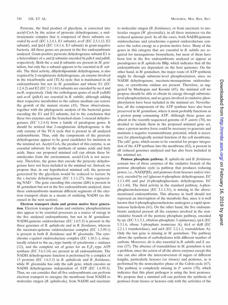
Pyruvate, the final product of glycolysis, is converted intoacetyl-CoA by the action of pyruvate dehydrogenase, a mul-tienzymatic complex that is composed of three subunits en-coded by aceE (EC 1.2.4.1, E1 subunit), aceF (EC 2.3.1.12, E2subunit), and lpdA (EC 1.8.1.4, E3 subunit) in gram-negativebacteria. All three genes are present in the five endosymbiontsanalyzed. Gram-positive pyruvate dehydrogenase subunit E1 isa heterodimer of � and � subunits encoded by pdhA and pdhB,respectively. Both the � and � subunits are present in M. geni-talium, but only the � subunit appears to be essential in B. sub-tilis. The third activity, dihydrolipoamide dehydrogenase, is alsorequired by 2-oxoglutarate dehydrogenase, an enzyme involvedin the tricarboxylic acid (TCA) cycle that is maintained in allendosymbionts but not in M. genitalium and whose E1 (EC1.2.4.2) and E2 (EC 2.3.1.61) subunits are encoded by sucA andsucB, respectively. Only the orthologous genes of sucB (odhB)and aceE (pdhA) are essential in B. subtilis, although addingtheir respective metabolites to the culture medium can restorethe growth of the mutant strains (45). These observations,together with the phylogenetic analysis of the bacterial genesencoding the E1 and E2 subunits, led to the conclusion thatthese two enzymes and the branched-chain 2-oxoacid dehydro-genases (EC 1.2.4.4) form a family of paralogous genes. Itshould be noticed that 2-oxoglutarate dehydrogenase is theonly enzyme of the TCA cycle that is present in all analyzedendosymbionts. Thus, only the components of the pyruvatedehydrogenase appear to be good candidates for inclusion inthe minimal set. Acetyl-CoA, the product of this enzyme, is anessential substrate for the synthesis of amino acids and fattyacids. Since our proposed minimal cell can obtain these bi-omolecules from the environment, acetyl-CoA is not neces-sary. Therefore, the genes that encode the pyruvate dehydro-genase have not been included in the minimal set. Instead, wepropose that, in our hypothetical minimal cell, the pyruvategenerated by the glycolysis would be reduced to lactate bythe L-lactate dehydrogenase (EC 1.1.1.27), thus regenerat-ing NAD�. The gene encoding this enzyme (ldh) is present inM. genitalium but not in the five endosymbionts analyzed, sincethese endosymbionts maintain different segments of the elec-tron transport chain as a means of oxidizing NADH (as dis-cussed in the next section).
Electron transport chain and proton motive force genera-tion. Electron transport chains and oxidative phosphorylationalso appear to be essential processes as a source of energy inthe five analyzed endosymbionts, but not in M. genitalium.NADH:quinone oxidoreductase (EC 1.6:5.3) is present in thethree genomes of B. aphidicola and in B. floridanus, whereasthe succinate:quinone oxidoreductase complex (EC 1.3.99.1)is present in both B. floridanus and W. glossinidia. The cyto-chrome o:quinol oxidoreductase complex (EC 1.10.3.-), struc-turally related to the aa3-type family of cytochrome c oxidases(12), and the complete set of genes for an F0F1-type ATPsynthase (EC 3.6.3.14) are present in all endosymbionts. TheNADH dehydrogenase function is performed by a complex of13 proteins (EC 1.6.5.3) in B. aphidicola and B. floridanus,while W. glossinidia has only the ndh gene, which encodes anNADH dehydrogenase independent of ATP (EC 1.6.99.3).Thus, we can consider that all five endosymbionts can performelectron transport to energize the membrane from NADH tomolecular oxygen (B. aphidicola), from NADH and succinate
to molecular oxygen (B. floridanus), or from succinate to mo-lecular oxygen (W. glossinidia), in all three instances via thereduced quinone pool. In all the cases, both NADH:quinoneoxidoreductase and cytochrome o:quinol oxidoreductase con-serve the redox energy as a proton motive force. Many of thegenes in this category that are essential in B. subtilis are re-quired for menaquinone biosynthesis, but most of them havebeen lost in the five endosymbionts analyzed or appear aspseudogenes in B. aphidicola BBp, which indicates that all theendosymbionts are dependent on the host quinones. On theother hand, in M. genitalium, the major route of ATP synthesismight be through substrate-level phosphorylation, since noNADH dehydrogenase, succinate:menaquinone oxidoreduc-tase, or cytochrome oxidase are present. Therefore, as sug-gested by Mushegian and Koonin (65), the minimal cell wepropose should be able to obtain its energy through substrate-level phosphorylation, and no genes involved in oxidative phos-phorylation have been included in the minimal set. Neverthe-less, all the components of the ATP synthase have also beenpreserved in M. genitalium, where it most probably functions asa proton pump consuming ATP. Although these genes areabsent in the recently sequenced genome of P. asteris (70), wedecided to include them in the proposed minimal gene set,since a proton motive force could be necessary to generate andmaintain a negative transmembrane potential, which is neces-sary for physiologically normal function of the cell membrane.The ydiC gene, which seems to be essential for proper integra-tion of the ATP synthase into the membrane (82), is present inall reduced genomes analyzed and has also been included inthe minimal set.
Pentose phosphate pathway. B. aphidicola and B. floridanuscontain two of three enzymes of the oxidative branch of thepentose phosphate cycle (a pathway that produces reducingpower, i.e., NAD[P]H), and pentoses from hexoses and/or trio-ses), encoded by zwf (glucose-6-phosphate dehydrogenase, EC1.1.1.49) and gnd (6-phosphogluconate dehydrogenase, EC1.1.1.44). The third activity in the standard pathway, 6-phos-phogluconolactonase (EC 3.1.1.31), is missing in the above-mentioned endosymbionts. This absence in itself should notrepresent an interruption of the metabolic flux, since it is wellknown that 6-phosphogluconolactone undergoes a rapid spon-taneous hydrolysis (61). On the other hand, the five endosym-bionts analyzed present all the enzymes involved in the non-oxidative branch of the pentose phosphate pathway, encodedby rpe (EC 5.1.3.1, ribulose-phosphate 3-epimerase), rpiA (EC5.3.1.6, ribose 5-phosphate isomerase A), tktA or tktB (EC2.2.1.1 transketolase), and talA (EC 2.2.1.2, transaldolase A).Only the last gene is missing in M. genitalium. The pathwayallows the synthesis of carbohydrates with different number ofcarbons. Moreover, tkt is also essential in B. subtilis and S. au-reus (37). The absence of transaldolase in M. genitalium is nota problem, since the action of all the above enzymes except thisone can also allow the interconversion of sugars of differentlengths, particularly hexoses (or trioses) and pentoses, as isperformed by the nonreductive phase of the Calvin cycle (67).The pathway is completely missing in P. asteris (70), whichindicates that this plant pathogen is using the host pentoses.We propose that a minimal cell can perform the synthesis ofpentoses from trioses or hexoses only with the activities of the
530 GIL ET AL. MICROBIOL. MOL. BIOL. REV.
on February 16, 2019 by guest
http://mm
br.asm.org/
Dow
nloaded from

nonoxidative branch of the pentose phosphate pathway en-coded by rpe, rpiA, and tkt.
Biosynthesis of amino acids. Several genes necessary for thesynthesis of amino acids of the aspartate family are includedamong the essential genes of B. subtilis, probably reflecting thelack of such amino acids in the culture media or the absence ofthe proper transporters to import them into the cell. B. aphidi-cola and B. floridanus provide their host insect with essentialamino acids, and thus the genes involved in the biosyntheticpathways for those amino acids are present in their genomes.However, W. glossinidia has lost most of these genes, and theonly gene in this category found in M. genitalium is glyA. Thisgene is also involved in the biosynthesis of folate derivativesand has been included in the section an biosynthesis of cofac-tors (below). Thus, it can be assumed that amino acids can beprovided by the environment in the small genome microorgan-isms analyzed, and no genes of this category need to be in-cluded in the minimal set.
Biosynthesis of lipids. Fatty acid biosynthesis, the first stagein membrane lipid biogenesis, is catalyzed in most bacteria bythe type II fatty acid synthase system, composed of a series ofsmall, soluble proteins that are each encoded by a discretegene (74). Although many of the genes involved in this path-way are essential in E. coli and B. subtilis, all the genes involvedin the synthesis of malonyl-CoA have been lost in B. aphidicolaand M. genitalium. In fact, the fatty acid biosynthesis pathwayis incomplete in most reduced genomes analyzed, since onlyacpP and acpS (EC 2.7.8.7) are present in all of them. Thesefindings probably imply that fatty acids can be provided by theenvironment in bacteria with small genomes, and that ACP(the acyl carrier protein, encoded by acpP) is involved in fattyacids uptake and activation (i.e., synthesis of acyl-CoA). How-ever, since the only metabolic destination of the fatty acids wouldbe the biosynthesis of phospholipids, we propose that the ac-tivation should take place through a single step catalyzed byacyl-CoA synthase (EC 6.2.1.3) encoded by fadD (76).
One of the most surprising findings when the first B. aphidi-cola genome was sequenced was that this bacterium lacks allgenes responsible for phospholipid biosynthesis (except cls, thegene that encodes cardiolipin synthase, EC 2.7.8.-). The se-quencing of two more B. aphidicola genomes corroborated thisfinding. However, a set of 10 genes coding for a complete bio-synthetic pathway of the major bacterial phospholipids (i.e.,phosphatidylethanolamine, phosphatidylglycerol, and cardi-olipin) from triosephosphate and acyl-CoA is present inW. glossinidia, all except one (plsB, EC 2.3.1.15) are present inB. floridanus, and some of them are essential in B. subtilis. Ahypothetical phospholipid biosynthetic pathway from glyceroland exogenous fatty acids has been proposed by Mushegianand Koonin (65) for M. genitalium, albeit with an unreasonablysmall number of steps. These facts probably indicate that thecell surface in B. aphidicola is fragile, maybe due to its pro-longed intracellular life inside vacuole-like host-derived or-ganelles. In contrast, pathogenic bacteria, such as M. genita-lium, and endosymbionts that live free in the cytosol of the hostcell (which is the case for B. floridanus and W. glossinidia) needa more structured and flexible surface to protect themselvesfrom the host cell. Since phospholipids are an indispensablecomponent of the formation of the membrane lipid bilayer, itseems reasonable to assume that B. aphidicola imports them
from the host cell. However, we consider phospholipid biosyn-thesis one of the essential functions to be performed by aminimal cell, and therefore we have included in our minimalgene set all the genes necessary for the biosynthesis of phos-phatidylethanolamine from dihydroxyacetone phosphate andactivated fatty acids. We assume for our minimal cell a theo-retical lipid bilayer made only of phosphatidylethanolamine,the main phospholipid present in bacterial membranes (it rep-resents 70 to 80% of all zwitterionic phospholipids in E. coli).The observation that E. coli cells lacking the quantitativelymajor acidic phospholipids (phosphatidylglycerol and cardio-lipin) are viable (42) also supports this choice. The nonessen-tiality of some metabolic steps or the absence of some genescan be explained by metabolic redundancy, nonorthologoussubstitutions, or even the presence of other kinds of phospho-lipids acting as functional surrogates (14).
lgt, which encodes a prolipoprotein diacylglycerol transfer-ase (EC 2.4.99.-) involved in the first step of the synthesis oflipoproteins, is conserved in all analyzed reduced genomes butis absent in P. asteris (70). Although it was annotated as a pseu-dogene in B. aphidicola BSg (80), the corresponding openreading frame contains a frameshift 29 nucleotides after thestart codon of the orthologous gene, but there is an alternativestart a few nucleotides downstream, rendering a protein of 246amino acids that has lost only the first transmembrane domain.The presence of this gene in the small genomes analyzed can beexplained because all such genomes contain genes that encodeat least one lipoprotein. However, since none of them has beenincluded in the minimal set, lgt has not been included either.
Biosynthesis of nucleotides. Among the genes related tonucleic acids metabolism, several genes involved in the synthe-sis of purines and pyrimidines are essential in B. subtilis andare present in all analyzed genomes; these include nrdE andnrdF (or the homologous genes nrdA and nrdB, encoding thetwo subunits of the ribonucleoside diphosphate reductase,EC 1.17.4.1), together with trxA and trxB (thioredoxin andthioredoxin reductase, EC 1.8.1.9), adk (adenylate kinase, EC2.7.4.3), gmk (guanylate kinase, EC 2.7.4.8), tmk (thymidylatekinase, EC 2.7.4.9), and prs (phosphoribosylpyrophosphatesynthase, EC 2.7.6.1). The nrdI gene, encoding a putative ribo-nucleoside diphosphate reductase that is essential in B. subtilis,has no orthologue in the five endosymbionts analyzed. How-ever, its presence in the M. genitalium genome may explain whynrdE seems to be nonessential in mycoplasmas (34).
All bacteria with small genomes analyzed in this study cansynthesize purine nucleotides, but with different metabolicsolutions. The enzyme ribose-phosphate diphosphokinase(EC 2.7.6.8) synthesizes 5-phosphoribosyl-alpha-1-diphosphate(PRPP) from ribose 5-phosphate and ATP, the starting pointfor nucleotide biosynthesis. This activity is encoded by the prsAgene, orthologous to the essential prs gene in B. subtilis, that ispresent in all the reduced genomes analyzed. The ppa gene,encoding an inorganic pyrophosphatase (EC 3.6.1.1), is alsopresent in all analyzed genomes and is essential in B. subtilisand E. coli. The gene product drives anabolic fluxes by pyro-phosphate hydrolysis in various biochemical reactions, includ-ing all reactions that involve PRPP. Purine monophosphatenucleotides can then be synthesized by using two alternativepathways. In all five endosymbionts, 1-(5�-phosphoribosyl)-5-amino-4-imidazolecarboxamide (AICAR) can be synthesized
VOL. 68, 2004 MINIMAL BACTERIAL GENE SET 531
on February 16, 2019 by guest
http://mm
br.asm.org/
Dow
nloaded from

de novo and used for the synthesis of the purine nucleotides. InW. glossinidia, AICAR biosynthesis is performed by a standardpathway starting with PRPP and glutamine, involving the prod-ucts of the purF (EC 2.4.2.14), purD (EC 6.3.4.13), purL (EC6.3.5.3), purM (EC 6.3.3.1), purE and purK (EC 4.1.1.21), purC(EC 6.3.2.6), and purB (EC 4.3.2.2) genes, although the stepusually catalyzed by the protein encoded by purN (EC 2.1.1.2)has been replaced by the enzyme 5�-phosphoribosylglyci-namide transformylase 2 (EC 2.1.2.-) (68) encoded by purT, ahomologue of purN (58). On the other hand, both B. aphidicolaand B. floridanus synthesize AICAR through the four firststeps of the histidine biosynthetic pathway starting with PRPPand ATP, using the set of activities encoded by hisG (EC2.4.2.17), hisI (EC 3.6.1.31 and 3.5.4.19), and his A (EC5.3.1.16). AMP and GMP are then synthesized from AICARthrough a pathway involving the products of the purA (EC6.3.4.4), purB (EC 4.3.2.2), and purH (EC 3.5.4.10) genes andthe guaC (EC 1.7.1.7) (in B. aphidicola) or guaA (EC 1.1.1.205)and guaB (EC 6.3.5.2) (in B. floridanus and W. glossinidia)genes. M. genitalium has a different and costless metabolic
solution, since it can use the enzyme hypoxanthine phospho-ribosyltransferase (EC 2.4.2.8, encoded by the gene hpt) for thesynthesis of GMP and AMP from PRPP and guanine or ade-nine, respectively, as may also be the case for B. aphidicolaBAp and BSg. The kinases encoded by adk and gmk are con-served in all reduced genomes analyzed, allowing the synthesisof nucleoside diphosphates from the nucleoside monophos-phates. Purine nucleotide diphosphate can be phosphorylatedby the kinase encoded by ndk or by the glycolytic enzymepyruvate kinase (EC 2.7.1.40), encoded by pykA, in B. aphidi-cola and M. genitalium, which lack ndk. The ribonucleosidediphosphate reductase function can be performed by the prod-ucts of the nrdA and nrdB genes or the nrdE and nrdF genes(depending on the analyzed organism), with the help of theproducts of trxA and trxB, to obtain the corresponding deoxyri-bonucleoside diphosphate (Fig. 2).
Pyrimidine metabolism is not so highly conserved in theanalyzed bacteria with small genomes. Only a few genes in-volved in the synthesis of nucleoside monophosphates havebeen maintained in all of them simultaneously: tmk, pyrG (CTP
FIG. 2. A minimal nucleotide metabolism based on salvage pathways. Activated ribonucleotides and deoxyribonucleotides are obtained fromfree bases (A, G, and U), PRPP, ATP-dependent phosphorylating reactions, and NADH-dependent reduction. White boxes indicate the individualenzymatic activity (EC number and coding gene). Other colors are used as in Fig. 1.
532 GIL ET AL. MICROBIOL. MOL. BIOL. REV.
on February 16, 2019 by guest
http://mm
br.asm.org/
Dow
nloaded from

synthase, EC 6.3.4.2), and dut (EC 3.6.1.23) (or its correspond-ing suggested homologue in M. genitalium, MG268) (65). Theupp gene (uracil phosphoribosyl transferase, EC 2.4.2.9), nec-essary for the incorporation of free uracil for the synthesis ofactivated ribonucleotides, is present only in B. floridanus andM. genitalium. It appears that B. aphidicola and W. glossinidiacan obtain the pyrimidine nucleotides from the cytoplasm ofthe host cell, probably as nucleoside monophosphates. How-ever, since we have not included specific transporters for suchmolecules in our minimal cell, we consider that in a hypothet-ical minimal cell, free uracil can be provided by the environ-ment and used to synthesize all the necessary pyrimidine nu-cleotides, using the costless pathway as described in Fig. 2. Theconversion of pyrimidine monophosphate to pyrimidine diphos-phate needs the action of one kinase, encoded by cmk (EC2.7.4.14) or tmk (EC 2.7.4.9). At least one of these genesappears in all genomes sequenced to date; i.e., chlamydiae andmycobacteria contain only cmk, while archaea, B. aphidicola,and some �- and ε-proteobacteria have only tmk. Therefore,only tmk has been included in the minimal gene set. Pyrimidinenucleoside diphosphates cannot be phosphorylated by the ki-nase encoded by pykA, and therefore in our metabolic map the
activity encoded by ndk plays an important role in catalyzingthe transfer of the gamma-phosphate from the nucleoside tri-phosphate to a variety of nucleoside diphosphates. This broadspecificity has been demonstrated in several experimental sys-tems including prokaryotic cells (see the primary literature inthe BRENDA database).
Biosynthesis of cofactors. Most of the genes involved in thebiosynthesis of cofactors have been lost in the reduced ge-nomes analyzed, and only a few genes included in this categorythat are essential in B. subtilis or E. coli are present in all ofthem. However, the presence of biosynthetic pathways in bac-teria with reduced genomes or their essentiality in laboratory-cultured bacteria reflects the chemical complexity of the envi-ronment. If a microorganism has free availability of coenzymeprecursors (i.e., vitamins), it can use short and simple salvagepathways rather than long and complicated de novo biosynthe-ses. As a general criterion, we propose that our minimal met-abolic chart starts from elaborate molecules, such as vitamins,and it contains only the minimum number of steps to incorpo-rate them as enzymatic cofactors (Fig. 3). Only the cofactorsthat are necessary for the correct catalytic function of the en-zymes present in our proposed minimal cell have been included.
FIG. 3. Biosynthesis of cofactors. A metabolism of essential cofactors used by the minimal cell starting with precursors (i.e., vitamins):nicotinamide, riboflavin, thiamine, pyridoxal, patothenic acid, methionine, and folic acid. Yellow boxes indicate the enzymes that need eachcofactor for their correct function. Other colors and symbols are as in Fig. 1 and 2. PP, pyridoxal-phosphate.
VOL. 68, 2004 MINIMAL BACTERIAL GENE SET 533
on February 16, 2019 by guest
http://mm
br.asm.org/
Dow
nloaded from
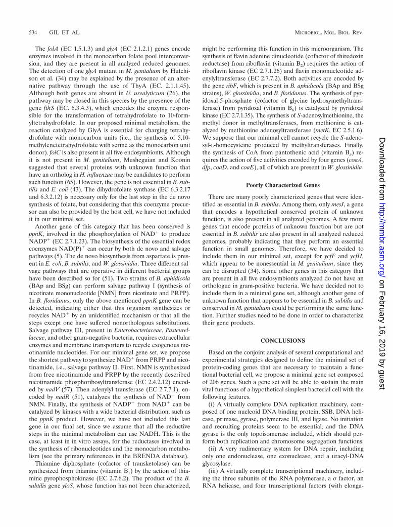
The folA (EC 1.5.1.3) and glyA (EC 2.1.2.1) genes encodeenzymes involved in the monocarbon folate pool interconver-sion, and they are present in all analyzed reduced genomes.The detection of one glyA mutant in M. genitalium by Hutchi-son et al. (34) may be explained by the presence of an alter-native pathway through the use of ThyA (EC. 2.1.1.45).Although both genes are absent in U. urealyticum (26), thepathway may be closed in this species by the presence of thegene fthS (EC. 6.3.4.3), which encodes the enzyme respon-sible for the transformation of tetrahydrofolate to 10-form-yltetrahydrofolate. In our proposed minimal metabolism, thereaction catalyzed by GlyA is essential for charging tetrahy-drofolate with monocarbon units (i.e., the synthesis of 5,10-methylenetetrahydrofolate with serine as the monocarbon unitdonor). folC is also present in all five endosymbionts. Althoughit is not present in M. genitalium, Musheguian and Kooninsuggested that several proteins with unknown function thathave an ortholog in H. influenzae may be candidates to performsuch function (65). However, the gene is not essential in B. sub-tilis and E. coli (43). The dihydrofolate synthase (EC 6.3.2.17and 6.3.2.12) is necessary only for the last step in the de novosynthesis of folate, but considering that this coenzyme precur-sor can also be provided by the host cell, we have not includedit in our minimal set.
Another gene of this category that has been conserved isppnK, involved in the phosphorylation of NAD� to produceNADP� (EC 2.7.1.23). The biosynthesis of the essential redoxcoenzymes NAD(P)� can occur by both de novo and salvagepathways (5). The de novo biosynthesis from aspartate is pres-ent in E. coli, B. subtilis, and W. glossinidia. Three different sal-vage pathways that are operative in different bacterial groupshave been described so for (51). Two strains of B. aphidicola(BAp and BSg) can perform salvage pathway I (synthesis ofnicotinate mononucleotide [NMN] from nicotinate and PRPP).In B. floridanus, only the above-mentioned ppnK gene can bedetected, indicating either that this organism synthesizes orrecycles NAD� by an unidentified mechanism or that all thesteps except one have suffered nonorthologous substitutions.Salvage pathway III, present in Enterobacteriaceae, Pasteurel-laceae, and other gram-negative bacteria, requires extracellularenzymes and membrane transporters to recycle exogenous nic-otinamide nucleotides. For our minimal gene set, we proposethe shortest pathway to synthesize NAD� from PRPP and nico-tinamide, i.e., salvage pathway II. First, NMN is synthesizedfrom free nicotinamide and PRPP by the recently describednicotinamide phosphoribosyltransferase (EC 2.4.2.12) encod-ed by nadV (57). Then adenylyl transferase (EC 2.7.7.1), en-coded by nadR (51), catalyzes the synthesis of NAD� fromNMN. Finally, the synthesis of NADP� from NAD� can becatalyzed by kinases with a wide bacterial distribution, such asthe ppnK product. However, we have not included this lastgene in our final set, since we assume that all the reductivesteps in the minimal metabolism can use NADH. This is thecase, at least in in vitro assays, for the reductases involved inthe synthesis of ribonucleotides and the monocarbon metabo-lism (see the primary references in the BRENDA database).
Thiamine diphosphate (cofactor of transketolase) can besynthesized from thiamine (vitamin B1) by the action of thia-mine pyrophosphokinase (EC 2.7.6.2). The product of the B.subtilis gene yloS, whose function has not been characterized,
might be performing this function in this microorganism. Thesynthesis of flavin adenine dinucleotide (cofactor of thiredoxinreductase) from riboflavin (vitamin B2) requires the action ofriboflavin kinase (EC 2.7.1.26) and flavin mononucleotide ad-enylyltransferase (EC 2.7.7.2). Both activities are encoded bythe gene ribF, which is present in B. aphidicola (BAp and BSgstrains), W. glossinidia, and B. floridanus. The synthesis of pyr-idoxal-5-phosphate (cofactor of glycine hydroxymethyltrans-ferase) from pyridoxal (vitamin B6) is catalyzed by pyridoxalkinase (EC 2.7.1.35). The synthesis of S-adenosylmethionine, themethyl donor in methyltransferases, from methionine is cat-alyzed by methionine adenosyltransferase (metK, EC 2.5.1.6).We suppose that our minimal cell cannot recycle the S-adeno-syl-L-homocysteine produced by methyltransferases. Finally,the synthesis of CoA from pantothenic acid (vitamin B5) re-quires the action of five activities encoded by four genes (coaA,dfp, coaD, and coaE), all of which are present in W. glossinidia.
Poorly Characterized Genes
There are many poorly characterized genes that were iden-tified as essential in B. subtilis. Among them, only mesJ, a genethat encodes a hypothetical conserved protein of unknownfunction, is also present in all analyzed genomes. A few moregenes that encode proteins of unknown function but are notessential in B. subtilis are also present in all analyzed reducedgenomes, probably indicating that they perform an essentialfunction in small genomes. Therefore, we have decided toinclude them in our minimal set, except for ycfF and ycfH,which appear to be nonessential in M. genitalium, since theycan be disrupted (34). Some other genes in this category thatare present in all five endosymbionts analyzed do not have anorthologue in gram-positive bacteria. We have decided not toinclude them in a minimal gene set, although another gene ofunknown function that appears to be essential in B. subtilis andconserved in M. genitalium could be performing the same func-tion. Further studies need to be done in order to characterizetheir gene products.
CONCLUSIONS
Based on the conjoint analysis of several computational andexperimental strategies designed to define the minimal set ofprotein-coding genes that are necessary to maintain a func-tional bacterial cell, we propose a minimal gene set composedof 206 genes. Such a gene set will be able to sustain the mainvital functions of a hypothetical simplest bacterial cell with thefollowing features.
(i) A virtually complete DNA replication machinery, com-posed of one nucleoid DNA binding protein, SSB, DNA heli-case, primase, gyrase, polymerase III, and ligase. No initiationand recruiting proteins seem to be essential, and the DNAgyrase is the only topoisomerase included, which should per-form both replication and chromosome segregation functions.
(ii) A very rudimentary system for DNA repair, includingonly one endonuclease, one exonuclease, and a uracyl-DNAglycosylase.
(iii) A virtually complete transcriptional machinery, includ-ing the three subunits of the RNA polymerase, a � factor, anRNA helicase, and four transcriptional factors (with elonga-
534 GIL ET AL. MICROBIOL. MOL. BIOL. REV.
on February 16, 2019 by guest
http://mm
br.asm.org/
Dow
nloaded from

tion, antitermination, and transcription-translation couplingfunctions). Regulation of transcription does not appear to beessential in bacteria with reduced genomes, and therefore theminimal gene set does not contain any transcriptional regula-tors.
(iv) A nearly complete translational system. It contains the20 aminoacyl-tRNA synthases, a methionyl-tRNA formyltrans-ferase, five enzymes involved in tRNA maturation and modi-fication, 50 ribosomal proteins (31 proteins for the large ribo-somal subunit and 19 proteins for the small one), six proteinsnecessary for ribosome function and maturation (four of whichare GTP binding proteins whose specific function is not wellknown), 12 translation factors, and 2 RNases involved in RNAdegradation.
(v) Protein-processing, -folding, secretion, and degradationfunctions are performed by at least three proteins for post-translational modification, two molecular chaperone systems(GroEL/S and DnaK/DnaJ/GrpE), six components of thetranslocase machinery (including the signal recognition parti-cle, its receptor, the three essential components of the trans-locase channel, and a signal peptidase), one endopeptidase,and two proteases.
(vi) Cell division can be driven by FtsZ only, consideringthat, in a protected environment, the cell wall might not benecessary for cellular structure.
(vii) A basic substrate transport machinery cannot be clearlydefined, based on our current knowledge. Although it appearsthat several cation and ABC transporters are always present inall analyzed bacteria, we have included in the minimal set onlya PTS for glucose transport and a phosphate transporter. Fur-ther analysis should be performed to define a more completeset of transporters.
(viii) The energetic metabolism is based on ATP synthesis byglycolytic substrate-level phosphorylation.
(ix) The nonoxidative branch of the pentose pathway con-tains three enzymes (ribulose-phosphate epimerase, ribose-phosphate isomerase, and transketolase), allowing the synthe-sis of pentoses (PRPP) from trioses or hexoses.
(x) No biosynthetic pathways for amino acids, since we sup-pose that they can be provided by the environment.
(xi) Lipid biosynthesis is reduced to the biosynthesis of phos-phatidylethanolamine from the glycolytic intermediate dihy-droxyacetone phosphate and activated fatty acids provided bythe environment.
(xii) Nucleotide biosynthesis proceeds through the salvagepathways, from PRPP and the free bases adenine, guanine, anduracil, which are obtained from the environment.
(xiii) Most cofactor precursors (i.e., vitamins) are providedby the environment. Our proposed minimal cell performs onlythe steps for the syntheses of the strictly necessary coenzymestetrahydrofolate, NAD�, flavin aderine dinucleotide, thiaminediphosphate, pyridoxal phosphate, and CoA.
Several attempts have been made to try to define the char-acteristics of a minimal cell. A computer model of a suchminimal cell was performed a few years ago and was called theE-CELL Project (81). The proposed virtual self-surviving cell(SSC) contained only 105 protein-coding genes that allowedthe cell to maintain protein and membrane structure. The onlyfunctions considered in this virtual cell were glycolysis (usingexogenous glucose to obtain ATP), phospholipid biosynthesis
(from exogenous fatty acids and glycerol), transcription, andtranslation. Therefore, this virtual minimal cell is able to main-tain metabolic homeostasis but not to reproduce and evolve.Among the 105 genes proposed for SSC, 97 are included in ourproposed minimal set. The only differences involve the fmtgene (methionyl-tRNA formyltransferase) and the genes pro-posed in SSC for phospholipid biosynthesis, since we haveincluded a different pathway for this purpose. Another com-putational approach obtained by comparative analysis of 21complete genomes of bacteria, archaea, and eukaryotes (48)suggested that a bare-bones set of about 150 genes would besufficient to maintain basal systems for transcription, transla-tion, and replication, a reduced repair machinery, a small set ofmolecular chaperones, an intermediate metabolism reduced toglycolysis, a primitive transport system, and no cell wall. Ourproposed minimal cell has a similar number of genes devotedto such functions. However, we also included in our minimalset the genes involved in a nonoxidative pentose phosphatepathway, the maintenance of the membrane proton motiveforce, and nucleotide and coenzyme syntheses, although someof these biosynthetic pathways are not present in some bacteriawith reduced genomes. The lack of such pathways reflects thatthese bacteria are very dependent on their hosts due to anirreversible degenerative process of their metabolic abilities.Therefore, we think they should be present in a hypotheticalminimal cell able to perform all the necessary reactions tomaintain a minimal and coherent metabolic functionality, al-though it remains questionable whether such a minimal cellcould survive under any realistic conditions.
At any rate, we should accept that there is no conceptual orexperimental support for the existence of one particular formof minimal cell, at least from a metabolic point of view. In thissense, our conclusions must be regarded as provisional. Dif-ferent approaches, ours among others, should converge in sev-eral solutions (35, 49). Finally, one must keep in mind that thiskind of research has little relevance for the study of the originof life, since it is impossible to identify any of the above-mentioned diverse solutions with the one adopted by the moreprimitive cells (63). This is especially true in the cases where abacterium-centered approach is followed, as described in thispaper. Any attempt to universalize the conclusions would nec-essarily include the comparison with archaeal genomes, morespecifically the smallest ones (84). We are sure that futurestudies will highlight a diversity of minimal ecologically depen-dent metabolic charts supporting a universal genetic machin-ery. In a more technological vein, the development of moresophisticated techniques for genomic engineering, togetherwith the continued efforts in defining the minimal gene set,could help to achieve the exciting goal of experimentally con-structing a minimal living cell (73).
ACKNOWLEDGMENTS
We thank Antonio Lazcano and Amparo Latorre for their sugges-tions and critical reading of the manuscript.
This work was supported by Ministerio de Ciencia y Tecnologıa,Spain (project BFM2003-00305), and Generalitat Valenciana, Spain(project grupos 03/204). R.G. is a recipient of a contract in the Ramony Cajal Program from the Ministerio de Ciencia y Tecnologıa, Spain.
REFERENCES
1. Akerley, B. J., E. J. Rubin, V. L. Novick, K. Amaya, N. Judson, and J. J.Mekalanos. 2002. A genome-scale analysis for identification of genes re-
VOL. 68, 2004 MINIMAL BACTERIAL GENE SET 535
on February 16, 2019 by guest
http://mm
br.asm.org/
Dow
nloaded from

quired for growth or survival of Haemophilus influenzae. Proc. Natl. Acad.Sci. USA 99:966–971.
2. Akman, L., A. Yamashita, H. Watanabe, K. Oshima, T. Shiba, M. Hattori,and S. Aksoy. 2002. Genome sequence of the endocellular obligate symbiontof tsetse flies, Wigglesworthia glossinidia. Nat. Genet. 32:402–407.
3. Ang, D. and C. Georgopoulos. 1989. The heat-shock-regulated grpE gene ofEscherichia coli is required for bacterial growth at all temperatures but isdispensable in certain mutant backgrounds. J. Bacteriol. 171:2748–2755.
4. Arigoni, F., F. Talabot, M. Peitsch, M. D. Edgerton, E. Meldrum, E. Allet,R. Fish, T. Jamotte, M. L. Curchod, and H. Loferer. 1998. A genome-basedapproach for the identification of essential bacterial genes. Nat. Biotechnol.16:851–856.
5. Begley, T. P., C. Kinsland, R. A. Mehl, A. Osterman, and P. Dorrestein. 2001.The biosynthesis of nicotinamide adenine dinucleotides in bacteria. Vitam.Horm. 61:103–109.
6. Berger, J. M., D. Fass, J. C. Wang, and S. C. Harrison. 1998. Structuralsimilarities between topoisomerases that cleave one or both strands. Proc.Natl. Acad. Sci. USA 95:7876–7881.
7. Bregeon, D., V. Colot, M. Radman, and F. Taddei. 2001. Translationalmisreading: a tRNA modification counteracts a �2 ribosomal frameshift.Genes Dev. 15:2295–2306.
8. Brown, W. J. and D. D. Rockey. 2000. Identification of an antigen localizedto an apparent septum within dividing chlamidiae. Infect. Immun. 68:708–715.
9. Cabedo, H., F. Macian, M. Villarroya, J. C. Escudero, M. Martınez-Vicente,E. Knecht, and M. E. Armengod. 1999. The Escherichia coli trmE (mnmE)gene, involved in tRNA modification, codes for an evolutionarily conservedGTPase with unusual biochemical properties. EMBO J. 18:7063–7076.
10. Caldas, T., E. Binet, P. Bouloc, A. Costa, J. Desgres, and G. Richarme. 2000.The FtsJ/RrmJ heat shock protein of Escherichia coli is a 23 S ribosomalRNA methyltransferase. J. Biol. Chem. 22:16414–16419.
11. Caldon, C. E., and P. E. March. 2003. Function of the universally conservedbacterial GTPases. Curr. Opin. Microbiol. 6:135–139.
12. Chepuri, V., L. Lemieux, D. C. Au, and R. B. Gennis. 1990. The sequence ofthe cyo operon indicates substantial structural similarities between cyto-chrome o ubiquinol oxidase of Escherichia coli and the aa3-type family ofcytochrome c oxidases. J. Biol. Chem. 265:11185–11192.
13. Clements, J. M., R. P. Beckett, A. Brown, G. Catlin, M. Lobell, S. Palan, W.Thomas, M. Whittaker, S. Wood, S. Salama, P. J. Baker, H. F. Rodgers, V.Barynin, D. W. Rice, and M. G. Hunter. 2001. Antibiotic activity and char-acterization of BB-3497, a novel peptide deformylase inhibitor. Antimicrob.Agents Chemother. 45:563–570.
14. Cronan, J. E. 2003. Bacterial membrane lipids: where do we stand? Annu.Rev. Microbiol. 57:203–224.
15. Cruz-Vera, L. R., J. M. Galindo, and G. Guarneros. 2002. Transcriptionalanalysis of the gene encoding peptidyl-tRNA hydrolase in E. coli. Microbi-ology 148:3457–3466.
16. Cruz-Vera, L. R., I. Toledo, J. Hernandez-Sanchez, and G. Guarneros. 2000.Molecular basis for the temperature sensitivity of Escherichia coli pth(Ts).J. Bacteriol. 182:1523–1528.
17. Cummings, H. S., and J. W. Hershey. 1994. Translation initiation factor IF1is essential for cell viability in Escherichia coli. J. Bacteriol. 176:198–205.
18. Curnow, A. W., K. W. Hong, R. Yuan, S. I. Kim, O. Martins, W. Winkler,T. M. Henkin, and D. Soll. 1997. Glu-tRNAGln amidotransferase: a novelheterotrimeric enzyme required for correct decoding of glutamine codonsduring translation. Proc. Natl. Acad. Sci. USA 94:11819–11826.
19. Deckert, G., P. V. Warren, T. Gaasterland, W. G. Young, A. L. Lenox, D. E.Graham, R. Overbeek, M. A. Snead, M. Keller, M. Aujay, R. Huber, R. A.Feldman, J. M. Short, G. J. Olsen, and R. V. Swanson. 1998. The completegenome of the hyperthermophilic bacterium Aquifex aeolicus. Nature 392:353–358.
20. Fares, M. A., M. X. Ruiz-Gonzalez, A. Moya, S. F. Elena, and E. Barrio.2002. GroEL as an enabler for bacterial endosymbiotic lifestyle. Nature 417:398.
21. Fleischmann, R. D., M. D. Adams, O. White, R. A. Clayton, E. F. Kirkness,A. R. Kerlavage, C. J. Bult, J. F. Tomb, B. A. Dougherty, J. M. Merrick, K.McKenney, G. Sutton, W. FitzHugh, C. Fields, J. D. Gocayne, J. Scott, R.Shirley, L.-I. Liu, A. Glodek, J. M. Kelley, J. F. Weidman, C. A. Phillips, T.Spriggs, E. Hedblom, M. D. Cotton, T. R. Utterback, M. C. Hanna, D. T.Nguyen, D. M. Saudek, R. C. Brandon, L. D. Fine, J. L. Fritchman, J. L.Fuhrmann, N. S. M. Geoghagen, C. L. Gnehm, L. A. McDonald, K. V. Small,C. M. Fraser, H. O. Smith, and J. C. Venter. 1995. Whole-genome randomsequencing and assembly of Haemophilus influenzae Rd. Science 269:496–512.
22. Forsyth, R. A., R. J. Haselbeck, K. L. Ohlsen, R. T. Yamamoto, H. Xu, J. D.Trawick, D. Wall, L. Wang, V. Brown-Driver, J. M. Froelich, G. C. Kedar, P.King, M. McCarthy, C. Malone, B. Miniser, D. Robbins, Z. Tan, Z.-Y. Zhu,G. Carr, D. A. Mosca, C. Zamudio, J. G. Foulkes, and J. W. Zyskind. 2002.A genome-wide strategy for the identification of essential genes in Staphy-lococcus aureus. Mol. Microbiol. 43:1387–1400.
23. Fraser, C. M., J. D. Gocayne, O. White, M. D. Adams, R. A. Clayton, R. D.Fleischmann, C. J. Bult, A. R. Kerlavage, G. Sutton, J. M. Kelley, J. L.Fritchman, J. F. Weidman, K. V. Small, M. Sandusky, J. Fuhrmann, D.
Nguyen, T. R. Utterback, D. M. Saudek, C. A. Phillips, J. M. Merrick, J. F.Tomb, B. A. Dougherty, K. F. Bott, P. C. Hu, T. S. Lucier, S. N. Peterson,H. O. Smith, C. A. Hutchison III, and J. C. Venter. 1995. The minimal genecomplement of Mycoplasma genitalium. Science 270:397–403.
24. Gerdes, S. Y., M. D. Schole, M. D’Souza, A. Bernal, M. V. Baev, M. Farrell,O. V. Kurnasov, M. D. Daugherty, F. Mseeh, B. M. Polanuyer, J. W. Camp-bell, S. Anantha, K. Y. Shatalin, S. A. K. Chowdhury, M. Y. Fonstein, andA. L. Osterman. 2002. From genetic footprinting to antimicrobial drug tar-gets: examples in cofactor biosynthetic pathways. J. Bacteriol. 184:4555–4572.
25. Gerdes, S. Y., M. D. Scholle, J. W. Campbell, G. Balazsi, E. Ravasz, M. D.Daugherty, A. L. Somera, N. C. Kyrpides, I. Anderson, M. S. Gelfand, A.Bhattacharya, V. Kapatral, M. D’Souza, M. V. Baev, Y. Grechkin, F. Mseeh,M. Y. Fonstein, R. Overbeek, A. L. Barabasi, Z. N. Oltvai, and A. L. Oster-man. 2003. Experimental determination and system level analysis of essentialgenes in Escherichia coli MG1655. J. Bacteriol. 185:5673–5684.
26. Glass, J. I., E. J. Lefkowitz, J. S. Glass, C. R. Heiner, E. Y. Chen, and G. H.Cassell. 2000. The complete sequence of the mucosal pathogen Ureaplasmaurealyticum. Nature 407:757–762.
27. Gil, R., B. Sabater-Munoz, A. Latorre, F. J. Silva, and A. Moya. 2002.Extreme genome reduction in Buchnera spp.: towards the minimal genomeneeded for symbiotic life. Proc. Natl. Acad. Sci. USA 99:4454–4458.
28. Gil, R., F. J. Silva, E. Zientz, F. Delmotte, F. Gonzalez-Candelas, A. Latorre,C. Rausell, J. Kamerbeek, J. Gadau, B. Holldobler, R. C. H. J. van Ham, R.Gross, and A. Moya. 2003. The genome sequence of Blochmannia floridanus:comparative analysis of reduced genomes. Proc. Natl. Acad. Sci. USA 100:9388–9393.
29. Green, S. M., T. Malik, I. G. Giles, and W. T. Drabble. 1996. The purB geneof Escherichia coli K-12 is located in an operon. Microbiology 142:3219–3230.
30. Guillon, J. M., Y. Mechulam, J. M. Schmitter, S. Blanquet, and G. Fayat.1992. Disruption of the gene for Met-tRNA(fMet) formyltransferase se-verely impairs growth of Escherichia coli. J. Bacteriol. 174:4294–4301.
31. Herring, C. D., J. D. Glasner, and F. R. Blattner. 2003. Gene replacementwithout selection: regulated suppression of amber mutations in Escherichiacoli. Gene 331:153–163.
32. Higgins, C. F. 2001. ABC transporters: physiology, structure and mecha-nism—an overview. Res. Microbiol. 152:205–210.
33. Hughes, D. 1990. Both genes for EF-Tu in Salmonella typhimurium areindividually dispensable for growth. J. Mol. Biol. 215:41–51.
34. Hutchison, C. A., III, S. N. Peterson, S. R. Gill, R. T. Cline, O. White, C. M.Fraser, H. O. Smith, and J. C. Venter. 1999. Global transposon mutagenesisand a minimal mycoplasma genome. Science 286:2165–2169.
35. Huynen, M. 2000. Constructing a minimal genome. Trends Genet. 16:116.36. Ito, K., M. Uno, and Y. Nakamura. 1998. Single amino acid substitution in
prokaryote polypeptide release factor 2 permits it to terminate translation atall three stop codons. Proc. Natl. Acad. Sci. USA 95:8165–8169.
37. Ji, Y., B. Zhang, S. F. van Horn, P. Warren, G. Woodnutt, M. K. R. Burnham,and M. Rosemberg. 2001. Identification of critical staphylococcal genes usingconditional phenotypes generated by antisense RNA. Science 293:2266–2269.
38. Judson, N., and J. J. Mekalanos. 2000. Transposon-based approaches toidentify essential bacterial genes. Trends Microbiol. 8:521–526.
39. Jung, Y. H., I. Park, and Y. Lee. 1992. Alteration of RNA I metabolism in atemperature-sensitive Escherichia coli rnpA mutant strain. Biochem. Bio-phys. Res. Commun. 186:1463–1470.
40. Kambampati, R., and C. T. Lauhon. 2003. MnmA and IscS are required forin vitro 2-thiouridine biosynthesis in Escherichia coli. Biochemistry 42:1109–1117.
41. Keiler, K. C., L. Shapiro, and K. P. Williams. 2000. tmRNAs that encode pro-teolysis-inducing tags are found in all known bacterial genomes: a two-piecetmRNA functions in Caulobacter. Proc. Natl. Acad. Sci. USA 97:7778–7783.
42. Kikuchi, S., I. Shibuya, and K. Matsumoto. 2000. Viability of an Escherichiacoli pgsA null mutant lacking detectable phosphatidylglycerol and cardio-lipin. J. Bacteriol. 182:371–376.
43. Kimlova, L. J., C. Pyne, K. Keshavjee, J. Huy, G. Beebakhee, and A. L.Bognar. 1991. Mutagenesis of the folC gene encoding folylpolyglutamatesynthetase-dihydrofolate synthetase in Escherichia coli. Arch. Biochem. Bio-phys. 284:9–16.
44. Klasson, L., and S. G. Andersson. 2004. Evolution of minimal-gene-sets inhost-dependent bacteria. Trends Microbiol. 12:37–43.
45. Kobayashi, K., S. D. Ehrlich, A. Albertini, G. Amati, K. K. Andersen, M.Arnaud, K. Asai, S. Ashikaga, S. Aymerich, P. Bessieres, F. Boland, S. C.Brignell, S. Bron, K. Bunai, J. Chapuis, L. C. Christiansen, A. Danchin, M.Debarbouille, E. Dervyn, E. Deuerling, K. Devine, S. K. Devine, O. Dreesen,J. Errington, S. Fillinger, S. J. Foster, Y. Fujita, A. Galizzi, R. Gardan, C.Eschevins, T. Fukushima, K. Haga, C. R. Harwood, M. Hecker, D. Hosoya,M. F. Hullo, H. Kakeshita, D. Karamata, Y. Kasahara, F. Kawamura, K.Koga, P. Koski, R. Kuwana, D. Imamura, M. Ishimaru, S. Ishikawa, I. Ishio,D. Le Coq, A. Masson, C. Mauel, R. Meima, R. P. Mellado, A. Moir, S.Moriya, E. Nagakawa, H. Nanamiya, S. Nakai, P. Nygaard, M. Ogura, T.Ohanan, M. O’Reilly, M. O’Rourke, Z. Pragai, H. M. Pooley, G. Rapoport,J. P. Rawlins, L. A. Rivas, C. Rivolta, A. Sadaie, Y. Sadaie, M. Sarvas, T.Sato, H. H. Saxild, E. Scanlan, W. Schumann, J. F. M. L. Seegers, J.Sekiguchi, A. Sekowska, S. J. Seror, M. Simon, P. Stragier, R. Studer, H.
536 GIL ET AL. MICROBIOL. MOL. BIOL. REV.
on February 16, 2019 by guest
http://mm
br.asm.org/
Dow
nloaded from
Takamatsu, T. Tanaka, M. Takeuchi, H. B. Thomaides, V. Vagner, J. M. vanDijl, K. Watabe, A. Wipat, H. Yamamoto, M. Yamamoto, Y. Yamamoto, K. Ya-mane, K. Yata, K. Yoshida, H. Yoshikawa, U. Zuber, and N. Ogasawara. 2003.Essential Bacillus subtilis genes. Proc. Natl. Acad. Sci. USA 100:4678–4683.
46. Kogoma, T. 1997. Stable DNA replication: interplay between DNA replica-tion, homologous recombination, and transcription. Microbiol. Mol. Biol.Rev. 61:212–238.
47. Konieczny, I. 2003. Strategies for helicase recruitment and loading in bac-teria. EMBO Rep. 4:37–41.
48. Koonin, E. V. 2000. How many genes can make a cell: the minimal-gene-setconcept. Annu. Rev. Genomics Hum. Genet. 1:99–116.
49. Koonin, E. V. 2003. Comparative genomics, minimal gene-sets and the lastuniversal common ancestor. Nat. Rev. Microbiol. 1:127–136.
50. Kunst, F., N. Ogasawara, I. Moszer, A. M. Albertini, G. Alloni, V. Azevedo,M. G. Bertero, P. Bessieres, A. Bolotin, S. Borchert, R. Borriss, L. Boursier,A. Brans, M. Braun, S. C. Brignell, S. Bron, S. Brouillet, C. V. Bruschi, B.Caldwell, V. Capuano, N. M. Carter, S.-K. Choi, J.-J. Codani, I. F. Conner-ton, N. J. Cummings, R. A. Daniel, F. Denizot, K. M. Devine, A. Dusterhoft,S. D. Ehrlich, P. T. Emmerson, K. D. Entian, J. Errington, C. Fabret, E.Ferrari, D. Foulger, C. Fritz, M. Fujita, Y. Fujita, S. Fuma, A. Galizzi, N.Galleron, S.-Y. Ghim, P. Glaser, A. Goffeau, E. J. Golightly, G. Grandi, G.Guiseppi, B. J. Guy, K. Haga, J. Haiech, C. R. Harwood, A. Henaut, H.Hilbert, S. Holsappel, S. Hosono, M.-F. Hullo, M. Itaya, L. Jones, B. Joris,D. Karamata, Y. Kasahara, M. Klaerr-Blanchard, C. Klein, Y. Kobayashi, P.Koetter, G. Koningstein, S. Krogh, M. Kumano, K. Kurita, A. Lapidus, S.Lardinois, J. Lauber, V. Lazarevic, S.-M. Lee, A. Levine, H. Liu, S. Masuda,C. Mauel, C. Medigue, N. Medina, R. P. Mellado, M. Mizuno, D. Moestl, S.Nakai, M. Noback, D. Noone, M. O’reilly, K. Ogawa, A. Ogiwara, B. Oudega,S.-H. Park, V. Parro, T. M. Pohl, D. Portetelle, S. Porwollik, A. M. Prescott,E. Presecan, P. Pujic, B. Purnelle, G. Rapoport, M. Rey, S. Reynolds, M.Rieger, C. Rivolta, E. Rocha, B. Roche, M. Rose, Y. Sadaie, T. Sato, E.Scanlan, S. Schleich, R. Schroeter, F. Scoffone, J. Sekiguchi, A. Sekowska,S. J. Seror, P. Serror, B.-S. Shin, B. Soldo, A. Sorokin, E. Tacconi, T. Takagi,H. Takahashi, K. Takemaru, M. Takeuchi, A. Tamakoshi, T. Tanaka, P.Terpstra, A. Tognoni, V. Tosato, S. Uchiyama, M. Vandenbol, F. Vannier, A.Vassarotti, A. Viari, R. Wambutt, E. Wedler, H. Wedler, T. Weitzenegger, P.Winters, A. Wipat, H. Yamamoto, K. Yamane, K. Yasumoto, K. Yata, K.Yoshida, H.-F. Yoshikawa, E. Zumstein, H. Yoshikawa, and A. Danchin.1997. The complete genome sequence of the Gram-positive bacterium Ba-cillus subtilis. Nature 390:249–256.
51. Kursanov, O. V., B. M. Polanuyer, S. Ananta, R. Sloutsky, A. Tam, S. Y.Gerdes, and A. L. Ostedrman. 2002 Ribosylnicotinamide kinase domain ofNadR protein: identification and implications in NAD biosynthesis. J. Bac-teriol. 184:6906–6917.
52. Leung, H. C. E., T. G. Hagervall, G. R. Bjork, and M. E. Winkler. 1998. Geneticlocations and database accession numbers of RNA-modifying and -editing en-zymes, p. 561–567. In H. Grosjean and R. Benne (ed.), Modification andediting of RNA. American Society for Microbiology, Washington, D.C.
53. Luisi, P. L., T. Oberholzer, and A. Lazcano. 2002. The notion of a DNAminimal cell: a general discourse and some guidelines for an experimentalapproach. Helv. Chim. Acta 85:1759–1777.
54. Margolin, W. 2000. Themes and variations in prokaryotic cell division.FEMS Microbiol. Rev. 24:531–548.
55. Margolis, P., C. Hackbarth, S. Lopez, M. Maniar, W. Wang, Z. Yuan, R.White, and J. Trias. 2001. Resistance of Streptococcus pneumoniae to de-formylase inhibitors is due to mutations in defB. Antimicrob. Agents Che-mother. 45:2432–2435.
56. Margolis, P. S., C. J. Hackbarth, D. C. Young, W. Wang, D. Chen, Z. Yuan,R. White, and J. Trias. 2000. Peptide deformylase in Staphylococcus aureus:resistance to inhibition is mediated by mutations in the formyltransferasegene. Antimicrob. Agents Chemother. 44:1825–1831.
57. Martin, P. R., R. J. Shea, and M. H. Mulks. 2001. Identification of a plasmid-encoded gene from Haemophilus ducreyi which confers NAD independence.J. Bacteriol. 183:1168–1174.
58. Masslewski, A., J. M. Smith, and S. J. Benkovic. 1994. Cloning and charac-terization of a new purine biosynthetic enzyme: a non-folate glycinamide ribo-nucleotide transformylase from Escherichia coli. Biochemistry 33:2531–2537.
59. Mazel, D., S. Pochet, and P. Marliere. 1994. Genetic characterization ofpolypeptide deformylase, a distinctive enzyme of eubacterial translation.EMBO J. 13:914–923.
60. Meyer, T. H., J. F. Menetret, R. Breitling, K. R. Miller, C. W. Akey, and T. A.Rapoport. 1999. The bacterial SecY/E translocation complex forms channel-like structures similar to those of the eukaryotic Sec61p complex. J. Mol.Biol. 285:1789–1800.
61. Miclet, E., V. Stoven, P. A. M. Michels, F. R. Opperdoes, J. Y. Lallemand,and F. Duffieux. 2001. NMR spectroscopic analysis of the first two steps ofthe pentose-phosphate pathway elucidates the role of 6-phosphogluconolac-tonase. J. Biol. Chem. 276:34840–34846.
62. Moran, N. A., and J. J. Wernegreen. 2000. Lifestyle evolution in symbioticbacteria: insights from genomics. Trends Ecol. Evol. 15:321–326.
63. Morange, M. 2003. La vie expliquee? 50 ans apres la double helix. OdileJacob, Paris, France.
64. Mori, H., K. Isono, T. Horiuchi, and T. Miki. 2000. Functional genomics ofEscherichia coli in Japan. Res. Microbiol. 151:121–128.
65. Mushegian, A. R., and E. V. Koonin. 1996. A minimal gene set for cellularlife derived by comparison of complete bacterial genomes. Proc. Natl. Acad.Sci. USA 93:10268–10273.
66. Newton, D. T., C. Creuzenet, and D. Mangroo. 1999. Formylation is notessential for initiation of protein synthesis in all eubacteria. J. Biol. Chem.274:22143–22146.
67. Nuno, J. C., I. Sanchez-Valdenebro, C. Perez-Iratxeta, E. Melendez-Hevia,and F. Montero. 1997. Network organization of cell metabolism: monosac-charide interconversion. Biochem. J. 325:103–111.
68. Nygaard, P., and J. M. Smith. 1993. Evidence for a novel glycinamide ribo-nucleotide transformylase in Escherichia coli. J. Bacteriol. 175:3591–3597.
69. Ogura, T., T. Tomoyasu, T. Yuki, S. Morimura, K. J. Begg, W. D. Donachie,H. Mori, H. Niki, and S. Hiraga. 1991. Structure and function of the ftsHgene in Escherichia coli. Res. Microbiol. 142:279–282.
70. Oshima, K., S. Kakizawa, H. Nishigawa, H.-Y. Jung, W. Wei, S. Suzuki, R.Arashida, D. Nakata, S. Miyata, M. Ugaki, and S. Namba. 2004. Reductiveevolution suggested from the complete genome sequence of a plant-patho-genic phytoplasma. Nat. Genet. 36:27–29.
71. Paciorek, J., K. Kardys, B. Lobacz, and K. I. Wolska. 1997. Escherichia colidefects caused by null mutations in dnaK and dnaJ genes. Acta Microbiol.Pol. 46:7–17.
72. Persson, B. C., G. O. Bylund, D. E. Berg, and P. M. Wikstrom. 1995.Functional analysis of the ffh-trmD region of the Escherichia coli chromo-some by using reverse genetics. J. Bacteriol. 177:5554–5560.
73. Pohorille, A., and D. Deamer. 2002. Artificial cells: prospects for biotech-nology. Trends Biotechnol. 20:123–128.
74. Rock, C. O., and S. Jackowski. 2002. Forty years of bacterial fatty acidsynthesis. Biochem. Biophys. Res. Commun. 292:1155–1166.
75. Schatz, P. J., K. L. Bieker, K. M. Ottemann, T. J. Silhavy, and J. Beckwith.1991. One of three transmembrane stretches is sufficient for the functioningof the SecE protein, a membrane component of the E. coli secretion ma-chinery. EMBO J. 10:1749–1757.
76. Schmelter, T., B. L. Trigatti, G. E. Gerber, and D. Mangroo. 2004. Biochem-ical demonstration of the involvement of fatty acyl-CoA synthetase in fatty acidtranslocation across the plasma membrane. J. Biol. Chem. 279:24163–24170.
77. Shigenobu, S., H. Watanabe, M. Hattori, Y. Sakaki, and H. Ishikawa. 2000.Genome sequence of the endocellular bacterial symbiont of aphids Buchnerasp. APS. Nature 407:81–86.
78. Silva, F. J., A. Latorre, and A. Moya. 2003. Why are the genomes of endo-symbiotic bacteria so stable? Trends Genet. 19:176–180.
79. Smalley, D. J., M. Whiteley, and T. Conway. 2003. In search of the minimalEscherichia coli genome. Trends Microbiol. 11:6–8.
80. Tamas, I., L. Klasson, B. Canback, A. K. Naslund, A. S. Eriksson, J. J. Werne-green, J. P. Sandstrom, N. A. Moran, and S. G. E. Andersson. 2002. 50 millionyears of genomic stasis in endosymbiotic bacteria. Science 296:2376–2379.
81. Tomita, M., K. Hashimoto, K. Takahashi, T. S. Shimizu, Y. Matsuzaki, F.Miyoshi, K. Saito, S. Tanida, K. Yugi, J. C. Venter, and C. A. Hutchison III.1999. E-CELL: software environment for whole-cell simulation. Bioinfor-matics 15:72–84.
82. van der Laan, M., M. L. Urbanus, C. M. ten Hagen-Jongman, N. Nouwen, B.Oudega, N. Harms, A. J. M. Driessen, and J. Luirink. 2003. A conservedfunction of YidC in the biogenesis of respiratory chain complexes. Proc.Natl. Acad. Sci. USA 100:5801–5806.
83. van Ham, R. C. H. J., J. Kamerbeek, C. Palacios, C. Rausell, F. Abascal, U.Bastolla, J. M. Fernandez, L. Jimenez, M. Postigo, F. J. Silva, J. Tamames, E.Viguera, A. Latorre, A. Valencia, F. Moran, and A. Moya. 2003. Reductive ge-nome evolution in Buchnera aphidicola. Proc. Natl. Acad. Sci. USA 100:581–586.
84. Waters, E., M. J. Hohn, I. Ahel, D. E. Graham, M. D. Adams, M. Barnstead,K. Y. Beeson, L. Bibbs, R. Bolanos, M. Keller, K. Kretz, X. Lin, E. Mathur,J. Ni, M. Podar, T. Richardson, G. G. Sutton, M. Simon, D. Soll, K. O.Stetter, J. M. Short, and M. Noordewier. 2003. The genome of Nano-archaeum equitans: insights into early archaeal evolution and derived para-sitism. Proc. Natl. Acad. Sci. USA 100:12984–12988.
85. Weitao, T., K. Nordstrom, and S. Dasgupta. 1999. Mutual suppression ofmukB and seqA phenotypes might arise from their opposing influences onthe Escherichia coli nucleoid structure. Mol. Microbiol. 34:157–168.
86. Yu, X.-C., and W. Margolin. 1999. FtsZ ring clusters in min and partitionmutants: role of both the Min system and the nucleoid in regulating FtsZring localization. Mol. Microbiol. 32:315–326.
87. Zuo, Y., and M. P. Deutscher. 2001. Exoribonuclease superfamilies: struc-tural analysis and phylogenetic distribution. Nucleic Acids Res. 29:1017–1026.
88. Zuurmond, A. M., A. K. Rundlof, and B. Kraal. 1999. Either of the chro-mosomal tuf genes of E. coli K-12 can be deleted without loss of cell viability.Mol. Gen. Genet. 260:603–607.
VOL. 68, 2004 MINIMAL BACTERIAL GENE SET 537
on February 16, 2019 by guest
http://mm
br.asm.org/
Dow
nloaded from