determinants of phage host range in staphylococcus species · which staphylococcal pathogenicity...
TRANSCRIPT

Determinants of Phage Host Range in Staphylococcus Species
Abraham G. Moller,a,c Jodi A. Lindsay,b Timothy D. Readc
aProgram in Microbiology and Molecular Genetics (MMG), Graduate Division of Biological and Biomedical Sciences (GDBBS), Emory University School of Medicine,Atlanta, Georgia, USA
bInstitute of Infection and Immunity, St. George’s, University of London, London, United KingdomcDivision of Infectious Diseases, Department of Medicine, Emory University School of Medicine, Atlanta, Georgia, USA
ABSTRACT Bacteria in the genus Staphylococcus are important targets for phagetherapy due to their prevalence as pathogens and increasing antibiotic resis-tance. Here we review Staphylococcus outer surface features and specific phageresistance mechanisms that define the host range, the set of strains that an indi-vidual phage can potentially infect. Phage infection goes through five distinctphases: attachment, uptake, biosynthesis, assembly, and lysis. Adsorption inhibi-tion, encompassing outer surface teichoic acid receptor alteration, elimination, orocclusion, limits successful phage attachment and entry. Restriction-modificationsystems (in particular, type I and IV systems), which target phage DNA inside thecell, serve as the major barriers to biosynthesis as well as transduction and hori-zontal gene transfer between clonal complexes and species. Resistance to latestages of infection occurs through mechanisms such as assembly interference, inwhich staphylococcal pathogenicity islands siphon away superinfecting phageproteins to package their own DNA. While genes responsible for teichoic acidbiosynthesis, capsule, and restriction-modification are found in most Staphylococ-cus strains, a variety of other host range determinants (e.g., clustered regularlyinterspaced short palindromic repeats, abortive infection, and superinfection im-munity) are sporadic. The fitness costs of phage resistance through teichoic acidstructure alteration could make staphylococcal phage therapies promising, buthost range prediction is complex because of the large number of genes in-volved, and the roles of many of these are unknown. In addition, little is knownabout the genetic determinants that contribute to host range expansion in thephages themselves. Future research must identify host range determinants, char-acterize resistance development during infection and treatment, and examinepopulation-wide genetic background effects on resistance selection.
KEYWORDS CRISPR, host range, phage resistance, phage therapy, staphylococci
The Staphylococcus genus includes commensals and pathogens of humans andanimals. Staphylococcus aureus and S. epidermidis, in particular, cause diverse
infections in humans and have become increasingly antibiotic resistant over the past 70years. Diseases range from food poisoning to skin and soft tissue infections, pneumo-nia, osteomyelitis, endocarditis, and septic shock. S. aureus is carried by between 20%(persistently) and 60% (intermittently) of the human population (1), primarily on theskin and upper respiratory tract. Methicillin-resistant S. aureus (MRSA) emerged in themid-1960s (2), and methicillin resistance has reduced the options for treatment withbeta-lactam antibiotics. The combination of high carriage rates, diverse pathologies,prevalent antimicrobial resistance, and a lack of a licensed vaccine (3) makes staphy-lococcal species important targets for new therapies.
Bacteriophages (phages) are natural killers of Staphylococcus bacteria, lysing bacte-rial cells through expression of holins, which permeabilize the membrane and release
Citation Moller AG, Lindsay JA, Read TD. 2019.Determinants of phage host range inStaphylococcus species. Appl Environ Microbiol85:e00209-19. https://doi.org/10.1128/AEM.00209-19.
Editor Karyn N. Johnson, University ofQueensland
Copyright © 2019 Moller et al. This is an open-access article distributed under the terms ofthe Creative Commons Attribution 4.0International license.
Address correspondence to Timothy D. Read,[email protected].
Accepted manuscript posted online 22March 2019Published
MINIREVIEW
crossm
June 2019 Volume 85 Issue 11 e00209-19 aem.asm.org 1Applied and Environmental Microbiology
16 May 2019
on January 26, 2020 by guesthttp://aem
.asm.org/
Dow
nloaded from

endolysins (4, 5) that degrade the peptidoglycan of the cell wall (6). Phage therapy isa promising alternative to antibiotics for treating infections because of the largenumber of diverse phages with low toxicity to humans and nontarget species (7, 8).
Phage therapy has a long history, reaching back before the antibiotic era to shortlyafter the discovery of phages themselves by Frederick Twort and Felix d’Herelle in the1910s (9–11). While overshadowed by the subsequent discovery of antibiotics andgenerally abandoned in the West for many years, phage therapy persisted as a bacterialtreatment in eastern Europe and the nations that composed the former Soviet Union(9, 10). There, phage cocktails were developed for the treatment of sepsis, osteomy-elitis, and burn wounds, among other staphylococcal diseases, with complete recoveryreported in some cases (12). Polish and Soviet studies showed that phage lysateseffectively treated staphylococcal skin and lung infections (13). More recently, theemergence of multidrug resistance in bacterial pathogens renewed interest in phagetherapy and phage biology (8, 14). Safety studies on the staphylococcal phage lysate(SPL) as well as phage cocktails containing S. aureus-specific phages indicated that theyhad no adverse effects when administered intranasally, intravenously, orally, topically,or subcutaneously (14). Phages have also been recently approved by the FDA as atreatment to clear another Gram-positive species (Listeria monocytogenes) present infood (15) and approved as personalized treatment for burn wound infections (16).
All known staphylococcal phages are members of the order Caudovirales with lineardouble-stranded DNA virion genomes. Staphylococcal phages are divided into threefamilies with distinctive morphologies: the long, noncontractile-tailed Siphoviridae, thecontractile-tailed Myoviridae, and the short, noncontractile-tailed Podoviridae (17, 18).Siphoviridae genomes are 39 to 43 kb in size, while those of the Myoviridae are 120 to140 kb and those of the Podoviridae are 16 to 18 kb (17). Currently reported Siphoviridaeare typically temperate phages that encode lysogeny functions within a genomicmodule, while reported Myoviridae and Podoviridae are virulent. The virulent phages arethe strongest potential candidates for phage therapy, given that they are not known tolysogenize and, thus, obligately kill their targets. Lytic staphylococcal phages havesurprisingly broad host ranges (19–22), antibiofilm activity (19, 23), and various degreesof effectiveness against infection (24–26). The Siphoviridae are agents of horizontalgene transfer (HGT) through transduction (27) into recipient strains (17) and activationof staphylococcal pathogenicity islands (SaPIs) (28). The Siphoviridae have been subdi-vided into integrase types based on the sequence of the integrase gene, necessary forlysogenic insertion into the chromosome (17, 29). Phages of certain integrase typesintroduce specific virulence factors (17). Integrase type 3 (Sa3int) phages encode theimmune evasion cluster (IEC), which includes the staphylokinase (sak), staphylococcalcomplement inhibitor (scn), chemotaxis inhibitory protein (chp), and enterotoxin S (sea).In addition, Sa2int phages often encode Panton-Valentine leukocidin (lukFS-PV), whileSa1int phages often encode exfoliative toxin A (eta). Temperate staphylococcal phagescan also disrupt chromosomal virulence factors (17). Sa3int and Sa6int phages, forexample, integrate into sites in the beta-hemolysin (hlb) or lipase (geh) gene, respec-tively (30, 31).
No single phage can kill every Staphylococcus strain. Instead, each phage has aparticular host range, defined as the set of strains permissive for its infection. Hostrange can be limited by active host resistance mechanisms, such as clustered regularlyinterspaced short palindromic repeat (CRISPR) or restriction-modification (R-M) systemsthat actively suppress phage infection, or by passive mechanisms, such as the loss ofreceptors for phage adsorption. It is unclear whether these host range-limiting factorshave arisen through specific adaptation against phage infection or are by-products ofselection against other stresses. There are, however, specific phage mechanisms coun-teracting host resistance that serve to broaden the phage host range. Phage host rangehas great importance to phage therapy because it defines the potential scope oftreatable strains, thus informing the selection of phages for rational, personalizedcocktail development.
Mechanisms of resistance to phages have previously been reviewed across bacteria
Minireview Applied and Environmental Microbiology
June 2019 Volume 85 Issue 11 e00209-19 aem.asm.org 2
on January 26, 2020 by guesthttp://aem
.asm.org/
Dow
nloaded from

generally (32, 33) and in lactic acid bacteria (34), but our article focuses on the particularfeatures of Staphylococcus (Fig. 1). By far, the majority of the literature has focused ontwo species: S. epidermidis and, especially, S. aureus. However, we include studies onother species (e.g., S. simulans), where appropriate. We then reflect on the possibleconsequences of resistance on phage host range and potential phage therapy forstaphylococcal infections, given that phage resistance elements determine host rangeand, thus, provide one criterion for phage efficacy in therapy. We also consider theevolutionary trade-offs of phage resistance in a therapeutic context due to the potentialeffects of phage resistance on either virulence or antibiotic resistance.
Host resistance can occur at different points in the phage life cycle (Fig. 1) (32, 33).There are no reports in Staphylococcus of mechanisms that limit the host range at theuptake and host lysis phases. We therefore concentrate on the attachment, biosynthe-sis, and assembly phases.
ATTACHMENTWall teichoic acid is the primary staphylococcal phage receptor. Attachment of
phages to the outside of the Staphylococcus cell (Fig. 2A) is the first stage of infection(Fig. 1). Staphylococcus may be resistant to phage adsorption if the receptor moleculeis not present, not recognized by the phage, or blocked. Mutations that alter compo-nents of the outer surface can have the effect of inhibiting adsorption and, thus,conferring resistance. Through genetic and biochemical studies on a small range ofstaphylococcal phages, the polyribitol phosphate (poly-RboP) polymer of wall teichoicacid (WTA) or N-acetylglucosamine (GlcNAc) modifications at the 4 positions of ribitolphosphate monomers in WTA appear to be the primary targets (35–41).
In an early S. aureus phage resistance study published in 1969, N-methyl-N=-nitro-N-nitrosoguanidine-mutagenized strain H (multilocus sequence type 30 [ST30]) (https://www.phe-culturecollections.org.uk/collections/nctc-3000-project.aspx) phage-resistantmutants were selected by plating on agar plates containing lawns of 52A (siphovirus)(40). Mutants also found to be resistant to phage K (myovirus) were deficient inN-acetylglucosamine, cell wall phosphorus, and ester-linked D-alanine in their enve-lopes, presumably due to a loss of wall teichoic acid production. Further biochemical
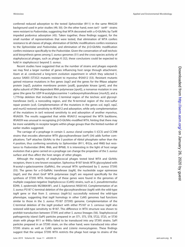
FIG 1 Stages of phage infection and corresponding examples of resistance mechanisms at each stage.Examples not yet identified in the staphylococci are listed in red.
Minireview Applied and Environmental Microbiology
June 2019 Volume 85 Issue 11 e00209-19 aem.asm.org 3
on January 26, 2020 by guesthttp://aem
.asm.org/
Dow
nloaded from

characterization showed that the mutants lacked UDP-GlcNAc:polyribitol phosphatetransferase activity and WTA. Counterintuitively, they did show the relevant biochem-ical activity for the last known step in WTA biosynthesis (phosphoribitol transferase[TarL]; Fig. 2B) (38). This surprising result suggested that the double-resistant mutantsproduced ribitol phosphate but either failed to properly polymerize WTA or attach it tothe cell wall. These mutants had pleiotropic phenotypic differences from their parentstrain (41), including longer generation times than their parent; cell growth in clumps;irregular, rough, gray colonies; and increased levels of wall-bound autolysin. A laterstudy characterizing spontaneous S. aureus strain A170 (ST45) mutants resistant tosiphovirus MSa found similar phenotypic defects (43), and biochemical assays alsoshowed that resistance was likely due to the lack of GlcNAc-modified WTA.
Genes responsible for phage adsorption were identified in a series of elegantmolecular genetic studies performed in the RN4220 (ST8) (44) background (35, 36, 45).Deletion of undecaprenyl-phosphate N-acetylglucosaminyl 1-phosphate transferase(tagO), the first gene involved in WTA biosynthesis, conferred resistance and reducedadsorption to the tested Myoviridae (�812 and �K), while a mutant with a transposoninsertion mutation in the tarM gene had resistance and reduced adsorption to Sipho-viridae (�Sa2mw, �47, �13, and �77). Complementation of wild-type alleles rescuedthese phenotypes (35). TarM is a glycosyltransferase responsible for attaching �-O-GlcNAc to the 4 position of the ribitol phosphate WTA monomer (46, 47). The tarMmutant was previously shown to lack GlcNAc-modified WTA in its envelope (46). TarS,the glycosyltransferase responsible for attaching �-O-GlcNAc to the 4 position of theribitol phosphate WTA monomer (48), was specifically required for podovirus adsorp-tion (45). Deletion of tarS conferred resistance and reduced adsorption to the testedPodoviridae (�44AHJD, �66, and �P68) (45), but only deletion of both tarS and tarM
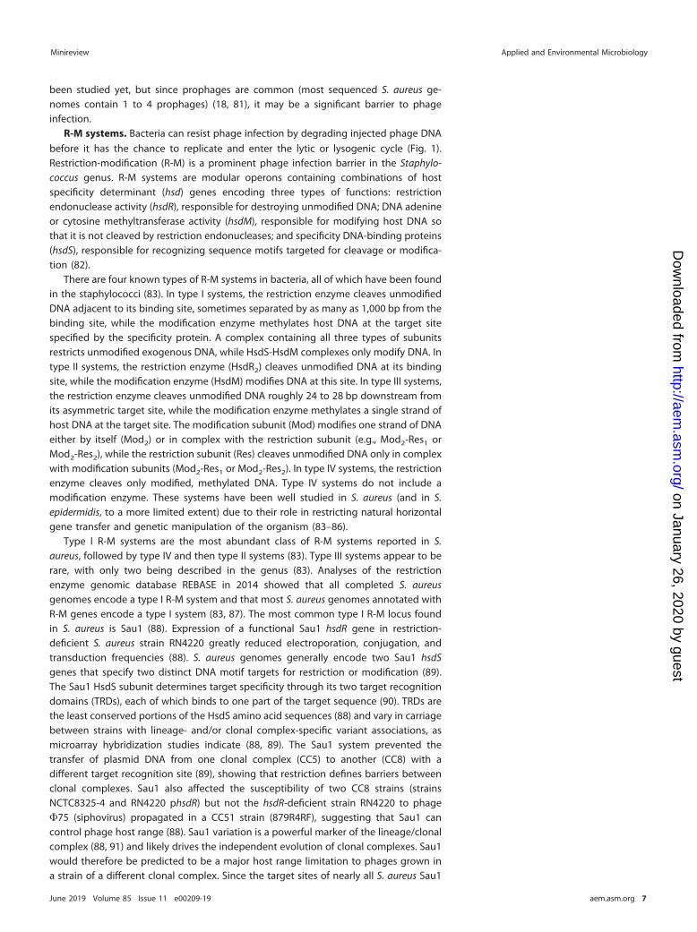
FIG 2 (A) Structure of the staphylococcal cell envelope. Lipoteichoic acid is shown in orange (glycerol phosphate), a surface protein is in black, wall teichoicacid is in orange (glycerol phosphate) and yellow (ribitol phosphate), capsule is in blue, and cell wall carbohydrates are in green (N-acetylglucosamine [GlcNAc])and purple (N-acetylmuramic acid [MurNAc]). Staphylococcal phages bind WTA and/or its ribitol phosphate modifications (i.e., GlcNAc). (B) Outline of the wallteichoic acid (WTA) biosynthesis pathway, with the proteins corresponding to each step listed in the blue arrows. Abbreviations are defined as follows: C55-P,undecaprenyl phosphate; GlcNAc, N-acetylglucosamine; UDP-GlcNAc, uridine-5-diphosphate-N-acetylglucosamine; ManNAc, N-acetylmannosamine; UDP-ManNAc, uridine-5-diphosphate-N-acetylmannosamine; Gro-P, glycerol phosphate; CDP-Gro, cytidyl diphosphate-glycerol; Rbo-P, ribitol phosphate; CDP-Rbo,cytidyl diphosphate-ribitol; ABC, ATP-binding cassette; and LCP, LytR-CpsA-Psr.
Minireview Applied and Environmental Microbiology
June 2019 Volume 85 Issue 11 e00209-19 aem.asm.org 4
on January 26, 2020 by guesthttp://aem
.asm.org/
Dow
nloaded from
conferred reduced adsorption to the tested Siphoviridae (�11) in the same RN4220background used in prior studies (49, 50). On the other hand, even tarS� tarM� strainswere resistant to Podoviridae, suggesting that WTA decorated with �-O-GlcNAc by TarMimpeded podovirus adsorption (45). Taken together, these findings suggest, for thesmall number of representatives that were tested, that elimination of WTA confersresistance to all classes of phage, elimination of GlcNAc modifications confers resistanceto the Siphoviridae and Podoviridae, and elimination of the �-O-GlcNAc modificationconfers resistance specifically to the Podoviridae. Given the conservation of wall teichoicacid biosynthesis genes among S. aureus genomes (51) and the cross-species activity ofstaphylococcal phages, such as phage K (52), these conclusions could be expected tohold in staphylococci beyond S. aureus.
Recent studies have suggested that as the number of strains and phages expandswe may find a larger number of genes influencing host range through attachment.Azam et al. conducted a long-term evolution experiment in which they selected S.aureus SA003 (ST352) mutants resistant to myovirus �SA012 (53). Resistant mutantsgained missense mutations in five genes (tagO and the genes for the RNase adapterprotein [rapZ], putative membrane protein [yozB], guanylate kinase [gmk], and thealpha subunit of DNA-dependent RNA polymerase [rpoA]), a nonsense mutation in onegene (the gene for UDP-N-acetylglucosamine 1-carboxyvinyltransferase [murA2]), and a1,779-bp deletion that included the C-terminal region of the teichoic acid glycosyl-transferase (tarS), a noncoding region, and the N-terminal region of the iron-sulfurrepair protein (scd). Complementation of the mutations in the genes scd, tagO, rapZ,and murA2 restored sensitivity to �SA012 and adsorption, while only complementationof the mutations in tarS restored sensitivity to and adsorption of another myovirus,�SA039. The results suggested that while �SA012 recognized the WTA backbone,�SA039 was unusual in recognizing �-O-GlcNAc-modified WTA, hinting that there maybe more variability in receptor targets within phage groups than the limited number ofearlier studies suggested.
The carriage of a prophage in certain S. aureus clonal complex 5 (CC5) and CC398strains that encodes alternative WTA glycosyltransferase (tarP) (54) adds further com-plications. TarP attaches GlcNAc to the 3 position of ribitol phosphate rather than the4 position, thus conferring sensitivity to Siphoviridae (�11, �52a, and �80) but resis-tance to Podoviridae (�44, �66, and �P68). It is interesting in the light of host rangeevolution that a gene carried on a prophage can change the properties of the S. aureussurface and thus affect the host ranges of other phages.
Although the majority of staphylococcal phages tested bind WTA and GlcNAcreceptors, there is one known exception. Siphovirus �187 binds WTA glycosylated withN-acetyl-D-galactosamine (GalNAc), the unusual WTA synthesized by S. aureus ST395(55). The genes for �-O-GalNAc transferase (tagN), the nucleotide sugar epimerase(tagV), and the short GroP WTA polymerase (tagF) are required specifically for thesynthesis of ST395 WTA. Homologs of these genes were found in the genomes ofmultiple coagulase-negative Staphylococcus (CoNS) strains, such as S. pseudintermediusED99, S. epidermidis M23864:W1, and S. lugdunensis N920143. Complementation of anS. aureus PS187 C-terminal deletion of the glycosyltransferase (tagN) with the wild-typetagN gene or that from S. carnosus (tagN-Sc) successfully restored the wild-typephenotype, suggesting that tagN homologs in other CoNS genomes had functionssimilar to those in the S. aureus PS187 (ST395) genome. Complementation of theC-terminal deletion of the tagN product with either PS187 or S. carnosus tagN alsorestored wild-type sensitivity to �187. This difference in WTA structure was shown toprohibit transduction between ST395 and other S. aureus lineages (56). Staphylococcalpathogenicity island (SaPI) particles prepared in an ST1, ST5, ST8, ST22, ST25, or ST30strain with phage �11 or �80� failed to be transduced into any ST395 strains. SaPIparticles prepared in an ST395 strain, on the other hand, were transduced into otherST395 strains as well as CoNS species and Listeria monocytogenes. These findingssuggest that the unique ST395 WTA restricts the phage host range to strains of the
Minireview Applied and Environmental Microbiology
June 2019 Volume 85 Issue 11 e00209-19 aem.asm.org 5
on January 26, 2020 by guesthttp://aem
.asm.org/
Dow
nloaded from

same sequence type or Gram-positive bacteria with a related WTA structure, such asListeria monocytogenes.
There has been one study showing that staphylococcal phages (siphovirus �SLT)can bind lipoteichoic acid (LTA), the lipid-anchored, polyglycerol phosphate (GroP)teichoic acid polymer (57) (Fig. 2A). However, subsequent elimination of LTA biosyn-thesis through ltaS deletion had no effect on adsorption of or sensitivity to phage (35),and therefore, the potential significance of LTA as an alternative receptor is currentlyunknown.
Effects of surface proteins and extracellular polysaccharides on attachment.Although proteins serve as receptors for many Gram-positive bacterial phages (forexample, the YueB receptor for Bacillus subtilis phage SPP1 [58]), there is no evidenceto suggest S. aureus proteins serve as S. aureus phage receptors. Phage interactionprotein (Pip) homologs exist throughout the Gram-positive bacteria, serving as proteinreceptors to which phages irreversibly bind (59). There are Pip surface protein ho-mologs anchored to the staphylococcal cell wall through the action of the sortaseenzyme in Staphylococcus (60, 61). However, deletion of neither the Pip homologs inRN4220 (ST8) (49) nor sortase A in Newman (ST254) (62, 63) affected sensitivity tophage �11 or phages �NM1, �NM2, and �NM4, respectively.
Some classes of proteins or extracellular polysaccharides have been shown to blockphage adsorption in the staphylococci through occlusion of the WTA receptors. Over-production of surface protein A in S. aureus was shown to reduce phage adsorptionthrough this mechanism (64), but work on surface protein occlusion remains limited.Capsule type 1 and 2 strains—strains M (ST1254) (https://www.phe-culturecollections.org.uk/collections/nctc-3000-project.aspx) and Smith diffuse (ST707) (https://www.phe-culturecollections.org.uk/collections/nctc-3000-project.aspx), respectively—were shownto occlude adsorption (65), but strains of the most common capsule types, types 5 and8, showed inconclusive results (66, 67). Differences in capsule thickness between strainsmay account for these variable results. Type 1 and 2 strains are mucoid and heavilyencapsulated, while type 5 and 8 strains are nonmucoid, despite encapsulation (68).The CoNS species Staphylococcus simulans also showed capsule-dependent inhibitionof phage adsorption (69).
The exopolysaccharides (EPS) of staphylococcal biofilms have not been shownto occlude adsorption. Surface proteins, such as biofilm-associated protein (Bap),exopolysaccharides (polysaccharide intercellular adhesin [PIA], composed of poly-N-acetylglucosamine [PNAG] and synthesized by the products of the icaADBC operon),and extracellular DNA (eDNA) compose staphylococcal biofilms, which can form byPIA-dependent or protein (Bap)-dependent mechanisms (70, 71). Other surface proteinsmore common than Bap can also mediate biofilm formation, such as FnbA/FnbB (72,73) and SasG (74) in S. aureus and Aap in S. epidermidis (70). Both S. aureus (19, 75) andS. epidermidis (52, 76, 77) biofilms are susceptible to phage predation. Phage resistancein staphylococcal biofilms may instead be associated with altered biofilm diffusion ormetabolism. Studies on S. epidermidis suggested that susceptibility to phages is similarin biofilms and stationary-phase cultures (52). Phages may, in fact, promote bacterialpersistence in S. aureus biofilms by releasing nutrients from lysed cells for the remain-ing live ones to utilize (78).
BIOSYNTHESISSuperinfection immunity. Staphylococcal temperate phages encode homologs of
the cI repressor (17, 18). In Escherichia coli, this protein represses expression of the lyticcycle in newly infecting phages with the same cI protein-binding sites, thus stoppingnew infections through a mechanism called superinfection immunity. Molecular andevolutionary studies on the E. coli phage lambda model suggest that many superin-fection immunity groups (in which member temperate phages confer immunity to eachother upon integration) coexist in nature (79), with cI repressor-operator coevolutiondriving the emergence of new immunity groups (80). Superinfection immunity as adetermining factor in phage host range in staphylococcal species appears not to have
Minireview Applied and Environmental Microbiology
June 2019 Volume 85 Issue 11 e00209-19 aem.asm.org 6
on January 26, 2020 by guesthttp://aem
.asm.org/
Dow
nloaded from
been studied yet, but since prophages are common (most sequenced S. aureus ge-nomes contain 1 to 4 prophages) (18, 81), it may be a significant barrier to phageinfection.
R-M systems. Bacteria can resist phage infection by degrading injected phage DNAbefore it has the chance to replicate and enter the lytic or lysogenic cycle (Fig. 1).Restriction-modification (R-M) is a prominent phage infection barrier in the Staphylo-coccus genus. R-M systems are modular operons containing combinations of hostspecificity determinant (hsd) genes encoding three types of functions: restrictionendonuclease activity (hsdR), responsible for destroying unmodified DNA; DNA adenineor cytosine methyltransferase activity (hsdM), responsible for modifying host DNA sothat it is not cleaved by restriction endonucleases; and specificity DNA-binding proteins(hsdS), responsible for recognizing sequence motifs targeted for cleavage or modifica-tion (82).
There are four known types of R-M systems in bacteria, all of which have been foundin the staphylococci (83). In type I systems, the restriction enzyme cleaves unmodifiedDNA adjacent to its binding site, sometimes separated by as many as 1,000 bp from thebinding site, while the modification enzyme methylates host DNA at the target sitespecified by the specificity protein. A complex containing all three types of subunitsrestricts unmodified exogenous DNA, while HsdS-HsdM complexes only modify DNA. Intype II systems, the restriction enzyme (HsdR2) cleaves unmodified DNA at its bindingsite, while the modification enzyme (HsdM) modifies DNA at this site. In type III systems,the restriction enzyme cleaves unmodified DNA roughly 24 to 28 bp downstream fromits asymmetric target site, while the modification enzyme methylates a single strand ofhost DNA at the target site. The modification subunit (Mod) modifies one strand of DNAeither by itself (Mod2) or in complex with the restriction subunit (e.g., Mod2-Res1 orMod2-Res2), while the restriction subunit (Res) cleaves unmodified DNA only in complexwith modification subunits (Mod2-Res1 or Mod2-Res2). In type IV systems, the restrictionenzyme cleaves only modified, methylated DNA. Type IV systems do not include amodification enzyme. These systems have been well studied in S. aureus (and in S.epidermidis, to a more limited extent) due to their role in restricting natural horizontalgene transfer and genetic manipulation of the organism (83–86).
Type I R-M systems are the most abundant class of R-M systems reported in S.aureus, followed by type IV and then type II systems (83). Type III systems appear to berare, with only two being described in the genus (83). Analyses of the restrictionenzyme genomic database REBASE in 2014 showed that all completed S. aureusgenomes encode a type I R-M system and that most S. aureus genomes annotated withR-M genes encode a type I system (83, 87). The most common type I R-M locus foundin S. aureus is Sau1 (88). Expression of a functional Sau1 hsdR gene in restriction-deficient S. aureus strain RN4220 greatly reduced electroporation, conjugation, andtransduction frequencies (88). S. aureus genomes generally encode two Sau1 hsdSgenes that specify two distinct DNA motif targets for restriction or modification (89).The Sau1 HsdS subunit determines target specificity through its two target recognitiondomains (TRDs), each of which binds to one part of the target sequence (90). TRDs arethe least conserved portions of the HsdS amino acid sequences (88) and vary in carriagebetween strains with lineage- and/or clonal complex-specific variant associations, asmicroarray hybridization studies indicate (88, 89). The Sau1 system prevented thetransfer of plasmid DNA from one clonal complex (CC5) to another (CC8) with adifferent target recognition site (89), showing that restriction defines barriers betweenclonal complexes. Sau1 also affected the susceptibility of two CC8 strains (strainsNCTC8325-4 and RN4220 phsdR) but not the hsdR-deficient strain RN4220 to phage�75 (siphovirus) propagated in a CC51 strain (879R4RF), suggesting that Sau1 cancontrol phage host range (88). Sau1 variation is a powerful marker of the lineage/clonalcomplex (88, 91) and likely drives the independent evolution of clonal complexes. Sau1would therefore be predicted to be a major host range limitation to phages grown ina strain of a different clonal complex. Since the target sites of nearly all S. aureus Sau1
Minireview Applied and Environmental Microbiology
June 2019 Volume 85 Issue 11 e00209-19 aem.asm.org 7
on January 26, 2020 by guesthttp://aem
.asm.org/
Dow
nloaded from
R-M systems from each of the different clonal complexes have now been identified (90),it should be possible to bioinformatically predict the Sau1-defined clonal complex hostrange of any sequenced bacteriophage.
The type IV R-M system SauUSI is estimated to be found in 90% of S. aureus strains(83, 92) and, in combination with Sau1, presents an effective restriction barrier forresisting phage infection (93). SauUSI specifically restricts DNA methylated or hy-droxymethylated at the C-5 position of cytosine (92). The preferred binding site forSauUSI is Sm5CNGS, where S represents either cytosine or guanine (92). Type II R-Msystems have been estimated to be present in �33% of strains and display a range oftarget sites (83, 94–96). The most common type II R-M system found in S. aureus iscalled Sau3A (94). The Sau3A restriction enzyme cleaves 5= to the guanine in unmod-ified 5=-GATC-3= sequences. The Sau3A modification enzyme, on the other hand,methylates the restriction site at the C-5 position of cytosine (97). Some type II systems,such as Sau42I, are encoded by phages. Sau42I is an example of a type IIS R-M system,which binds asymmetric DNA sequences and cleaves outside the recognition site,unlike most type II systems (82). Unlike type I and type IV systems, type II systems areoften carried on mobile genetic elements, which are capable of frequent transferbetween strains and which are not conserved among all members of the same clonalcomplex, so they present a more strain-specific and variable limit to host range (87).Certain S. aureus type II R-M systems (e.g., Sau96I) serve to negate the type IV SauUSIsystem because they methylate cytosines and guanines in sequences that SauUSItargets for cleavage. This is an interesting example of how R-M systems acquired byHGT can have unpredictable interactions with existing systems.
If unmodified phages can survive restriction enzyme degradation upon cell entry, thephage DNA molecules acquire protective DNA methylation as they replicate. While survivalof restriction can happen stochastically at high multiplicities of infection, phages have alsobeen shown to have evolved or acquired adaptations for restriction evasion. Antirestrictionmechanisms include restriction site alteration, restriction site occlusion, indirect subversionof restriction-modification activity, and direct inhibition of restriction-modification systems(98). Restriction site alteration can include both incorporation of alternative bases, such as5-hydroxymethyluracil (5hmU) and 5-hydroxymethylcytosine (5hmC), and loss of restrictionsites through selection. A clear example of the latter in the staphylococci is the eliminationof GATC sites in the 140-kb phage K genome, enabling its avoidance of Sau3A restriction(99). Another example is the evolution of particular antimicrobial resistance-carrying con-jugative plasmids which have lost specific Sau1 R-M sites, allowing their transfer betweencommon MRSA lineages (88). Restriction site occlusion refers to DNA-binding proteinspreventing restriction enzymes from binding and digesting DNA (98, 100, 101). R-Msubversion occurs through either stimulation of host modification enzymes or destructionof restriction cofactors (e.g., S-adenosylmethionine [SAM]) (98, 102, 103). R-M inhibitionoccurs most often in type I systems (but also in some type II systems) through the bindingof specific antirestriction proteins, such as ArdA, ArdB, and Ocr (98, 104, 105). There is noliterature specifically characterizing antirestriction in Staphylococcus, but an E. coli ardAhomolog has been identified in the staphylococcal Tn916 and Tn5801 transposons (106).
CRISPR systems. Clustered regularly interspaced short palindromic repeats (CRISPRs)confer immunity to phage infection through the cleavage of extrinsic DNA in a sequence-specific manner. Unlike R-M systems, which target specific DNA sequence motifs, CRISPRsadaptively incorporate target sequences from phages that they have destroyed to increasethe efficiency of protection. After integrating short segments of foreign DNA as spacers ofCRISPR arrays, CRISPR-associated (Cas) nucleases process the transcribed CRISPR array RNAinto CRISPR RNAs (crRNAs), used to target new incursions of identical foreign DNA elementsfor destruction (107, 108). Surveys of S. aureus and S. epidermidis genomes indicate thatCRISPRs are not common in these species (109, 110). These surveys looked for the presenceof the cas6 and cas9 genes, which encode nucleases required for type I/III and type IICRISPR-mediated resistance, respectively. Cas6 is an endoribonuclease found in type I andIII CRISPR systems that cleaves pre-crRNA transcripts within the 3= end of the repeat region
Minireview Applied and Environmental Microbiology
June 2019 Volume 85 Issue 11 e00209-19 aem.asm.org 8
on January 26, 2020 by guesthttp://aem
.asm.org/
Dow
nloaded from
to produce mature guide crRNAs (111, 112), while Cas9 is an endonuclease found in typeII CRISPR systems that cleaves DNA in a crRNA-guided manner (112, 113). Only 12 of 300published S. epidermidis genomes searched encoded the Cas6 nuclease, 18 of 130 S.epidermidis isolates from Denmark (Copenhagen University Hospital) tested positive forcas6 via PCR, and 14 of nearly 5,000 published S. aureus genomes encoded CRISPR/Cassystems (109). Another study specifically examining S. aureus found that 2 of 32 S. aureusstrains encoded CRISPR-Cas systems (110). These CRISPRs were similar to those found intwo S. lugdunensis strains, suggesting that they were recombined with S. lugdunensis orderived from a common ancestor (110). CRISPR/Cas systems have also occasionally beenreported in strains of other species (S. capitis, S. schleiferi, S. intermedius, S. argenteus, and S.microti) (109). Only a single S. aureus strain has been reported to encode Cas9, which isfound in a staphylococcal cassette chromosome mec element (SCCmec)-like region (114).Nonetheless, CRISPR systems have been shown to be important in resisting the introduc-tion of foreign DNA in S. epidermidis RP62a (115, 116). Anti-CRISPR mechanisms, such asproteins that prevent CRISPR-Cas systems from binding DNA target sites, are being discov-ered in many phages (117–119), although they have not yet been discovered in thosespecific for staphylococci. Currently discovered anti-CRISPR mechanisms have been shownto target both type I and type II CRISPR systems (117–120).
ASSEMBLY
Assembly interference is the parasitization of superinfecting phage by chromosomalphage-like elements and has been demonstrated experimentally in S. aureus pathoge-nicity island (SaPI)-helper phage interactions. SaPIs encode important virulence factors,such as toxic shock syndrome toxin (TSST), but are mobilized only by superinfectinghelper siphoviruses (28, 121). The Dut dUTPase protein expressed by helper phagesderepresses the Stl SaPI repressor, activating the SaPI lytic cycle (28). The derepressedSaPIs then take advantage of the superinfection to proliferate at the expense of thehelper phage. SaPIs interfere with helper phage assembly through several mechanisms(122): remodeling phage capsid proteins to fit the small SaPI genome (123–127),encoding phage packaging interference (Ppi) proteins that prevent helper phage DNApackaging into new SaPI particles (123), and disrupting phage late gene activation(128). All known SaPIs encode phage packaging interference (Ppi) proteins, whichdivert phage DNA packaging toward SaPIs by inhibiting helper phage terminase smallsubunits (TerSP) but not the corresponding SaPI subunits (TerSS) (123). Ppi proteins aredivided into two classes based on sequences that differ in helper phage specificity.Class I interferes with �80� and �11, while class II interferes with �12 (123). ThePtiM-modulated PtiA and the PtiB SaPI2 proteins inhibit expression of the LtrC-activated phage 80 late gene operon (packaging and lysis genes), thus interfering withlater steps of the helper phage life cycle (128). The SaPI particles then go on to infectnew S. aureus hosts, integrating their DNA into the chromosome instead of killing thecell. Helper phages and SaPIs are thought to gain and lose resistance to each other ina Red Queen scenario, given the observed rapid coevolution of their respective dut andstl genes (129). SaPIs are found throughout Staphylococcus species and beyond; there-fore, they may be a common strain-specific modifier of siphovirus infection potential.
OTHER PHAGE HOST RANGE-LIMITING FACTORS
Several uncommon or less well understood mechanisms may contribute to phagehost range limitation in Staphylococcus. One abortive infection (Abi) system, theeukaryote-like serine/threonine kinase Stk2, has been characterized in S. aureus and S.epidermidis (130). In this case, siphovirus infection results in self-induced killing of thehost cell, preventing the amplification and spread of phages in the population. Stk2 wasfound to be activated by a phage protein of unknown function and caused cell deathby phosphorylating host proteins involved in diverse core cellular functions. Only S.epidermidis RP62A and a few S. aureus strains encode Stk2, however, suggesting limitedgenus-wide importance. The recent long-term evolution study on S. aureus strain SA003uncovered two genes involved in postadsorption resistance to myovirus �SA012 (53).
Minireview Applied and Environmental Microbiology
June 2019 Volume 85 Issue 11 e00209-19 aem.asm.org 9
on January 26, 2020 by guesthttp://aem
.asm.org/
Dow
nloaded from

Missense mutations in guanylate kinase and the alpha subunit of DNA-dependent RNApolymerase conferred resistance but not corresponding decreases in the adsorptionrate, suggesting some postadsorption role in resisting infection. More phage resistancesystems likely remain undiscovered. A genome-wide association study of 207 clinicalMRSA strains and 12 phage preparations identified 167 gene families putatively asso-ciated with phage-bacterium interactions (131). While these families includedrestriction-modification genes, transcriptional regulators, and genes of prophage andSaPI origin, most were accessory gene families of unknown function.
PHAGE HOST RANGE IN STAPHYLOCOCCUS IS DETERMINED BY A HIERARCHICALCOMBINATION OF HOST FACTORS
In summary, we have described how the host range of a Staphylococcus phage isdetermined by a combination of both host- and phage-encoded genes, as well as theepigenetic DNA methylation patterns conferred on its DNA from the last strain that itinfected. Bacterium-encoded factors can be conceived of as affecting host range atdifferent levels within the species (Fig. 3). At the highest level, most phages’ target forreceptor binding (WTA) is highly conserved across Staphylococcus species. Strains withunusual WTAs, such as S. aureus ST395 and CoNS strains with poly-GroP WTA (55, 56),would be expected to be genetically isolated within the genus. Type I R-M hsdSallotypes and capsule type are conserved between most strains of the same clonalcomplex (CC) but differ between isolates of different CC groups and thus contribute todefining the host range in a large subset of S. aureus strains. At the level of individualstrains, inserted prophages and SaPIs, Stk2, type II systems acquired by HGT, and otheras yet unknown functions may all serve to limit the host range. We know even lessabout phage-encoded systems that counteract host resistance. The finding that lyticphages (Myoviridae and Podoviridae) tend to have broader host ranges than Siphoviri-dae when challenged against the same set of Staphylococcus strains suggests that theformer encode an array of uncharacterized genes that work against host defenses.
FUTURE DIRECTIONS
Although much progress has been made in the past 5 decades toward understand-ing the mechanisms that define staphylococcal phage host range, numerous important
FIG 3 Phage host range for an individual strain is the combination of multiple factors that have different levels ofconservation within the species. This is illustrated by a hypothetical phylogenetic tree. Mechanisms can be presentthroughout strains (1, most conserved; red), present in many strains but with considerable allelic variation (2,conserved but polymorphic; shades of green), or present in a few strains, possibly with allelic variation (3a to 3c,less conserved with potential polymorphism; blue, purple, and yellow, respectively). Branches where mechanismsevolved by mutation or homologous recombination, in the case of mechanisms 1 and 2, or were acquired by HGT,in the case of mechanisms 3a to 3c, are annotated with colored stars. The table on the right summarizes themechanisms (1 to 3c) present in each strain (strains A to J) using shaded boxes with corresponding colors. StrainJ has a mutation that results in the null phenotype for the red mechanism. Host range is the result of thecombination of mechanisms present, so strains A to C as well as F, H, and I would be predicted to have identicalhost ranges, but phage-specific factors could also introduce variability.
Minireview Applied and Environmental Microbiology
June 2019 Volume 85 Issue 11 e00209-19 aem.asm.org 10
on January 26, 2020 by guesthttp://aem
.asm.org/
Dow
nloaded from
questions remain. We need to know more about species other than S. aureus and S.epidermidis, and even within these species, we need to make sure that rarer andnon-methicillin-resistant strains are included in studies (132). We also need to ensurethat our collections reflect the true diversity of phages that infect Staphylococcusspecies. Even within the two main species, only a relatively small number of phageshave been tested. This will lead us to consider the questions of phage ecology whenunderstanding what types of phages are found in different environments and withwhat abundance.
Discovering novel phage resistance mechanisms would aid the effort to understanddeterminants of host range. Many phage resistance mechanisms have been identifiedand characterized in other Gram-positive bacteria and other bacteria generally but notin the staphylococci. Superinfection exclusion (Sie) and abortive infection (Abi) systems,for example, are well characterized in the lactococci (133–135). In addition, a recentpublication describes some 26 new antiphage defense systems identified in bacteria(136), not including the recently discovered bacteriophage exclusion (BREX) and de-fense island system associated with restriction-modification (DISARM) phage defenses(137–139). Five of the 10 verified, newly discovered antiphage defense systems (Tho-eris, Hachiman, Gabija, Lamassu, and Kiwa) have orthologs in staphylococcal genomes(136).
Understanding phage host range to the point that we can make accurate predic-tions based on the host genome will be important for developing phage therapiesagainst Staphylococcus strains. Ideally, cocktail formulations for therapy consist ofphages with broad, nonoverlapping host ranges against the target species (or clonalcomplex) to be treated. As there are many more genome sequences available thanstrains that can be tested for sensitivity in the laboratory (e.g., �40,000 for S. aureus)(140), with a predictive model we could run in silico tests on genome sequences tomodel the efficacy of the cocktail. With the potential for genome sequencing to beused in the future as a primary clinical diagnostic, we could modify the cocktail tocontain phages that specifically target the bacterium causing the infection.
Knowledge of phage host range will also lead us to understand the fitness costs ofresistance and its potential trade-offs with the virulence and antibiotic resistance ofStaphylococcus. Strains with null mutations in biosynthetic genes are rare, given WTA’sroles in cell division, autolysis, virulence, and antibiotic resistance (36, 37). Althoughknocking out the genes involved in the first two steps of WTA biosynthesis has nofitness cost in S. aureus (at least under laboratory conditions) (141, 142), WTA has manycritical physiological roles, especially in environments subject to phage therapy. Staph-ylococcal WTA is required for nasal colonization (141, 143), cell division (41, 43),regulating autolysis (144, 145), lysozyme resistance through cell wall cross-linking (132,146), resistance to cationic antimicrobial peptides and fatty acids (147, 148), and biofilmformation (149). WTA-altered or -negative phage-resistant mutants would, in turn,become less virulent (43) and even antibiotic sensitive, which would make them highlyunfit in the natural habitat colonizing mammalian hosts or in an infection site subjectto treatment. Given that methicillin resistance requires WTA (50), phage– beta-lactamcombination therapies could be particularly promising. Mutants resistant to eitherphages or beta-lactams would be sensitive to the other treatment, assuming that theinfecting strain is sensitive to the phage treatment. Nonetheless, as we note for hostrange, strains containing minor but fitness-neutral resistance mechanisms, such as R-Msystems, may be the most recalcitrant to phage therapy. Staphylococcal phage thera-pies must then overcome both immediate, emerging mutational resistance and intrin-sic resistance mechanisms (e.g., R-M systems) specific to strains or clonal complexes.These resistance limitations, however, could be overcome by selecting phage hostrange mutants that escaped host resistance mechanisms, thus isolating more usefulphages that would form more effective phage cocktails (150, 151).
The phage-resistant mutants isolated so far, such as those described in the adsorp-tion studies, were typically selected in rich, aerated laboratory medium. The conse-quences for fitness of the same mutations occurring during in vivo infection might be
Minireview Applied and Environmental Microbiology
June 2019 Volume 85 Issue 11 e00209-19 aem.asm.org 11
on January 26, 2020 by guesthttp://aem
.asm.org/
Dow
nloaded from
more severe. In addition, both the relevance of various resistance mechanisms in vivoand the effect of strain genetic background on resistance selection, especially on aspecies-wide scale, have been left unexamined in most previous work. One study inmammalian hosts showed that the environment altered phage transfer frequency andselection (152), leading to the spread of prophage and the selection of phage resistanceby superimmunity. In laboratory media, the phage transfer frequency was lower andthe spread of prophage was less pronounced (152). It will be important to know bothhow quickly and in which loci mutations emerge, as well as the more general distri-bution of resistance gene families.
Finally, it is interesting to consider what phage host range studies reveal about thehosts themselves. Staphylococci seem to be unusual among Gram-positive bacteria inrequiring conserved WTA receptors for attachment and having no reported role forprotein receptors. Differences in the outer surface of Staphylococcus and/or a feature ofthe phage ecology within the genus may account for this fact. Another interestingquestion is why CRISPRs play a lesser role for intercepting extrinsic phage DNA thanR-M systems in this genus compared with others. It could be that CRISPR systems havea finite capacity for carrying fragments of mobile genetic elements, while R-M systemscan attack a wider range of incoming DNA, relevant to rapidly evolving populations.Future studies that probe these questions may reveal some of the differential evolu-tionary forces that shape the genomes of pathogenic bacteria.
CONCLUSIONS
Staphylococcal phage resistance mechanisms have been identified at three stages ofinfection (attachment, biosynthesis, and assembly) and regulate host range in a hierarchicalmanner, depending on mechanism conservation. We need further studies to objectivelyidentify the contribution of individual phage resistance mechanisms to host range. Suchwork would provide the information needed not only to formulate phage cocktails effectiveagainst a wide variety of strains but also to overcome remaining obstacles to cocktaildevelopment (e.g., highly effective R-M or Abi systems). Future studies relevant to phagetherapy should also characterize phage resistance development during infection andtherapy as well as the effects of resistance on mutant fitness. Taken together, this futurework will inform the rational design of phage cocktails to treat staphylococcal infectionsalone or in combination with antibiotics.
ACKNOWLEDGMENTSWe thank Michelle Su and Robert Petit for critically reading the manuscript and
providing helpful comments.A.G.M. was supported by the National Science Foundation (NSF) Graduate Research
Fellowship Program (GRFP). J.A.L. was supported by the Medical Research Council(grant MR/P028322/1). T.D.R. was supported by National Institutes of Health (NIH) grantAI121860.
REFERENCES1. Kluytmans J, van Belkum A, Verbrugh H. 1997. Nasal carriage of Staph-
ylococcus aureus: epidemiology, underlying mechanisms, and associ-ated risks. Clin Microbiol Rev 10:505–520. https://doi.org/10.1128/CMR.10.3.505.
2. Kong EF, Johnson JK, Jabra-Rizk MA. 2016. Community-associatedmethicillin-resistant Staphylococcus aureus: an enemy amidst us.PLoS Pathog 12:e1005837. https://doi.org/10.1371/journal.ppat.1005837.
3. Bagnoli F, Bertholet S, Grandi G. 2012. Inferring reasons for the failureof Staphylococcus aureus vaccines in clinical trials. Front Cell InfectMicrobiol 2:16. https://doi.org/10.3389/fcimb.2012.00016.
4. Wang I-N, Smith DL, Young R. 2000. Holins: the protein clocks ofbacteriophage infections. Annu Rev Microbiol 54:799 – 825. https://doi.org/10.1146/annurev.micro.54.1.799.
5. Young R, Bläsi U. 1995. Holins: form and function in bacteriophage lysis.FEMS Microbiol Rev 17:195–205.
6. Loessner MJ. 2005. Bacteriophage endolysins— current state of re-search and applications. Curr Opin Microbiol 8:480 – 487. https://doi.org/10.1016/j.mib.2005.06.002.
7. Pirisi A. 2000. Phage therapy—advantages over antibiotics? Lancet356:1418. https://doi.org/10.1016/S0140-6736(05)74059-9.
8. Nobrega FL, Costa AR, Kluskens LD, Azeredo J. 2015. Revisiting phagetherapy: new applications for old resources. Trends Microbiol 23:185–191. https://doi.org/10.1016/j.tim.2015.01.006.
9. Stone R. 2002. Stalin’s forgotten cure. Science 298:728 –731. https://doi.org/10.1126/science.298.5594.728.
10. Bradbury J. 2004. My enemy’s enemy is my friend. Lancet 363:624 – 625.https://doi.org/10.1016/S0140-6736(04)15629-8.
11. Kutter E, De Vos D, Gvasalia G, Alavidze Z, Gogokhia L, Kuhl S, AbedonST. 2010. Phage therapy in clinical practice: treatment of humaninfections. Curr Pharm Biotechnol 11:69 – 86. https://doi.org/10.2174/138920110790725401.
Minireview Applied and Environmental Microbiology
June 2019 Volume 85 Issue 11 e00209-19 aem.asm.org 12
on January 26, 2020 by guesthttp://aem
.asm.org/
Dow
nloaded from
12. Kutateladze M, Adamia R. 2010. Bacteriophages as potential new ther-apeutics to replace or supplement antibiotics. Trends Biotechnol 28:591–595. https://doi.org/10.1016/j.tibtech.2010.08.001.
13. Sulakvelidze A, Alavidze Z, Morris JG. 2001. Bacteriophage therapy.Antimicrob Agents Chemother 45:649 – 659. https://doi.org/10.1128/AAC.45.3.649-659.2001.
14. Lu TK, Koeris MS. 2011. The next generation of bacteriophage therapy.Curr Opin Microbiol 14:524 –531. https://doi.org/10.1016/j.mib.2011.07.028.
15. Ly-Chatain MH. 2014. The factors affecting effectiveness of treatment inphages therapy. Front Microbiol 5:51. https://doi.org/10.3389/fmicb.2014.00051.
16. Jault P, Leclerc T, Jennes S, Pirnay JP, Que Y-A, Resch G, Rousseau AF,Ravat F, Carsin H, Le Floch R, Schaal JV, Soler C, Fevre C, Arnaud I,Bretaudeau L, Gabard J. 2018. Efficacy and tolerability of a cocktail ofbacteriophages to treat burn wounds infected by Pseudomonasaeruginosa (PhagoBurn): a randomised, controlled, double-blind phase1/2 trial. Lancet Infect Dis 19:35– 45. https://doi.org/10.1016/S1473-3099(18)30482-1.
17. Xia G, Wolz C. 2014. Phages of Staphylococcus aureus and their impacton host evolution. Infect Genet Evol 21:593– 601. https://doi.org/10.1016/j.meegid.2013.04.022.
18. Deghorain M, Van Melderen L. 2012. The staphylococci phages family:an overview. Viruses 4:3316 –3335. https://doi.org/10.3390/v4123316.
19. Alves DR, Gaudion A, Bean JE, Esteban PP, Arnot TC, Harper DR, Kot W,Hansen LH, Enright MC, Jenkins A. 2014. Combined use of bacterio-phage K and a novel bacteriophage to reduce Staphylococcus aureusbiofilm formation. Appl Environ Microbiol 80:6694 – 6703. https://doi.org/10.1128/AEM.01789-14.
20. Hsieh S-E, Lo H-H, Chen S-T, Lee M-C, Tseng Y-H. 2011. Wide hostrange and strong lytic activity of Staphylococcus aureus lytic phageStau2. Appl Environ Microbiol 77:756 –761. https://doi.org/10.1128/AEM.01848-10.
21. Synnott AJ, Kuang Y, Kurimoto M, Yamamichi K, Iwano H, Tanji Y. 2009.Isolation from sewage influent and characterization of novel Staphylo-coccus aureus bacteriophages with wide host ranges and potent lyticcapabilities. Appl Environ Microbiol 75:4483– 4490. https://doi.org/10.1128/AEM.02641-08.
22. O’Flaherty S, Ross RP, Meaney W, Fitzgerald GF, Elbreki MF, Coffey A.2005. Potential of the polyvalent anti-Staphylococcus bacteriophageK for control of antibiotic-resistant staphylococci from hospitals.Appl Environ Microbiol 71:1836 –1842. https://doi.org/10.1128/AEM.71.4.1836-1842.2005.
23. Gutiérrez D, Briers Y, Rodríguez-Rubio L, Martínez B, Rodríguez A,Lavigne R, García P. 2015. Role of the pre-neck appendage protein(Dpo7) from phage vB_SepiS-phiIPLA7 as an anti-biofilm agent instaphylococcal species. Front Microbiol 6:1315. https://doi.org/10.3389/fmicb.2015.01315.
24. Matsuzaki S, Yasuda M, Nishikawa H, Kuroda M, Ujihara T, Shuin T, ShenY, Jin Z, Fujimoto S, Nasimuzzaman MD, Wakiguchi H, Sugihara S,Sugiura T, Koda S, Muraoka A, Imai S. 2003. Experimental protectionof mice against lethal Staphylococcus aureus infection by novelbacteriophage �MR11. J Infect Dis 187:613– 624. https://doi.org/10.1086/374001.
25. Wills QF, Kerrigan C, Soothill JS. 2005. Experimental bacteriophageprotection against Staphylococcus aureus abscesses in a rabbit model.Antimicrob Agents Chemother 49:1220 –1221. https://doi.org/10.1128/AAC.49.3.1220-1221.2005.
26. Verstappen KM, Tulinski P, Duim B, Fluit AC, Carney J, van Nes A,Wagenaar JA. 2016. The effectiveness of bacteriophages againstmethicillin-resistant Staphylococcus aureus ST398 nasal coloniza-tion in pigs. PLoS One 11:e0160242. https://doi.org/10.1371/journal.pone.0160242.
27. Chen J, Quiles-Puchalt N, Chiang YN, Bacigalupe R, Fillol-Salom A, CheeMSJ, Fitzgerald JR, Penadés JR. 2018. Genome hypermobility by lateraltransduction. Science 362:207–212. https://doi.org/10.1126/science.aat5867.
28. Novick RP, Christie GE, Penadés JR. 2010. The phage-related chromo-somal islands of Gram-positive bacteria. Nat Rev Microbiol 8:541–551.https://doi.org/10.1038/nrmicro2393.
29. McCarthy AJ, Witney AA, Lindsay JA. 2012. Staphylococcus aureustemperate bacteriophage: carriage and horizontal gene transfer islineage associated. Front Cell Infect Microbiol 2:6. https://doi.org/10.3389/fcimb.2012.00006.
30. Carroll JD, Cafferkey MT, Coleman DC. 1993. Serotype F double- andtriple-converting phage insertionally inactivate the Staphylococcus au-reus �-toxin determinant by a common molecular mechanism. FEMSMicrobiol Lett 106:147–155. https://doi.org/10.1111/j.1574-6968.1993.tb05951.x.
31. Iandolo JJ, Worrell V, Groicher KH, Qian Y, Tian R, Kenton S, Dorman A,Ji H, Lin S, Loh P, Qi S, Zhu H, Roe BA. 2002. Comparative analysis of thegenomes of the temperate bacteriophages �11, �12 and �13 of Staph-ylococcus aureus 8325. Gene 289:109 –118. https://doi.org/10.1016/S0378-1119(02)00481-X.
32. Labrie SJ, Samson JE, Moineau S. 2010. Bacteriophage resistancemechanisms. Nat Rev Microbiol 8:317–327. https://doi.org/10.1038/nrmicro2315.
33. Hyman P, Abedon ST. 2010. Bacteriophage host range and bacterialresistance. Adv Appl Microbiol 70:217–248. https://doi.org/10.1016/S0065-2164(10)70007-1.
34. Allison GE, Klaenhammer TR. 1998. Phage resistance mechanisms inlactic acid bacteria. Int Dairy J 8:207–226. https://doi.org/10.1016/S0958-6946(98)00043-0.
35. Xia G, Corrigan RM, Winstel V, Goerke C, Gründling A, Peschel A. 2011.Wall teichoic acid-dependent adsorption of staphylococcal siphovirusand myovirus. J Bacteriol 193:4006 – 4009. https://doi.org/10.1128/JB.01412-10.
36. Xia G, Kohler T, Peschel A. 2010. The wall teichoic acid and lipoteichoicacid polymers of Staphylococcus aureus. Int J Med Microbiol 300:148 –154. https://doi.org/10.1016/j.ijmm.2009.10.001.
37. Brown S, Santa Maria JP, Walker S. 2013. Wall teichoic acids of Gram-positive bacteria. Annu Rev Microbiol 67:313–336. https://doi.org/10.1146/annurev-micro-092412-155620.
38. Shaw DRD, Mirelman D, Chatterjee AN, Park JT. 1970. Ribitol teichoicacid synthesis in bacteriophage-resistant mutants of Staphylococcusaureus H. J Biol Chem 245:5101–5106.
39. Shaw DRD, Chatterjee AN. 1971. O-Acetyl groups as a component ofthe bacteriophage receptor on Staphylococcus aureus cell walls. JBacteriol 108:584 –585.
40. Chatterjee AN. 1969. Use of bacteriophage-resistant mutants to studythe nature of the bacteriophage receptor site of Staphylococcus au-reus. J Bacteriol 98:519 –527.
41. Chatterjee AN, Mirelman D, Singer HJ, Park JT. 1969. Properties of anovel pleiotropic bacteriophage-resistant mutant of Staphylococcusaureus H1. J Bacteriol 100:846 – 853.
42. Reference deleted.43. Capparelli R, Nocerino N, Lanzetta R, Silipo A, Amoresano A, Gian-
grande C, Becker K, Blaiotta G, Evidente A, Cimmino A, Iannaccone M,Parlato M, Medaglia C, Roperto S, Roperto F, Ramunno L, Iannelli D.2010. Bacteriophage-resistant Staphylococcus aureus mutant confersbroad immunity against staphylococcal infection in mice. PLoS One5:e11720. https://doi.org/10.1371/journal.pone.0011720.
44. Nair D, Memmi G, Hernandez D, Bard J, Beaume M, Gill S, Francois P,Cheung AL. 2011. Whole-genome sequencing of Staphylococcus au-reus strain RN4220, a key laboratory strain used in virulence research,identifies mutations that affect not only virulence factors but also thefitness of the strain. J Bacteriol 193:2332–2335. https://doi.org/10.1128/JB.00027-11.
45. Li X, Gerlach D, Du X, Larsen J, Stegger M, Kühner P, Peschel A, Xia G,Winstel V. 2015. An accessory wall teichoic acid glycosyltransferaseprotects Staphylococcus aureus from the lytic activity of Podoviridae.Sci Rep 5:17219. https://doi.org/10.1038/srep17219.
46. Xia G, Maier L, Sanchez-Carballo P, Li M, Otto M, Holst O, Peschel A.2010. Glycosylation of wall teichoic acid in Staphylococcus aureus byTarM. J Biol Chem 285:13405–13415. https://doi.org/10.1074/jbc.M109.096172.
47. Sobhanifar S, Worrall LJ, Gruninger RJ, Wasney GA, Blaukopf M, Bau-mann L, Lameignere E, Solomonson M, Brown ED, Withers SG,Strynadka N. 2015. Structure and mechanism of Staphylococcus aureusTarM, the wall teichoic acid �-glycosyltransferase. Proc Natl Acad SciU S A 112:E576 –E585. https://doi.org/10.1073/pnas.1418084112.
48. Sobhanifar S, Worrall LJ, King DT, Wasney GA, Baumann L, Gale RT,Nosella M, Brown ED, Withers SG, Strynadka N. 2016. Structure andmechanism of Staphylococcus aureus TarS, the wall teichoic acid�-glycosyltransferase involved in methicillin resistance. PLoS Pathog12:e1006067. https://doi.org/10.1371/journal.ppat.1006067.
49. Li X, Koç C, Kühner P, Stierhof Y-D, Krismer B, Enright MC, Penadés JR,Wolz C, Stehle T, Cambillau C, Peschel A, Xia G. 2016. An essential role
Minireview Applied and Environmental Microbiology
June 2019 Volume 85 Issue 11 e00209-19 aem.asm.org 13
on January 26, 2020 by guesthttp://aem
.asm.org/
Dow
nloaded from

for the baseplate protein Gp45 in phage adsorption to Staphylococcusaureus. Sci Rep 6:26455. https://doi.org/10.1038/srep26455.
50. Brown S, Xia G, Luhachack LG, Campbell J, Meredith TC, Chen C, WinstelV, Gekeler C, Irazoqui JE, Peschel A, Walker S. 2012. Methicillin resis-tance in Staphylococcus aureus requires glycosylated wall teichoicacids. Proc Natl Acad Sci U S A 109:18909 –18914. https://doi.org/10.1073/pnas.1209126109.
51. Qian Z, Yin Y, Zhang Y, Lu L, Li Y, Jiang Y. 2006. Genomic character-ization of ribitol teichoic acid synthesis in Staphylococcus aureus:genes, genomic organization and gene duplication. BMC Genomics7:74. https://doi.org/10.1186/1471-2164-7-74.
52. Cerca N, Oliveira R, Azeredo J. 2007. Susceptibility of Staphylococcusepidermidis planktonic cells and biofilms to the lytic action of staph-ylococcus bacteriophage K. Lett Appl Microbiol 45:313–317. https://doi.org/10.1111/j.1472-765X.2007.02190.x.
53. Azam AH, Hoshiga F, Takeuchi I, Miyanaga K, Tanji Y. 2018. Analysis ofphage resistance in Staphylococcus aureus SA003 reveals differentbinding mechanisms for the closely related Twort-like phages �SA012and �SA039. Appl Microbiol Biotechnol 102:8963– 8977. https://doi.org/10.1007/s00253-018-9269-x.
54. Gerlach D, Guo Y, Castro CD, Kim S-H, Schlatterer K, Xu F-F, Pereira C,Seeberger PH, Ali S, Codée J, Sirisarn W, Schulte B, Wolz C, Larsen J,Molinaro A, Lee BL, Xia G, Stehle T, Peschel A. 2018. Methicillin-resistantStaphylococcus aureus alters cell wall glycosylation to evade immunity.Nature 563:705–709. https://doi.org/10.1038/s41586-018-0730-x.
55. Winstel V, Sanchez-Carballo P, Holst O, Xia G, Peschel A. 2014. Biosyn-thesis of the unique wall teichoic acid of Staphylococcus aureus lineageST395. mBio 5:e00869-14. https://doi.org/10.1128/mBio.00869-14.
56. Winstel V, Liang C, Sanchez-Carballo P, Steglich M, Munar M, BrökerBM, Penadés JR, Nübel U, Holst O, Dandekar T, Peschel A, Xia G.2013. Wall teichoic acid structure governs horizontal gene transferbetween major bacterial pathogens. Nat Commun 4:2345. https://doi.org/10.1038/ncomms3345.
57. Kaneko J, Narita-Yamada S, Wakabayashi Y, Kamio Y. 2009. Identifica-tion of ORF636 in phage �SLT carrying Panton-Valentine leukocidingenes, acting as an adhesion protein for a poly(glycerophosphate)chain of lipoteichoic acid on the cell surface of Staphylococcus aureus.J Bacteriol 191:4674 – 4680. https://doi.org/10.1128/JB.01793-08.
58. Baptista C, Santos MA, São-José C. 2008. Phage SPP1 reversible adsorp-tion to Bacillus subtilis cell wall teichoic acids accelerates virus recog-nition of membrane receptor YueB. J Bacteriol 190:4989 – 4996. https://doi.org/10.1128/JB.00349-08.
59. Duerkop BA, Huo W, Bhardwaj P, Palmer KL, Hooper LV. 2016. Molec-ular basis for lytic bacteriophage resistance in enterococci. mBio7:e01304-16. https://doi.org/10.1128/mBio.01304-16.
60. Mazmanian SK, Liu G, Ton-That H, Schneewind O. 1999. Staphylo-coccus aureus sortase, an enzyme that anchors surface proteins tothe cell wall. Science 285:760 –763. https://doi.org/10.1126/science.285.5428.760.
61. Ton-That H, Liu G, Mazmanian SK, Faull KF, Schneewind O. 1999.Purification and characterization of sortase, the transpeptidase thatcleaves surface proteins of Staphylococcus aureus at the LPXTG motif.Proc Natl Acad Sci U S A 96:12424 –12429. https://doi.org/10.1073/pnas.96.22.12424.
62. Baba T, Bae T, Schneewind O, Takeuchi F, Hiramatsu K. 2008. Genomesequence of Staphylococcus aureus strain Newman and comparativeanalysis of staphylococcal genomes: polymorphism and evolution oftwo major pathogenicity islands. J Bacteriol 190:300 –310. https://doi.org/10.1128/JB.01000-07.
63. Bae T, Baba T, Hiramatsu K, Schneewind O. 2006. Prophages of Staphylo-coccus aureus Newman and their contribution to virulence. Mol Microbiol62:1035–1047. https://doi.org/10.1111/j.1365-2958.2006.05441.x.
64. Nordström K, Forsgren A. 1974. Effect of protein A on adsorption ofbacteriophages to Staphylococcus aureus. J Virol 14:198 –202.
65. Wilkinson BJ, Holmes KM. 1979. Staphylococcus aureus cell surface:capsule as a barrier to bacteriophage adsorption. Infect Immun 23:549 –552.
66. Sutra L, Audurier A, Poutrel B. 1988. Relationship between capsulartypes 5 and 8 and phage types in Staphylococcus aureus isolates fromcow, goat and ewe milk. FEMS Microbiol Lett 55:83– 85. https://doi.org/10.1111/j.1574-6968.1988.tb02802.x.
67. Sompolinsky D, Samra Z, Karakawa WW, Vann WF, Schneerson R, MalikZ. 1985. Encapsulation and capsular types in isolates of Staphylococcus
aureus from different sources and relationship to phage types. J ClinMicrobiol 22:828 – 834.
68. O’Riordan K, Lee JC. 2004. Staphylococcus aureus capsular polysaccha-rides. Clin Microbiol Rev 17:218 –234. https://doi.org/10.1128/CMR.17.1.218-234.2004.
69. Ohshima Y, Schumacher-Perdreau F, Peters G, Pulverer G. 1988. Therole of capsule as a barrier to bacteriophage adsorption in an encap-sulated Staphylococcus simulans strain. Med Microbiol Immunol 177:229 –233.
70. O’Gara JP. 2007. ica and beyond: biofilm mechanisms and regulation inStaphylococcus epidermidis and Staphylococcus aureus. FEMS MicrobiolLett 270:179–188. https://doi.org/10.1111/j.1574-6968.2007.00688.x.
71. Götz F. 2002. Staphylococcus and biofilms. Mol Microbiol 43:1367–1378. https://doi.org/10.1046/j.1365-2958.2002.02827.x.
72. McCourt J, O’Halloran DP, McCarthy H, O’Gara JP, Geoghegan JA. 2014.Fibronectin-binding proteins are required for biofilm formation bycommunity-associated methicillin-resistant Staphylococcus aureusstrain LAC. FEMS Microbiol Lett 353:157–164. https://doi.org/10.1111/1574-6968.12424.
73. O’Neill E, Pozzi C, Houston P, Humphreys H, Robinson DA, Loughman A,Foster TJ, O’Gara JP. 2008. A novel Staphylococcus aureus biofilmphenotype mediated by the fibronectin-binding proteins, FnBPA andFnBPB. J Bacteriol 190:3835–3850. https://doi.org/10.1128/JB.00167-08.
74. Geoghegan JA, Corrigan RM, Gruszka DT, Speziale P, O’Gara JP, Potts JR,Foster TJ. 2010. Role of surface protein SasG in biofilm formation byStaphylococcus aureus. J Bacteriol 192:5663–5673. https://doi.org/10.1128/JB.00628-10.
75. Kelly D, McAuliffe O, Ross RP, Coffey A. 2012. Prevention of Staphylo-coccus aureus biofilm formation and reduction in established biofilmdensity using a combination of phage K and modified derivatives. LettAppl Microbiol 54:286 –291. https://doi.org/10.1111/j.1472-765X.2012.03205.x.
76. Curtin JJ, Donlan RM. 2006. Using bacteriophages to reduce formationof catheter-associated biofilms by Staphylococcus epidermidis. Antimi-crob Agents Chemother 50:1268 –1275. https://doi.org/10.1128/AAC.50.4.1268-1275.2006.
77. Gutiérrez D, Martínez B, Rodríguez A, García P. 2012. Genomic charac-terization of two Staphylococcus epidermidis bacteriophages with anti-biofilm potential. BMC Genomics 13:228. https://doi.org/10.1186/1471-2164-13-228.
78. Resch A, Fehrenbacher B, Eisele K, Schaller M, Götz F. 2005. Phagerelease from biofilm and planktonic Staphylococcus aureus cells. FEMSMicrobiol Lett 252:89 –96. https://doi.org/10.1016/j.femsle.2005.08.048.
79. Kameyama L, Fernandez L, Calderon J, Ortiz-Rojas A, Patterson TA.1999. Characterization of wild lambdoid bacteriophages: detection of awide distribution of phage immunity groups and identification of aNus-dependent, nonlambdoid phage group. Virology 263:100 –111.https://doi.org/10.1006/viro.1999.9888.
80. Berngruber TW, Weissing FJ, Gandon S. 2010. Inhibition of superinfec-tion and the evolution of viral latency. J Virol 84:10200 –10208. https://doi.org/10.1128/JVI.00865-10.
81. Goerke C, Pantucek R, Holtfreter S, Schulte B, Zink M, Grumann D,Bröker BM, Doskar J, Wolz C. 2009. Diversity of prophages in dominantStaphylococcus aureus clonal lineages. J Bacteriol 191:3462–3468.https://doi.org/10.1128/JB.01804-08.
82. Wilson GG, Murray NE. 1991. Restriction and modification systems.Annu Rev Genet 25:585– 627. https://doi.org/10.1146/annurev.ge.25.120191.003101.
83. Sadykov MR. 2014. Restriction-modification systems as a barrier forgenetic manipulation of Staphylococcus aureus, p 9 –23. In Bose JL (ed),The genetic manipulation of staphylococci. Springer, New York, NY.
84. Monk IR, Shah IM, Xu M, Tan M-W, Foster TJ. 2012. Transforming theuntransformable: application of direct transformation to manipulategenetically Staphylococcus aureus and Staphylococcus epidermidis.mBio 3:e00277-11. https://doi.org/10.1128/mBio.00277-11.
85. Monk IR, Foster TJ. 2012. Genetic manipulation of staphylococci—breaking through the barrier. Front Cell Infect Microbiol 2:49. https://doi.org/10.3389/fcimb.2012.00049.
86. Costa SK, Donegan NP, Corvaglia A-R, François P, Cheung AL. 2017.Bypassing the restriction system to improve transformation of Staph-ylococcus epidermidis. J Bacteriol 199:e00271-17. https://doi.org/10.1128/JB.00271-17.
87. Roberts RJ, Vincze T, Posfai J, Macelis D. 2015. REBASE—a database for
Minireview Applied and Environmental Microbiology
June 2019 Volume 85 Issue 11 e00209-19 aem.asm.org 14
on January 26, 2020 by guesthttp://aem
.asm.org/
Dow
nloaded from

DNA restriction and modification: enzymes, genes and genomes. Nu-cleic Acids Res 43:D298 –D299. https://doi.org/10.1093/nar/gku1046.
88. Waldron DE, Lindsay JA. 2006. Sau1: a novel lineage-specific type Irestriction-modification system that blocks horizontal gene transferinto Staphylococcus aureus and between S. aureus isolates of dif-ferent lineages. J Bacteriol 188:5578 –5585. https://doi.org/10.1128/JB.00418-06.
89. Roberts GA, Houston PJ, White JH, Chen K, Stephanou AS, Cooper LP,Dryden DTF, Lindsay JA. 2013. Impact of target site distribution for typeI restriction enzymes on the evolution of methicillin-resistant Staphy-lococcus aureus (MRSA) populations. Nucleic Acids Res 41:7472–7484.https://doi.org/10.1093/nar/gkt535.
90. Cooper LP, Roberts GA, White JH, Luyten YA, Bower EKM, MorganRD, Roberts RJ, Lindsay JA, Dryden D. 2017. DNA target recognitiondomains in the type I restriction and modification systems of Staph-ylococcus aureus. Nucleic Acids Res 45:3395–3406. https://doi.org/10.1093/nar/gkx067.
91. Stegger M, Lindsay JA, Moodley A, Skov R, Broens EM, Guardabassi L.2011. Rapid PCR detection of Staphylococcus aureus clonal complex398 by targeting the restriction-modification system carrying sau1-hsdS1. J Clin Microbiol 49:732–734. https://doi.org/10.1128/JCM.01970-10.
92. Xu S, Corvaglia AR, Chan S-H, Zheng Y, Linder P. 2011. A type IVmodification-dependent restriction enzyme SauUSI from Staphylococ-cus aureus subsp. aureus USA300. Nucleic Acids Res 39:5597–5610.https://doi.org/10.1093/nar/gkr098.
93. Veiga H, Pinho MG. 2009. Inactivation of the SauI type I restriction-modification system is not sufficient to generate Staphylococcus au-reus strains capable of efficiently accepting foreign DNA. Appl EnvironMicrobiol 75:3034 –3038. https://doi.org/10.1128/AEM.01862-08.
94. Seeber S, Kessler C, Götz F. 1990. Cloning, expression and character-ization of the Sau3AI restriction and modification genes in Staphylo-coccus carnosus TM300. Gene 94:37– 43. https://doi.org/10.1016/0378-1119(90)90465-4.
95. Sussenbach JS, Steenbergh PH, Rost JA, van Leeuwen WJ, van EmbdenJD. 1978. A second site-specific restriction endonuclease from Staphy-lococcus aureus. Nucleic Acids Res 5:1153–1163. https://doi.org/10.1093/nar/5.4.1153.
96. Dempsey RM. 2005. Sau42I, a BcgI-like restriction-modification systemencoded by the Staphylococcus aureus quadruple-converting phage 42.Microbiology 151:1301–1311. https://doi.org/10.1099/mic.0.27646-0.
97. Lebenka A, Rachkus I. 1989. DNA-methylase Sau 3A: isolation andvarious properties. Biokhimiia 54:1009 –1014. (In Russian.)
98. Tock MR, Dryden DT. 2005. The biology of restriction and anti-restriction. Curr Opin Microbiol 8:466 – 472. https://doi.org/10.1016/j.mib.2005.06.003.
99. O’Flaherty S, Coffey A, Edwards R, Meaney W, Fitzgerald GF, Ross RP.2004. Genome of staphylococcal phage K: a new lineage of Myoviridaeinfecting Gram-positive bacteria with a low G�C content. J Bacteriol186:2862–2871. https://doi.org/10.1128/JB.186.9.2862-2871.2004.
100. Iida S, Streiff MB, Bickle TA, Arber W. 1987. Two DNA antirestrictionsystems of bacteriophage P1, darA, and darB: characterization of darA�
phages. Virology 157:156 –166. https://doi.org/10.1016/0042-6822(87)90324-2.
101. Kruger DH, Barcak GJ, Reuter M, Smith HO. 1988. EcoRII can be acti-vated to cleave refractory DNA recognition sites. Nucleic Acids Res16:3997– 4008. https://doi.org/10.1093/nar/16.9.3997.
102. Zabeau M, Friedman S, Van Montagu M, Schell J. 1980. The ral geneof phage �. Mol Gen Genet 179:63–73. https://doi.org/10.1007/BF00268447.
103. Studier FW, Movva NR. 1976. SAMase gene of bacteriophage T3 isresponsible for overcoming host restriction. J Virol 19:136 –145.
104. Zavilgelsky GB, Kotova VY, Rastorguev SM. 2008. Comparative analysis ofanti-restriction activities of ArdA (ColIb-P9) and Ocr (T7) proteins. Biochem-istry (Mosc) 73:906. https://doi.org/10.1134/S0006297908080087.
105. McMahon SA, Roberts GA, Johnson KA, Cooper LP, Liu H, White JH,Carter LG, Sanghvi B, Oke M, Walkinshaw MD, Blakely GW, Naismith JH,Dryden D. 2009. Extensive DNA mimicry by the ArdA anti-restrictionprotein and its role in the spread of antibiotic resistance. Nucleic AcidsRes 37:4887– 4897. https://doi.org/10.1093/nar/gkp478.
106. Sansevere EA, Robinson DA. 2017. Staphylococci on ICE: overlookedagents of horizontal gene transfer. Mob Genet Elements 7:1–10. https://doi.org/10.1080/2159256X.2017.1368433.
107. Barrangou R, Marraffini LA. 2014. CRISPR-Cas systems: prokaryotes
upgrade to adaptive immunity. Mol Cell 54:234 –244. https://doi.org/10.1016/j.molcel.2014.03.011.
108. Barrangou R, Fremaux C, Deveau H, Richards M, Boyaval P, Moineau S,Romero DA, Horvath P. 2007. CRISPR provides acquired resistanceagainst viruses in prokaryotes. Science 315:1709 –1712. https://doi.org/10.1126/science.1138140.
109. Li Q, Xie X, Yin K, Tang Y, Zhou X, Chen Y, Xia J, Hu Y, Ingmer H, Li Y,Jiao X. 2016. Characterization of CRISPR-Cas system in clinical Staphy-lococcus epidermidis strains revealed its potential association withbacterial infection sites. Microbiol Res 193:103–110. https://doi.org/10.1016/j.micres.2016.09.003.
110. Yang S, Liu J, Shao F, Wang P, Duan G, Yang H. 2015. Analysis of thefeatures of 45 identified CRISPR loci in 32 Staphylococcus aureus.Biochem Biophys Res Commun 464:894 –900. https://doi.org/10.1016/j.bbrc.2015.07.062.
111. Carte J, Wang R, Li H, Terns RM, Terns MP. 2008. Cas6 is an endoribo-nuclease that generates guide RNAs for invader defense in prokaryotes.Genes Dev 22:3489 –3496. https://doi.org/10.1101/gad.1742908.
112. Jiang F, Doudna JA. 2015. The structural biology of CRISPR-Cas systems.Curr Opin Struct Biol 30:100 –111. https://doi.org/10.1016/j.sbi.2015.02.002.
113. Gasiunas G, Barrangou R, Horvath P, Siksnys V. 2012. Cas9-crRNAribonucleoprotein complex mediates specific DNA cleavage for adap-tive immunity in bacteria. Proc Natl Acad Sci U S A 109:E2579 –E2586.https://doi.org/10.1073/pnas.1208507109.
114. Kinnevey PM, Shore AC, Brennan GI, Sullivan DJ, Ehricht R, Monecke S,Slickers P, Coleman DC. 2013. Emergence of sequence type 779methicillin-resistant Staphylococcus aureus harboring a novel pseudostaphylococcal cassette chromosome mec (SCCmec)-SCC-SCCCRISPRcomposite element in Irish hospitals. Antimicrob Agents Chemother57:524 –531. https://doi.org/10.1128/AAC.01689-12.
115. Gill SR, Fouts DE, Archer GL, Mongodin EF, DeBoy RT, Ravel J, PaulsenIT, Kolonay JF, Brinkac L, Beanan M, Dodson RJ, Daugherty SC, MadupuR, Angiuoli SV, Durkin AS, Haft DH, Vamathevan J, Khouri H, UtterbackT, Lee C, Dimitrov G, Jiang L, Qin H, Weidman J, Tran K, Kang K, HanceIR, Nelson KE, Fraser CM. 2005. Insights on evolution of virulence andresistance from the complete genome analysis of an early methicillin-resistant Staphylococcus aureus strain and a biofilm-producingmethicillin-resistant Staphylococcus epidermidis strain. J Bacteriol 187:2426 –2438. https://doi.org/10.1128/JB.187.7.2426-2438.2005.
116. Marraffini LA, Sontheimer EJ. 2008. CRISPR interference limits horizon-tal gene transfer in staphylococci by targeting DNA. Science 322:1843–1845. https://doi.org/10.1126/science.1165771.
117. Bondy-Denomy J, Garcia B, Strum S, Du M, Rollins MF, Hidalgo-Reyes Y,Wiedenheft B, Maxwell KL, Davidson AR. 2015. Multiple mechanisms forCRISPR-Cas inhibition by anti-CRISPR proteins. Nature 526:136 –139.https://doi.org/10.1038/nature15254.
118. Pawluk A, Staals RHJ, Taylor C, Watson BNJ, Saha S, Fineran PC, MaxwellKL, Davidson AR. 2016. Inactivation of CRISPR-Cas systems by anti-CRISPR proteins in diverse bacterial species. Nat Microbiol 1:16085.https://doi.org/10.1038/nmicrobiol.2016.85.
119. Maxwell KL. 2016. Phages fight back: inactivation of the CRISPR-Casbacterial immune system by anti-CRISPR proteins. PLoS Pathog 12:e1005282. https://doi.org/10.1371/journal.ppat.1005282.
120. Rauch BJ, Silvis MR, Hultquist JF, Waters CS, McGregor MJ, Krogan NJ,Bondy-Denomy J. 2017. Inhibition of CRISPR-Cas9 with bacteriophageproteins. Cell 168:150 –158.e10. https://doi.org/10.1016/j.cell.2016.12.009.
121. Lindsay JA, Ruzin A, Ross HF, Kurepina N, Novick RP. 1998. The gene fortoxic shock toxin is carried by a family of mobile pathogenicity islandsin Staphylococcus aureus. Mol Microbiol 29:527–543. https://doi.org/10.1046/j.1365-2958.1998.00947.x.
122. Seed KD. 2015. Battling phages: how bacteria defend against viralattack. PLoS Pathog 11:e1004847. https://doi.org/10.1371/journal.ppat.1004847.
123. Ram G, Chen J, Kumar K, Ross HF, Ubeda C, Damle PK, Lane KD,Penadés JR, Christie GE, Novick RP. 2012. Staphylococcal pathogenicityisland interference with helper phage reproduction is a paradigm ofmolecular parasitism. Proc Natl Acad Sci U S A 109:16300 –16305.https://doi.org/10.1073/pnas.1204615109.
124. Ruzin A, Lindsay J, Novick RP. 2001. Molecular genetics of SaPI1—amobile pathogenicity island in Staphylococcus aureus. Mol Microbiol41:365–377. https://doi.org/10.1046/j.1365-2958.2001.02488.x.
125. Damle PK, Wall EA, Spilman MS, Dearborn AD, Ram G, Novick RP,
Minireview Applied and Environmental Microbiology
June 2019 Volume 85 Issue 11 e00209-19 aem.asm.org 15
on January 26, 2020 by guesthttp://aem
.asm.org/
Dow
nloaded from
Dokland T, Christie GE. 2012. The roles of SaPI1 proteins gp7 (CpmA)and gp6 (CpmB) in capsid size determination and helper phage inter-ference. Virology 432:277–282. https://doi.org/10.1016/j.virol.2012.05.026.
126. Ubeda C, Maiques E, Barry P, Matthews A, Tormo MA, Lasa I, Novick RP,Penadés JR. 2008. SaPI mutations affecting replication and transfer andenabling autonomous replication in the absence of helper phage. MolMicrobiol 67:493–503. https://doi.org/10.1111/j.1365-2958.2007.06027.x.
127. Poliakov A, Chang JR, Spilman MS, Damle PK, Christie GE, Mobley JA,Dokland T. 2008. Capsid size determination by Staphylococcus aureuspathogenicity island SaPI1 involves specific incorporation of SaPI1proteins into procapsids. J Mol Biol 380:465– 475. https://doi.org/10.1016/j.jmb.2008.04.065.
128. Ram G, Chen J, Ross HF, Novick RP. 2014. Precisely modulated patho-genicity island interference with late phage gene transcription. ProcNatl Acad Sci U S A 111:14536 –14541. https://doi.org/10.1073/pnas.1406749111.
129. Frígols B, Quiles-Puchalt N, Mir-Sanchis I, Donderis J, Elena SF, BucklingA, Novick RP, Marina A, Penadés JR. 2015. Virus satellites drive viralevolution and ecology. PLoS Genet 11:e1005609. https://doi.org/10.1371/journal.pgen.1005609.
130. Depardieu F, Didier J-P, Bernheim A, Sherlock A, Molina H, Duclos B,Bikard D. 2016. A eukaryotic-like serine/threonine kinase protectsstaphylococci against phages. Cell Host Microbe 20:471– 481. https://doi.org/10.1016/j.chom.2016.08.010.
131. Zschach H, Larsen MV, Hasman H, Westh H, Nielsen M, MiedzybrodzkiR, Jonczyk-Matysiak E, Weber-Dabrowska B, Górski A. 2018. Use of aregression model to study host-genomic determinants of phage sus-ceptibility in MRSA. Antibiotics (Basel) 7:E9. https://doi.org/10.3390/antibiotics7010009.
132. Atilano ML, Pereira PM, Yates J, Reed P, Veiga H, Pinho MG, Filipe SR.2010. Teichoic acids are temporal and spatial regulators of peptidogly-can cross-linking in Staphylococcus aureus. Proc Natl Acad Sci U S A107:18991–18996. https://doi.org/10.1073/pnas.1004304107.
133. Mahony J, McGrath S, Fitzgerald GF, van Sinderen D. 2008. Identifica-tion and characterization of lactococcal-prophage-carried superinfec-tion exclusion genes. Appl Environ Microbiol 74:6206 – 6215. https://doi.org/10.1128/AEM.01053-08.
134. Sun X, Göhler A, Heller KJ, Neve H. 2006. The ltp gene of temperateStreptococcus thermophilus phage TP-J34 confers superinfection ex-clusion to Streptococcus thermophilus and Lactococcus lactis. Virology350:146 –157. https://doi.org/10.1016/j.virol.2006.03.001.
135. Chopin M-C, Chopin A, Bidnenko E. 2005. Phage abortive infection inlactococci: variations on a theme. Curr Opin Microbiol 8:473– 479.https://doi.org/10.1016/j.mib.2005.06.006.
136. Doron S, Melamed S, Ofir G, Leavitt A, Lopatina A, Keren M, Amitai G,Sorek R. 2018. Systematic discovery of antiphage defense systems inthe microbial pangenome. Science 359:eaar4120. https://doi.org/10.1126/science.aar4120.
137. Goldfarb T, Sberro H, Weinstock E, Cohen O, Doron S, Charpak�AmikamY, Afik S, Ofir G, Sorek R. 2015. BREX is a novel phage resistance systemwidespread in microbial genomes. EMBO J 34:169 –183. https://doi.org/10.15252/embj.201489455.
138. Barrangou R, van der Oost J. 2015. Bacteriophage exclusion, a newdefense system. EMBO J 34:134 –135. https://doi.org/10.15252/embj.201490620.
139. Ofir G, Melamed S, Sberro H, Mukamel Z, Silverman S, Yaakov G, Doron
S, Sorek R. 2018. DISARM is a widespread bacterial defence system withbroad anti-phage activities. Nat Microbiol 3:90 –98. https://doi.org/10.1038/s41564-017-0051-0.
140. Petit RA, III, Read TD. 2018. Staphylococcus aureus viewed from theperspective of 40,000� genomes. PeerJ 6:e5261. https://doi.org/10.7717/peerj.5261.
141. Weidenmaier C, Kokai-Kun JF, Kristian SA, Chanturiya T, Kalbacher H,Gross M, Nicholson G, Neumeister B, Mond JJ, Peschel A. 2004. Role ofteichoic acids in Staphylococcus aureus nasal colonization, a major riskfactor in nosocomial infections. Nat Med 10:243–245. https://doi.org/10.1038/nm991.
142. D’Elia MA, Henderson JA, Beveridge TJ, Heinrichs DE, Brown ED.2009. The N-acetylmannosamine transferase catalyzes the first com-mitted step of teichoic acid assembly in Bacillus subtilis and Staph-ylococcus aureus. J Bacteriol 191:4030 – 4034. https://doi.org/10.1128/JB.00611-08.
143. Winstel V, Kühner P, Salomon F, Larsen J, Skov R, Hoffmann W, PeschelA, Weidenmaier C. 2015. Wall teichoic acid glycosylation governsStaphylococcus aureus nasal colonization. mBio 6:e00632-15. https://doi.org/10.1128/mBio.00632-15.
144. Schlag M, Biswas R, Krismer B, Kohler T, Zoll S, Yu W, Schwarz H, PeschelA, Götz F. 2010. Role of staphylococcal wall teichoic acid in targetingthe major autolysin Atl. Mol Microbiol 75:864 – 873. https://doi.org/10.1111/j.1365-2958.2009.07007.x.
145. Biswas R, Martinez RE, Göhring N, Schlag M, Josten M, Xia G, Hegler F,Gekeler C, Gleske A-K, Götz F, Sahl H-G, Kappler A, Peschel A. 2012.Proton-binding capacity of Staphylococcus aureus wall teichoic acidand its role in controlling autolysin activity. PLoS One 7:e41415. https://doi.org/10.1371/journal.pone.0041415.
146. Bera A, Biswas R, Herbert S, Kulauzovic E, Weidenmaier C, Peschel A,Götz F. 2007. Influence of wall teichoic acid on lysozyme resistancein Staphylococcus aureus. J Bacteriol 189:280 –283. https://doi.org/10.1128/JB.01221-06.
147. Kohler T, Weidenmaier C, Peschel A. 2009. Wall teichoic acid protectsStaphylococcus aureus against antimicrobial fatty acids from humanskin. J Bacteriol 191:4482– 4484. https://doi.org/10.1128/JB.00221-09.
148. Peschel A, Otto M, Jack RW, Kalbacher H, Jung G, Götz F. 1999.Inactivation of the dlt operon in Staphylococcus aureus confers sensi-tivity to defensins, protegrins, and other antimicrobial peptides. J BiolChem 274:8405– 8410. https://doi.org/10.1074/jbc.274.13.8405.
149. Holland LM, Conlon B, O’Gara JP. 2011. Mutation of tagO reveals anessential role for wall teichoic acids in Staphylococcus epidermidisbiofilm development. Microbiology 157:408 – 418. https://doi.org/10.1099/mic.0.042234-0.
150. Yu L, Wang S, Guo Z, Liu H, Sun D, Yan G, Hu D, Du C, Feng X, Han W,Gu J, Sun C, Lei L. 2018. A guard-killer phage cocktail effectively lysesthe host and inhibits the development of phage-resistant strains ofEscherichia coli. Appl Microbiol Biotechnol 102:971–983. https://doi.org/10.1007/s00253-017-8591-z.
151. Gu J, Liu X, Li Y, Han W, Lei L, Yang Y, Zhao H, Gao Y, Song J, Lu R, SunC, Feng X. 2012. A method for generation phage cocktail with greattherapeutic potential. PLoS One 7:e31698. https://doi.org/10.1371/journal.pone.0031698.
152. McCarthy AJ, Loeffler A, Witney AA, Gould KA, Lloyd DH, Lindsay JA.2014. Extensive horizontal gene transfer during Staphylococcus aureusco-colonization in vivo. Genome Biol Evol 6:2697–2708. https://doi.org/10.1093/gbe/evu214.
Minireview Applied and Environmental Microbiology
June 2019 Volume 85 Issue 11 e00209-19 aem.asm.org 16
on January 26, 2020 by guesthttp://aem
.asm.org/
Dow
nloaded from