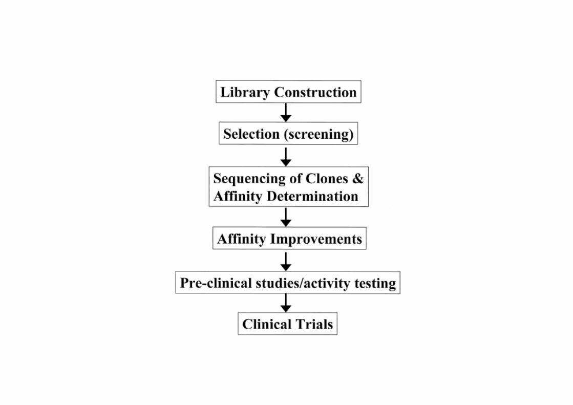
phage display technology - uni- · pdf fileprinciple of the phage display technology . ......
TRANSCRIPT
Phage display technology
Ursula Dietrich
Georg-Speyer-Haus
Georg-Speyer-Haus Institute for Biomedical Research Frankfurt, Germany

Selection technique based on the presentation of peptides or proteins on the surface of bacteriophages. The DNA sequence encoding the peptide is fused to a gene coding for a surface protein of the phage resulting in physical linkage between DNA sequence and peptide sequence. This allows easy cloning of DNA sequences for random peptide libraries into a phage vector and rapid identification of selected peptides by sequencing of the DNA encoding the peptide inserts.
G.P. Smith, 1985
G.P. Smith, 1985
Principle of the phage display technology
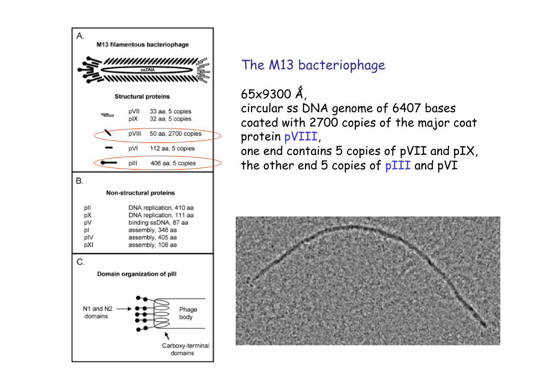
The M13 bacteriophage
65x9300 Ǻ, circular ss DNA genome of 6407 bases coated with 2700 copies of the major coat protein pVIII, one end contains 5 copies of pVII and pIX, the other end 5 copies of pIII and pVI

Infection begins through attachment of the pIII protein to the bacterial F-pilus (bacterial hosts for phage propagation need the F-factor!). After retraction of the F-pilus the pIII protein binds to the bacterial TolA membrane protein. The phage DNA is being transferred into the host cell and converted into a double stranded DNA by bacterial enzymes. With the help of two phage proteins, pII and pX, the replicative dsDNA is amplified through a rolling circle mechanism leading to the accumulation of ssDNA forms. This ssDNA is covered by accumulating pV protein, which then prevents the conversion to dsDNA. At the membrane, a complex of pI, pIV and pXI cover the DNA packaging structure and lead to assembly of the phage particle and its release from the cell in a non-lytic manner. During relase pV is exchanged for pVIII and the minor coat proteins are added.
bact
eria
l en
zym
es
The M13 replication cycle
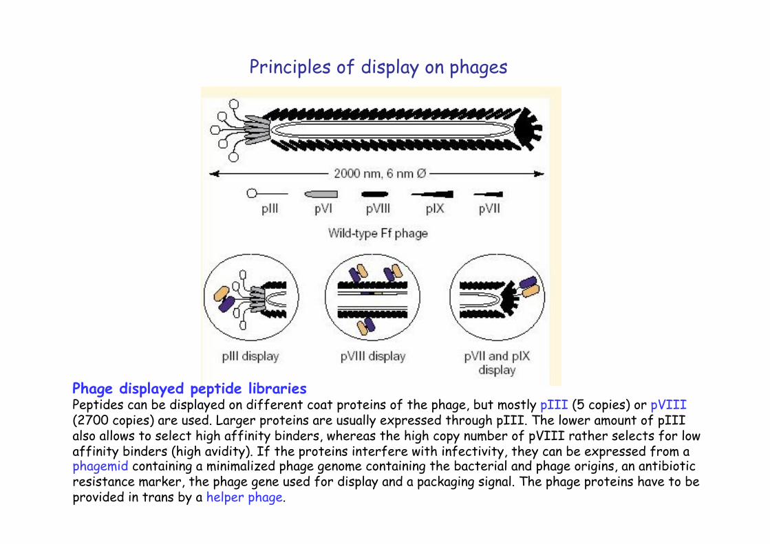
Phage displayed peptide libraries Peptides can be displayed on different coat proteins of the phage, but mostly pIII (5 copies) or pVIII (2700 copies) are used. Larger proteins are usually expressed through pIII. The lower amount of pIII also allows to select high affinity binders, whereas the high copy number of pVIII rather selects for low affinity binders (high avidity). If the proteins interfere with infectivity, they can be expressed from a phagemid containing a minimalized phage genome containing the bacterial and phage origins, an antibiotic resistance marker, the phage gene used for display and a packaging signal. The phage proteins have to be provided in trans by a helper phage.
Principles of display on phages
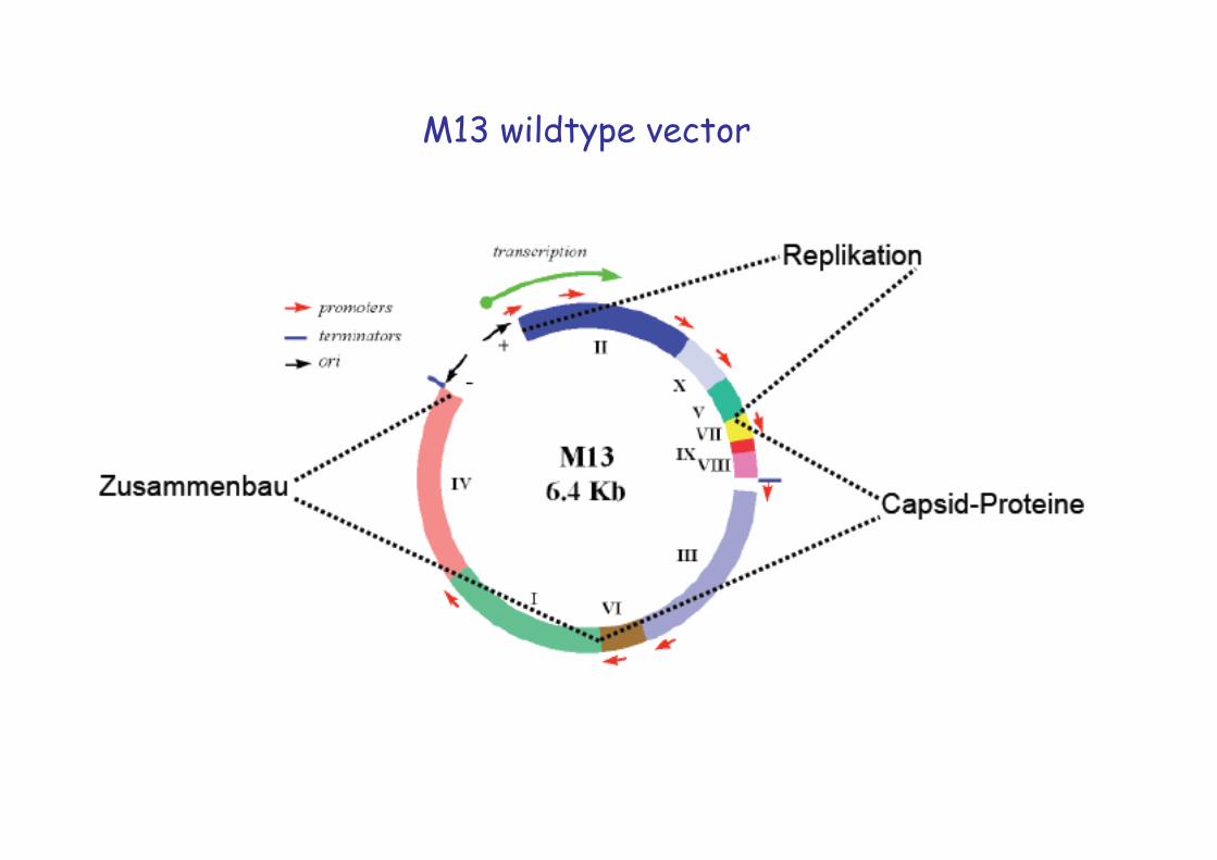
M13 wildtype vector
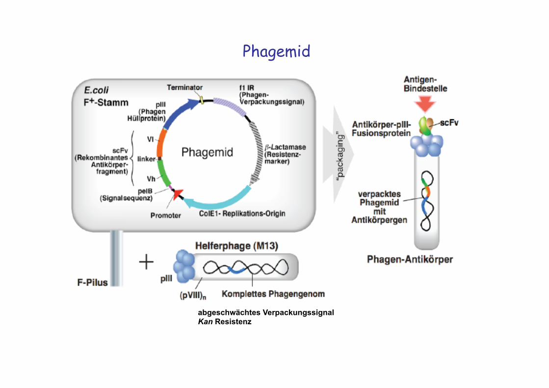
Phagemid
abgeschwächtes Verpackungssignal Kan Resistenz
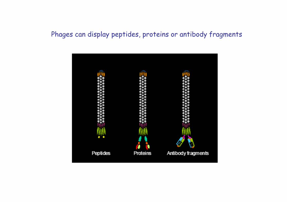
Phages can display peptides, proteins or antibody fragments
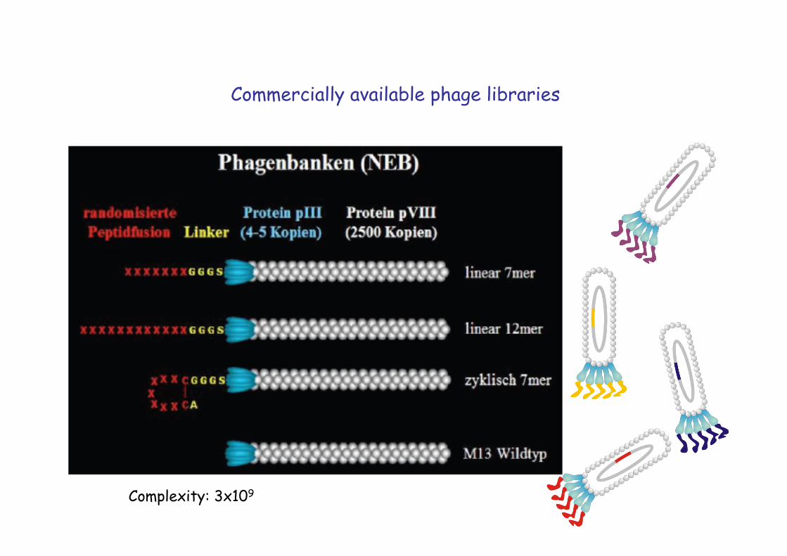
Commercially available phage libraries
Complexity: 3x10⁹
M. Hust et al , Appl Microbiol Biotechnol 2008 �
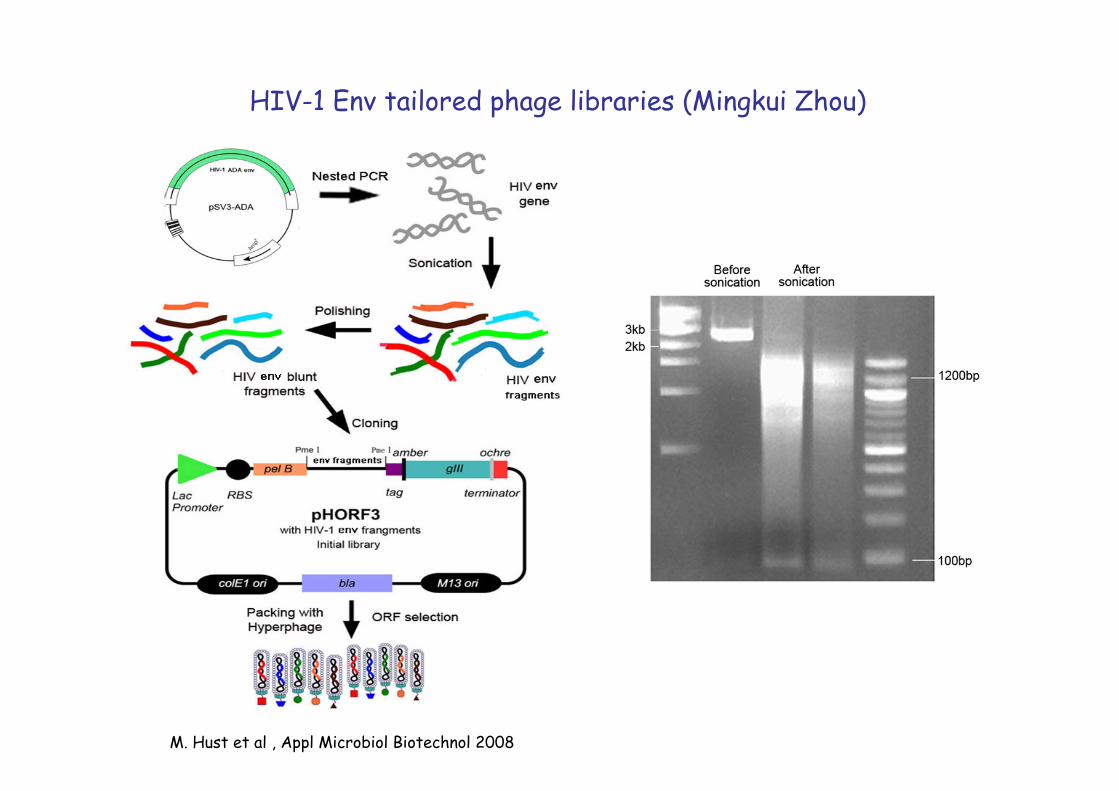
HIV-1 Env tailored phage libraries (Mingkui Zhou)
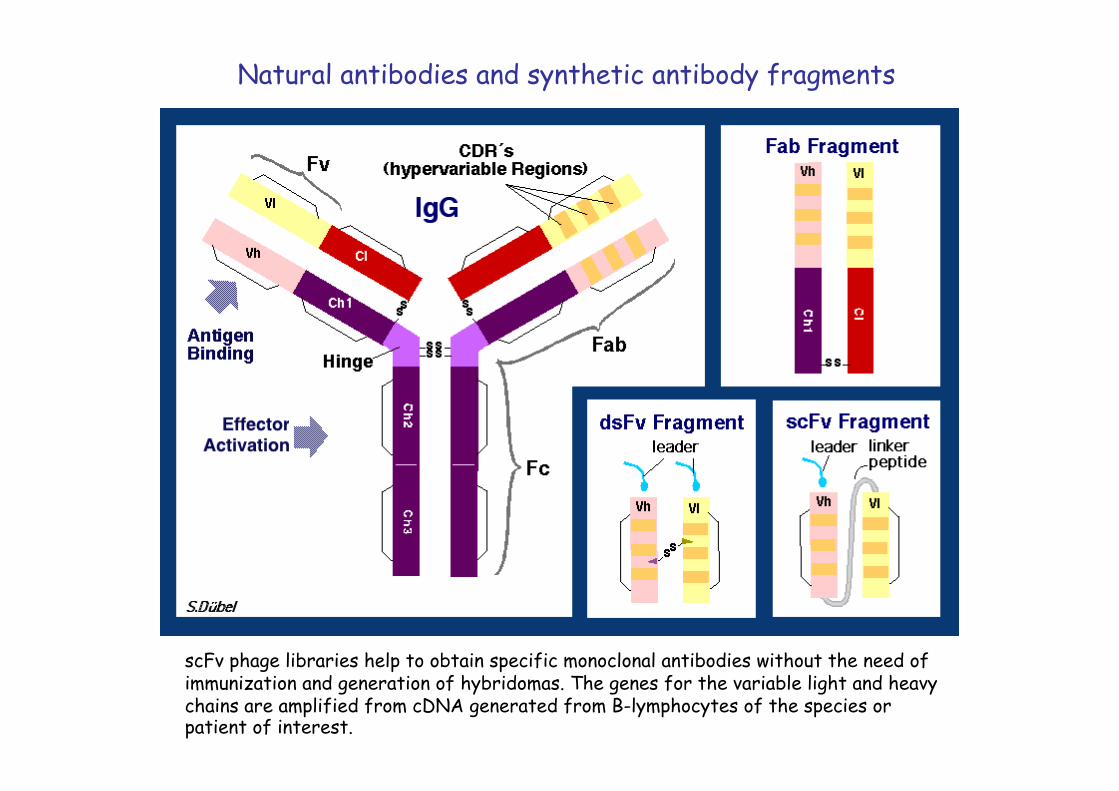
scFv phage libraries help to obtain specific monoclonal antibodies without the need of immunization and generation of hybridomas. The genes for the variable light and heavy chains are amplified from cDNA generated from B-lymphocytes of the species or patient of interest.
Natural antibodies and synthetic antibody fragments
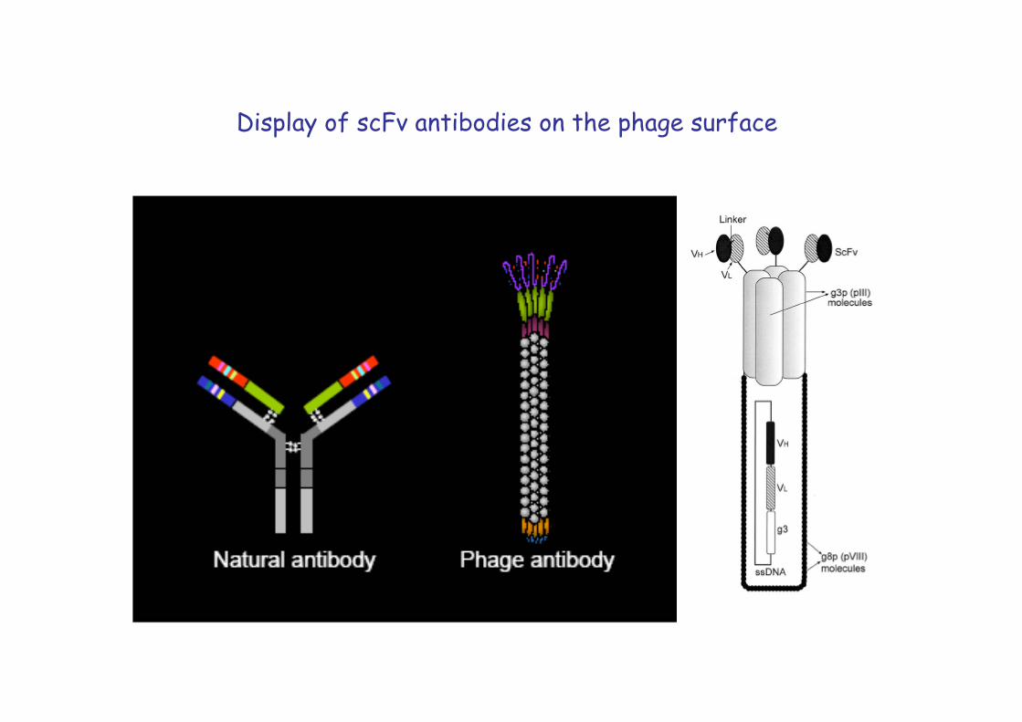
Display of scFv antibodies on the phage surface
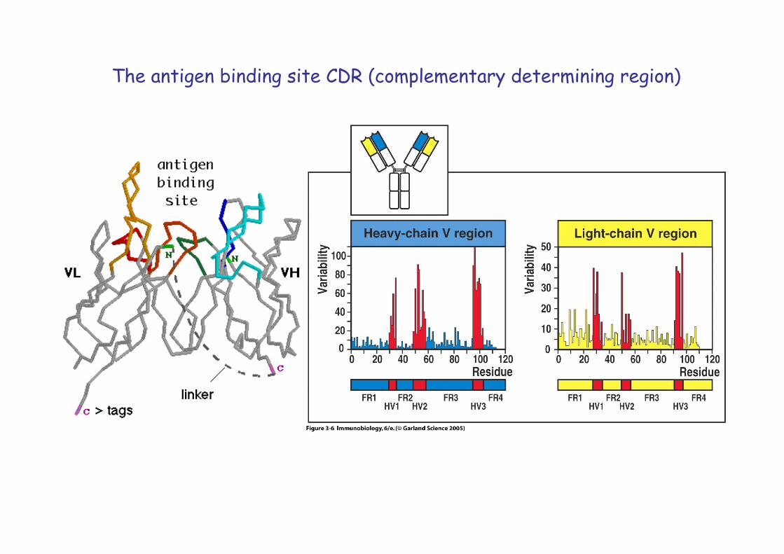
The antigen binding site CDR (complementary determining region)
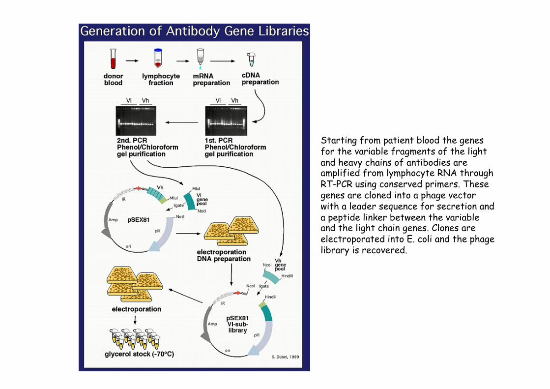
Starting from patient blood the genes for the variable fragments of the light and heavy chains of antibodies are amplified from lymphocyte RNA through RT-PCR using conserved primers. These genes are cloned into a phage vector with a leader sequence for secretion and a peptide linker between the variable and the light chain genes. Clones are electroporated into E. coli and the phage library is recovered.

Biopanning: selecting phages for a specific target
Several rounds of positive and negative selections
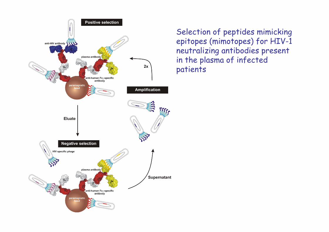
Selection of peptides mimicking epitopes (mimotopes) for HIV-1 neutralizing antibodies present in the plasma of infected patients
E
plasma-ab (HIV+ vs. HIV-)
anti-M13-HRP-ab
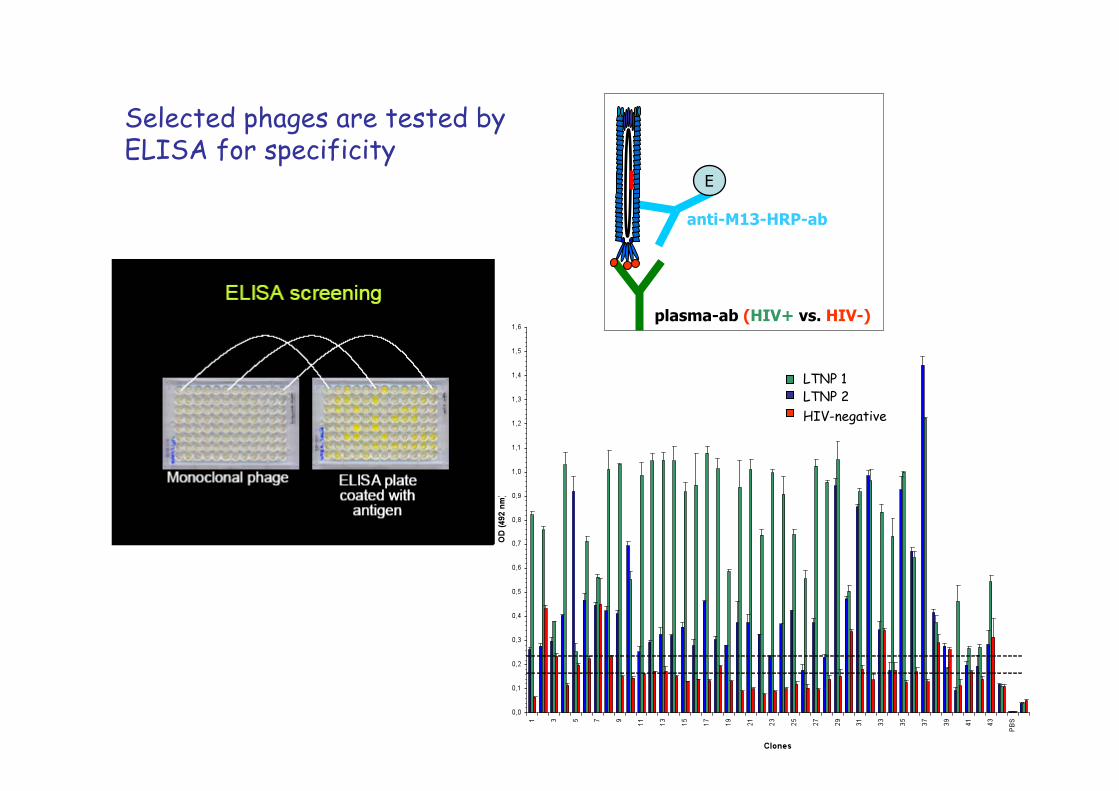
Selected phages are tested by ELISA for specificity
LTNP 1 LTNP 2 HIV-negative

Phage clone 2-7.2 (MH04/01) sequence: LLTTNKD L452 not exposed,L453 recessed overlapping the CD4bs: T283,T455,N280,K282,D279
CD4bs CCR5bs
Linear peptide sequences can be localized as conformational epitopes overlapping the CD4-binding site by 3DEX
Phage clone 12-12.9 sequence: TATPDLTLYYMPG overlapping the CD4bs: T283,A281,T455,D368,P369
3DEX program: Humbert et al., J Comp Chem 2005, 26 (9),879-887
Y Y HIV-1
3A9 5C7
CD4
gp41 gp120
mab 17b
CD4i epitope
CCR5
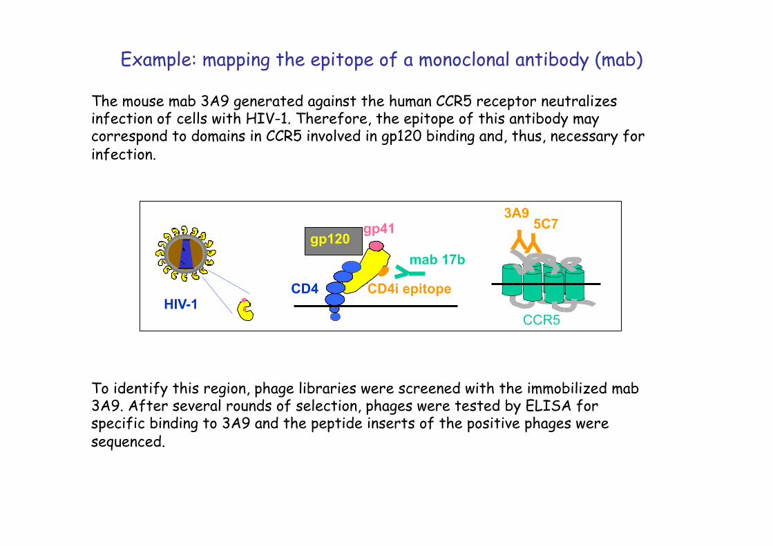
Example: mapping the epitope of a monoclonal antibody (mab)
The mouse mab 3A9 generated against the human CCR5 receptor neutralizes infection of cells with HIV-1. Therefore, the epitope of this antibody may correspond to domains in CCR5 involved in gp120 binding and, thus, necessary for infection.
To identify this region, phage libraries were screened with the immobilized mab 3A9. After several rounds of selection, phages were tested by ELISA for specific binding to 3A9 and the peptide inserts of the positive phages were sequenced.

positive selection α -mouse-MB
negative selection
amplification
2x
elute
mab 3A9
supernatant
mu IgG
3. positive selection
titering of phages
E
anti-M13-ab anti -mouse HRP
pick single phage clones
positive clones ss-DNA preparation
and sequencing
capture ELISA
Biopanning: Selection phages binding Mab 3A9

3A9 mab 5C7 mab N-terminus
C-terminus
S I Y
D F G N-terminus
C-terminus
P H W D L R
3A9: C-HASIYDFGS-C 5C7: C-PHWLRDLRV-C 3A9 and 5C7 mimotopes selected from phage display libraries
Phagotopes selected with HIV-1 neutralizing mabs 3A9 and 5C7 can be aligned as conformational eptiopes to extracellular domains of CCR5
A dominant C-HASIYDFGS-C peptide was selected, which could be aligned to extracellular domains of CCR5.
This discontinous epitope mimics a region essential for HIV-1 infection, as exchanges of the corresponding amino acid codons for alanine in the CCR5 cDNA result in drastically reduced infection of cells transfected with the mutant cDNAs.
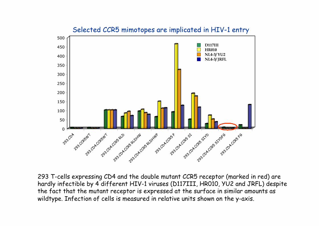
293 T-cells expressing CD4 and the double mutant CCR5 receptor (marked in red) are hardly infectible by 4 different HIV-1 viruses (D117III, HR010, YU2 and JRFL) despite the fact that the mutant receptor is expressed at the surface in similar amounts as wildtype. Infection of cells is measured in relative units shown on the y-axis.
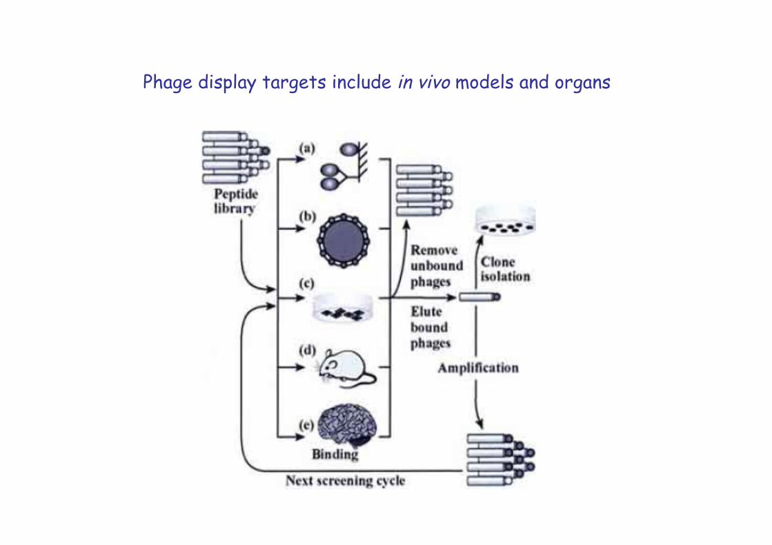
Phage display targets include in vivo models and organs

Tumor-specific phages can be selected in vivo in mice by performing first a negative selection in a normal mouse followed by phage recovery from the blood, and subsequent positive selections with the recovered phages in tumor bearing mice
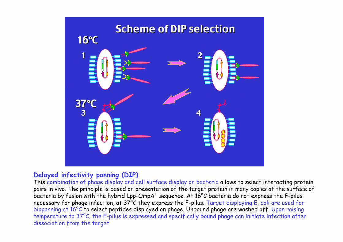
Delayed infectivity panning (DIP) This combination of phage display and cell surface display on bacteria allows to select interacting protein pairs in vivo. The principle is based on presentation of the target protein in many copies at the surface of bacteria by fusion with the hybrid Lpp-OmpA´ sequence. At 16°C bacteria do not express the F-pilus necessary for phage infection, at 37°C they express the F-pilus. Target displaying E. coli are used for biopanning at 16°C to select peptides displayed on phage. Unbound phage are washed off. Upon raising temperature to 37°C, the F-pilus is expressed and specifically bound phage can initiate infection after dissociation from the target.

Mirror image phage display
Natural proteins are composed of L-amino acids. In view of therapeutic applications of the selected peptides, D-peptides are preferable as they are not cleaved by natural proteases and are not immuno-genic. To obtain D-peptides binding to an L-target, the D-form of the L-target is first synthesized. This D-form mirrors the original L-form. Then L-peptides are selected from the biological phage libraries with the D-form as target and D-peptides are synthesized from the selected L-peptides.