design and characterization of liposomes containing long-chain n-acylpes for brain delivery:...
TRANSCRIPT
Design and Characterization ofLiposomes Containing Long-ChainN-AcylPEs for Brain Delivery:Penetration of LiposomesIncorporating GM1 into the Rat Brain
Margarita Mora,1 Maria-Luisa Sagristá,1
Domenico Trombetta,2 Francesco P. Bonina,3
Anna De Pasquale,2 and Antonella Saija2,4
Received June 12, 2002; accepted July 1, 2002
Purpose. To develop a suitable liposomal carrier to encapsulate neu-roactive compounds that are stable enough to carry them to the brainacross the blood-brain barrier with the appropriate surface characteris-tics for an effective targeting and for an active membrane transport.Methods. Liposomes containing glycosides and a fusogenic lipid wereprepared by extrusion. Photon correlation spectroscopy, fluorescencespectroscopy, and differential scanning calorimetry were used tocharacterize liposomal preparations. Tissue distribution was deter-mined by using 3H-cholesterylhexadecylether as a marker.Results. The incorporation of glycoside determinants and N-palmitoylphosphatidylethanolamine gives liposomes with similar ini-tial size, trapped volume, negative surface charge, bilayer fluidity, andmelting temperature, except for monosialoganglioside-containing li-posomes, which showed less negative surface charge and the highestsize, trapped volume and melting temperature. All glycosilated for-mulations gave liposomes able to retain up to the 95% of encapsu-lated carboxyfluorescein after 90 min at physiologic temperatureeven in the presence of serum. Monosialoganglioside liposomes wererecovered in the cortex, basal ganglia, and mesencephalon of bothbrain hemispheres. The liver uptake was higher for sulfatide- andglucose-liposomes, whereas the higher blood levels were observed forglucose- and mannose-liposomes.
Conclusions. These results show the suitability of such liposomalformulations to hold encapsulated drugs. Moreover, the brain uptakeof monosialoganglioside liposomes makes them good candidates asdrug delivery systems to the brain.
KEY WORDS: liposomes; N-acylPEs; brain delivery; permeability;serum.
INTRODUCTION
The blood-brain barrier (BBB) restricts the brain uptakeof many valuable hydrophilic drugs and limits their efficacy inthe treatment of brain diseases because of the presence oftight junctions, high metabolic capacity, low pinocytic vesicu-lar traffic, and efficient efflux mechanisms. The lipophilic na-ture of the BBB permits only small lipid-soluble drugs to passthrough this barrier (1) and enter the brain via diffusion.Some essential compounds, such as amino acids (2), glucose(3), and iron transferrin (4), need specific carriers to permeateinto the brain, and several saturable transport systems havealso been reported for peptides (5). Therefore, different con-cepts are required to develop systems to facilitate the trans-port of poorly permeable drugs across the BBB for effectivemanagement of brain disorders, and different drug deliverystrategies have been proposed to improve drug delivery to thebrain (6).
In this way, colloidal carriers, such as nanoparticles andliposomes, which may take advantage of the biochemicaltransport systems that are also present in the BBB, have beenconsidered (7). However, one of the main problems in thetargeted drug delivery is the rapid opsonization and uptake ofthe injected carrier systems by the reticuloendothelial system,by macrophages in liver and spleen. This problem can beovercome by the use of ideal liposome (or nanoparticle) for-mulations containing the following features: stability in theblood, controlled circulation lifetime, disease site localizationand target cell-specific binding and delivery. Liposomes areversatile drug delivery carriers that have proven to be usefulin reducing toxicity and enhancing the activity of a variety ofpharmacologically active compounds. High phase-transitionlipids, high cholesterol content, and small percentages of com-ponents, such as monosialoganglioside (GM1), phosphatidylinositol, or polyethyleneglycol, normally comprise long-circulating liposomes. The modification of the liposomal sur-face with hydrophilic molecules increases the blood circula-tion time of the particulate by reducing the binding of plasmaproteins (8).
Our work has focused on developing a suitable liposomalcarrier to encapsulate neuroactive compounds that are stableenough to carry them to the brain (sterically stabilized lipo-somes) with the appropriate surface characteristics for an ef-fective targeting and for an active membrane transport acrossthe BBB (site-specific delivery carriers). In this way, it hasbeen reported that chemical conjugation of potentially centralnervous system (CNS)-active drugs with tyrosine or glucose(9), the addition of sugar moieties to bioactive peptides (10),and the use of saccharide determinants (11) represents a suc-cessful means of improving their brain delivery. Mannose-labeled liposomes are efficiently incorporated into the mousebrain, lysosomes, and glial cells, suggesting that mannose canbe recognized by the cells of the BBB (12).
To achieve an efficient transfer of the liposomal contentinto the cells, we have considered the preparation of lipo-
1 Deparment of Biochemistry and Molecular Biology, Faculty ofChemistry, University of Barcelona, Martı i Franques 1, 08028-Barcelona, Spain
2 Department Farmaco-Biologico, School of Pharmacy, University ofMessina, Contrada Annunziata, 98168-Messina, Italy
3 Department of Pharmaceutical Sciences, Faculty of Pharmacy, Uni-versity of Catania, Viale A. Doria 6, 95125-Catania, Italy
4 To whom correspondence should be addressed. (e-mail:[email protected])
ABBREVIATIONS: BBB, blood brain barrier; CF, carboxyfluores-cein; Chol, cholesterol; CNS, central nervous system; DDSs, drugdelivery systems; DMPC, dimyristoylphosphatidylcholine; DPH, 1,6-diphenyl-1,3,5-hexatriene; DSC, differential scanning calorimetry;EPC, egg phosphatidylcholine; EPE, egg phosphatidylethanolamine;GLU, p-aminophenyl-�-D-gluco-pyranoside; GM1, monosialogan-glioside; IUVs, intermediate unilamellar vesicles; MAN, p-aminophe-nyl-�-D-manno-pyranoside; MLVs, multilamellar vesicles; N-acylPEs,N-acylphosphatidylethanolamines; NPPE, N-palmitoylphosphati-dylethanolamine; p, polarization; PBS, phosphate-buffered saline;PCS, photon correlation spectroscopy; RES, reticuloendothelial sys-tem; SUL, sulfatide; Tm, main transition temperature; TMA-DPH,1-(4-trimethylammoniumphenyl)-6-phenyl-1,3,5-hexatriene p-toluensulfonate; �Hcal, calorimetric enthalpy; �Scal, calorimetric en-tropy; �T1/2 , width at half peak height.
Pharmaceutical Research, Vol. 19, No. 10, October 2002 (© 2002) Research Paper
14300724-8741/02/1000-1430/0 © 2002 Plenum Publishing Corporation

somes containing N-acylphosphatidylethanolamines (N-acylPEs). Fusogenic liposomes can be prepared by using fu-sogenic lipids (13), by conjugation of fusogenic molecules toliposome membranes (14), or by incorporation of viral fusionproteins (15) or fusion peptides (16) to bilayers. We previ-ously showed the capacity of liposomes containing N-acylPEsto fuse in the presence of either monovalent or divalent cat-ions (17) and that the incorporation of N-acylPEs into eggphosphatidylcholine (EPC) liposomes decreases their perme-ability (18) and stabilizes them against leakage in the pres-ence of serum (19).
This article analyzes the effect of the incorporation ofGM1 or sulfatide (to reduce liposome uptake by the RES)and p-aminophenyl-�-D-mannopyranoside or p-aminophe-nyl-�-D-glucopyranoside (as recognition markers), in the ef-ficiency of N-acylPE containing intermediate unilamellarvesicles (IUVs) as a drug delivery system (DDS) to the ratbrain.
MATERIALS AND METHODS
Materials
Synthetic L-�-dimyristoylphosphatidylcholine (DMPC),transphosphatidylated egg phosphatidylethanolamine (EPE),and cholesterol (Chol) were purchased from Avanti PolarLipids (Birmingham, AL, USA). Bovine brain monosialogan-glioside (GM1) and sulfatide (SUL), p-aminophenyl-�-D-manno-pyranoside (MAN), p-aminophenyl-�-D-gluco-pyranoside (GLU), rat serum, sodium phosphate monobasicand dibasic were from Sigma Chemical Co. (St. Louis, MO,USA). Palmitoyl chloride for synthesis was from Merck(Schuchardt, Germany), Triton X-100 from Merck (Darm-stadt, Germany), 6(5)-carboxyfluorescein (CF) from East-man-Kodak (Rochester, NY, USA) and Sephadex G-50and Sephadex LH-20 from Pharmacia Biotech (Uppsala,Sweden). 1,6-Diphenyl-1,3,5-hexatriene (DPH) and its am-monium salt (TMA-DPH) were obtained from MolecularProbes (Eugene, OR, USA). 3H-cholesterylhexadecylether(50 Ci/mmol) was from NEN (Boston, MA, USA). All re-agents were analytical grade and Milli-Q water (MilliporeBedford, MA, USA, resistivity of 18 M� · cm) was used.
Synthesis and Characterization ofN-Palmitoylphosphatidylethanolamine
N-palmitoylphosphatidylethanolamine (NPPE) was syn-thesized by condensing palmitoyl chloride with purified trans-phosphatidylated EPE as described (18). The product waspurified by silicic acid column chromatography and prepara-tive TLC and characterized by proton-NMR and IR-spectroscopy (18). Lipid concentrations were determined byphosphorus analysis (20).
Vesicle Preparation
IUVs were prepared by extrusion following a standardprocedure (21). Multilamellar vesicles (MLVs) were preparedby hydration of dried lipid films, containing a fixed molarratio of DMPC, Chol, and NPPE and a fixed amount of GLU,MAN, GM1, or SUL. These components were mixed at thefollowing molar ratios: DMPC/Chol/NPPE (8:3:4), DMPC/Chol/NPPE/GLU (8:3:4:1.5), DMPC/Chol/NPPE/MAN (8:3:4:1.5), DMPC/Chol/NPPE/GM1 (8:3:4:1.5), and DMPC/Chol/
NPPE/SUL (8:3:4:1.5), to prepare the lipid films. The driedlipid film was hydrated with 10 mM phosphate buffer (pH 7.4)containing 140 mM NaCl, at a concentration of 1 mg lipid/mLfor 10 min at 50°C, and the lipid dispersion was frozen andthawed (five times) and sonicated (bath sonicator, 60 min,50°C). IUVs were prepared by passing repeatedly (10 timesfor each membrane) the MLV dispersion under pressurethrough different pore-sized polycarbonate membranes (Nu-cleopore; 400-, 200-, and 100-nm pore sizes) in an extrusiondevice from Lipex Biomembranes Inc. (Vancouver, Canada).When required, liposomes were prepared at a concentrationof 20 mg lipid/mL and were labeled with trace amounts of3H-cholesterylhexadecylether. The final concentration of themarker in the liposomal suspension was 10 �Ci/mL.
Vesicle Size Analysis
The size and size distribution of unilamellar vesicles weredetermined by photon correlation spectroscopy (PCS). APCS41 size analyzer (Malvern Autosizer IIc, Malvern Instru-ments, Malvern, Worcestershire, UK) and a 5-mW He-Nelaser (Spectra Physics, Darmstadt, Hessen, Germany), at anexcitation wavelength of 633 nm, were used. Data were col-lected with a Malvern 7032N 72 data channel correlator, andthe mean hydrodynamic diameter was calculated from a cu-mulant analysis of the intensity autocorrelation function. Be-fore measuring, vesicle dispersions were appropriately dilutedto avoid multiple scattering. The influence of such dilution onsize measurement was previously proved to be nonsignificant(unpublished results).
�-Potential Measurement
�-potential measurements were performed on MLV sus-pensions in 10 mM phosphate buffer (pH 7.4) containing 140mM NaCl, by laser-Doppler anemometzy in a ZetaSizer 4(Malvern Instruments). The optic unit contained a 5-mW He-lium-Neon laser, a ZET5104 electrophoresis cell (4-nm diam-eter quartz capillary), a sample handling unit, a multi-8 bitcorrelator with 72 data channels, and 7 monitor channels withvariable time expansion. The method uses the autocorrelationfunction of the light scattered in colloid solution measured bya photon-counting system. The measurements were per-formed at 25°C on MLV suspensions (0.5 mg/mL). To checkthe Malvern device, a carboxy-modified polystyrene latexsample, with a �-potential of −55 mV at 25°C, was used beforeeach set of measurements.
Determination of the Entrapped Volume
The volume of the aqueous space of IUVs was deter-mined by measuring the fluorescence at 520 nm of an aliquotof the IUVs suspension, free of non-encapsulated CF. Thesample (80 �L) was diluted in 2 mL of 10 mM PBS (pH 7.4),in the presence of 150 �L of a 10% Triton X-100 solution. CFconcentration was determined by comparison with a standardcurve. Lipid concentration was quantified by phosphorusanalysis using ammonium ferrothiocyanate (22).
Calorimetric Measurements
Calorimetric measurements were performed by a MettlerDSC-30 differential scanning calorimeter. The temperaturescale was calibrated by indium, undecan, and water, and thetransitional enthalpies were calibrated with indium. The tem-perature of the maximum of the transition endotherm (Tm)
NPPE-Containing Liposomes for Brain Delivery 1431

and the enthalpy (�Hcal) were determined with a MettlerTC10A TA processor. The processor analyzes the heat flowcurves by integration of the area under the peak over a dy-namic baseline type X8. The cooperativity of the transitionwas evaluated, in an approximate manner, from the widths athalf-peak heights (°C) of the main transition endotherms(�T1/2) due the heterogeneous chain composition of lipids.The pellet (100 �L) (∼7 mg of lipid), obtained by centrifuga-tion of the MLV dispersion, was transferred to a 160-�Laluminum sample pan. Several heating and cooling cycleswere performed to equilibrate the samples before to run thedefinitive calorimetric scans. The heating rate was 2°C min−1,and only the heating scans were analyzed (10–60°C). A 140-�L aluminum pan, filled with 100 �L of 10 mM PBS (pH 7.4),was used as reference. After calorimetric scans, the phospho-lipid amount was determined by the method of Stewart (22).
Determination of Membrane Fluidity
Membrane fluidity was determined by fluorescence po-larization measurements of the fluorescent probes DPH andTMA-DPH. The incorporation of the fluorophores was per-formed during the annealing process by adding 2 �L of a 2mM tetrahydrofurane DPH or TMA-DPH solution to 3 mLof the preformed IUVs (0.35 mg/mL) to get a lipid-probemolar ratio of 375:1. The suspension was incubated in thedark (60 min) at T > Tm of the lipids, with gentle stirring andfiltered (0.45-�m pore-membrane filter), before the assay.Excitation and emission wavelengths were 360 and 430 nm,respectively. Measures were performed at different tempera-tures (20–60°C), in a Kontron SFM 25 spectrofluorimeter(optical arrangement in the “L” format). The sample wasexcited with vertically polarized light, and fluorescence inten-sities were recorded with the analyzing polarizer oriented par-allel (IVV) and perpendicular (IVH) to the excitation polar-izer. The polarization, p, was calculated from:
p =Ivv − IVH
Ivv + GIVH
where G is an instrumental correction factor. Samples wereilluminated by using narrow slits for the shortest time possibleand were maintained in the dark between two consecutivemeasures to prevent decreases in fluorescence intensity dueto the formation of dark isomers of the probes.
Permeability Experiments
Liposome permeability was examined by the fluores-cence technique using CF described by Weinstein et al. (23).The dye was purified by Sephadex LH-20 column chromatog-raphy and acid precipitation (pH 4.5) (24). IUVs were pre-pared as described above, but with a 10 mM phosphate-buffered solution (PBS) (pH 7.4) containing 0.82% NaCl and50 mM carboxyfluorescein. Non-encapsulated dye was re-moved by Sephadex G-50 column chromatography (20 × 1cm). The composition of eluted liposomes was analyzed (20)and found to be the same as that of the film used for theirpreparation. CF release was monitored on a Kontron SFM25spectrofluorimeter by using excitation and emission wave-lengths of 492 and 520 nm, respectively. Small aliquots of theeluted liposomes (80 �L, 100 �g lipid/mL) were added toeach cuvette (total volume 2 mL), and the fluorescence was
measured for 90 min. A 0.416 �M CF solution was used tocalibrate the 100% fluorescence value. The permeability ex-periments were performed at 30, 37, 42, and 47°C, in theabsence and in the presence of 12.5 or 125 �L rat serum/mLof aqueous volume in the cuvette. The cuvettes were main-tained at the assay temperature by a circulating water bath.The rates of CF leakage are expressed as the percentage oftotal CF released:
%CFreleased = � Ft − F0
FT − F0� × 100
where Ft is the fluorescence intensity at a specified time, F0 isthe fluorescence at zero time, and FT is the total fluorescenceobtained by adding 150 �L of a 10% (v/v) Triton X-100(Merk) solution. FT was corrected for the dilution introduc-ed by the addition of Triton. Incubation of liposomes withhigher concentration of Triton did not affect the value of FT,indicating that the release of dye from the liposomes wascomplete.
Tissue Distribution Studies
The experiments were performed on male adult Sprague-Dawley rats (320–350 g body weight; Charles River, Milan,Italy). The animals were maintained under normal controlledlighting and temperature conditions and allowed free accessto food and water until used.
On the day of the experiment, the animals were anaes-thetized with urethane (1.2 g/kg intraperitoneally; injectionvolume: 0.5 mL/100 g body weight). Short polyethylene cath-eters were inserted in the left common carotid artery for ret-rograde injection of liposome suspension and in the left femo-ral artery for blood sampling (25). Ten minutes after comple-tion of surgical procedures, an aliquot of liposome suspension(20 mg lipid/mL) containing 3H-cholesterylhexadecylether (2�Ci/200 �L/rat) was injected through the carotid artery. Atthe end of the experimental period (10 min), a large bloodvolume was collected in polyethylene tubes, to measure thewhole-blood isotope concentration, and, immediately after,the animals were killed by rapid i.v. injection of 1 mL ofsaturated KCl solution. The brain and the liver were rapidlyremoved; cerebral tissue specimens (cortex, mesencephalon,and basal ganglia) were dissected from each hemisphere.During the experiment, hematocrit values were monitored.
Tissue samples were placed in preweighed vials that wereimmediately reweighed. Sample solubilization was accom-plished by adding 1 mL of Soluene 300 (Packard) and incu-bating the vials at 60°C overnight. Each vial was then filledwith 5 mL of Hionic-Fluor (Packard). A 20-�L whole-bloodsample was digested in 0.5 mL of Soluene 300 and counted in6 mL of Hionic Fluor acidified with 0.5 mL of 0.5 M HCl.Beta-counting was performed by a Packard TRI-CARB2100TR liquid scintillation counter. Single sample quenchingwas monitored by the external standard method. Samplecounts were corrected for background and quenching.
Results of in vivo experiments are expressed as means ±SD and were analyzed for statistical significance by Student’st test for unpaired data. Statistical significance was acceptedwhere p < 0.05.
Mora et al.1432

RESULTS AND DISCUSSION
Characterization of Liposomes ContainingGlycoside Determinants
Physical size and surface potential of liposomes, quantityof entrapped solutes, thermodynamic parameters, and bilayerfluidity were determined to characterize the liposomal sys-tems. The size and fluidity effects of liposome on their bio-distribution and accumulation in cancer tissues after an i.v.administration has been shown (26), and the knowledge ofthese parameters is necessary for evaluating the results ofexperiments using phospholipid vesicles.
Liposomes, prepared as for permeability measurements,were analyzed for size and size distribution by PCS. The meanhydrodynamic diameter (Table I) of control liposomes didnot change with the incorporation of p-aminophenyl-�-D-gluco-pyranoside and sulfatide, whereas 7 and 15% size in-creases were observed with p-aminophenyl-�-D-manno-pyranoside and monosialoganglioside, respectively. The poly-dispersity index was always < 0.2, being the mosthomogeneous suspension that contains aminophenyl-�-D-manno-pyranoside.
To assess the stability of the liposomal suspensions, theirsize and size distribution were also measured after 3 weeks ofstorage. The results (Fig. 1) show a small size increase for allthe dispersions during the first week, but an important sizeincrease for liposomes containing aminophenyl-�-D-gluco-pyranoside, aminophenyl-�-D-manno-pyranoside, and mono-sialoganglioside after 3 weeks of storage. The instability ofglucose- and mannose-containing liposomes can be attributedto some undesirable interaction between the aminophenyl-carbohydrate and other bilayer components because DPPC/Chol/NPPE liposomes are more stable. Note that Umezawaand Eto (12) described the preparation of lecithin/cholesterol/aminophenyl-�-D-manno-pyranoside (7:2:1) liposomes butdid not indicate the stability of the liposomal systems. Theinstability of the liposomes containing monosialogangliosidecan be explained by the fact that its incorporation into NPPE-containing bilayers may introduce significant structuralchanges, as revealed in our previous studies on bilayer ar-rangement by 31P-NMR (27), showing the decrease in thefluidity of the bilayers after GM1 incorporation.
The �-potential values for MLVs are summarized inTable I. The negative value of the �- potential obtained forDPPC/Chol/NPPE liposomes is in agreement with the anionicnature of NPPE, due to the substitution of the amine group of
EPE by an amide bond in NPPE. The incorporation of theaminophenyl-carbohydrates into the bilayer did not modifythe �-potential, as could be expected for the uncharged natureof the carbohydrates. Despite the presence of a negative sul-fate group in sulfatide, at the pH of the medium, their incor-poration into the bilayer did not modify the �-potential. Thedecrease in the negative charge of liposomes with the additionof monosialoganglioside (with a negative sialic acid group)could be explained by the shielding of the negative charge ofthe polar head of NPPE by the voluminous polar head of theganglioside.
The aqueous space volume (Table I) increased with theaddition of monosialoganglioside to the liposomal bilayers,but this parameter was not modified with the incorporation of
Table I. Characteristic Parameters of the Aqueous Dispersions of Liposomes Containing Glycoside Determinants
Sample composition(molar ratio) DH
a (nm) Polydispersity indexb �-Potentialc (mV)Trapped volumed
(�l/�mol)
DMPC/Chol/NPPE (8:3:4) 106.3 ± 1.5 0.173 ± 0.015 −27.5 ± 0.9 1.40 ± 0.08DMPC/Chol/NPPE/GLU (8:3:4:1.5) 105.4 ± 12.3 0.102 ± 0.098 −27.8 ± 0.8 1.49 ± 0.15DMPC/Chol/NPPE/MAN (8:3:4:1.5) 113.3 ± 9.1 0.059 ± 0.003 −28.2 ± 1.3 1.40 ± 0.13DMPC/Chol/NPPE/GM1 (8:3:4:1.5) 121.5 ± 10.9 0.144 ± 0.010 −15.5 ± 0.6 2.17 ± 0.29DMPC/Chol/NPPE/SUL (8:3:4:1.5) 102.8 ± 9.7 0.119 ± 0.010 −27.4 ± 0.7 1.48 ± 0.09
a Diameter calculated from dynamic light-scattering data expressed as Z average mean.b Indicates the homogeneity of liposomal suspensions.c Determined by laser-Doppler anemometry.d Determined by carboxyfluorescein dye retention and expressed as �L CF 50 mM/�mol lipid. Data are expressed as means ± SD of three
independent experiments.
Fig. 1. Effect of time storage in liposome size. The vesicle diameterwas measured by dynamic light scattering immediately after lipo-somes preparation and during the first 3 weeks of storage. Liposomeswere prepared by using: (�) DMPC/Chol/NPPE (8:3:4), (�) DMPC/Chol/NPPE/GLU (8:3:4:1.5), (�) DMPC/Chol/NPPE/MAN (8:3:4:1.5), (�) DMPC/Chol/NPPE/GM1 (8:3:4:1.5), and (�) DMPC/Chol/NPPE/SUL (8:3:4:1.5). Total lipid concentration was in all cases 0.1mg/mL. The values in the plots correspond to the mean values ofthree independent experiments. The coefficients of variation rangedfrom 2 to 15%.
NPPE-Containing Liposomes for Brain Delivery 1433
the other glycoside determinants. This result is in agreementwith the greater size for DPPC/Chol/NPPE/GM1 liposomes incomparison with DPPC/Chol/NPPE, DPPC/Chol/NPPE/GLU, and DPPC/Chol/NPPE/SUL liposomes, which showeda similar size by photon correlation spectroscopy. The great-est size and aqueous space volume of DPPC/Chol/NPPE/GM1 liposomes could be the result of the increased bilayerrigidity due to the incorporation of GM1 into NPPE-containing bilayers and the resulting steric hindrance to giveliposomes with high curvature radium. The same entrappedvolume for DPPC/Chol/NPPE/MAN and control liposomescould be explained by the lesser degree of heterogeneity ofthe liposomes containing aminophenyl-mannose.
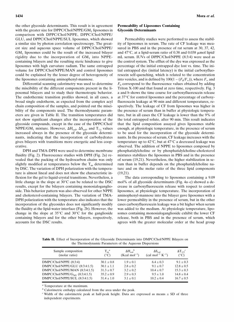
Differential scanning calorimetry was used to determinethe miscibility of the different components present in the li-posomal bilayers and to study their thermotropic behavior.The endothermic transition profiles showed, in all cases, abroad single endotherm, as expected from the complex acylchain composition of the samples, and pointed out the misci-bility of the components used. The thermodynamic param-eters are given in Table II. The transition temperatures didnot show significant changes after the incorporation of theglycoside compounds, except in the case of the DPPC/Chol/NPPE/GM1 mixture. However, �Hcal, �Scal, and T1/2 valuesincreased always in the presence of the glycoside determi-nants, indicating that the incorporation of carbohydratesgives bilayers with transitions more energetic and less coop-erative.
DPH and TMA-DPH were used to determine membranefluidity (Fig. 2). Fluorescence studies with DPH (Fig. 2a) re-vealed that the packing of the hydrocarbon chains was onlyslightly modified at temperatures below the Tm determinedby DSC. The variation of DPH polarization with the tempera-ture is almost lineal and does not show the characteristic in-flexion for the gel to liquid-crystal transitions. Nevertheless, alittle change in the slope at 30°C can be related to the DSCresults, except for the bilayers containing monosialoganglio-side. This behavior pattern was also observed for other NPPEand cholesterol-containing bilayers. The variation of TMA-DPH polarization with the temperature also indicates that theincorporation of the glycosides does not significantly modifythe fluidity at the lipid-water interface (Fig. 2b). However, thechange in the slope at 35°C and 30°C for the gangliosidecontaining bilayers and for the other bilayers, respectively,accounts for the DSC results.
Permeability of Liposomes ContainingGlycoside Determinants
Permeability studies were performed to assess the stabil-ity of liposomes in serum. The rate of CF leakage was mea-sured in PBS and in the presence of rat serum at 30, 37, 42,and 47°C, at a lipid-serum ratio of 0.38 and 0.038 �mol lipid/mL serum. IUVs of DPPC/Chol/NPPE (8:3:4) were used asthe control system. The efflux of the dye was expressed as thepercentage of the initial entrapped dye lost vs. time. The ini-tial entrapped dye (initial latency) is the initial carboxyfluo-rescein self-quenching, which is related to the concentrationinto vesicles, and is defined by 100(1 − (F0/F�)), where F� andF0 correspond to the fluorescence values obtained by addingTriton X-100 and that found at zero time, respectively. Fig. 3a and b shows the time course for carboxyfluorescein releaseat 37°C for control liposomes and the percentage of carboxy-fluorescein leakage at 90 min and different temperatures, re-spectively. The leakage of CF from liposomes was higher inthe presence of serum than in buffer at physiologic tempera-ture, but in all cases the CF leakage is lower than the 9% ofthe total entrapped solute, after 90 min. This result indicatesthat the lipid composition assayed gives liposomes stableenough, at physiologic temperature, in the presence of serumto be used for the incorporation of the glycoside determi-nants. In the presence of serum, CF leakage increases with thetemperature up to 42°C, but at 47°C a decreased leakage wasobserved. The addition of NPPE to liposomes composed byphosphatidylcholine or by phosphatidylcholine-cholesterolmixtures stabilizes the liposomes in PBS and in the presenceof serum (19,21). Nevertheless, the higher stabilization in se-rum than in buffer depends on the phosphatidylcholine na-ture and on the molar ratio of the three lipid components(19,21).
The data corresponding to liposomes containing a 9.09mol% of all glycoside determinants (Fig. 4a–c) showed a de-crease in carboxyfluorescein release with respect to controlliposomes, at physiologic temperature. The incorporation ofaminophenyl-mannose into the bilayer gave liposomes with alower permeability in the presence of serum, but in the othercases carboxyfluorescein leakage was a bit higher when serumwas added to the medium. At physiologic temperature, lipo-somes containing monosialoganglioside exhibit the lower CFrelease, both in PBS and in the presence of serum, whichagrees with the greater molecular order at the head group
Table II. Effect of Incorporation of the Glycoside Determinants into DMPC/Chol/NPPE Bilayers onthe Thermodynamic Parameters of the Aqueous Dispersions
Sample composition(molar ratio)
Tma
(°C)�Hcal
b
(Kcal mol−1)�Scal
(cal mol−1 K−1)�T1/2
c
(°C)
DMPC/Chol/NPPE (8:3:4) 30.1 ± 0.8 1.9 ± 0.1 6.4 ± 0.3 9.1 ± 0.3DMPC/Chol/NPPE/GLU (8:3:4:1.5) 30.1 ± 1.1 2.8 ± 0.2 9.1 ± 0.7 12.8 ± 0.5DMPC/Chol/NPPE/MAN (8:3:4:1.5) 31.3 ± 0.7 3.2 ± 0.2 10.4 ± 0.7 15.3 ± 0.3DMPC/Chol/NPPE/GM1 (8:3:4:1.5) 35.2 ± 0.9 2.9 ± 0.3 9.5 ± 1.0 14.8 ± 0.4DMPC/Chol/NPPE/SUL (8:3:4:1.5) 31.4 ± 1.0 3.1 ± 0.1 10.2 ± 0.4 16.7 ± 0.5
a Temperature at the maximum.b Calorimetric enthalpy calculated from the area under the peak.c Width of the calorimetric peak at half-peak height. Data are expressed as means ± SD of three
independent experiments.
Mora et al.1434

level and with the higher main transition temperature of thissystem. This result could be the consequence of the rigidifyingeffect of GM1. The results showed that all the glycosylatedformulations assayed gave liposomes with very little perme-ability (< 4.5% at 90 min) at physiologic temperature. Theseresults point out the high ability of such liposomal formula-tions to hold the encapsulated material and their potential usefor drug delivery.
The effect of the temperature on the behavior of glyco-sylated liposomes was also shown by changes in their perme-ability properties (Fig. 5). In general, there was a progressiveincrease in carboxyfluorescein release when temperature in-
creased, both in PBS and in the presence of rat serum. Nev-ertheless, the temperature-leakage relationship depends onthe glycoside nature and on the medium. Thus, at 30°C, thepercentage of leakage after 90 min, was always lower than3%, and the CF released was slightly higher in the presence of
Fig. 2. Influence of liposome composition on the packing degree ofthe acyl chains in the bilayer. Temperature profiles corresponding tothe fluorescence polarisation of DPH (a) and TMA-DPH (b) incor-porated into lipid bilayer. The results indicated in the plots are themeans of individual experiments performed in triplicate and the co-efficients of variation ranged from 2 to 4%. Lipid concentration was0.35 mg/mL. (�) DMPC/Chol/NPPE (8:3:4), (�) DMPC/Chol/NPPE/GLU (8:3:4:1.5), (�) DMPC/Chol/NPPE/MAN (8:3:4:1.5), (�)DMPC/Chol/NPPE/GM1 (8:3:4:1.5), and (�) DMPC/Chol/NPPE/SUL (8:3:4:1.5).
Fig. 3. Effect of serum and temperature in carboxyfluorescein effluxfrom DMPC/Chol/NPPE (8:3:4) liposomes. Efflux was measured dur-ing 1.5-h periods in the absence (�) and in the presence of rat serumat lipid serum ratio of 0.38 mmols lipid/mL serum (�) and 0.038mmols lipid/mL serum (�). (a) Time curves of carboxyfluoresceinefflux at 37°C expressed as the percentage of initial trapped solutelost over a given time. (b) Carboxyfluorescein efflux at 90 min ex-pressed as the percentage of initial trapped solute at different tem-peratures. Lipid concentration in the spectrofluorimeter cuvettes was4 �g/mL. The results shown are the means of individual experimentsperformed in triplicate. The variation coefficients ranged between 3and 12% for (a) and between 4 and 9% for (b).
NPPE-Containing Liposomes for Brain Delivery 1435

serum when liposomes contained GM1. At 37°C, CF effluxwas higher in the presence of serum, except for liposomescontaining mannose. At 42°C, a decrease in the permeabilityin the presence of serum was observed for glucose-containingliposomes. And finally, at 47°C, a stabilization of liposomes inthe presence of serum was observed in all cases except for lipo-somes containing GM1 at a lipid-serum ratio of 0.038 mmollipid/mL serum. Figure 5 also shows that, after 90 min, CF leak-age in the presence of serum, at temperature up to 47°C, neversurpasses the 8% of the total dye entrapped into liposomes.
Targeting of Liposomes Containing GlycosideDeterminants to the Brain
The more interesting results were observed after injec-tion of GM1 liposomes (Table III). A brain tracer uptakehigher for GM1 liposomes than for control liposomes wasrecovered in the cortex, basal ganglia, and mesencephalon ofboth hemispheres; conversely, no significant changes were ob-served in liver uptake and blood concentration of the tracercontained in GM1 vesicles. Thus, GM1 liposomes appear goodcandidates as a DDS to the brain. GM1 is endowed withneurotrophic and neuroprotective properties (28) and the useof GM1 liposomes might be particularly favorable in certaincerebral pathologic conditions, acting this component of theliposomal system synergically together with the liposome-encapsulated drug.
Furthermore, our results seem to exclude that the ob-served best brain uptake of [H3] is due to repeated liposomepassages through the cerebral districts, given that no increasein blood tracer levels was observed. Of course, further experi-
ments are needed to clarify if GM1 liposomes might be se-questered in the brain vessel endothelium.
Concerning MAN, GLU and SUL liposomes, no signifi-cant increase in brain uptake of tracer was found in compari-son with control liposomes. The liver uptake of tracer washigher in rats treated with SUL and GLU liposomes than inthose receiving control liposomes, but the difference was onlystatistically significant in the first case. Moreover, higherblood levels of tracer were observed in GLU- and MAN-liposomes injected rats than in control animals.
These results could mean that glucose and, particularly,mannose incorporation might improve the survival of lipo-somes in blood circulation (so functioning, perhaps, as circu-lating reservoirs), but not their selective delivery to the brain,also because of an unfavorable competition with endogenoussubstrates. Conversely, a better proclivity to liver sequestra-tion may be supposed for GLU and, especially, SUL lipo-somes. Consistent with this finding, glycosylated (particularlygalactosylated and mannosylated) liposomes recently haveproved to be a promising approach to obtain selective hepaticdrug delivery (11,29), and sulfatide-containing reverse-phaseevaporation vesicles seemed able to target the encapsulateddrug into the liver (30).
CONCLUSIONS
This work describes the preparation and characterizationof liposomal carriers to encapsulate neuroactive compoundsand to carry them to the brain across the blood-brain barrier.The incorporation of glycoside determinants, for an effective
Fig. 5. Carboxyfluorescein efflux of liposomes containing glycosidedeterminants at 90 min expressed as the percentage of initial trappedsolute at different temperatures. Efflux was measured after a 90-minliposome incubation in the absence (�) and in the presence of ratserum at lipid serum ratio of 0.38 mmols lipid/mL serum (�) and0.038 mmols lipid/mL serum (�). Efflux is expressed as the percent-age of initial trapped solute lost at different temperatures. Molar ratioof bilayer components was as follows: (a) DMPC/Chol/NPPE/MAN(8:3:4:1.5), (b) DMPC/Chol/NPPE/GLU (8:3:4:1.5), (c) DMPC/Chol/NPPE/GM1 (8:3:4:1.5), and (d) DMPC/Chol/NPPE/SUL (8:3:4:1.5).Lipid concentration in the spectrofluorimeter cuvettes was 4 �g/mL.The results shown are the means of individual experiments per-formed in triplicate. The coefficients of variation ranged from2 to 8%.
Fig. 4. Time curves of carboxyfluorescein efflux from liposomes con-taining glycoside determinants. Efflux was measured during 1.5-h pe-riods at 37°C in the absence (�) and in the presence of rat serum atlipid serum ratio of 0.38 mmols lipid/mL serum (�) and 0.038 mmolslipid/mL serum (�). Efflux is expressed as the percentage of initialtrapped solute lost over a given time. Molar ratio of bilayer compo-nents was as follows: (a) DMPC/Chol/NPPE/MAN (8:3:4:1.5), (b)DMPC/Chol/NPPE/GLU (8:3:4:1.5), (c) DMPC/Chol/NPPE/GM1 (8:3:4:1.5), and (d) DMPC/Chol/NPPE/SUL (8:3:4:1.5). Lipid concen-tration in the spectrofluorimeter cuvettes was 4 �g/mL. The resultsshown are the means of individual experiments performed in tripli-cate. The coefficients of variation ranged from 2 to 11%.
Mora et al.1436

targeting, and a fusogenic lipid, to facilitate the transfer of theencapsulated material into the cells, has been considered. Theresults show that all glycosylated formulations made lipo-somes able to retain up to 95% of the encapsulated carboxy-fluorescein after 90 min at physiologic temperature, even inthe presence of serum.
Biodistribution studies show that GM1 liposomes aregood candidates as a DDS to the brain. Nevertheless, glucoseand, particularly, mannose incorporation might improve thesurvival in blood circulation of liposomes acting as circulatingreservoirs, but not their selective delivery to the brain. Con-versely, a better proclivity to liver sequestration may be sup-posed for GLU and, especially, SUL liposomes.
Both the potential fusogenic characteristics and the long-circulating behavior of these NPPE- and glycoside-containingliposomes are now being considered on the basis of our pre-vious studies in this regard (21) and of the results of thepermeability and biodistribution experiments.
ACKNOWLEDGMENTS
This work was supported by a grant from DGICYT(Spain) (PB94-0911-A). The authors thank the University ofMessina for the postdoctoral fellowship given to Dr. Do-menico Trombetta.
REFERENCES
1. W. M. Pardridge. New approaches to drug delivery through theblood brain barrier. Trends Biotechnol. 12:239–245 (1994).
2. W. H. Oldendorf and J. Szabo. Amino acid assignment to one ofthree blood-brain barrier amino acid carriers. Am. J. Physiol.230:94–98 (1976).
3. A. Gjedde. High- and low-affinity transport of D-glucose fromblood to brain. J. Neurochem. 36:1463–1471 (1981).
4. W. A. Jefferies, M. R. Brandon, S. V. Hunt, A. F. Williams, K. C.Gatter, and D. Y. Mason. Transferrin receptors on endotheliumof brain capillaries. Nature 312:162–163 (1984).
5. A. J. Kastin, W. Pan, L. M. Maness, and W. A. Banks. Peptidescrossing the blood-brain barrier: some unusual observations.Brain Res. 848:96–100 (1999).
6. R. A. Kroll and A. A. Neuwelt. Outwitting the blood-brain bar-rier for therapeutic purposes: osmotic opening and other means.Neurosurgery 42:1083–1100 (1998).
7. A. Prokop. Bioartificial organs in the twenty-first century: Nano-biological devices. Ann. NY Acad. Sci. 944:472–490 (2001).
8. D. Papahadjopoulos, T. M. Allen, A. Gabizon, E. Mayhew, K.Matthay, S. K. Huang, K. D. Lee, M. C. Woodle, D. D. Lasic, C.Redemann, and F. J. Martin. Sterically stabilized liposomes: Im-
provements in pharmacokinetics and antitumor therapeutic effi-cacy. Proc. Natl. Acad. Sci. USA 88:11460–11464 (1991).
9. F. P. Bonina, L. Arenare, F. Palagiano, A. Saija, F. Nava, D.Trombetta, and P. Caprariis. Synthesis, stability and pharmaco-logical evaluation of nipecotic acid prodrugs. J. Pharm. Sci. 88:561–567 (1999).
10. R. D. Egleton, S. A. Mitchell, J. D. Huber, J. Janders, D. Stro-pova, R. Polt, H. I. Yamamura, V. J. Hruby, and T. P. Davis.Improved bioavailability to the brain of glycosylated Met-enkephalin analogs. Brain Res. 881:37–46 (2000).
11. S. Kawakami, J. Wong, A. Sato, Y. Hattori, F. Yamashita, and M.Hashida. Biodistribution characteristics of mannosylated, fuco-sylated and galactosylated liposomes in mice. Biochim. Biophys.Acta 1524:258–265 (2000).
12. F. Umezawa and Y. Eto. Liposome targeting to mouse brain:mannose as a recognition marker. Biochim. Biophys. Res. Com-mun. 153:1038–1044 (1988).
13. T. Shangguan, C. C. Pak, S. Ali, A. S. Janoff, and P. Meers.Cation-dependent fusogenicity of an N-acyl phosphatidylethanol-amine. Biochim. Biophys. Acta 1368:171–183 (1998).
14. K. Kono, M. Iwamoto, R. Nishikawa, H. Yanagie, and T. Taka-gishi. Design of fusogenic liposomes using a poly(ethylene glycol)derivative having amino grups. J. Control. Release 68:225–235(2000).
15. H. Haller, C. Maasch, D. Dragun, M. Wellner, M. Vonjantalipin-ski, and F. C. Luft. Antisense oligodesoxynucleotide strategies inrenal and cardiovascular disease. Kidney Int. 53:1550–1558(1998).
16. C. Puyal, L. Maurin, G. Miruel, A. Bienvenüe, and J. Philippot.Design of a short membrane-destabilizing peptide covalentlybound to liposomes. Biochim. Biophys. Acta 1195:259–266(1994).
17. M. Mora, F. Mir, M. A. De Madariaga, and M. L. Sagristá. Ag-gregation and fusion of vesicles composed of N-palmitoyl deriva-tives of membrane phospholipids. Lipids 35:513–524 (2000).
18. J. C. Domingo, M. Mora, and M. A. De Madariaga. Incorpora-tion of N-acylethanolamine pohospholipids into egg phosphati-dylcholine vesicles: characterization and permeability propertiesof the binary systems. Biochim. Biophys. Acta 1148:308–316(1993).
19. M. Mercadal, J. C. Domingo, M. Bermúdez, M. Mora, and M. A.De Madariaga. N-Palmitoylphosphatidylethanolamine stabilizesliposomes in the presence of human serum: Effect of lipidic com-position and system characterization. Biochim. Biophys. Acta1235:281–288 (1995).
20. G. Rouser, A. N. Siakotos, and S. Fleischer. Quantitative analysisof phospholipids by thin layer chromatography and phosphorusanalysis of spots. Lipids 1:85–86 (1966).
21. M. L. Sagristá, M. Bermúdez, M. A. De Madariaga, and M. Mora.N-acylaminophospholipids give negative charge and fusogenicproperties to lipid bilayers. Use of N-palmitoylphosphatidyletha-nolamine to obtain long-circulating and fusogenic liposomes toencapsulate tuberculostatic drugs. In: S. G. Pandalai (ed.), RecentResearch Development in Lipids Research, Vol. 3. TransworldResearch Network, Trivandrum, India, 1999, pp. 127–158.
Table III. Biodistribution of DMPC/Chol/NPPE Liposomes Containing Different Glycoside Determinants in Healthy Rats
Tissue
[H3] Tissue uptakea
Control liposomes Glucose liposomes Mannose liposomes GM1 liposomes Sulfatide liposomes
Cortex Right 0.11 ± 0.04 Right 0.14 ± 0.03 Right 0.15 ± 0.01 Right 0.48 ± 0.11b Right 0.08 ± 0.02Left 0.16 ± 0.05 Left 0.33 ± 0.07b Left 0.16 ± 0.02 Left 3.25 ± 0.82b Left 0.10 ± 0.02
Basal ganglia + Right 0.08 ± 0.03 Right 0.12 ± 0.03 Right 0.14 ± 0.01 Right 0.26 ± 0.06b Right 0.08 ± 0.01mesencephalon Left 0.10 ± 0.06 Left 0.24 ± 0.10 Left 0.14 ± 0.01 Left 2.48 ± 0.70b Left 0.09 ± 0.02
Liver 7.16 ± 1.42 10.88 ± 2.56 7.39 ± 1.45 4.66 ± 0.76 18.66 ± 4.25c
Whole blood 17.32 ± 2.50 26.45 ± 3.89c 101.97 ± 28.9c 20.66 ± 2.78 19.32 ± 2.86
a Tissue uptake was calculated by the following equation: (tissue dpm/g tissue) × (g rat body weight/total injected dpm). Data are expressedas means ± SD of four experiments at least.
b p < 0.05 vs. the respective.c p < 0.05 vs. control, of control.
NPPE-Containing Liposomes for Brain Delivery 1437

22. J. C. M. Stewart. Colorimetric determination of phospholipidswith ammonium ferrocyanate. Anal. Biochem. 104:10–14 (1980).
23. J. N. Weinstein, E. Ralston, L. D. Leserman, R. D. Klausner, P.Dragsten, P. Henkart, and R. Blumental. Self-quenching of car-boxyfluorescein fluorescence: uses in studying liposome stabilityand liposome cell interaction. In: G. Gregoriadis (ed.), LiposomeTechnology, Vol. 3, CRC Press, Boca Raton, Florida 1984, pp.183–204.
24. J. C. Domingo, F. Rosell, M. Mora, and M. A. De Madariaga.Importance of the purification grade of 5(6)-carboxyfluoresceinon the stability and permeability properties of N-acylphos-phatidylethanolamine liposomes. Biochem. Soc. Trans. 17:997–999 (1989).
25. A. Saija, P. Princi, M. Lanza, M. Scalese, E. Aramnejad, and A.De Sarro. Systemic cytokine administration can affect blood-brain barrier permeability in the rat. Life Sci. 56:775–784 (1995).
26. K. Hoshiyama, A. Nagayasa, Y. Yamagiwa, T. Nishida, H. Ha-rashima, and H. Kiwada. Effects of the size and fluidity of lipo-
somes on their accumulation in tumors: A presumption of theirinteraction with tumors. Int. J. Pharmaceutics 121:195–203 (1995).
27. M. Bermúdez, E. Martınez, M. Mora, M. L. Sagristá, and M. A.De Madariaga. Molecular and physicochemical aspects of theinteractions of the tuberculostatics ofloxacin and rifampicin withliposomal bilayers: A 31P-NMR and DSC study. Colloids Surf.158:59–66 (1999).
28. J. M. Wells, R. F. Ventura, P. B. Eisenhauer, D. C. McKenna, R.E. Fine, and M. D. Ullman. Transport of GM1 and GM1 innerester across an in vitro model of the blood-brain barrier. Neuro-sci. Lett. 217:121–124 (1996).
29. Y. Hattori, S. Kawakami, F. Yamashita, and M. Hashida. Con-trolled biodistribution of galactosylated liposomes and incorpo-rated probucol in hepatocyte-selective drug targeting. J. Control.Release 69:369–377 (2000).
30. D. Chen and K. H. Lee. Biodistribution of calcitonin encapsu-lated in liposomes in mice with particular reference to the centralnervous system. Biochim. Biophys. Acta 1158:244–250 (1993).
Mora et al.1438