desempeño clínico de perros con enfermedad articular
TRANSCRIPT
331Vet. Méx., 38 (3) 2007
Desempeño clínico de perros con enfermedad articular degenerativa de rodilla tratados con ácido hialurónico
y sulfato de condroitina
Clinical performance of dogs with knee degenerative joint disease treated with hyaluronic acid and chondroitin
sulfate
Recibido el 24 de octubre de 2005 y aceptado el 3 de junio de 2006.*Universidade Federal de Minas Gerais, Avenida Antônio Carlos 6627,CP 567, CEP30161-970, Belo Horizonte, Minas Gerais, Brasil, Tel.: (55)31-34992236, fax: 34992059, correo electrónico: [email protected]**Departamento de Cirugía de Pequeños Animales, Universidade Federal de Minas Gerais, Avenida Antônio Carlos 6627, CP 567, CEP30161-970, Belo Horizonte, Minas Gerais, Brasil, Tel.: (55)31-34992236, fax: 34992059, correo electrónico: [email protected] (autor para correspondencia).***Área de Anestesiología en Pequeños Animales, Universidade Federal de Minas, Gerais, Avenida Antônio Carlos, 6627,CP 567, CEP30161-970, Belo Horizonte, Minas Gerais, Brasil, Tel.: (55)31-34992236, fax: 34992059, [email protected]: Cleuza Maria de Faria Rezende, Departamento de Cirugía de Pequeños Animales, Universidade Federal de Minas Gerais, Brasil, Avenida Antônio Carlos 6627, CP 567, CEP30161-970, Belo Horizonte, Minas Gerais, Brasil, Tel.:(55)31-34992236, fax: 34992059, correo electrónico: [email protected]
Abstract
The aim of this study was the assessment of hyaluronic acid and chondroitin sulfate association in the treatment of degenerative joint disease (DJD) in dogs. Ten mongrel dogs underwent sectioning of cranial cruciate ligament, and after 21 days, development of DJD was confi rmed by arthroscopy. Arthrotomy substitution of cranial cruciate ligament was carried out in all animals. The treatment started with hyaluronic acid (HA) plus chondroitin sulfate (CS) on fi ve animals, while remaining fi ve animals were used as a control group. Gait was alternatively recorded for ninety days after cranial cruciate ligament substitution The animals treated with hyaluronic acid and chondroitin sulfate showed lesser muscular atrophy, decrease in lameness degree and earlier return on physical activity.
Key words: HYALURONIC ACID, CHONDROITIN SULFATE, DEGENERATIVE JOINT DISEASE, DOG.
Resumen
El objetivo del presente trabajo fue evaluar la asociación de ácido hialurónico y sulfato de condroitina en el tratamiento de la enfermedad articular degenerativa (EAD) en caninos. En diez perros sin raza defi nida se llevó a cabo la sección del ligamento cruzado craneal, y se confi rmó el desenvolvimiento de la EAD después de 21 días. El ligamento cruzado craneal se sustituyó mediante artrotomía en todos los animales. El tratamiento se inició con ácido hialurónico (AH) y sulfato de condroitina (SC) en cinco perros, el resto se usó como grupo testigo. La deambulación fue evaluada de manera alternada durante 90 días posteriores a la sustitución del ligamento cruzado craneal. Los animales tratados con ácido hialurónico y sulfato de condroitina mostraron menor grado de atrofi a muscular, disminución del grado de claudicación y retorno precoz a la actividad física.
Palabras clave: ÁCIDO HIALURÓNICO, SULFATO DE CONDROITINA, ENFERMEDAD ARTICULAR DEGENERA-TIVA, CANINO.
Sergio Andrés Arias Serrato* Cleuza Maria de Faria Rezende** Eliane Gonçalves Melo***

332
Introduction
The cranial cruciate ligament rupture of the knee represents the fi rst cause of degenerative joint disease (DJD) in the canine.1 The DJD is
a progressive alteration, usually seen in small animal orthopedic clinic that leads to joint function loss.2,3 The main features of this affection are: synovitis, peri-articular fi brosis, subchondral bone remodelation and fi brillation with cartilage ulceration and eburna-tion. Nevertheless, the DJD, in which the treatment remains palliative in controlling pain and infl amma-tion,2 is classifi ed as a non infl ammatory disease, even with the fact of low intensity infl ammatory reaction.4
Recently, a trend towards a therapeutic use of nutraceutics, like chondroitin sulfate, glucosamine and hyaluronic acid (DJD modifi ers or chondropro-tectors) in the treatment of DJD, has increased.5-7
Hyaluronic acid and chondroitin sulfate are drugs commonly used in equine and canine DJD control and good clinical and histologic results in this spe-cies have been reported.6,8-23 However, hyaluronic acid and chondroitin sulfate therapeutic use has always been isolated, so that no reports on clinical response and benefi ts showing therapeutic association of these drugs have been found. Some studies suggest the asso-ciation between nutraceutics in the canine DJD.7,20,24 The degenerative process cure or reversion is the scope of this research to date. Hence, as individual use of hyaluronic acid and chondroitin sulfate have given favorable results, a synergic response is expected when drugs are associated to DJD therapy.
The aim of this study is to evaluate the physical performance of dogs with DJD as a result of cranial cruciate ligament rupture and treated with hyaluronic acid with the addition of chondroitin sulfate
Material and methods
Ten adult mongrel dogs from the Belo Horizonte city hall pound (Brazil) were used (eight males and two females). Weight variation was 17 to 25 kilograms. The animals went through an adaptation period of fi fteen days after which clinical examinations, such as hemogram and serologic tests for leishmaniasis were carried out. Animals with the best clinical condi-tion and which tested negative for leishmaniasis were selected. Therefore, desparasitation and vaccinations against rabies, parvovirus, leptospira, distemper and canine infectious hepatitis were done.
The left hind limb was prepared in order to per-form orthopedic surgery. With a previous 12-hour- fasting period, pre-anesthetic medication using atropin sulfate (0.044 mg/kg, SC) was administered and after 15 minutes, xilazine chlorhydrate (2.0 mg/
Introducción
La ruptura del ligamento cruzado craneal de la rodilla representa la primera causa de enfer-medad articular degenerativa en caninos.1 La
EAD constituye una alteración progresiva y común en la clínica ortopédica de pequeños animales, y resulta en pérdida de la función articular.2,3 Sus caracterís-ticas principales son: sinovitis, fi brosis periarticular, remodelación del hueso subcondral y fi brilación con ulceración y eburnación cartilaginosa. Sin embargo, con la evidencia de reacción infl amatoria de baja intensidad, la EAD se clasifi ca como alteración no infl amatoria,4 cuyo tratamiento es paliativo y consiste en el control del dolor e infl amación.2
Recientemente, el uso terapéutico de nutracéuti-cos en el tratamiento de la EAD, como el sulfato de condroitina, glucosamina y ácido hialurónico, ha cobrado interés, debido a que actúan como agentes modifi cadores de la osteoartrosis o condroprotecto-res.5-7 El ácido hialurónico y el sulfato de condroitina son agentes utilizados comúnmente en el control de la EAD en equinos y caninos, en los que se notifi can resultados clínicos e histológicos favorables.6,8-23 Sin embargo, se han empleado generalmente de forma individual, por ello en la literatura consultada no se encontró información acerca de la respuesta clínica y benefi cios obtenidos con asociaciones terapéuticas de estos fármacos en el perro. Algunos estudios sugieren utilizar asociaciones de nutracéuticos para el manejo de la EAD en esa especie.7,20,24 La cura o reversión del proceso degenerativo constituyen motivos de investi-gación actual. De esta forma, como el uso individual de ácido hialurónico o sulfato de condroitina ha mos-trado resultados favorables, se espera una respuesta sinérgica en los efectos benéfi cos cuando se asocian los fármacos en la terapia de la enfermedad articular degenerativa.
El objetivo de este trabajo es evaluar el desem-peño físico de caninos con EAD de rodilla, inducida mediante ruptura de ligamento cruzado craneal y tra-tados con ácido hialurónico y sulfato de condroitina.
Material y métodos
Se utilizaron diez perros adultos sin raza defi nida (ocho machos y dos hembras), provenientes de la perrera de la alcaldía Municipal de Belo Horizonte, Brasil, con peso entre 17 y 25 kilogramos. Los ani-males permanecieron por un periodo de adaptación de 15 días, durante el cual se les realizaron exáme-nes clínicos, hemograma y pruebas serológicas para leishmaniasis. Se seleccionaron los animales en con-dición clínica favorable y negativos a la enfermedad, luego fueron desparasitados y vacunados (rabia, par-

333Vet. Méx., 38 (3) 2007
kg IM) was also administered. After ten minutes, anesthetic induction was performed using sodic pen-thobarbital 2.5% (12,5 mg/kg, IV) and maintenance was done with halothane. Thirty minutes before the surgery, sodic cefalotin (30 mg/kg ,IV) was given in order to perform antimicrobial chemoprophylaxis. The animals were positioned in dorsal recumbent position and synovial liquid was aspirated from the left knee, the cranial cruciate ligament was transected by arthroscopic means.* After twenty one days of the ligament section, an arthroscopic evaluation was made to confi rm DJD development. The ligament was substituted, performing an intra-capsular technique reported by Schawalder25 that uses autogenic fascia lata. Lateral arthrotomy and fascia lata fl ap were per-formed. The fl ap was inserted in the caudal region of the joint capsule through the joint space. A diagonal tunnel in the tibia was performed, starting at the point of cranial cruciate ligament insertion and ending at the tibial crest. The fl ap was sutured to the bone with a polyglecaprone suture. The animals wore a Thomas crutch after the surgery for fi fteen days, and then were randomly divided in two groups of fi ve animals:
• Group 1 : control group: animals did not receive chondroitin sulfate** and hyaluronic acid*** asso-ciation.• Group 2: formed by treated animals with chon-droitin sulfate plus hyaluronic acid following this protocol: 240 mg/dog (2 mL) of chondroitin sul-fate, IM completing six applications and hyaluronic acid 20 mg/dog IV, in intervals of fi ve days, totaling three applications. The fi rst application was carried out the day of substitution of the cranial cruciate ligament.
The animals were assessed daily, after removing the crutch, with respect to clinical aspects, gait, clau-dication severity and limb support based on classifi ca-tions described in the literature22,26,27 (Table 1).
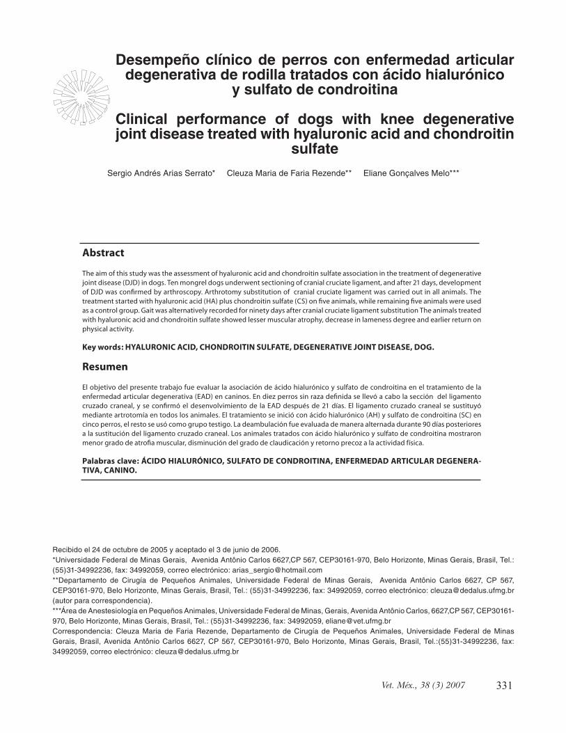
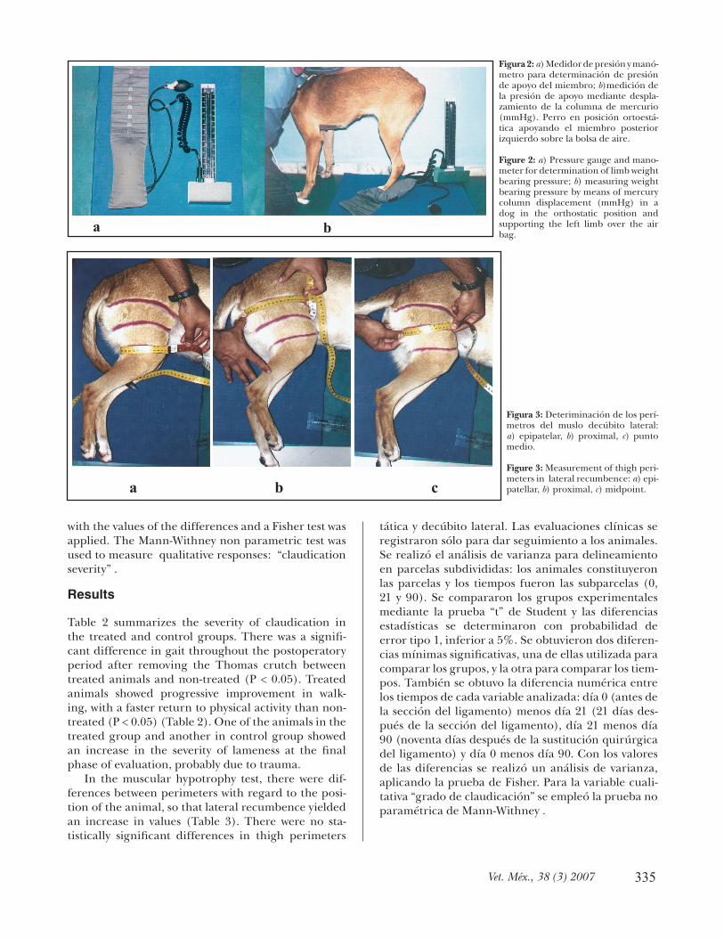
The range of knee fl exion and extension was deter-mined using a plastic goniometer (femoro-tibio-patel-lar angle, FTPA) (Figures 1a y b); the support pressure of the operated limb by means of column mercury displacement measured in millimeters (mm Hg) (Fig-ures 2a y b) and the thigh girth with a measuring tape (Figure 3). These tests were administered before the arthroscopic section of the ligament (0 day), twenty one days after section (21 day) and ninety days after cranial cruciate substitution (90 day). The thigh girth was measured on three locations: epipatellar perimeter, proximal perimeter (approximately three centimeters distal to the greater trocanter ) and the midpoint between the proximal and epipatellar (Fig-ures 3a, b and c ). These perimeters were measured in orthostatic position and lateral recumbence. Clini-cal evaluation was exclusively registered for monitor-
vovirosis, leptospirosis, distemper, hepatitis infecciosa canina).
El miembro posterior izquierdo fue preparado para cirugía ortopédica; previo ayuno de 12 horas, se continuó con medicación preanestésica con sulfato de atropina (0.044 mg/kg, vía subcutánea); 15 minutos después se administró clorhidrato de xilazina (2.0 mg/kg vía intramuscular), y diez minutos más tarde se realizó inducción anestésica con pentobarbital sódico al 2.5% en dosis de 12.5 mg/kg de peso vivo vía intra-venosa y se mantuvo anestesia general con halotano. Treinta minutos antes de la cirugía se realizó quimio-profi laxia antimicrobiana con cefalotina sódica (30 mg/kg de peso vivo vía intravenosa). Los animales fueron colocados en decúbito dorsal y se les extrajo líquido sinovial de la rodilla izquierda, para realizar la evaluación artroscópica* y seccionar el ligamento cruzado craneal. Veintiún días después de este pro-cedimiento quirúrgico, se realizó nueva evaluación artroscópica, verifi cándose el desenvolvimiento de la EAD. El ligamento se sustituyó por fascia lata autó-gena, siguiendo la técnica descrita por Schawalder.25 Se realizó artrotomía lateral, divulsión y disección de un retajo de la fascia lata. El retajo fue insertado por la porción caudal de la articulación, atravesándolo hasta la región intraarticular. Se realizó un túnel óseo de forma diagonal en la tibia, partiendo desde la región de inserción del ligamento cruzado craneal hasta la cresta. La fascia se fi jó por medio de sutura de poligle-caprone. Posteriormente a la cirugía se colocó muleta de Thomas durante 15 días y se distribuyeron los ani-males de forma aleatoria en dos grupos de cinco inte-grantes cada uno:
• Grupo 1: constituyó el grupo testigo, cuyos ani-males no recibieron la asociación de sulfato de con-droitina** y ácido hialurónico.***• Grupo 2: formado por animales tratados con sul-fato de condroitina y ácido hialurónico según pro-tocolo a seguir: 240 mg/perro (2 mL) de sulfato de condroitina, vía intramuscular en intervalos de cinco días hasta completar seis aplicaciones, y 20 mg/perro de ácido hialurónico, vía endovenosa, en intervalos de cinco días, hasta completar tres aplica-ciones. La primera aplicación se realizó el día de la sustitución del ligamento cruzado craneal.
Los animales fueron evaluados diariamente, des-pués de retirada la muleta, en cuanto a aspectos clí-nicos, características de deambulación, apoyo del miembro y grado de claudicación, con base en clasifi -caciones descritas en la literatura22,26,27 (Cuadro 1).
Se determinó la amplitud de fl exión y extensión
*Storz, Karl Storz Endoscopy, artroscopio de 2.7 mm de diámetro con ángulo de 30 grados.**Artroglycan, Syntek, S.A.***Legend, Bayer, S.A.

334
ing the animals. Variance analysis (ANOVA) in the split plot confi guration was applied. Animals were the plots and the sub-plots were times of evaluation (0, 21 and 90). Experimental groups were compared through the T student test and statistical differences were determined with error (type 1)probability infe-rior to 5%. Two minimal signifi cant differences were obtained, one for group comparisons and another for time comparisons. Numerical differences between the times of each analyzed variable were determined this way: day 0 (before ligament section) minus day 21 (21 days after ligament section), day 21 minus day 90 (ninety days after surgical substitution of the liga-ment) and day 0 minus day 90. An ANOVA was done
del miembro (ángulo fémoro-tibio-patelar, AFTP) mediante goniómetro de plástico (Figuras 1a y b); la presión de apoyo del miembro operado, mediante des-plazamiento de columna de mercurio en milímetros (mm Hg) (Figuras 2a y b), y el perímetro del muslo con cinta métrica (Figura 3). Las medidas se toma-ron antes de la sección artroscópica (día 0), 21 días después de la sección (día 21) y 90 días posteriores a la sustitución del ligamento cruzado craneal (día 90). El perímetro del muslo se midió en tres ubicaciones: perímetro epipatelar, perímetro proximal (casi a tres centímetros distal del trocánter mayor) y en el punto medio entre las dos anteriores (Figuras 3a, b y c ). Esos perímetros se midieron en las posiciones ortoes-
Cuadro 1
CARACTERÍSTICAS DE LA CLAUDICACIÓN EN GRADOS PARA EVALUACIÓN CLÍNICA DESPUÉS DEL TRATAMIENTO CON ÁCIDO HIALURÓNICO Y
SULFATO DE CONDROITINA DEGREES FOR CLINICAL EVALUATION OF LAMENESS AFTER TREATMENT
WITH HYALURONIC ACID AND CHONDROITIN SULFATE
Lameness degree Description
0 Normal limb weight bearing
1 Walks without lameness, normal in station, but sporadically lame when running without elevating the limb
.
2 Intermittent lameness use of the limb with total weight bearing.
3 Lameness when walking, partial weight bearing and elevating it when running
4 Non-frequent and intermittent use and limb weight bearing in orthostatic position and when walking it does not support the weight
5 No limb use.
a b
Figura 1: a) Goniómetro utilizado para deter-minación del ángulo fémoro-tibio-patelar; b)determinación del ángulo fémoro-tibio-patelar (AFPT) en decúbito lateral.
Figure 1: a) Goniometer for stifl e angle deter-mination; b) determination of stifl e angle (AFTP) in lateral recumbence.
335Vet. Méx., 38 (3) 2007
with the values of the differences and a Fisher test was applied. The Mann-Withney non parametric test was used to measure qualitative responses: “claudication severity” .
Results
Table 2 summarizes the severity of claudication in the treated and control groups. There was a signifi -cant difference in gait throughout the postoperatory period after removing the Thomas crutch between treated animals and non-treated (P < 0.05). Treated animals showed progressive improvement in walk-ing, with a faster return to physical activity than non-treated (P < 0.05) (Table 2). One of the animals in the treated group and another in control group showed an increase in the severity of lameness at the fi nal phase of evaluation, probably due to trauma.
In the muscular hypotrophy test, there were dif-ferences between perimeters with regard to the posi-tion of the animal, so that lateral recumbence yielded an increase in values (Table 3). There were no sta-tistically signifi cant differences in thigh perimeters
tática y decúbito lateral. Las evaluaciones clínicas se registraron sólo para dar seguimiento a los animales. Se realizó el análisis de varianza para delineamiento en parcelas subdivididas: los animales constituyeron las parcelas y los tiempos fueron las subparcelas (0, 21 y 90). Se compararon los grupos experimentales mediante la prueba “t” de Student y las diferencias estadísticas se determinaron con probabilidad de error tipo 1, inferior a 5%. Se obtuvieron dos diferen-cias mínimas signifi cativas, una de ellas utilizada para comparar los grupos, y la otra para comparar los tiem-pos. También se obtuvo la diferencia numérica entre los tiempos de cada variable analizada: día 0 (antes de la sección del ligamento) menos día 21 (21 días des-pués de la sección del ligamento), día 21 menos día 90 (noventa días después de la sustitución quirúrgica del ligamento) y día 0 menos día 90. Con los valores de las diferencias se realizó un análisis de varianza, aplicando la prueba de Fisher. Para la variable cuali-tativa “grado de claudicación” se empleó la prueba no paramétrica de Mann-Withney .
a b
a b c
Figura 2: a) Medidor de presión y manó-metro para determinación de presión de apoyo del miembro; b)medición de la presión de apoyo mediante despla-zamiento de la columna de mercurio (mmHg). Perro en posición ortoestá-tica apoyando el miembro posterior izquierdo sobre la bolsa de aire.
Figure 2: a) Pressure gauge and mano-meter for determination of limb weight bearing pressure; b) measuring weight bearing pressure by means of mercury column displacement (mmHg) in a dog in the orthostatic position and supporting the left limb over the air bag.
Figura 3: Deteriminación de los perí-metros del muslo decúbito lateral: a) epipatelar, b) proximal, c) punto medio.
Figure 3: Measurement of thigh peri-meters in lateral recumbence: a) epi-patellar, b) proximal, c) midpoint.

336
between groups when considering, individual evalu-ation time (Tables 3 and 5). Nevertheless, there was signifi cant difference between perimeters in different times (0, 21 and 90) under a specifi c treatment (Table 3). The epipatellar perimeter in the control group on the orthostatic position showed signifi cant atrophy on day 21, but with a partial mass recuperation on day 90. Same results were observed in the treated groups at this position. Epipatellar perimeter in lateral recum-bence in control group did not have signifi cant atro-phy on day 21; but on day 90 when compared to day 0, there was signifi cant statistic atrophy. The treated group did not show signifi cant differences throughout the evaluated times.
The proximal perimeters of control group in the orthostatic position had signifi cant atrophy on day 21 which was maintained up to day 90. There was no atro-phy in the treated group throughout the evaluated times. In lateral recumbence both groups presented signifi cant atrophy on day 21; both groups maintained this degree of atrophy until day 90.
There was signifi cant atrophy at the midpoint perimeter of control group in orthostatic position on day 21 which was maintained until day 90. Animals in the treated group did not show signifi cant atrophy in orthostatic position during the evaluated times. Measures in lateral recumbence of the control group revealed signifi cant atrophy on day 21 and even more on day 90. In the treated group, atrophy was signifi -cant on day 21 which was maintained up to day 90.
The values of FTPA in fl exion and extension in orthostatic position are presented in Table 4. In this position, a signifi cant difference between experimen-tal groups was noticed at time 0, whereas there were no differences at times 21 and 90. The FTPA in fl ex-ion and lateral recumbence showed signifi cant differ-ences between control and treated groups, denotating greater limb fl exion in treated animals (Table 4). In relation to the limb extension range, there was no difference between groups at the evaluated times. However, the behavior of each group, at times, was sta-tistically different. The animals in the control group showed an increasing resistance to limb extension on days 0 and 21, followed by partial recuperation on day 90. The treated group showed statistical equivalence in limb extension range, at all times throughout the evaluation. This refl ects lack of resistance to exten-sion in this group.
The mean differences between values on days (0, 21 and 90) are presented in Table 5, which shows signifi -cant difference between treatments in range of fl exion of treated animals (HA + CS). Comparisons between days 0 and 90 in non-treated animals revealed an aver-age decrease fl exion of 3.2 degrees and a increase of 9.8 degrees in treated animals on day 90.
Resultados
En el Cuadro 2 están relacionados los grados de clau-dicación de animales tratados y del grupo testigo. Después de retirar la muleta, hubo diferencia signifi -cativa en la ambulación de animales tratados y no tra-tados. Durante el periodo posoperatorio (P < 0.05), se evidenció mejora progresiva en la marcha de los animales tratados, con retorno más precoz a la activi-dad física que los no tratados (P < 0.05) (Cuadro 2). En uno de los animales del grupo tratado y en otro del grupo no tratado, se observó aumento en el grado de claudicación casi al fi nal del estudio, posiblemente debido a un trauma.
En las determinaciones de hipotrofi a muscular se observó diferencia entre los perímetros, de acuerdo con la posición del animal, y se observaron valores mayores en la posición decúbito lateral (Cuadro 3). No hubo diferencia estadística signifi cativa entre los grupos en cuanto a los perímetros del muslo en un mismo tiempo medido (Cuadros 3 y 5). Sin embargo, hubo diferencia signifi cativa entre los perímetros en los diferentes tiempos 0, 21 y 90 bajo un mismo tra-tamiento (Cuadro 3). En el perímetro epipatelar del grupo testigo y en posición ortoestática, se observó un grado de atrofi a signifi cativo el día 21, y se recuperó parcialmente la masa muscular el día 90. En esta posi-ción se observó igual comportamiento en el grupo tra-tado. En decúbito lateral y en el perímetro epipatelar del grupo testigo no se mostró grado signifi cativo de atrofi a el día 21; se observó atrofi a estadísticamente signifi cativa el día 90 con respecto al día 0. El grupo tratado no mostró diferencia signifi cativa en los tiem-pos evaluados.
En el perímetro proximal del grupo testigo y en posición ortoestática, se observó atrofi a signifi cativa el día 21, el grado de atrofi a se mantuvo hasta el día 90. En el grupo tratado no se observó atrofi a signifi -cativa en los tiempos evaluados. En decúbito lateral, tanto el grupo testigo como el tratado mostraron atro-fi a signifi cativa el día 21, ambos grupos mantuvieron el grado de atrofi a hasta el día 90.
En el perímetro del punto medio del grupo testigo y en posición ortoestática, se observó atrofi a signifi -cativa el día 21, la cual se mantuvo hasta el día 90. En el grupo tratado no se presentó atrofi a signifi ca-tiva en los tiempos evaluados cuando se midieron los perímetros en la posición ortoestática. Las medidas en decúbito lateral del grupo testigo revelaron atro-fi a signifi cativa el día 21 y aún mayor el día 90. En el grupo tratado hubo atrofi a signifi cativa el día 21, que se mantuvo hasta el día 90.
Las medidas del AFTP en fl exión, extensión y en posición ortostática se presentan en el Cuadro 4. En la posición ortoestática hubo diferencia signifi cativa

337Vet. Méx., 38 (3) 2007
Cuadro 2
GRADOS DE CLAUDICACIÓN DE ANIMALES EN LOS GRUPOS TESTIGO Y TRATADO CON ÁCIDO HIALURÓNICO Y SULFATO DE CONDROITINA
LAMENESS DEGREES FROM ANIMALS OF CONTROL AND TREATED WITH HYALURONIC ACID AND CHONDROITIN SULFATE
Lameness degrees
Days after substitution
HA + CS Group animals
Control group animals
*16 5 3 4 3 3 5 5 5 5 5 18 5 3 4 3 3 5 5 5 5 5 20 5 3 4 3 3 5 4 5 5 5 22 5 3 3 3 3 5 4 5 5 5 24 4 3 3 2 3 5 4 5 5 5 26 4 2 3 2 3 4 4 5 5 5 28 4 2 3 2 3 4 4 5 4 5
**30 3 2 3 2 3 4 4 5 4 5 32 3 2 2 2 3 4 4 5 4 5 34 3 2 2 2 3 4 4 5 4 5 36 3 2 2 2 3 3 3 5 4 4 38 3 2 2 2 3 3 2 5 3 4 40 3 2 2 2 3 3 2 4 3 4 42 3 2 1 1 3 3 2 4 3 4 44 2 2 1 1 3 3 2 4 3 4 46 2 2 1 1 3 3 2 4 2 4 48 2 2 0 0 3 2 2 4 2 4 50 2 2 0 0 3 2 2 4 2 4 52 2 2 0 0 2 2 2 4 2 3 54 2 2 0 0 2 2 2 4 2 3 56 2 2 0 0 2 2 2 4 2 2 58 2 2 0 0 2 2 2 4 2 2 60 2 2 0 0 1 2 2 4 2 2 62 2 2 0 0 1 2 2 4 2 2 64 2 2 0 0 1 2 2 4 2 2 66 2 2 0 0 1 2 2 4 2 2 68 2 2 0 0 1 2 2 4 2 2 70 2 2 0 0 1 2 2 3 2 2 72 2 2 0 0 1 2 2 3 2 2 74 2 2 0 0 1 2 2 3 2 4 76 3 2 0 0 1 2 2 2 2 5 78 5 2 0 0 1 2 2 2 2 5 80 5 2 0 0 1 2 2 2 2 5 82 4 1 0 0 0 2 2 2 2 5 84 3 1 0 0 0 2 2 2 2 4 86 3 1 0 0 0 2 2 2 2 4 88 3 1 0 0 0 2 2 2 2 4 90 2 1 0 0 0 2 2 3 2 3
*First day after withdrawing de Thomas crutch. **End of treatment with HA+CS.

338
Cuadro 3
MEDIAS DE LOS PERÍMETROS DEL MUSLO EN CENTÍMETROS, EN TRES DIFERENTES TIEMPOS QUIRÚRGICOS, EN POSICIÓN ORTOSTÁTICA Y EN DECÚBITO LATERAL, EN
PERROS NO TRATADOS Y TRATADOS CON ÁCIDO HIALURÓNICO Y SULFATO DE CONDROITINA.
AVERAGE OF THIGH PERIMETERS IN CENTIMETERS, THREE DIFFERENT SURGICAL TIMES, IN THE ORTHOSTATIC POSITION AND LATERAL RECUMBENCE OF DOGS
UNTREATED AND TREATED WITH HYALURONIC ACID AND CHONDROITIN SULFATE
Position Orthostatic Lateral recumbence
Perimeter Days 0 21 90 0 21 90
Epipatellar¹ Control 25.8aA 24.4bA 25.4abA 27.4aA 26.6abA 25.8bA
HA + CS 26.2aA 25bA 25.8abA 27.6aA 27.3aA 26.4aA
Midthigh² Control 32.2aA 28.8bA 28.2bA 38.4aA 31.8bA 29cA
HA + CS 31.6aA 30aA 30.2aA 34.4aA 32.5bA 32.4bA
Proximal³ Control 36.4aA 33bA 34bA 38.4aA 34.8bA 34bA
HA + CS 37.2aA 35.8aA 36aA 40.6aA 37.2bA 37.2bA
Averages with equal very small letters in the rows and equal capital letters in the columns are statistically equal. CV in orthostatic position of 1= 6.4% columns and 3.3% in rows, 2 = 9.4% columns and 4.9%in rows, 3 = 7.7% columns and 4.1% in rows. CV in lateral position of 1 = 7.9% columns and 3.5% in rows, 2 = 10% columns and 3.8% in rows, 3 = 8.0% columns and 5.2% in rows. dms in orthostatic position for comparison between groups of 1 = 2.6, 2 = 4.5 and 3 = 4.3. In lateral recumbence 1 = 3.3, 2 = 4.9, 3 = 4.9. Dms in orthostatic position for comparison between times of 1 = 1.15, 2 = 2.0 and 3 = 2.0. In lateral position 1 = 1.3, 2 = 1.7, 3 = 2.6. Day 0 = Before section of cranial cruciate ligament.
Day 21 = 21 days after ligament section. Day 90 = 90 days after ligament substitution.
There was no statistical difference between treated animals and non-treated with respect to the pressure exerted by the limb. This fact was confi rmed because of the low F value in the table of time differences (Table 5). Nonetheless, there was a statistically signifi cant dif-ference inside each experimental group throughout the times evaluated (Table 6). In the control group, there was no signifi cant decrease in limb pressure from day 0 to 21, but a decrease between 21 and 90. In treated animals there was signifi cant decrease in limb pressure from day 0 to 21 which was maintained up to day 90.
Discussion
Observed results showed that animals treated with hyaluronic acid (HA) and chondroitin sulfate (CS) association had better physical and orthopedic per-formance than animals only treated by means of sur-gical stabilization. Similar studies accomplished by Melo et al.,6 which used separately HA or CS, did not
entre los grupos experimentales en el tiempo 0, y no se registró diferencia signifi cativa en los tiempos 21 y 90. Las mediciones del AFTP en fl exión y en la posición decúbito lateral, mostraron diferencia estadística-mente signifi cativa entre el grupo testigo y el tratado; en los animales tratados se observó mayor fl exión del miembro (Cuadro 4). En cuanto a la amplitud de extensión no existió diferencia signifi cativa entre el grupo testigo y el tratado en los tiempos medidos. Sin embargo, el comportamiento dentro del mismo grupo en cada tiempo fue estadísticamente diferente. El grupo testigo mostró aumento de la resistencia a la extensión del día 0 al 21, con recuperación parcial el día 90. El grupo tratado mostró equivalencia estadís-tica en el grado de extensión del miembro en todos los tiempos, ello refl eja ausencia de resistencia a la extensión.
La media de las diferencias entre los valores los días 0, 21 y 90, se presenta en el Cuadro 5, que mues-tra diferencia signifi cativa entre los tratamientos en cuanto a la magnitud de fl exión en animales tratados

339Vet. Méx., 38 (3) 2007
fi nd signifi cant differences between treated and non-treated animals. In this study, the outcome suggests favorable effects as a result of HA and CS association in dogs’ gait with cranial cruciate rupture and DJD development in the early and late post-operative term. The results can be explained because of the benefi ts reported by each drug.4,18,19,21,22,,28-31
The values of thigh perimeter are a result of limb activity, when there is muscular hypotrophy, as con-sequence of a decreased physical activity, for which it is an indicator of performance and limb use in the dog.32 The thigh perimeter with lesser infl uence, more sensitive and appropriate in assessing the severity of muscular hypotrophy was the midpoint thigh perim-eter. This perimeter was measured around the more prominent mass region of the thigh, where the syno-vial capsule does not exist and the small movements of the limb (fl exion or extension) did not interfere with the measurement.
Values from the midpoint thigh perimeter in ortho-static and lateral recumbence positions revealed mus-
con AH + SC. Al comparar los resultados de los días 0 y 90 en animales no tratados, se observó reducción de la fl exión de 3.2 grados en promedio, y en anima-les tratados se observó aumento de la fl exión de 9.8 grados, en promedio, con respecto al día 0.
No se estableció diferencia estadística entre ani-males tratados y no tratados en relación con el grado de presión ejercida por el miembro operado, con-fi rmado por el bajo valor de F en el cuadro de dife-rencias entre los tiempos (Cuadro 5). No obstante, se registró diferencia estadísticamente signifi cativa entre los tiempos dentro de un grupo experimental cuando fueron evaluados aisladamente (Cuadro 6). En el grupo testigo no hubo disminución signifi cativa de la presión de apoyo del miembro entre los días 0 y 21, por lo que se estableció diferencia entre los días 0 y 90. En los animales tratados hubo disminución signi-fi cativa en la presión de apoyo entre los días 0 y 21; la diferencia estadísticamente signifi cativa se mantuvo hasta el día 90.
Cuadro 4
MEDIAS DE LOS ÁNGULOS DE FLEXIÓN Y EXTENSIÓN DE LA ARTICULACIÓN FÉMORO-TIBIO-PATELAR EN POSICIÓN
ORTOSTÁTICA Y DECÚBITO LATERAL, DE PERROS NO TRATADOS Y TRATADOS CON ÁCIDO HIALURÓNICO Y SULFATO DE
CONDROITINA
AVERAGE OF ANGLES IN FLEXION AND EXTENSION OF STIFLE JOINT IN THE ORTHOSTATIC POSITION AND LATERAL
RECUMBENCE OF DOGS UNTREATED AND TREATED WITH HYALURONIC ACID AND CHONDROITIN SULFATE
Position Group/ days 0 21 90
Orthostátic¹ Control 39.8aB 48.6aA 46aA
HA + CS 51.2aA 53aA 54.6aA
Lateral recumbence
Flexion² Control 141aA 138,8aA 137.6aB
HA + CS 140.4bA 138bA 150.2aA
Extension³ Control 18.2bA 39.2aA 26.8abA
HA + CS 27.8aA 40.2aA 34.6aA
Averages with equal very small letters in the rows and equal capital letters in the columns are statistically equal. CV of 1 = 14.6% columns and 16.7% in rows, 2 = 2.3% columns and 3.9% rows, 3 = 27.8% columns and 36.2% filas. Dms of 1 for comparison between groups = 13.8 and between times = 11. In lateral recumbence, dms for groups comparison of 2 = 7.9, 3 = 17.7; comparison between times of 2 = 7.6, 3 = 15.2. Day 0 = Before section of cranial cruciate ligament. Day 21 = 21 days after ligament section. Day 90 = 90 days after ligament substitution.

340
Cuadro 5
VALORES MEDIOS DE LAS DIFERENCIAS NUMÉRICAS DE LOS PERÍMETROS, DE LA PRESIÓN Y AFTP EN PERROS TRATADOS CON ÁCIDO HIALURÓNICO Y SULFATO DE CONDROITINA Y NO TRATADOS, DURANTE LOS PERIODOS 0-21, 21-90 Y 0-90 DÍAS
AVERAGES OF NUMERICAL DIFFERENCES OF PERIMETERS, PRESSURE AND AFTP OF DOGS TREATED WITH HYALURONIC ACID AND CHONDROITIN SULFATE AND
UNTREATED IN TIMES 0-21, 21-90 AND 0-90 DAYS
Days
Orthostatic position
0-21 21-90 0-90
Variable C HA+CS Fisher C HA+CS Fisher C HA+CS Fisher
Epipatellar perimeter 1.4 1.2 0.05 –0.2 – 0.8 0.75 0.4 0.4 0
Proximal perimeter 3.4 1.4 1.88 –1 –0.2 0.33 2.2 1.2 1.13
Midpoint 3.4 1.6 2.13 0.6 –0.2 0.37 4 1.4 10.24
Goniometer
orthostatic pos.
–8.8 –1.8 0.66 2.6 2 0.02 –6.2 –3.4 0.13
Pressure mm Hg 8.8 20.8 3.15 2.4 –6.6 1.55 7.2 14.2 0.67
Lateral recumbence
Epipatellar perimeter 0.8 0.2 0.52 0.8 1 0.035 1.6 1.2 0.4
Proximal perimeter 3.6 3.4 0.02 0.8 0 0.15 4.4 3.4 0.33
Midpoint 2.6 1.6 0.89 2.8 –0.4 8.53 5.4 3.4 2.32
Goniometer flexion 2.2 2.4 0.002 1.2 –12.2 4.92 3.4 –9.8 11.02*
Goniometer extension –19 –12.4 0.33 12.4 5.6 0.80 –8.6 –6.8 0.05 * = Significant in Fisher F test with error type 1 probability < 0.05 C = control group, HA+CS = hyaluronic acid and chondroitin sulfate treated group, Fisher = obtained value with Fisher test.
Cuadro 6
MEDIAS DE LA PRESIÓN DE APOYO MEDIDA EN MILÍMETROS DE MERCURIO EN PERROS NO TRATADOS Y TRATADOS CON ÁCIDO
HIALURÓNICO Y SULFATO DE CONDROITINA
AVERAGES OF PRESSURE WEIGHT BEARING MEASURED IN DISPLACED MERCURY MILIMETERS OF DOGS UNTREATED AND
TREATED WITH HYALURONIC ACID AND CHONDROITIN SULFATE Group/ days 0 21 90
34.4aA 25.6abA 23.2bA
43.2aA 22.4bA 29bA
Averages with equal very small letters in the rows and equal capital letters in the columns are statistically equal. . CV = 25.8% columns and 26.8% in rows. Dms for groups comparison = 14.2, and between times = 10.7. Day 0 = Before section of cranial cruciate ligament. Day 21 = 21 days after ligament section. Day 90 = 90 days after ligament substitution
Control
HA + CS

341Vet. Méx., 38 (3) 2007
cular hypotrophy 21 days after arthroscopic section of cranial cruciate ligament, probably as a consequence of DJD development. Despite the non-signifi cant value obtained from the treated group in orthostatic position, this value was located close to the inferior numeric limit that confers statistical signifi cance between the times 0 and 21, showing atrophy. Ben-efi ts using the drugs were verifi ed when results from the midpoint thigh perimeter were analyzed up to day 90. On this day, there was an increase in hypotrophy in non-treated animals and maintenance in the extent of muscle mass loss in treated animals. Similar results were observed in the proximal thigh perimeter.
It must be taken into account that the perimeters were infl uenced due to animal position, level of mus-cular tension and presence of synovial capsule. In lat-eral recumbence, the values increased due to muscle relaxing. In the case of epipatellar perimeter and as reported by Johnson et al.,33 it is probable that post surgical thickening of the synovial capsule infl uenced the measurement, leading to an apparent recupera-tion of the thigh mass that was observed in both experimental groups in orthostatic position.
Considering the variation coeffi cient and the obtained minimum signifi cant differences in this study, it was observed that the degree of muscular atrophy 21 and 110 days (day 90) after the section of the cranial cruciate ligament was inferior to 10% and 20%, respectively. Clinically, the atrophy was more evident in the contra lateral limb due to compensa-tory hypertrophy. These results are similar to those found by Campbell et al.,34 at which greater atrophy of the member was noticed two to three months after the rupture, without substitution of ligament. Neverthe-less, the atrophy degree does not differ with the surgi-cal substitution of the ligament, as it was observed in this study and in studies made by Hoelzler et al.32 with dogs with a ruptured ligament and underwent liga-ment substitution.
The lack of signifi cant difference between the mus-cular perimeters in dogs treated and untreated in a static time of evaluation, was due to the high variation of the morphologic characteristics of the thigh in ani-mals of each experimental group. This comparison was not relevant to the objective of this study, since the comparison of the perimeters is important when it is made in the same animals and through the evaluated times. The split plot statistic delineation was effi cient for this comparison.
In tests of fl exion and extension, it was noticed that animals treated with HA and CS did not show statisti-cally signifi cant differences in the extension, due to the high variation coeffi cient obtained in this variable; in fl exion, the animals of the control group showed resis-tance. The results suggest a more favorable evolution
Discusión
Los resultados observados muestran que los animales tratados con ácido hialurónico y sulfato de condroi-tina en asociación, presentan mejor desempeño físico y ortopédico que cuando son sometidos únicamente a intervención quirúrgica. En estudios similares realizados por Melo et al.,6 en los cuales los anima-les fueron tratados con ácido hialurónico o sulfato de condroitina, no fue posible establecer diferencias estadísticamente signifi cativas entre animales trata-dos y no tratados. En este estudio, los resultados de la ambulación de perros con ruptura de ligamento cruzado y desenvolvimiento de EAD, tratados con AH y SC, sugieren un efecto favorable cuando se asocian los fármacos en el posoperatorio inmediato y tardío. Los efectos benéfi cos como resultado de la terapia con cada fármaco ya se han descrito en la lite-ratura.4,18,19,21,22,28-31
Los valores de los perímetros del muslo resultan de la actividad ejercitada por el miembro, cuando hay hipotrofi a muscular, como consecuencia de la disminución de la actividad física, por lo que es un indicador de desuso y bajo desempeño.32 El períme-tro del muslo menos infl uido, más sensible y más ade-cuado para determinar el grado de atrofi a muscular fue el perímetro del punto medio. Este perímetro fue determinado en la región más prominente del muslo, donde no se presenta cápsula articular, y la masa mus-cular no es alterada por las pequeñas diferencias en la posición del miembro (fl exión o extensión).
Los datos obtenidos del perímetro medio del muslo en la posición ortoestática y en decúbito lateral mostra-ron hipotrofi a del miembro 21 días después de la sec-ción artroscópica del ligamento cruzado craneal, en virtud del desenvolvimiento de la EAD. El valor obte-nido en la posición ortoestática del grupo tratado no fue estadísticamente diferente al día 0. Sin embargo, este valor fue cercano al límite inferior numérico que establece diferencia estadística signifi cativa entre los dos tiempos (0 y 21), denotando atrofi a. El benefi cio obtenido con los fármacos se confi rma al analizar los resultados de perimetría muscular en el punto medio, en los cuales se mostró aumento de la hipotrofi a a los 90 días, en los animales no tratados; el grado de atro-fi a muscular en los tratados se mantuvo. Resultados semejantes a los del perímetro medio se observaron en los datos del perímetro proximal.
Debe tomarse en cuenta que los perímetros fueron infl uidos por la posición del animal, el grado de ten-sión muscular y la presencia de cápsula articular. En la posición decúbito lateral se observaron valores incrementados como consecuencia del relajamiento muscular. En cuanto al perímetro epipatelar es pro-bable que los resultados hayan sido infl uidos por el

342
for treated animals, since while observing in detail the data and comparing the experimental groups, it is verifi ed that the behavior of the variables is better in the treated group than in the non-treated.
Satisfactory performances in limb fl exion and extension, either surgically32 or therapeutically (HA or CS) treated animals have been described in lit-erature.6,8,15,35-38 The satisfactory results of treated animals in fl exion and extension parameters can be explained by the regulation properties of articular microhomeostasis, macrohomeostasis and miniho-meostasis described in the viscosupplementation con-cept reported by Balazs and Denlinger39 using HA, and the reported analgesic and anti-infl ammatory effects using both drugs.3,17,20,37-48 Arias et al.49 reported an anti-infl ammatory effect with the HA-CS association, while fi nding absence of lymphoplasmocytic infi ltrate in synovial membranes of dogs treated with the asso-ciation of drugs; however, progression of DJD was not eliminated.
In both experimental groups, the results of the FTPA in fl exion and extension on day 0 were infl u-enced by the initial adaptation of the animals to the experimental manipulation, resulting in smaller values in fl exion and larger in extension. On the other days, values of fl exion were also infl uenced by the muscular atrophy, since the decrease of the mus-cular perimeter of the thigh increased the degree of fl exion of the limb. It is probable that the greater value obtained in fl exion at day 90 in the treated group, may be a consequence of the return in fl exion limb nor-mality. An improvement of limb fl exion in untreated animals at days 21 and 90, was also observed in this study. This could be the consequence of the surgical stabilization.
Several authors have suggested an increase in limb weight bearing in animals treated with nutraceu-tics.8,9,37,40,50 In this study, the results of animals under medical treatment did not demonstrate signifi cant improvement of limb weight bearing in pressure test. The value obtained from the control group between days 0 and 21 was next to the bottom limit that estab-lishes statistical signifi cant difference between these days. Thus a reduction of weight bearing in both groups at day 21 was revealed, though without return to normality. When making a correlation between gait characteristics of treated dogs with degrees of pres-sure exerted by the limb, it was evident that the car-rying capacity of weight was not full in the last days of observation. It is probable that the weight supported by the operated limb was distributed by the others as reported by Auer et al.40 and Gingerich et al.50 in horses. This variable could be more accurate deter-mined by means of measurement of the weight bear-ing in force plate in future studies.
engrosamiento posquirúrgico de la cápsula articular, como lo cita Johnson et al.,33 lo que llevó a una apa-rente recuperación de la masa muscular observada en ambos grupos experimentales en la posición ortoes-tática.
Teniendo en cuenta el coefi ciente de variación y las diferencias mínimas signifi cativas obtenidas en este estudio, se observó que el grado de atrofi a mus-cular 21 y 110 días (día 90) después de seccionar el ligamento cruzado craneal, fue inferior a 10% y 20%, respectivamente. Clínicamente, la atrofi a se mostró más evidente en el miembro contralateral, debido a la hipertrofi a compensatoria. Estos hallazgos son seme-jantes a los encontrados por Campbell et al.,34 en los cuales se registró mayor atrofi a del miembro dos a tres meses después de la ruptura, sin sustitución de liga-mento. Sin embargo, el grado de atrofi a no difi ere con la sustitución quirúrgica del ligamento, como se observó aquí y como se constata en estudios de Hoel-zler et al.32 en perros con ruptura de ligamento y some-tidos a sustitución ligamentaria.
La falta de diferencia signifi cativa cuando se esta-blece la comparación entre los perímetros musculares de animales tratados y no tratados en un tiempo está-tico de evaluación, se debió a la alta variación de las características morfológicas del muslo en los animales de cada grupo experimental. Esta comparación no fue de relevancia para el objetivo de este estudio, ya que la comparación de los perímetros es relevante cuando se hace en los mismos animales y a través de los tiempos evaluados. El delineamiento en parcelas subdivididas se mostró efi ciente en esa comparación.
En las pruebas de fl exión y extensión se observó que los animales tratados con AH y SC no mostraron diferencia estadísticamente signifi cativa en la exten-sión debido al alto coefi ciente de variación obtenido en esta variable; en la fl exión, los animales del grupo testigo mostraron resistencia. Los resultados sugieren una evolución más favorable para los animales trata-dos, ya que al observar en detalle los datos y compa-rando los grupos experimentales, se verifi ca que el comportamiento de las variables es mejor en el grupo tratado que en el no tratado.
Estos desempeños satisfactorios en la fl exión y extensión del miembro en animales tratados apenas quirúrgicamente32 o terapéuticamente con AH o SC, ya se han descrito en la literatura.6,8,15,35-38 Los resultados satisfactorios en los parámetros de fl exión y extensión de los animales tratados se explican por las propieda-des de regulación de la microhomeostasis, macroho-meostasis y minihomeostasis articular descritas en el concepto de viscosuplementación relatado por Balazs y Denlinger39 con AH, y por los efectos analgésicos y antiinfl amatorios citados en la literatura con el uso de ambos fármacos.3,17,20,37-48 Arias et al.49 demostraron el

343Vet. Méx., 38 (3) 2007
The results obtained in the tests suggest that the physical performance and gait of treated animals are better than those of untreated. The treated animals could move better and had a faster return to physical activities. However, there was no full recovery of the muscle mass, which was probably due to the lack of total support of the weight and DJD progression.
It is concluded that using HA-CS association in dogs with stifl e degenerative joint disease due to the rupture of the cranial cruciate ligament led to a better physical performance, lesser degree of muscle atro-phy, decreased degree of lameness and a faster return to function in the postoperative period in treated ani-mals. The results of this study also suggest that there is a clinical synergetic effect between the drugs when they are used in association with canine stifl e arthro-sis.
Acknowledgements
The authors wish to thank Adaulfo Arias Cotes and Inversora Arias Serrato S.C.S from Colombia for the fi nancial support, and professors of Minas Gerais Fed-eral University; Roberto Baracat de Araujo, Renato Cesar Sachetto Tôrres and Ivan Barbosa Sampaio for their cooperation in this study.
Referencias
efecto antiinfl amatorio de la asociación de AH y SC al encontrarse ausencia de infi ltrado linfoplasmocítico en las membranas sinoviales de caninos tratados con asociación de los fármacos, pero no se logró la elimi-nación de la progresión de la EAD.
Debe considerarse que los resultados del AFTP en fl exión y extensión del miembro el día 0 fueron infl ui-dos en ambos grupos experimentales por la mínima adaptación inicial de los animales a la manipulación experimental, lo que resultó en valores menores en la fl exión y mayores en extensión el día 0. En los demás tiempos, los valores de fl exión también fueron dis-cretamente infl uidos por la atrofi a muscular, ya que la disminución del perímetro muscular en el muslo aumentó el grado de fl exión del miembro. Es probable que el valor superior obtenido en fl exión el día 90 en el grupo tratado, sea consecuencia del retorno al valor de normalidad de fl exión para este miembro. En este estudio también se observó mejoría en la fl exión de los animales no tratados los días 21 y 90. Esto último pudo deberse a la estabilización quirúrgica.
Varios autores han sugerido un aumento en el apoyo del miembro en animales tratados con nutra-céuticos.8,9,37,40,50 En los resultados de la prueba de pre-sión obtenidos en este trabajo, no se refl ejó mejoría signifi cativa en el apoyo del miembro bajo tratamiento médico. El valor presentado en el grupo testigo entre los días 0 y 21 se encontró próximo al límite inferior numérico que establece diferencia estadística signi-fi cativa entre los dos tiempos, determinándose una disminución de apoyo en ambos grupos al día 21, la cual no retornó a la normalidad. Al realizarse una correlación entre las características de ambulación de perros tratados con el grado de presión ejercida por el miembro, es evidente que el soporte de peso en los últimos días de observación no fue total. Es probable que el peso no soportado por el miembro operado sea distribuido en los demás como lo citan Auer et al.40 y Gingerich et al.50 en equinos, pues esta variable podría determinarse en estudios posteriores con mayor pre-cisión a través de estudios de medición del apoyo en plataforma de fuerza.
Con los resultados obtenidos en las diferentes pruebas realizadas es evidente que el desempeño físico y las características de ambulación de animales tratados son mejores que los observados en animales no tratados, puesto que los animales tratados consi-guen moverse mejor y retornan a la actividad física en menor tiempo. No obstante, no hubo total recupera-ción de la masa muscular, quizá debido a la falta de soporte total del peso y progresión de la EAD.
Se concluye que el uso de ácido hialurónico aso-ciado con el sulfato de condroitina, en perros con enfermedad articular degenerativa de rodilla debida a ruptura de ligamento cruzado craneal, resulta
1.
2.
3.
4.
5.
6.
7.
Harasen G. Canine cranial cruciate ligament rupture in profi le. Can Vet J 2003; 44: 845-846.Anderson MA, Slater MR, Hammad TA. Results of a survey of small animal practitioners on the perceived clinical effi cacy and safety of an oral nutraceutical. Prev Vet Med 1999; 38: 65-73.Kuroki K, Cook JL, Kreeger JM. Mechanisms of action and potential uses of hyaluronan in dogs with osteoar-thritis. J Am Vet Med Assoc 2002; 221: 944-950. Hulse D. Treatment methods for pain in the osteoar-thritic patient. Vet Clin North Am 1998; 28: 361-375.Johnson KA, Hulse DA, Hart RC, Kochevart D, Chu Q. Effects of an orally administered mixture of chondroi-tin sulfate, glucosamine hydrochloride and manganese ascorbate on synovial fl uid chondroitin sulfate 3B3 and 7D4 epitope in a canine cruciate ligament transaction model of osteoarthritis. Osteoarthritis cartilage 2001; 9: 14-21.Melo EG, Rezende CM, Gomes MG, Freitas PM, Arias SA. Sulfato de condroitina e hialuronato de sódio no tratamento da doença articular degenerativa experi-mental em cães. Aspectos clínicos e radiológicos. Arq Brás Méd Vet Zootec 2003; 55: 35-43.McCarthy GM, Donovan JO, Jones B, McAllister H, Seed M et al. Randomized double blind , positive controlled trial to assess the effi cacy of glucosamine/chondroitin sulfate for the treatment of dogs with osteoarthritis. Vet J 2006: (in press).

344
25.
26.
27.
28.
29.
30.
31.
32.
33.
34.
en mejor desempeño físico, menor grado de atrofi a muscular, disminución del grado de claudicación y retorno más rápido a la actividad física en el periodo posoperatorio de animales tratados. Los resultados de este trabajo también sugieren que hay un efecto clínico sinérgico entre los fármacos cuando se admi-nistran juntos en la terapia de caninos con artrosis de rodilla.
Agradecimientos
Los autores agradecen a Adaulfo Arias Cotes e Inver-sora Arias Serrato S.C.S., de Colombia, por el apoyo fi nanciero, y a los profesores de la Universidad Federal de Minas Gerais, Roberto Baracat de Araujo, Renato César Sachetto Tôrres e Ivan Barbosa Sampaio, por su colaboración en este trabajo.
8.
9.
10.
11.
12.
13.
14.
15.
16.
17.
18.
19.
20.
21.
22.
23.
24.
Phillips MW. Intra-articular sodium hyaluronate in the horse: a clinical trial. Proc Am Assoc Equine Pract 1980; 26:389-394.Ruth DT, Swites BJ. Comparision of the effectiveness of intra-articular hyaluronic acid and conventional ther-apy for the treatment of naturally occurring arthritic conditions in horses. Equine Pract 1985; 7: 25-29.Abatangelo G, Botti P, Bue M, Gei V, Samson JC, Cortivo R et al. Intraarticular sodium hyaluronate injections in the pond nuki experimental model of osteoarthrithis in dogs. Clin Orthop Relat Res 1989; 241:278-284.Schiavinato A, Lini E, Guidolin D, Pezzoli G, Botti P, Martelli M et al. Intraarticular sodium hyaluronate injections in the pond nuki experimental model of osteoarthrithis in dogs, Clin Orthop Relat Res 1989; 241:278-284.Howard RD, McILwraith CW. Sodium hyaluronate in the treatment of equine joint disease. Compend Contin Educ 1993; 15: 473-481.Keller WG, Aron DN, Rowland GN, Odend´hal S, Brown J. The effect of trans-stiffl e external skeletal fi xation and hyaluronic acid therapy on articular cartilage in the dog. Vet Surg 1994; 23:119-128.Robles AM, Wainsten L. Mecanismo de acción y uso de precursores de glicosaminoglicanos en la artrosis. Sel Vet 1995; 3:118-121.Moore MG. Promising responses to a new oral treat-ment for degenerative joint disorders. Canine Pract 1996; 21:7- 11.Palmer JL, Bertone AL. Joint biomechanics in the pathogenesis of traumatic arthritis. In: McILwraith CW, Trotter GW, editors. Joint Disease in the Horse, Philadelphia : Saunders, 1996; Chapter 7: 104-119.McILwraith CW. Use of sodium hyaluronate (Hyaluro-nan) in equine joint disease. Equine Vet Educ 1997; 9:296-304.Campos JÁ. Efi cácia do hialuronato de sódio no trata-mento sintomático da displasia coxo-femoral em cães. A Hora Vet 1998, 17:59-61.Gannon JR. Clinical experiences with intravenous use of sodium hyaluronate in racing Greyhounds. Aust Vet J 1998; 76:474-475.Lipiello L, Idouraine A, McNamara PS, Barr SC, McLaughlin RM. Cartilage stimulatory and antipro-teolytic activity is present in sera of dogs treated with a chondroprotective agent. Canine Pract 1999; 24:18-19.Loneux P, Balligand M. L’ ostéoarthrose chez le chien. Ann Med Vet 1999;143: 153-160.Souza RL, Raiser AG, Rios MV, Guimarães LD, Araújo L, Leotte AM et al. Precursores de glicosaminoglica-nos na reparação articular após trauma iatrogênico no joelho de cães. Clín Vet 1999; 4: 33-38.White GW, Stites T, Hamm J, Pool R. Evaluation of the effi cacy of various preparations of sodium hyaluronate in an induced equine carpitis model. J Equine Vet Sci 1999; 19: 331. Homandberg GA, Guo MS, Ray CL, Ding BS. Mixtures of glucosamine and chondroitin sulfate reverse fi bro-nectin fragment mediated damage to cartilage more
effectively than either agent alone. Osteoarthritis Car-tilage 2006; 14: 793-806.Schawalder P. Eigene methoden zur operativen rekon-struktion bei rupturen des voederen und hinteren krenzbandes. Kleintierpraxis1989; 7: 323-330.Roy RG, Wallace LJ, Johnston GR, Wickstrom SL. A retrospective evaluation of stiffl e osteoarthritis in dogs with bilateral medial patellar luxation and unilateral surgical repair. Vet Surg 1992; 21:475-479.Doig PA, Purbrick KA, Hare JE, McKeown DB. Clini-cal effi cacy and tolerance of meloxicam in dogs with chronic osteoarthritis. Can Vet J 2000; 41: 296-300.Hanson RR. Mode of action of oral chondroprotective agents. Canine Pract 1996; 21: 24.Hanson RR, Smalley LR, Huff GK, White S, Hammad TA. Oral treatment with a glucosamine-chondroitin sulfate compound for degenerative joint disease in horses: 25 cases. Equine Pract 1997; 19:16-21.Bali JP, Cousse H, Neuzil E. Biochemical of the phar-macologic action of chondroitin sulfates on the osteo-articular system. Sem Arthr Rheum 2001; 31: 58-68.Dobenecker B, Beetz Y, Kienzle E. A placebocontrolled double blind study on the effect of nutraceuticals (chondroitin sulfate and mussel extract) in dogs with joint diseases as perceived by their owners. J Nutr 2002; 132: 1690s-1691s.Hoelzler MG, Milis DL, Francis DA, Weigel JP. Results of arthroscopic versus open arthrotomy for surgical management of cranial cruciate ligament defi cience in dogs. Vet Surg 2004; 33: 146-153.Johnson JM, Johnson AL, Pijanowski GJ, Kneller SK, Schaeffer DJ, Eurell JA et al. Rehabilitation of dogs with surgically treated cranial cruciate ligament-defi cient stifl es by use of electrical stimulation of muscles. Am J Vet Res 1997; 58:1473-1478.Campbell JR, Duff SRI, Gilbertson EM. The effect on the contralateral stifl e joint of sectioning of the cranial cruciate ligament in the dog. J Small Anim Pract 1982; 23:511-516.

345Vet. Méx., 38 (3) 2007
Dahlberg L, Lohmander LS, Ryd R. Intraarticular injections of hyaluronan in patients with cartilage abnormalities and knee pain. Arthritis Rheum 1994; 37:521-528.Frizziero L, Govoni E, Bacchini P. Intra-articular hyaluronic acid in the treatment of osteoarthritis of the knee: clinical and morphological study. Clin Exp Rheum 1998; 16:441-449.Gaustad G, Larsen S. Comparison of polysulphated gly-cosaminoglycan and sodium hyaluronate with placebo in treatment of traumatic arthritis in horses. Equine Vet J 1995; 27:356-362.Dorna V, Guerrero RC. Effects of oral and intramuscu-lar use of chondroitin sulfate in induced equine aseptic arthritis. J Equine Vet Sci 1998; 18:548-555.Balazs EA, Denlinger JL. Viscosupplementation: a new concept in the treatment of osteoarthritis. J Rheumatol 1993; 20 suppl. 39:3-9.Auer JA, Fackelman GE, Gingerich DA, Fetter AW. Effect of hyaluronic acid in naturally ocurring and experimental induced osteoarthritis. Am J Vet Res 1980; 41:568-574.Todhunter RJ, Yeh LA, Sheldon A, Grisanzio L, Walker SL, Burton-Wurster N et al. Effects of strosmelisyn activ-ity on proteoglycan degradation of canine articular explants. Am J Vet Res 1995; 56:1241-1247.Kasahara M. Effi cacy of intravenous administration of hyaluronic acid for experimentally induced arthritis in horses. Jpn J Vet Res 1997; 45:118.Listrat V, Ayral X, Patarnello F, Bonvarlet JP, Simonnet J, Amor B et al. Evaluación artroscópica de un potencial efecto modifi cador por el hialuronato de la estructura en artrosis de rodilla. Osteoarthritis Cartilage 1997; 5:153-160.
Huskisson EC, Donelly S. Hyaluronic acid in the treat-ment of osteoarthritis of the knee. Rheumatology 1999; 38: 60-607.Ronchetti IP, Guerra D, Taparelli F, Boraldi F, Ber-gamini G, Mori G et al. Morphological analysis of knee synovial membrane biopsies from a randomized con-trolled clinical study comparing the effects of sodium hyaluronate (Hyalgan®) and metilprednisolone ace-tate (Depomedrol®) in osteoarthritis. Rheumatology 2001; 40: 158-169.Moreland LW. Intra-articular hyaluronan (hyaluronic acid) and hylans for the treatment of osteoarthritis; mechanisms of action. Arthritis Res Ther 2003; 5: 54-67.Wang y, Prentice LF, Vitetta L, Wluka AE, Circuttini FM. The effect of nutritional supplements on osteoar-thritis of the knee. Rheumatology 2004; 9: 275-296.Vebelhart D, Malaise M, Marcolongo R, DeVathaire F, Piperno M. Intermittent treatment of knee osteoarthri-tis with oral chondroitin sulfate; a one year, random-ized, double blind, multicenter study versus placebo. Osteoarthritis Cartilage 2004; 12: 269-276.Arias SA, Rezende CM, Melo EG, Nunes VA, Correa JC. Avaliação radiological e artroscópica e histologia da membrane sinovial do joelho de cães tratados com associação de sulfato de condroitina e hialuronato de sódio, após doença articular degenerativa induzida experimentalmente. Arq Brás Méd Vet Zootec 2003; 55: 421-429. Gingerich DA, Auer JA, Fackelman GE. Effect of exog-enous hyaluronic acid on joint function in experimen-tally equine osteoarthritis: dosage titration studies. Res Vet Sci 1981; 30:192-197.
35.
36.
37.
38.
39.
40.
41.
42.
43.
44.
45.
46.
47.
48.
49.
50.