describing scale shapes of the male and female glossogobius aureus
TRANSCRIPT

Egypt. Acad. J. Biolog. Sci., 4(1): 47-58 (2012) B. Zoology Email: [email protected] ISSN: 2090 - 0759 Received: 22 / 4 /2012 www.eajbs.eg.net
Describing Scale Shapes of the Male and Female Glossogobius aureus Akihito and Meguro, 1975 from Tumaga River, Zamboanga City, Philippines
Dulce-Amor P. Matondo1, Mark Anthony J. Torres2, Jessie G. Gorospe3 and
Cesar G. Demayo2
1- Biology Department, College of Science and Mathematics of the Western Mindanao State University, Zamboanga City, 7000 Philippines
2- Department of Biological Sciences, College of Science and Mathematics of the MSU-Iligan Institute of Technology, Iligan City, Philippines
3- MSU-Naawan, Naawan, Misamis Oriental, Philippines
ABSTRACT The ultrastructures present in the scales of Glossogobius aureus were subjected
to morphological analysis using a Leica ES2 stereomicroscope in tandem with an Olympus digital camera with a 12.1 megapixel resolution and a 5x optical zoom. The female scales were typically of a ctenoid type whereas the male scales showed the presence of cycloid and ctenoid types. The study described 21 scale morphotypes in the male species while 24 morphotypes were described in the female species. These morphotypes were categorized into main, regenerated and specialized scale types. Differences in scale morphology between sexes are best described by the variation in the characteristics of its fully developed scales. Keywords: Ctenoid, cycloid, morphotypes, regenerated scales
INTRODUCTION Goby fishes belong to the order Perciformes and family Gobiidae. The species
are identified by the presence of a fused pelvic fin at the anterior portion of the body which serves as a suction disc to enable them to dwell at the bottom attaching themselves to rocks or reefs. They have distinctive 2 dorsal fins: dorso-anterior spines and dorso-posterior soft rays. These fishes comprised one of the major groups of fish widely distributed in marine, brackish and freshwater environment with more than 2,000 known species in more than 200 genera (Akihito and Meguro, 1975a). Its high level of endemism made gobies a significant factor in considering our local biodiversity (Bagorodsky et al., 2010; Sanda and Kovacic, 2009).
The problem of distinction between closely similar species of the same genus is best described in the case of Glossogobius aureus Akihito and Meguro, 1975 and G. giuris (Hamilton, 1822). The golden tank goby G. aureus has been misidentified as G. giuris in the past until Akihito and Meguro (1975b) has finally identified it as morphologically distinct from G. giuris and other goby species which almost have similar observable phenotypic characters. The aspects of body morphology in G. aureus have been described in detail by (Akihito and Meguro, 1975b) and the biology and its ecology have been accounted in the information provided (Akihito and Meguro, 1975a) but there is still a need to have other means of clearly described species.
The needs for accurate fish species description, identification and stock discrimination are very important in systematics and fish diversity conservation. Scale shape and its internal structures have proven through the years to be important in fish identification and fish population discrimination (Poulet et al., 2005; Richards and

Dulce-Amor P. Matondo et al. 48
Esteves, 1997; Fraisse, 1990; De Pontual and Prouzet, 1987; Casselman et al., 1981; Jarvis et al., 2005). Shapes of fish scales are species-specific (Ibañez and O’Higgins, 2011). Lepidological studies were not only used in the identification of fish in a population but also in the evaluation of pollution status of the aquatic environment (Esmaeili et al., 2007). Most of these studies however were done on commercial fish while some taxa were completely disregarded (Esmaeili and Gholami, 2011). Several studies have regarded the scales as a better alternative tool in studying the biology of the fish including sexual dimorphism (Tandon and Johal, 1994; Johal and Thomas, 2000; Johal, 2005; Esmaeili and Gholami, 2011).
The differences in morphology between male and female member of the same species are described as sexual dimorphism (Klappenbach, 2010). In fish, sexual dimorphism is always attributed to the idea that males are bigger than females and that males are more colourful than females but morphological variations have caused some problems in identification of the sexes (Poulet et. al., 2005; Cadrin, 2000). In this study, we describe the scales of both sexes of the golden tank goby Glossogobius aureus to have an idea about the extent of variations in the scales located in the different regions of the body of the fish. The major objective of this study is to assess whether the morphological structure of its scales could be useful in describing scale variation within and between sexes of the same species.
MATERIAL AND METHODS
A. Collection and Preparation of Samples G. aureus in the adult stage found no economic significance in Zamboanga City where they were found to be very abundant in the Tumaga River. Its fry however is used as one of the mixtures of food omelets by the people living in the area. Gobies caught from the Tumaga River served as pets for children living along river banks but are considered as pest in nearby fishponds.
Adult male and female G. aureus (Fig. 1) were collected from the Tumaga River in Zamboanga City, Philippines. Sex was determined through gonad examination.
Fig. 1: A female G. aureus (wt. 38 g; SL 114 mm; TL 142 mm) [Wt = weight, SL = standard length, TL = total length]. Fish body regions previously described by (Patterson et al., 2002) were used as
reference for area of scale collection in the fish body but slightly modified (Fig. 2). Scales were collected from each body regions with the use of a flat end forceps and placed in separate but properly labeled plastic petri dishes. These were soaked in a very dilute liquid detergent for 30 minutes, delicately scraped off adhering tissues, rinsed with tap water and allowed to air dry slightly before mounting on a pair of

Describing Scale Shapes of the Male and Female Glossogobius aureus 49
1”x3” glass slides to prevent curling and breaking. Slides were pressed hard against each other and taped on each end. The slides were allowed to stand for 24 hours to allow moisture to completely evaporate before they were photographed and examined using a stereomicroscope.
Fig. 2: An illustration depicting the fish body regions where scale samples were collected from. [A = head region, Bc, De, Fg, Hi, J = body regions] B. Photography of Preserved Fish Scales Images of fish scale were taken using an Olympus digital camera with a 12.1 megapixel resolution and a 5x optical zoom attached to a Leica ES2 stereomicroscope. C. Qualitative Analysis of Scale Morphology The general morphology of the scale was evaluated using parameters as described by Matondo et al. (2010) and Jawad (2005).
RESULTS AND DISCUSSION
A. Morphology of the Scale Scales of female G. aureus were of ctenoid type. Thin and sharp ctenii were
observed at the posterior margin of the scales (Fig. 3A). It is interesting to note that the scales of the male were of cycloid and ctenoid types (Fig. 3B, C). In fish with ctenoid scales, occurrence of both cycloid and ctenoid scales in a given fish body is possible because all ctenoid scales evolved from cycloid scales. During the developmental process of these scales, cycloid scales found above the lateral line do not develop ctenii until they attain the first year of their lives but in some scales in the head, cheek and opercle, they developed ctenii late in life or not at all. There are exceptions however, in some fish such as Apomotis cyanellus, ctenii developed in the late stage and involved only those scales found below the lateral line (Creaser, 1926). Fig. 3: A) A fully developed ctenoid scale present in both male and female G. aureus showing scale
features such as a distinct, posteriorly located focus (F), ctenii (Ct) at the posterior margin, primary radii (R), and circuli (C) within the interradial area (Ira). B) A cycloid scale without radii with a concentric disrupted circuli with a distinct centrally located focus. C) A cycloid scale with an indistinct off-centered focus, radii and concentric layer of circuli in the interradial area.

Dulce-Amor P. Matondo et al. 50
Cycloid scales were found on the head region (A) and the body region (Hi) of the male fish. For the ctenoid scales, structures such as radii, circuli, focus and ctenii are prominent. Between ctenoid and cycloid, the occurrence of the ctenii is the main difference. However, in the cycloid scales (Fig. 3B) obtained from the head region (A), absence of radii is very prominent while the cycloid scale (Fig. 3C) taken from the body region (Hi) have radii. Several researchers have agreed that radii formation is directly proportional to the mobility of the part where the scale is found (Taylor, 1914; Creaser, 1926). Since the head have limited mobility, this is possible. For the region Hi which is located below the lateral line towards the caudal fin, more radii were observed. This may be due to the constant movement of the caudal fin compared to the head region (Table 1). Radii are deep narrow grooves that run in a radial manner towards the focus (Matondo et al., 2010). Primary (1°), secondary (2°) and tertiary (3°) radii were observed in the scale architecture of both sexes of fish. Scales from regions A and Bc have wide lateral margins, 10 radii with very few 2° and 3° radii. Scales from regions De, Fg and Hi have few 1° radii, numerous 2° but few 3° if there is any and with narrower scale margins. Scales from region J showed a narrower lateral margin but have more 1° radii than scales from region (A). Table (1) shows the variation in the type and number of radii and the appearance of the circuli in the scales described from the different regions of the body of G. aureus.
Table 1: Variation in the number and type of radii and circuli appearance of the male and female G.
aureus. Body Regions Type of Radii/No. of Radii Circuli Appearance
Male and Female A Mostly 1° but few; very few 2° and 3° radii Distinct but disrupted Bc Mostly 1° but moderately few; very few 2° and 3° radii Distinct but disrupted De Mostly 2° and 3° radii; few 1° Distinct but disrupted Fg Mostly 2° and 3° radii; few 1° Distinct but disrupted Hi Mostly 2° and 3° radii; few 1° Distinct but disrupted J Mostly 1° but moderately few; very few 2° and 3° radii Distinct but disrupted
Scales in male and female G. aureus also showed variation in focus position
(Fig. 4). A fully developed scale has a distinct and posteriorly located focus (Fig. 4a) while regenerated scales (Fig. 4b, 4c) have varying size and location. Figure (4b) reflects a focus position inherent to a cycloid scale found in the head region.
Fig. 4: Varying focus position found in G. aureus. a) focus distinct and posteriorly located found in all
body regions of both sexes of the fish. b) focus distinct and centrally located present only in the male head region (A). c) focus small and indistinct, posteriorly located present in both sexes. d) a large amorphous focus located off-centered in the scale is prominent among male and female G.aureus.
Describing Scale Shapes of the Male and Female Glossogobius aureus 51
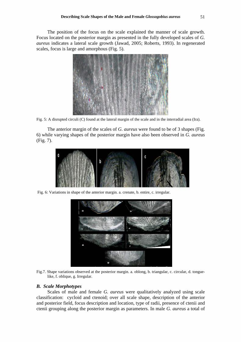
The position of the focus on the scale explained the manner of scale growth. Focus located on the posterior margin as presented in the fully developed scales of G. aureus indicates a lateral scale growth (Jawad, 2005; Roberts, 1993). In regenerated scales, focus is large and amorphous (Fig. 5).
Fig. 5: A disrupted circuli (C) found at the lateral margin of the scale and in the interradial area (Ira).
The anterior margin of the scales of G. aureus were found to be of 3 shapes (Fig. 6) while varying shapes of the posterior margin have also been observed in G. aureus (Fig. 7).
Fig. 6: Variations in shape of the anterior margin. a. crenate, b. entire, c. irregular.
Fig.7. Shape variations observed at the posterior margin. a. oblong, b. triangular, c. circular, d. tongue-
like, f. oblique, g. Irregular.
B. Scale Morphotypes Scales of male and female G. aureus were qualitatively analyzed using scale
classification: cycloid and ctenoid; over all scale shape, description of the anterior and posterior field, focus description and location, type of radii, presence of ctenii and ctenii grouping along the posterior margin as parameters. In male G. aureus a total of

Dulce-Amor P. Matondo et al. 52
21 scale morphotypes were observed (Figs. 8, 9, 11) while in females, there were 23 scale morphotypes described (Figs. 8, 10, 12).
Jawad (2005) claimed that scale shape variation do occur within different body regions in some species. These variations could be attributed on the rate of growth of the parts of the scale during scale development and that such developmental process could also be altered by environmental factors and the growth of the fish itself (Creaser, 1926). On the account of this principle, the various scale morphotypes identified were further classified into three categories: main scales, regenerated scales and the specialized scales. Main scales are the fully developed scales characterized by the presence of at least a 1° or a 1° and 2° radii, circuli, a ctenii (in ctenoid scales), an anterior, posterior and lateral fields and of a very distinct focus. Regenerated scales are characterized by the presence of 2° or 2° and 3° radii, circuli, a ctenii (in ctenoid scales), and an anterior, posterior and lateral fields. An unusual large indistinct or amorphous focus is an identifiable mark in these types of scales. Regenerated scales are developed as replacement of loss scales (Creaser, 1926). Specialized scales are scales that showed particular shapes and structures that prevent them from being categorized within the other major groups of scales (Chen, 2010). These specialized scales were observed to be confined within one region only.
For the main scale types, qualitative scale analysis of both male and female G. aureus revealed 3 morphotypes each of fully developed scales (Fig. 9).
Fig. 8: Main scale morphotypes in female (a) and male (b) G. aureus. Fig. 9: Different morphotypes of regenerated scales found in the different body regions of the male G.
aureus.

Describing Scale Shapes of the Male and Female Glossogobius aureus 53
Variation in scale morphology between sexes is best illustrated by the type 2 scale morphotypes as described in Table (2). As for the regenerated scales, males of G. aureus showed 14 regenerated scale morphotypes (Fig. 10) while 13 morphotypes were observed in females (Fig. 11). These regenerated scales were described, counted, and their distribution in the fish body are presented in Tables (3) (males) and (5) (females).
Fig. 10: Regenerated scales found in different body regions of the female G. aureus.
Fig. 11: Specialized scales in the male G. aureus.
Fig. 12: Specialized scales in female G. aureus.

Dulce-Amor P. Matondo et al. 54
Table 2: Description and distribution of main scale types in male and female G. aureus. Scale Type Description
Location No. of scales
examined Type 1 Male Female
small to medium ctenoid scales with a pentagonal scale shape; a crenate anterior margin with a triangular posterior field; 1°, 2° and 3° radii present with a distinct posteriorly located focus and 3 groups of ctenii on the posterior margin
All body regions
43 small to medium ctenoid scales with a pentagonal scale shape; a crenate anterior margin with a rounded posterior field; 1°, 2° and 3° radii present with a distinct posteriorly located focus and 3 groups of ctenii on the posterior margin
All body regions
49
Type 2 Male Female
small to medium size ctenoid scale with an oblong scale shape; a crenate anterior margin with an oblong posterior field; 10, 20 and 30 radii present with a distinct posteriorly located focus and 1 group of ctenii on the posterior margin
Regions A, Bc, Hi and J
21
small size ctenoid scales with a rounded scale shape; a crenate anterior margin with a triangular posterior field; 1°, 2° and 3° radii present with a distinct posteriorly located focus and 3 groups of ctenii on the posterior margin
Region A 12
Type 3 Male Female
small to medium size ctenoid scales with a cycloid scale shape; a crenate anterior margin with triangular posterior field; 1°, 2° and 3° radii present with a distinct posteriorly located focus and 3 groups of ctenii on the posterior margin
Regions A, Bc, De
16
small to medium size ctenoid scales with a cycloid scale shape; a crenate anterior margin with triangular posterior field; 1°, 2° and 3° radii present with a distinct posteriorly located focus and 3 groups of ctenii on the posterior margin
Regions A, Bc, De
9
C. Specialized Scales in Male and Female G. aureus
Table 3: Description and distribution of regenerated scale morphotypes in male G. aureus.
Scale Types
Description
Body Regions
No. of Scales
examined Type 1 Range from small to medium size ctenoid scale with a square-like scale shape, large-off centered
amorphous focus, a crenate anterior margin, an oblique posterior field, presence of 2° and 3° radii with 3 groups of ctenii growing on the posterior margin
Regions De, Hi
5
Type 2 Range from small to medium size ctenoid scale with a square-like scale shape, a large off-centered amorphous focus, a crenate anterior margin, a circularly-shaped posterior field, presence of 2° and 3° radii with 1 group of ctenii growing on the posterior margin
Regions De, Fg, Hi
8
Type 3 Range from small to medium size ctenoid scale with a square-like scale shape, large off –centered amorphous focus, a crenate anterior margin, a skewed-shaped posterior field, presence of 2° and 3° radii with 2 groups of ctenii growing on the posterior margin
Regions De, Fg, J
9
Type 4 Range from small to medium size ctenoid scale with a square-like scale shape, large off-centered amorphous focus, a crenate anterior margin, a tongue-like shaped posterior field, presence of 2° and 3° radii with 3 groups of ctenii growing on the posterior margin
Regions De, Fg
4
Type 5 Medium size ctenoid scale with a pentagonal scale shape, a large amorphous off-centered focus, a crenate anterior margin, a triangular-shaped posterior field, presence of 2° and 3° radii with 3 groups of ctenii growing on the posterior margin
Regions Bc, De
3
Type 6 Range from small to medium size ctenoid scale with an oval-like scale shape, a large off-centered amorphous focus, a crenate anterior margin, an oblique- shaped posterior field, presence of 2° and 3° radii with 3 groups of ctenii growing on the posterior margin
Regions De, Fg
2
Type 7 Range from small to medium size ctenoid scale with an oblong-like scale shape, large off-centered amorphous focus, a crenate anterior margin, an oblong shaped posterior field, presence of 2° and 3° radii with 1 group of ctenii growing on the posterior margin
Regions A, De, Hi
3
Type 8 Range from small to medium size ctenoid scale with an oblong-like scale shape, a small posteriorly located amorphous focus, a crenate anterior margin, an oblong shaped posterior field, presence of 2° and 30 radii with 1 group of ctenii growing on the posterior margin
Regions Fg, Hi
2
Type 9 Range from small to medium size ctenoid scale with a rectangular-like scale shape, large off-centered amorphous focus, a crenate anterior margin, a circular-shaped posterior field, presence of 2° and 3° radii with 1 group of ctenii growing on the posterior margin
Regions A, Bc, De, Fg
10
Type 10 Range from small to medium size ctenoid scale with an oblong-like scale shape a large off-centered amorphous focus, a crenate anterior margin, a skewed- shaped posterior field, presence of 2° and 3° radii with 1 group of ctenii growing on the posterior margin
Regions De, Fg, Hi
5
Type 11 Range from small to medium size ctenoid scale with an oblong-like scale shape characterized by a small posteriorly located amorphous focus, a crenate anterior margin, a skewed-shaped posterior field, presence of 2° and 3° radii with 1 group of ctenii growing on the posterior margin
Regions De, Fg, Hi
5
Type 12 Range from small to medium size ctenoid scale with a pentagonal-like scale shape characterized by a small posteriorly located amorphous focus, a crenate anterior margin, a triangular-shaped posterior field, presence of 2° and 3° radii with 3 groups of ctenii growing on the posterior margin
Regions De, Fg
6
Type 13 A medium size flabellate-shaped ctenoid scale characterized by a crenate anterior margin, a large off-centered amorphous focus with a truncate shape posterior margin where 1 group of ctenii was growing
Region De
2
Type 14 A small size irregularly shaped ctenoid scale characterized by a crenate anterior margin, a small amorphous posteriorly located focus with an irregularly shaped posterior margin where 2 groups of ctenii were growing
Region Fg 1

Describing Scale Shapes of the Male and Female Glossogobius aureus 55
Four types of specialized scales were found in male G. aureus while 7 morphotypes of specialized scales were described in the female. Illustrated in Figure (11) (male) and Fig. (12) (female), these specialized scales were described, counted, and its distribution in the fish body is provided in Tables (4) (males) and table (6) (females). Table 4: Description and distribution of specialized scales in male G. aureus.
Scale Types
Description
Body Region
No. of Scales
examined Type 1 small size cycloid scale with an oblong scale shape, a large-off centered amorphous focus, a slightly
crenate anterior margin, an skewed posterior field and presence of few 2° and 3° Region
A
1 Type 2 small size cycloid scale with a rounded-scale shape, a centrally located distinct focus, an entire
anterior margin, a cuneate-shaped posterior field and absence of radii Region
A
1 Type 3 small size cycloid scale with an oval scale shape, a large-off centered amorphous focus, a crenate
anterior margin, an oblong posterior field and presence of few 2° and 3° radii Region
Hi
2 Type 4 A small size obtuse-shaped ctenoid scale, a sinuate anterior margin, a distinct posteriorly located
focus with a skewed shape posterior margin and 1° , 2° and 3° radii Region
A
2
Table 5: Description and distribution of regenerated scale morphotypes in female G. aureus.
Scale Types
Description
Body Region
No. of Scales
examined Type 1 small size ctenoid scale with rounded scale shape, a large-off centered amorphous focus, a crenate
anterior margin, a circular shaped posterior field, presence of 2° and 3° radii with 1 group of ctenii growing on the posterior margin
Region A 2
Type 2 small size ctenoid scale with a square-like scale shape, a large off-centered amorphous focus, a crenate anterior margin, an oblique-shaped posterior field, presence of 2° and 3° radii with 3 groups of ctenii growing on the posterior margin
Region Hi
2
Type 3 small size ctenoid scale with a square-like scale shape, a large off-centered amorphous focus, a crenate anterior margin, a circular-shaped posterior field, presence of 2° and 3° radii with 1 group of ctenii growing on the posterior margin
Regions Hi, J
3
Type 4 small to medium size ctenoid scale with a square-like scale shape, a large off-centered amorphous focus, a crenate anterior margin, a skewed-shaped posterior field, presence of 2° and 3° radii with 2 groups of ctenii growing on the posterior margin
Regions Fg, Hi, J
9
Type 5 Medium size ctenoid scale with a square-like scale shape, a large off-centered amorphous focus, a crenate anterior margin, a tongue-like shaped posterior field, presence of 2° and 3° radii with 3 groups of ctenii growing on the posterior margin
Region Hi 1
Type 6 small to medium size ctenoid scale with an oblong scale shape, a large off-centered amorphous focus, a crenate anterior margin, an oblique-shaped posterior field, presence of 2° and 3° radii with 3 groups of ctenii growing on the posterior margin
Regions Bc, Hi, J
3
Type 7 small to medium size ctenoid scale with an oblong scale shape, a large off-centered amorphous focus, a crenate anterior margin, a circular-shaped posterior field, presence of 2° and 3° radii with 1 group of ctenii growing on the posterior margin
Regions A, Bc, De,
J
22
Type 8 small to medium size ctenoid scale with an oblong scale shape , a large off-centered amorphous focus, a crenate anterior margin, an oblong- shaped posterior field, presence of 20 and 30 radii with 1 group of ctenii growing on the posterior margin
Regions Bc, De, Fg, J
20
Type 9 small to medium size ctenoid scale with an oblong scale shape, a large off-centered amorphous focus, a crenate anterior margin, a skewed-shaped posterior field, presence of 2° and 3° radii with 2 groups of ctenii growing on the posterior margin
Region Hi
1
Type 10
small to medium size ctenoid scale with an oblong scale shape, a large off-centered amorphous focus, a crenate anterior margin, a tongue-like shaped posterior field, presence of 2° and 3° radii with 3 groups of ctenii growing on the posterior margin
Regions De, Fg,
2
Type 11
small to medium size ctenoid scale with an oval scale shape, a large off-centered amorphous focus, a crenate anterior margin, an oblong-shaped posterior field, presence of 2° and 3° radii with 1 group of ctenii growing on the posterior margin
Regions Hi, J
2
Type 12
small size ctenoid scale with an oblong scale shape, a large off-centered amorphous focus, a crenate anterior margin, a circular-shaped posterior field, presence of 2° and 3° radii with 2 groups of ctenii growing on the posterior margin
Region A
1
Type 13
small to medium size ctenoid scale with a pentagonal scale shape, a large off-centered amorphous focus, a crenate anterior margin, a skewed-shaped posterior field, presence of 2° and 3° radii with 2 groups of ctenii growing on the posterior margin
Regions Bc, De
2

Dulce-Amor P. Matondo et al. 56
Table 6: Description and distribution of specialized scale in female G. aureus. Scale Type
Description
Body region
No. of Scales
examined Type 1 small size ctenoid scale with an irregular scale shape, a distinct posteriorly located focus, a narrow
crenate anterior margin, an oblong shaped posterior field and presence of few 1°, 2° and 3° with 1 group of ctenii growing on the posterior margin
Region A
3
Type 2 small size ctenoid scale with an oval scale shape, a distinct posteriorly located focus, an irregular anterior margin, an oblong-shaped posterior field and presence of few 1°, 2° and 3° radii with 1 group of ctenii growing on the posterior margin
Region A
1
Type 3 small size ctenoid scale with a flabellate scale shape, distinct posteriorly located focus, a crenate anterior margin, a triangular posterior field and presence of few 1°, 2° and 3° radii 3 groups of ctenii growing on the posterior margin
Region A
1
Type 4 small size ctenoid scale with a flabellate scale shape, a distinct posteriorly located focus, an entire anterior margin, an oblong posterior field and presence of few 1°, 2° and 3° radii with 1 group of ctenii growing on the posterior margin
Region A
1
Type 5 small size ctenoid scale with an obcordate scale shape, a distinct posteriorly located focus, a depressed but entire anterior margin, an oblong-shaped posterior field and presence of few 1°, 2° and 3° radii with 1 group of ctenii growing on the posterior margin
Region Hi
1
Type 6 A small size oblong-like shaped ctenoid scale, a crenate anterior margin, a distinct posteriorly located focus with an irregularly shaped posterior margin where 2 groups of ctenii were growing
Region Hi
1
Type 7 A small size obtuse-shaped ctenoid scale , a crenate anterior margin, a distinct posteriorly located focus with a circular shape posterior margin and few 1° , 2° and 3° radii with 1 group of ctenii growing on the posterior margin
Region A
1
Results of this study have shown that qualitatively, morphological variation
existed within body regions in both sexes of G. aureus in terms of the type of scale, over all scale shape, focus description and location, presence of ctenii, shape of the posterior margin and the number of groupings of ctenii along the posterior margin. While all scales in the females were ctenoid, the males showed the presence of both ctenoid and cycloid scales. Shape variations in fully developed scales were also observed within males and females of G. aureus. Shape variation in fully developed scales between sexes was established providing an evidence that these fishes are sexually dimorphic. It is important to note however that in using scale shape morphology to describe sexual dimorphism in fishes, fully developed scales should be used for comparison as these scales have fewer variations to consider than with regenerated scales.
ACKNOWLEDGEMENT The senior author would like to thank the Commission of Higher Education
(CHED) for the scholarship grant and the Western Mindanao State University, Zamboanga City for the faculty privilege.
REFERENCES
Akihito and Meguro, K. (1975a). First record of Glossobius celebius from Japan. Japanese Journal of Icthyology, 21(4): 227-230, figs. 1-4. In Japanese.
Akihito and Meguro, K. (1975b). Description of a new gobiid fish G. aureus, with notes on related species of the genus. Japanese Journal of Icthyology, 22(3):127-142.
Bagorodsky, S., Kovacic, M. Ozen, O., and Bilecenogin, M. (2010). Records of two uncommon goby species (Millerigobius macrocephalus, Zebrus zebrus) from the Aegean Sea. Acta Adriatica, 51(2):217- 222.
Cadrin, S. (2000). Advances in morphometric identification of fishery stocks. Reviews in Fish Biology and Fisheries, 10: 91-112.
Describing Scale Shapes of the Male and Female Glossogobius aureus 57
Casselman, J., Collins, J., Crossman, E., Ihssen, P. and Spanger, G. (1981). Lake whitefish (Coregonus clupeaformis) stocks of the Ontario waters of Lake Huron. Canadian Journal of Fisheries and Aquatic Sciences, 38: 1772-1779.
Chen, D. (2010). Squamation in Andreolopis from the late Silurian of Sweden. A degree project in Biology, Master of Science, Biology Education Center, Uppsala University and Department of Organismal Biology, 31 pp.
Creaser, C. (1926). The structure and growth of the scales of fishes in relation to the interpretation of their life history, with special reference to the sunfish Eupomotis gibbosus. Museum of Zoology, University of Michigan, Miscellaneous Publications No. 17. 84pp. Published by the University, Ann Harbor, Michigan, December 16, 1926.
De Pontual, H. and Prouzet, P. (1987). Atlantic salmon, Salmo salar L., stock discrimination by scale shape analysis. Aquaculture and Fisheries Management, 18: 227-289.
Esmaeili, H. and Gholami, Z. (2011). Scanning electron microscopy of the scale morphology in cyprinid fish, Rutilus frisii kutum Kamenskii, 1901 (Actinopterygii: Cyprinidae). Iranian Journal of Fisheries Sciences, 10(1): 155-166.
Esmaeili, H.R., Hojat Ansari, T, and Teimory, A. (2007). Scale structure of a cyprinid fish, Capoeta damascina (Valenciennes in Cuvier and Valenciennes, 1842) using scanning electron microscope (SEM). Iranian Journal of Science and Technology, Transaction A, 31(A3): 255-262.
Fraisse, L. (1990). Essai de discrimination de deux populations de vandoise (Leuciscus leuciscus L., 1758) parétude de la forme de leurs écailles: aspects methodologiques. Hydroécologie Appliquée, pp. 135-149.
Ibañez, A. and O’Higgins, P. (2011). Identifying fish scales: The influence of allometry on scale shape and classification. Fisheries Research, 109(1): 54-60.
Jarvis, R., Klodowski, H. and Sheldon, S. (2005). New method of quantifying scale shape and an application to stock identification in Walleye (Stizostedion vitreum vitreum). Transactions of the American Fisheries Society, 107: 528- 543.
Jawad, L. (2005). Comparative scale morphology and squamation patterns in triplefins (Pisces: Teleostei: Perciformes: Tripterygiidae). Tuhinga, 16: 137-167.
Johal, M. (2005). Recent innovations in the determinations in age determination using hard parts in Indian freshwater fishes, pp. 91-98, In: New Horizons in Animal Sciences (Eds. Sobti, R. and Sharma, V.), Jalandhar, Punjab, Visual Publishing Company.
Johal, M. and Thomas, N. (2000). EMSI Bull. 1(1): 16-19. Klappenbach, L. (2010). Animal/Wildlife Guide: What is sexual dimorphism?
About.com Guide, a part of the New York Times Company. Matondo, D., Torres, M., Tabugo, S. and Demayo, C. (2010). Describing variations in
scales between sexes of the yellowstriped goatfish, Upeneus vittatus ( Forskal, 1775) (Perciformes: Mullidae). Egypt. Acad. J. biolog. Sci., 2(1): 37-50.
Patterson, R., Wright, C., Chang, A., Taylor, L., Lyons, P., Dallimore, A. and Kumar, A. (2002) Atlas of squamatological (fish scale) material in coastal British Columbia and an assessment of the utility of various scale types in paleofisheries reconstruction. Paleontologica Electronica, 4(1): 88pp.
Poulet, N., Reyjol, Y., Collier, H. and Lek, S. (2005). Does fish scale morphology allow the identification of populations at a local scale? A case study for rostrum dace Leuciscus leuciscus burdigalensis in River Viaur (SW) France, Aquat. Sci., 67: 122-127.

Dulce-Amor P. Matondo et al. 58
Richards, A. and Esteves, C. (1997). Stock-specific variation in scale morphology of atlantic striped Bass. Transactions of the American fishery Soc., 126: 908-918.
Roberts, C. (1993). Comparative morphology of spined scales and their phylogenetic significance in the Teleostei. Bulletin of Marine Science, 52: 60-113.
Sanda R. and Kovacic, M. (2009). Freshwater gobies in the Adriatic Drainage Basin of the Western Balkans. Annales. Ser. Hist. Nal., 19: 1-10.
Tandon, K. and Johal, M. (1994). Scales, a tool in fish biology, pp. 1-11, In: Advances in Fish Biology (Ed. Singh, H.), Delhi, India, Hindustan Publishing Corporation.
Taylor, H. (1914). The structure and growth of the scales of the squeteague and the pigfish as indicative of life history, U.S. Bureau of Fish., Bulletin 34: 289-330.