department of radiation oncology university of michigan
TRANSCRIPT

Department of Radiation OncologyUniversity of Michigan
Department of Radiation OncologyDepartment of Radiation OncologyUniversity of MichiganUniversity of Michigan
TheThe
Video and Surface PositioningVideo and Surface Positioning
Scott W. Hadley PhDScott W. Hadley PhD
I for one welcome our AI SUV overlords

AAPM Summer School 2006
ContributorsContributors
• Scott Hadley University of Michigan
• Scott Johnson Varian Medical Systems
• Shidong Li Henry Ford Hospital
• Marco Riboldi TMB Lab @ Politecnico di Milano
• Christoph Bert GSI & MGH

AAPM Summer School 2006
Goal of Patient PositioningGoal of Patient Positioning
• Reproduce patient position as seen in CT simulation– Aligning Skin Marks with Laser
– Matching Boney Anatomy with MV
– Target Positioning with US, markers, MV, kV, CBCT

AAPM Summer School 2006
Goal of Patient PositioningGoal of Patient Positioning
• Frequency of Position Verification
– First Day and weekly
– Adaptive protocol, image for several days, move to average then weekly
– Daily positioning or target localization

AAPM Summer School 2006
Advantages of Video & SurfaceAdvantages of Video & Surface
• No ionizing radiation– Daily use with no extra radiation
– Operators can be at couch side for positioning
• Video cameras see what we see
• Can be "no touch" and "no marks"

AAPM Summer School 2006
Advantages of Video & SurfaceAdvantages of Video & Surface
• Quantitative 3D couch positioning, 6 parameter rigid transformation
• Intrafraction patient monitoring
• 4D acquisition
• Non-rigid positioning possible

AAPM Summer School 2006
Disadvantages of Video & SurfaceDisadvantages of Video & Surface
• Positioning based on skin surface only– Skin surface as a surrogate for target
anatomy
• Can be obstructed by clothes, mask, other objects

AAPM Summer School 2006
History of Video TechniquesHistory of Video Techniques• 1972 Kelsey, C.A., R.G. Lane, and W.G. Connor, Measurement
of patient movement during radiation therapy. Radiology. 103(3): p. 697-8.
• 1975 Connor, W.G., et al., Patient repositioning and motion detection using a video cancellation system. Int J Radiat OncolBiol Phys. 1(1-2): p. 147-53
• 1977 Renner, W.D., et al., The use of photogrammetry in tissue compensator design. Part II: experimental verification of compensator design. Radiology,
• 1982 Andrew, J.W. and J.E. Aldrich, A microcomputer-based system for radiotherapy beam compensator design and patient contour plotting. Med Phys. 9(2): p. 279-83
AAPM Summer School 2006
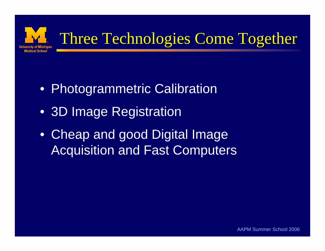
Three Technologies Come TogetherThree Technologies Come Together
• Photogrammetric Calibration
• 3D Image Registration
• Cheap and good Digital Image Acquisition and Fast Computers

AAPM Summer School 2006
Image Guided SurgeryImage Guided Surgery
Grimson, W.E.L., et al., An automatic registration method for frameless stereotaxy, image guided surgery, and enhanced reality visualization. IEEE Transactions on Medical Imaging, 1996. 15(2): p. 129-140

AAPM Summer School 2006
In Room (Spy) CamerasIn Room (Spy) Cameras
IsocenterIsocenter
AP cameraAP camera Left lateral cameraLeft lateral camera
Right lateral cameraRight lateral camera
Sagittal cameraSagittal camera

AAPM Summer School 2006
Live Video SubtractionLive Video Subtraction

AAPM Summer School 2006
Volume Rendered Ref’ ImagesVolume Rendered Ref’ Images
Volume Rendered Reference image from CT
Live Video of Patient on Table
Video Subtraction for positioning
AAPM Summer School 2006
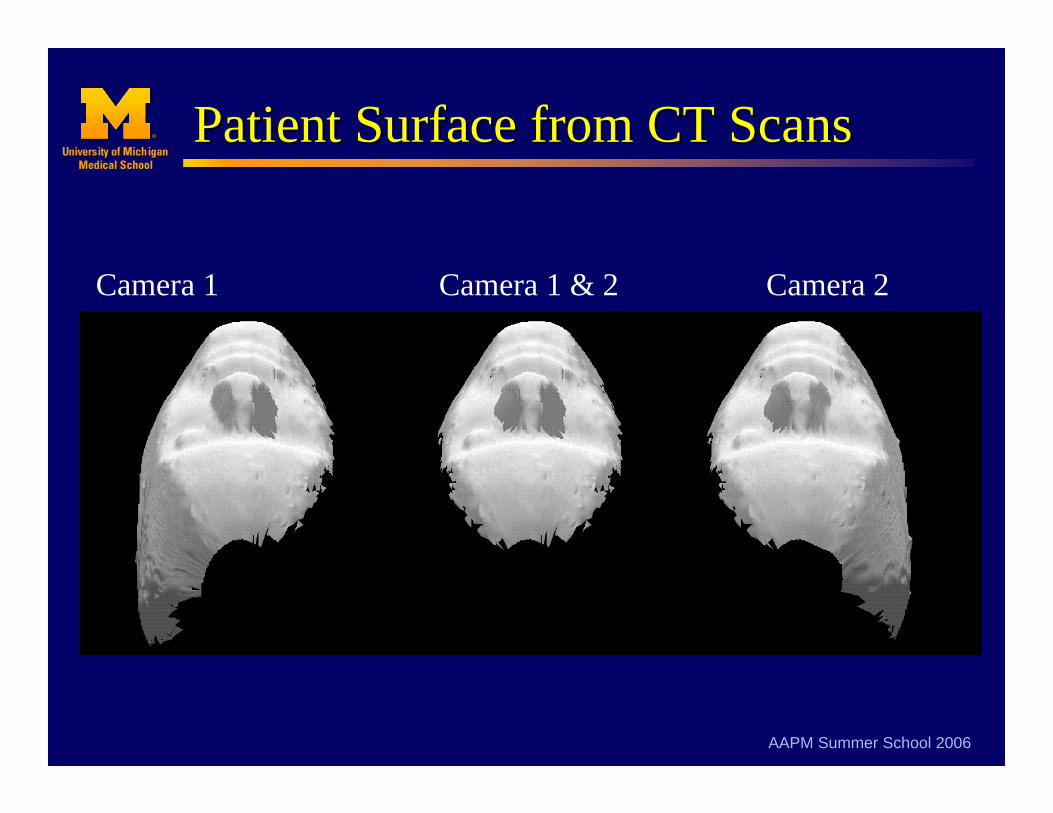
Patient Surface from CT ScansPatient Surface from CT Scans
Camera 1 Camera 2Camera 1 & 2

AAPM Summer School 2006
3D Surface to 2D Image Matching3D Surface to 2D Image Matching

AAPM Summer School 2006
Latest TechnologyLatest Technology
• 3D Surface Scanners– Video + Laser, interfermometry, light pattern
– Large FOV
– Multiple Frames per second
• Surface Registration software– 6 parameter rigid transformation
– Direct measurement of positioning error
AAPM Summer School 2006

AAPM Summer School 2006

AAPM Summer School 2006

AAPM Summer School 2006

AAPM Summer School 2006

AAPM Summer School 2006

Surface imaging at MGH
3D surface imaging for PBI setup
Christoph Bert1/2, Katie Metheany2,Karen Doppke2/3, Alphonse Taghian2/3,
Simon N. Powell2/3, George T.Y. Chen2/3
1Gesellschaft für Schwerionenforschung, Darmstadt, Germany2Massachusetts General Hospital, Boston, Ma
3Harvard Medical School, Cambridge, Ma

Surface imaging at MGH
Shape conservation CT-stereovision• “Patient”
contour TPS– Binary cube
fill →marching cubes →decimation
• Any surface image
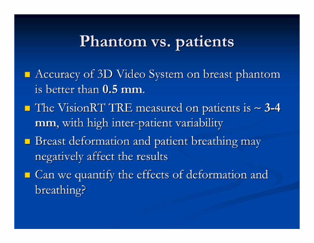
• Surface registration →distance projection →0.65mm RMS
+2-2mm

Surface imaging at MGH
Comparison to CT surface• Purpose: Check if CT, linac, and 3D
camera coordinate systems match• Methods:
– CT scan phantom, extract surface, DRRs– a) line up according to 3D surface ⇒
b) compare portfilm with DRR (lat.+a.p.)– b) ⇒ a)

Surface imaging at MGH
Surface data + Setup
3D surfaces and originMarkerblock

Surface imaging at MGH
Film samples
a.p. lateral

Surface imaging at MGH
Combined results - alignment

Surface imaging at MGH
Stability test - results

AAPM Summer School 2006
AAPM Summer School 2006

AAPM Summer School 2006
AAPM Summer School 2006

AAPM Summer School 2006

AAPM Summer School 2006

AAPM Summer School 2006

AAPM Summer School 2006

AAPM Summer School 2006

AAPM Summer School 2006
AAPM Summer School 2006

AAPM Summer School 2006

AAPM Summer School 2006
ConclusionsConclusions
• What role is it going to play– Laser replacement?
– Daily Targeting of underlying target?
– Monitor patient during treatment?
– Tracking and Gating?
If You're Considering Video/SurfaceIf You're Considering Video/Surface

AAPM Summer School 2006
ConclusionsConclusions
• What Body Site– Head / Intracranial works well.
– PBI near surface works well.
– PBI near chest wall X-ray chest wall.
– Prostate not so well but may be an aid to the therapists.
If You're Considering Video/SurfaceIf You're Considering Video/Surface
AAPM Summer School 2006
ConclusionsConclusions
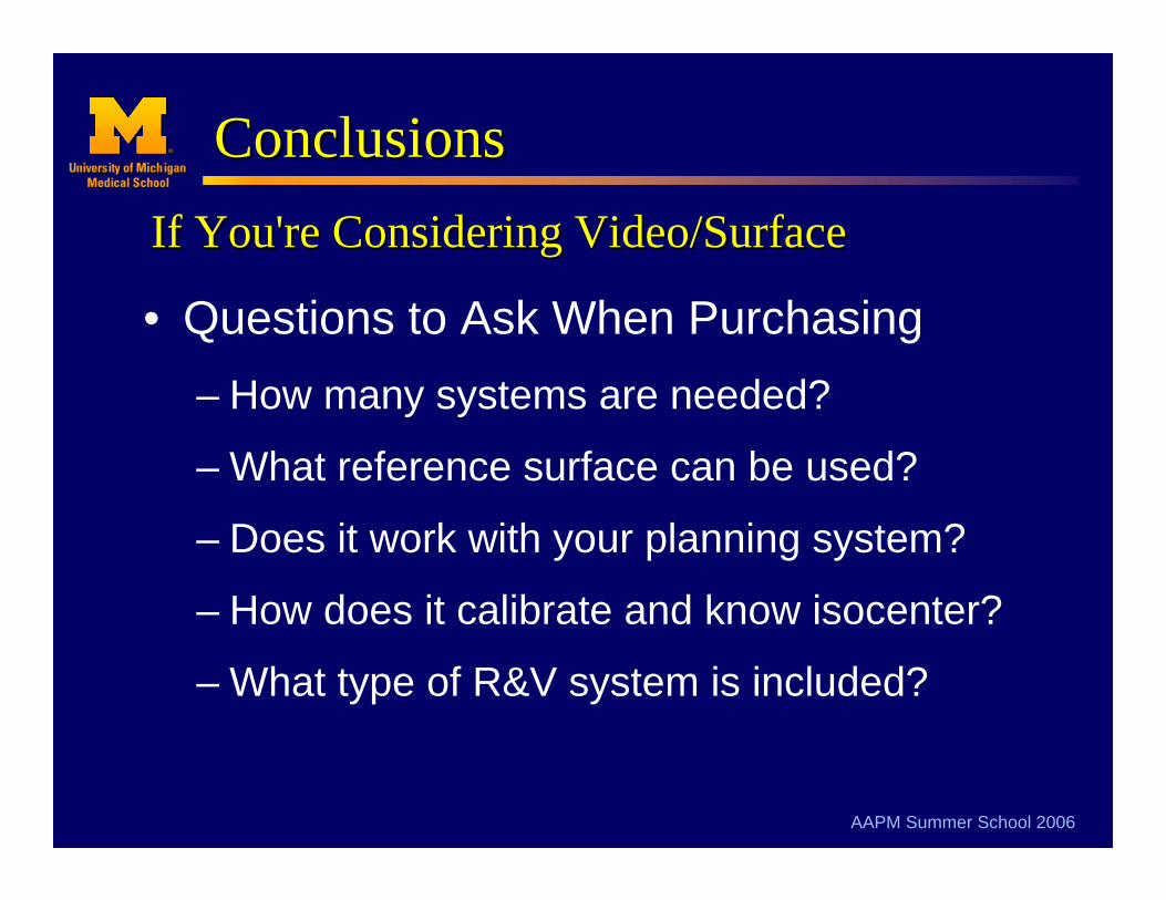
• Questions to Ask When Purchasing– How many systems are needed?
– What reference surface can be used?
– Does it work with your planning system?
– How does it calibrate and know isocenter?
– What type of R&V system is included?
If You're Considering Video/SurfaceIf You're Considering Video/Surface

AAPM Summer School 2006
ConclusionsConclusions
• Installation, Commissioning and QA– Manufacture’s CAP
– Commissioning• Known phantom and site specific
– QA• Daily isocenter check
• Longer term stability
If You're Considering Video/SurfaceIf You're Considering Video/Surface