radiation oncology advances
TRANSCRIPT

Radiation Oncology Advances
Mehta_FM.indd iMehta_FM.indd i 8/28/2007 6:24:27 PM8/28/2007 6:24:27 PM
Cancer Treatment and ResearchSteven T. Rosen, M.D., Series Editor
Arnold, A.J. (ed.): Endocrine Neoplasms. 1997. ISBN 0-7923-4354-9.Pollock, R.E. (ed.): Surgical Oncology. 1997. ISBN 0-7923-9900-5.Verweij, J., Pinedo, H.M., Suit, H.D. (eds): Soft Tissue Sarcomas: Present Achievements and Future Prospects. 1997. ISBN 0-7923-9913-7.Walterhouse, D.O., Cohn, S. L. (eds): Diagnostic and Therapeutic Advances in Pediatric Oncology. 1997. ISBN 0-7923-9978-1.Mittal, B.B., Purdy, J.A., Ang, K.K. (eds): Radiation Therapy. 1998. ISBN 0-7923-9981-1.Foon, K.A., Muss, H.B. (eds): Biological and Hormonal Therapies of Cancer. 1998. ISBN 0-7923-9997-8.Ozols, R.F. (ed.): Gynecologic Oncology. 1998. ISBN 0-7923-8070-3.Noskin, G.A. (ed.): Management of Infectious Complications in Cancer Patients. 1998. ISBN 0-7923-8150-5.Bennett, C.L. (ed.): Cancer Policy. 1998. ISBN 0-7923-8203-X.Benson, A.B. (ed.): Gastrointestinal Oncology. 1998. ISBN 0-7923-8205-6.Tallman, M.S., Gordon, L.I. (eds): Diagnostic and Therapeutic Advances in Hematologic Malignancies. 1998. ISBN 0-7923-8206-4.von Gunten, C.F. (ed.): Palliative Care and Rehabilitation of Cancer Patients. 1999. ISBN 0-7923-8525-X. Burt, R.K., Brush, M.M. (eds): Advances in Allogeneic Hematopoietic Stem Cell Transplantation. 1999. ISBN 0-7923-7714-1.Angelos, P. (ed.): Ethical Issues in Cancer Patient Care 2000. ISBN 0-7923-7726-5.Gradishar, W.J., Wood, W.C. (eds): Advances in Breast Cancer Management. 2000. ISBN 0-7923-7890-3.Sparano, J.A. (ed.): HIV & HTLV-I Associated Malignancies. 2001. ISBN 0-7923-7220-4.Ettinger, D.S. (ed.): Thoracic Oncology. 2001. ISBN 0-7923-7248-4.Bergan, R.C. (ed.): Cancer Chemoprevention. 2001. ISBN 0-7923-7259-X.Raza, A., Mundle, S.D. (eds): Myelodysplastic Syndromes & Secondary Acute Myelogenous Leukemia 2001. ISBN: 0-7923-7396.Talamonti, M.S. (ed.): Liver Directed Therapy for Primary and Metastatic Liver Tumors. 2001. ISBN 0-7923-7523-8.Stack, M.S., Fishman, D.A. (eds): Ovarian Cancer. 2001. ISBN 0-7923-7530-0.Bashey, A., Ball, E.D. (eds): Non-Myeloablative Allogeneic Transplantation. 2002. ISBN 0-7923-7646-3.Leong, S.P.L. (ed.): Atlas of Selective Sentinel Lymphadenectomy for Melanoma, Breast Cancer and Colon Cancer. 2002. ISBN 1-4020-7013-6.Andersson, B., Murray, D. (eds): Clinically Relevant Resistance in Cancer Chemotherapy. 2002. ISBN 1-4020-7200-7.Beam, C. (ed.): Biostatistical Applications in Cancer Research. 2002. ISBN 1-4020-7226-0.Brockstein, B., Masters, G. (eds): Head and Neck Cancer. 2003. ISBN 1-4020-7336-4.Frank, D.A. (ed.): Signal Transduction in Cancer. 2003. ISBN 1-4020-7340-2.
(continued after index)
Mehta_FM.indd iiMehta_FM.indd ii 8/28/2007 6:24:27 PM8/28/2007 6:24:27 PM

Radiation Oncology Advances
Søren M. Bentzen, Ph.D., D.Sc.ProfessorDepartment of Human OncologyUniversity of Wisconsin School of MedicineMadison, Wisconsin, USA
Paul M. Harari, M.D.Jack Fowler Professor and ChairmanDepartment of Human OncologyUniversity of Wisconsin School of MedicineMadison, Wisconsin, USA
Wolfgang A. Tomé, M.D., Ph.DAssociate ProfessorDepartment of Human OncologyUniversity of Wisconsin School of MedicineMadison, Wisconsin, USA
Minesh P. Mehta, M.D.ProfessorDepartment of Human OncologyUniversity of Wisconsin School of MedicineMadison, Wisconsin, USA
Mehta_FM.indd iiiMehta_FM.indd iii 8/28/2007 6:24:27 PM8/28/2007 6:24:27 PM
Edited by

Søren M. Bentzen, PhDPaul M. Harari, MDWolfgang A. Tomé, PhDMinesh P. Mehta, MD
University of WisconsinDepartment of Human Oncology600 Highland Avenue, K4/310Madison, WI 53792 USA
Series Editor:Steven T. RosenRobert H. Lurie Comprehensive Cancer CenterNorthwestern UniversityChicago, ILUSA
ISBN-13: 978-0387-36743-9 e-ISBN-13: 978-0387-36744-6
Library of Congress Control Number: 2007925437
All rights reserved. This work may not be translated or copied in whole or in part without the written permission of the publisher (Springer Science + Business Media, LLC, 233 Spring Street, New York, NY 10013, USA), except for brief excerpts in connection with reviews or scholarly analysis. Use in connection with any form of information storage and retrieval, electronic adaptation, computer software, or by similar or dissimilar methodology now known or hereafter developed is forbidden.The use in this publication of trade names, trademarks, service marks, and similar terms, even if they are not identified as such, is not to be taken as an expression of opinion as to whether or not they are subject to proprietary rights.
Printed on acid-free paper.
9 8 7 6 5 4 3 2 1
springer.com
Mehta_FM.indd ivMehta_FM.indd iv 8/28/2007 6:24:28 PM8/28/2007 6:24:28 PM
© 2008 Springer Science + Business Media, LLC

Contents
1 Radiation Oncology Advances: An Introduction ..................................... 1Søren M. Bentzen, Paul M. Harari, Wolfgang A. Tomé, and Minesh P. Mehta
Advances in Imaging and Theragnostic Radiation Oncology ....................... 1Advances in Molecular Biology and Targeted Therapies ............................. 2Advances in Treatment Delivery and Planning ............................................. 3Clinical Advances .......................................................................................... 4References ...................................................................................................... 4
Section I. Advances in Imaging and Biologically-Based Treatment Planning
2 Advanced Image-Guided External Beam Radiotherapy ......................... 7Thomas Rockwell Mackie and Wolfgang Tomé
Introduction .................................................................................................... 7Image Guidance for Defining Target Volumes ......................................... 9Image Guidance at the Time of Delivery ................................................... 13Optical Guidance ....................................................................................... 14Optical Tracking Systems .......................................................................... 15Optical Tracking in Fractionated Stereotactic Radiotherapy, Intracranial, and Head and Neck IMRT ..................................................... 16Optically Guided Ultrasound ..................................................................... 18In-Room CT Guidance ............................................................................... 20Image Guidance and Organ Motion ........................................................... 27Image Guidance for Follow-Up Imaging and Retreatments ...................... 29Summary .................................................................................................... 31
References ...................................................................................................... 32
v
Mehta_FM.indd vMehta_FM.indd v 8/28/2007 6:24:28 PM8/28/2007 6:24:28 PM

3 Dose Painting and Theragnostic Imaging: Towards the Prescription, Planning and Delivery of Biologically Targeted Dose Distributions in
External Beam Radiation Oncology ........................................................ 40Søren M. Bentzen
Radiation Theragnostics .............................................................................. 40From Anatomical to Biological Targeting in Radiation Therapy ............... 41From Target Selection and Delineation to 4D Dose Prescription ............... 42The Case for Nonuniform Theragnostic Dose Distributions....................... 43Precision Requirements ............................................................................... 46Targeting Hypoxia Using EBRT: Are We Ready for Dose Painting by Numbers? .................................................................................. 47
Hypoxia as a Cause of Clinical Failure of Radiation Therapy ................ 47Hypoxia Imaging ..................................................................................... 49Spatiotemporal Stability of the PET Hypoxia Map ................................. 52Dose Painting by Numbers ...................................................................... 55Dose Delivery and Expected Change in Outcome ................................... 56
Conclusion ................................................................................................... 56References .................................................................................................... 57
4 Molecular and Functional Imaging in Radiation Oncology .................. 62Robert Jeraj and M. Elizabeth Meyerand
Introduction .................................................................................................. 62Molecular and Functional Imaging Modalities ........................................... 63
Positron Emission Tomography ............................................................... 63Single Photon Emission Tomography ..................................................... 64Dynamic Contrast Enhanced Computer Tomography (DCE-CT) ........... 64Dynamic Contrast Enhanced Magnetic Resonance Imaging (DCE-MRI) ................................................................................ 64Magnetic Resonance Spectroscopy .......................................................... 65Optical Imaging ....................................................................................... 65Comparison Between Different Imaging Modalities ............................... 65
Molecular and Functional Imaging Targets................................................. 66Cellular Metabolism ................................................................................. 67Cellular Proliferation ............................................................................... 71Cellular Death .......................................................................................... 72Cellular Regulation .................................................................................. 73Tumor Microenvironment ........................................................................ 76
Future ........................................................................................................... 79References .................................................................................................... 81
vi Contents
Mehta_FM.indd viMehta_FM.indd vi 8/28/2007 6:24:28 PM8/28/2007 6:24:28 PM

Contents vii
5 Prognostic and Predictive Markers in Radiation Therapy: Focus on Prostate Cancer .................................................................................... 95
Mark A. Ritter
Introduction .................................................................................................. 95The Need for Biomarkers of Radiation Response in Prostate Cancer ........................................................................................ 95Optimal Biomarkers and Patient Cohort Characteristics............................. 96Evaluation of Candidate Markers ................................................................ 97
Biological Rationale ................................................................................. 97Biomarker Frequency ............................................................................... 100
Biomarker Assessment Methods ................................................................. 100Immunohistochemistry ................................................................................ 101Clinical Correlative Data in Prostate Cancer............................................... 102
Markers of Cell Cycle Control, DNA Repair and Apoptosis .................. 103Proliferation ............................................................................................. 103Hypoxia .................................................................................................... 104
Limitations of Existing Studies ................................................................... 104Future Studies and Directions ...................................................................... 105
Large Prospective Clinical Trials ............................................................. 105Biomarker-Based Adaptive Therapy ....................................................... 106
Conclusion ................................................................................................... 107References .................................................................................................... 107
Section II. Advances in Molecular Biology and Targeted Therapies
6 Overview of Cancer Molecular Radiobiology ......................................... 115Jann N. Sarkaria and Robert G. Bristow
Introduction .................................................................................................. 115Interaction of Radiation with Living Cells .................................................. 115Cellular Response to Ionizing Radiation ..................................................... 116Cell Cycle Arrest ......................................................................................... 116DNA Repair ................................................................................................. 118Apoptosis ..................................................................................................... 119Cell Survival Signaling ................................................................................ 120
Ras Signaling ........................................................................................... 120Receptor Tyrosine Kinases ...................................................................... 121mTOR Signaling ...................................................................................... 121
Targeting Housekeeping Proteins ................................................................ 122HSP90 Inhibitors ...................................................................................... 123HDAC Inhibitors ...................................................................................... 124Proteosome Inhibitors .............................................................................. 126
Conclusion ................................................................................................... 127References .................................................................................................... 128
Mehta_FM.indd viiMehta_FM.indd vii 8/28/2007 6:24:28 PM8/28/2007 6:24:28 PM

7 Clinical Application of EGFR Inhibitors in Head and Neck Squamous Cell Cancer .............................................................................. 132
Igor Astsaturov, Roger B. Cohen, and Paul M. Harari
Introduction .................................................................................................. 132EGFR Biology ............................................................................................. 133Anti-EGFR Monoclonal Antibodies ............................................................ 135
Radiation Plus Cetuximab For Locoregionally Advanced HNSCC ........ 135Cetuximab, Cisplatin, and Radiation in Locoregionally Advanced HNSCC ................................................................................... 137Cetuximab ± Chemotherapy in Recurrent and/or Metastatic HNSCC ....................................................................... 138Cetuximab with Chemotherapy in the First-Line Treatment of Patients with Recurrent and/or Metastatic HNSCC ............................ 139
EGFR Tyrosine Kinase Inhibitors (TKIs) ................................................... 140TKI Monotherapy in HNSCC .................................................................. 141TKIs in Combination with Radiation Therapy ........................................ 141TKIs with Dual Specificity ...................................................................... 142
Patient Selection .......................................................................................... 142Conclusions .................................................................................................. 143References .................................................................................................... 144
8 Advancement of Antiangiogenic and Vascular Disrupting Agents Combined with Radiation ......................................................................... 150
Deborah Citrin and Kevin Camphausen
Introduction .................................................................................................. 150Tumor Vasculature ...................................................................................... 150Targeting the Tumor Vasculature ................................................................ 152Antiangiogenic Agents ................................................................................ 152Vascular Disrupting Agents ......................................................................... 155Combining Antiangiogenic and Vascular Disrupting Agents with Radiation .............................................................................................. 157Antiangiogenic Agents and Radiation in the Laboratory ............................ 157
Angiogenesis Inhibitors and Radiation in the Clinic ............................... 159Vascular Disrupting Agents and Radiation in the Laboratory ................. 160Vascular Disrupting Agents with Radiation in the Clinic ....................... 161
Future Directions ......................................................................................... 162Conclusion ................................................................................................... 163References .................................................................................................... 164
9 Overcoming Therapeutic Resistance in Malignant Gliomas: Current Practices and Future Directions ............................................................... 169
Arnab Chakravarti and Kamalakannan Palanichamy
Introduction .................................................................................................. 169Signal Transduction Pathways Involved in Treatment Resistance ............. 169Angiogenesis Pathways ............................................................................... 171
viii Contents
Mehta_FM.indd viiiMehta_FM.indd viii 8/28/2007 6:24:28 PM8/28/2007 6:24:28 PM
Conventional Chemotherapeutic Agents in Malignant Gliomas ................. 173Biotherapeutic Strategies ............................................................................. 178
Antiepidermal Growth Factor Receptor (EGFR) Strategies .................... 178mTor Pathway Inhibition: CCI-779 ............................................................. 180Antiangiogenic Strategies ............................................................................ 181Summary ...................................................................................................... 182References .................................................................................................... 182
Section III. Advances in Treatment Delivery and Planning
10 Advances in Intensity-Modulated Radiotherapy Delivery .................... 189John D. Fenwick, Stephen W. Riley, and Alison J.D. Scott
Introduction .................................................................................................. 189Background .................................................................................................. 189Fixed-Field IMRT ........................................................................................ 190
Direct Aperture Optimisation and Jaws-Only Linear Accelerator IMRT ........................................................................ 195
Tomotherapy ................................................................................................ 195Axial Tomotherapy .................................................................................. 196Helical Tomotherapy ............................................................................... 197Future Developments ............................................................................... 202
CyberKnife ................................................................................................... 204Summary ...................................................................................................... 205References .................................................................................................... 206
11 Image-Based Modeling of Normal Tissue Complication Probability for Radiation Therapy ............................................................................... 211
Joseph O. Deasy and Issam El Naqa
Introduction .................................................................................................. 211NTCP Models: Tools or Toys? ................................................................ 212Why Image-Based NTCP Analysis? ........................................................ 214
Tissue Dose–Response Classification ......................................................... 215The Concepts of “Serial” and “Parallel” Tissue Dose–Response ............ 215Local vs. Global Organ Injuries ............................................................... 217
NTCP Models .............................................................................................. 218The Generalized Equivalent Uniform Dose Equation ............................. 219Basic Mathematical Features of Common NTCP Functions ................... 221Cluster Models ......................................................................................... 223A Data-Mining/Data-Driven Approach to NTCP Modeling ................... 223Selection of Relevant Input Variables ..................................................... 227
Selection of Model Functional Form ........................................................... 228Selection of Model Order ............................................................................ 229
Model Order Based on Information Theory ............................................ 229Model Order Based on Cross-validation Methods ................................... 231Model Variable Stability .......................................................................... 231
Contents ix
Mehta_FM.indd ixMehta_FM.indd ix 8/28/2007 6:24:28 PM8/28/2007 6:24:28 PM

Model Parameter Fitting .......................................................................... 231Image-Based Factors and Radiosensitivity Predictors ............................. 232
Some Critical NTCP Endpoints ................................................................... 233Late Rectal Toxicity Due to External Beam Prostate Cancer Treatment ..................................................................................... 233Radiation Pneumonitis Due to Thoracic Irradiation for Lung Cancer ............................................................................................. 234Xerostomia Due to Head and Neck Cancer Treatment ............................ 236Drawbacks to Treatment Planning Based on Dose–Volume Limits ....... 239Uncertainties in NTCP Models ................................................................ 240Incorporating Fractionation Sensitivity ................................................... 241
Summary ...................................................................................................... 244References .................................................................................................... 244
12 Optimization of Radiotherapy Using Biological Parameters ................ 253Yusung Kim and Wolfgang Tomé
Introduction .................................................................................................. 253The Need for Optimization Based on Biological Parameters .................. 255Radiobiological Models ........................................................................... 256
Biological Optimization ............................................................................... 260Subvolume-Based Radiobiological Models ............................................. 260
Impact of Diagnostic Accuracy on Biological Optimization ...................... 266Functional Imaging in Oncology ............................................................. 266Theragnostic Imaging in Risk-Adaptive Radiotherapy ........................... 266The Impact of Imaging Sensitivity on Risk-Adaptive Radiotherapy ....... 267Clinical Parameters Necessary for Biological Optimization ................... 268
Summary ...................................................................................................... 270References .................................................................................................... 271
Section IV. Clinical Advances
13 Combined Chemoradiotherapy Advances .............................................. 277Gordon Wong and Minesh P. Mehta
Introduction .................................................................................................. 277Head and Neck Cancers ............................................................................... 278Nonsmall Cell Lung Carcinoma .................................................................. 284Cervical Carcinoma ..................................................................................... 288Esophageal Carcinoma ................................................................................ 290Rectal Adenocarcinoma ............................................................................... 292Anal Squamous Cell Carcinoma .................................................................. 294Muscle Invasive Bladder Cancer ................................................................. 295Conclusion ................................................................................................... 296References .................................................................................................... 296
x Contents
Mehta_FM.indd xMehta_FM.indd x 8/28/2007 6:24:28 PM8/28/2007 6:24:28 PM

14 Cytoprotection for Radiation-Associated Normal Tissue Injury .............................................................................................. 302
Jingfang Mao, Oluwatoyosi A. Fatunase, and Lawrence B. Marks
Biologic Rationale of Cytoprotectors .......................................................... 302Assessment of Amifostine in Patients with Head and Neck Cancer ........... 304Assessment of Amifostine in Patients with Thoracic Tumors .................... 304
Lung Injury .............................................................................................. 304Esophageal Injury .................................................................................... 307
Assessment of Amifostine in Patients with Pelvic Tumors ........................ 307Assessment of Amifostine in Patients with Tumors at Other Sites ............ 307Impact of Amifostine on Tumor Control and Survival ............................... 309Amifostine-Related Toxicity ....................................................................... 309Administration of Amifostine ...................................................................... 309Other Cytoprotectors .................................................................................... 316Conclusion ................................................................................................... 318References .................................................................................................... 318
Index .................................................................................................................. 323
Contents xi
Mehta_FM.indd xiMehta_FM.indd xi 8/28/2007 6:24:28 PM8/28/2007 6:24:28 PM

Contributors
Igor Astsaturov, MDDepartment of Medical OncologyDivision of Medical ScienceFox Chase Cancer CenterPhiladelphia, PA, USA
Søren M. Bentzen, PhD, DScDepartment of Human Oncology and Department of Medical PhysicsUniversity of Wisconsin School of Medicine and Public HealthMadison, WI, USA
Robert G. Bristow, MD, PhD, FRCPCDepartment of Radiation OncologyPrincess Margaret Hospital – University Health NetworkToronto, ON, Canada M5G 2M9
Kevin Camphausen, MDRadiation Oncology BranchNational Cancer InstituteBethesda, MD, USA
Arnab Chakravarti, MDLaboratory of Cellular and Molecular Neuro-Radiation OncologyDepartment of Radiation OncologyMassachusetts General Hospital/Harvard Medical SchoolBoston, MA, USA
Deborah Citrin, MDRadiation Oncology BranchNational Cancer InstituteBethesda, MD, USA
xiii
Mehta_FM.indd xiiiMehta_FM.indd xiii 8/28/2007 6:24:29 PM8/28/2007 6:24:29 PM

xiv Contributors
Roger B. Cohen, MDDepartment of Medical OncologyDivision of Medical ScienceFox Chase Cancer CenterPhiladelphia, PA, USA
Joseph O. Deasy, PhDDepartment of Radiation OncologyDivision of Bioinformatics and Outcomes ResearchWashington University School of MedicineSt. Louis, MO, USA
Issam El Naqa, PhDDepartment of Radiation OncologyDivision of Bioinformatics and Outcomes ResearchWashington University School of MedicineSt. Louis, MO, USA
Oluwatoyosi A. Fatunase, BADepartment of Radiation OncologyDuke University Medical CenterDurham, NC, USA
John D. Fenwick, PhDMedical PhysicsClatterbridge Centre for OncologyBebington, Wirral, UK
Paul M. Harari, MDDepartment of Human OncologyUniversity of Wisconsin School of Medicine and Public HealthMadison, WI, USA
Robert Jeraj, PhDDepartment of Medical PhysicsUniversity of Wisconsin School of Medicine and Public HealthMadison, WI, USA
Yusung Kim, PhDDepartment of Human Oncology and Department of Medical PhysicsUniversity of Wisconsin School of Medicine and Public HealthMadison, WI, USA
Mehta_FM.indd xivMehta_FM.indd xiv 8/28/2007 6:24:29 PM8/28/2007 6:24:29 PM

Contributors xv
Thomas Rockwell Mackie, PhDDepartment of Human Oncology and Department of Medical PhysicsUniversity of Wisconsin School of Medicine and Public HealthMadison, WI, USAandTomoTherapy, Inc.Madison, WI, USA
Jingfang Mao, MDDepartment of Radiation OncologyDuke University Medical CenterDurham, NC, USAandDepartment of Radiation OncologyCancer Hospital, Fudan UniversityShanghai, People’s Republic of China
Lawrence B. Marks, MDDepartment of Radiation OncologyDuke University Medical CenterDurham, NC, USA
Minesh P. Mehta, MDDepartment of Human OncologyUniversity of Wisconsin School of Medicine and Public HealthMadison, WI, USA
M. Elizabeth Meyerand, PhDDepartment of Medical PhysicsUniversity of Wisconsin School of Medicine and Public HealthMadison, WI, USA
Kamalakannan Palanichamy, PhDDepartment of Radiation OncologyMassachusetts General HospitalBoston, MA, USA
Stephen W. Riley, MScMedical PhysicsClatterbridge Centre for OncologyBebington, Wirral, UK
Mehta_FM.indd xvMehta_FM.indd xv 8/28/2007 6:24:29 PM8/28/2007 6:24:29 PM

xvi Contributors
Mark A. Ritter, MD, PhDDepartment of Human OncologyUniversity of Wisconsin School of Medicine and Public HealthMadison, WI, USA
Jann N. Sarkaria, MDMayo ClinicDepartment of Radiation OncologyRochester, MN, USA
Alison J. D. Scott, MScMedical PhysicsClatterbridge Centre for OncologyBebington, Wirral, UK
Wolfgang A. Tomé, PhDDepartment of Human Oncology and Department of Medical PhysicsUniversity of Wisconsin School of Medicine and Public HealthMadison, WI, USA
Gordon Wong, MDDepartment of Human OncologyUniversity of Wisconsin School of Medicine and Public HealthMadison, WI, USA
Mehta_FM.indd xviMehta_FM.indd xvi 8/28/2007 6:24:29 PM8/28/2007 6:24:29 PM

1Radiation Oncology Advances: An Introduction
Søren M. Bentzen, Paul M. Harari, Wolfgang A. Tomé, and Minesh P. Mehta
Radiation therapy remains a mainstay of cancer treatment: it is estimated that around half of all cancer patients in Europe, in the US probably close to two-thirds, receive radiation therapy as part of their initial therapy, for salvage or for palliation. There are good reasons to expect the role of radiation therapy in the management of cancer to expand in the next decade. This is partly because of changing demo-graphics: the aging of the population means that an increasing fraction of cancer patients will be elderly and may present with comorbidities making nonsurgical management of their disease an attractive option. At the same time, more wide-spread implementation of cancer screening programs in the coming years means that more patients will present with early disease where persistent tumor control with organ and functional preservation are realistic treatment aims. But most of all, the role of radiation therapy is likely to expand as a result of a number of recent technological and biological advances that are rapidly changing the way we pre-scribe, plan and deliver radiation therapy alone or in combination with other modalities and these could potentially widen the indications for combination thera-pies involving ionizing radiation as one component. These advances are the topic of the current volume. Naturally, a volume like the current one can only highlight some selected areas of progress and we have chosen to organize these thematically under four broad, partly overlapping headings: Imaging and Theragnostic Radiation Oncology, Molecular Biology and Targeted Therapies, Treatment Delivery and Planning, and finally, Clinical Advances.
Advances in Imaging and Theragnostic Radiation Oncology
Ionizing radiation is a unique anticancer agent because it can be precisely modulated in space and time. This anatomical or geometrical targeting is so fundamental to delivering therapeutic radiation that we often do not even think about it; but very few solid malignancies could be controlled by radiation therapy if we had to deliver this systemically, i.e., by total body irradiation. The overall principle of trying to exclude normal tissues and structures from the irradiated volume means on the other hand, that the selection and delineation of target volumes, the prescription of
1
M.P. Mehta et al. (eds.), Radiation Oncology Advances.
Mehta_Ch01.indd 1Mehta_Ch01.indd 1 8/27/2007 6:43:03 PM8/27/2007 6:43:03 PM
© Springer 2008
2 S.M. Bentzen et al.
a given biologically equivalent dose to these targets and the subsequent verification that the dose is delivered as intended are integral components of high-precision radiation therapy.
Image Guided Radiotherapy (IGRT) is the use of imaging in the treatment room to improve the spatial precision of delivering radiation therapy and these technologies, reviewed by Mackie and Tomé in Chap. 2, are rapidly being introduced in centers all-over the world [1, 2] Some IGRT techniques allow the recording or at least the estimation of the delivered dose distribution which in turn may be used to adapt the dose distribution delivered in subsequent fractions in order to approach the initially prescribed distribution as closely as possible.
Theragnostics, a term formed from the Greek words for medical treatment and knowledge [3] refers to strategies for personalizing therapy based on individual-patient level biological images or biomarkers. Theragnostic imaging for radiation oncology is the use of molecular and functional imaging as a rational basis for delivering biologically targeted external beam radiation therapy. This represents a major extension of the role of imaging in radiation therapy. The ultimate realiza-tion of this principle is Dose Painting by Numbers, i.e., the voxel-based prescrip-tion of a nonuniform dose distribution estimated to optimize the efficacy:toxicity ratio in an individual patient. This novel treatment paradigm is discussed in Chap. 3 by Bentzen. Theragnostic imaging is becoming a realistic research aim thanks to the rapid advances in functional and molecular imaging, mainly Positron Emission Tomography (PET) and Magnetic Resonance Imaging (MRI), reviewed in some detail by Jeraj and Meyerand in Chap. 4. While these imaging modalities are of great value in traditional anatomical imaging, the current excitement stems from the fact that they can map molecular and phenotypical characteristics with a spatial resolution of a few millimeters. This is a relevant spatial scale for what can be reliably resolved when delivering a nonuniform dose distribution in radiation therapy.
Chapter 5 by Ritter reviews prognostic and predictive markers in prostate cancer. Again, rapid progress in this area may eventually lead to personalized prescriptions of dose fractionation or optimize the use of multiple modalities in managing indi-vidual cases. The chapter focuses on immunohistochemical markers in pre-treat-ment biopsies, where most published data are available, but an increasing number of studies are concerned with high-throughput assays in basic and clinical radiation research.
Advances in Molecular Biology and Targeted Therapies
Recent advances in our understanding of the molecular responses induced in cells by ionizing radiation are expertly reviewed by Sarkaria and Bristow (Chap. 6). Many of these pathways constitute drugable targets for inhibitors that might enhance the efficacy of radiation therapy. Enthusiasm for such strategies is fuelled by the first randomized controlled trial demonstrating a significant clinical benefit
Mehta_Ch01.indd 2Mehta_Ch01.indd 2 8/27/2007 6:43:03 PM8/27/2007 6:43:03 PM

1 Radiation Oncology Advances: An Introduction 3
from combining radiation with a molecular targeted agent – namely cetuximab, a recombinant mouse/human chimeric monoclonal antibody against the Epidermal Growth Factor (EGF) receptor [4]
Astsaturov, Cohen and Harari present a more indepth review of the biology of the EGF Receptor and its signaling pathway in Chap. 7 as a potential target in combination with fractionated radiotherapy for head and neck squamous cell carcinoma (HNSCC). A flurry of agents are under development for targeting EGFR and in March 2006, the US Food and Drugs Administration approved cetuximab combined with radiation as a primary treatment option for patients with loco-regionally advanced HNSCC in whom chemoradiation therapy is deemed not to be an option.
While blocking the EGF pathway during fractionated radiotherapy may seem an obvious strategy [5], the rationale for combining antiangiogenic and antivascular targeting agents with radiation may be less intuitive. However, recent research shows that there are several reasons why this could result in a therapeutic gain in practice. Chapter 8 is a review of this rapidly expanding field by Citrin and Camphausen.
Targeted agents combined with radiation may offer new opportunities in the treatment of central nervous system malignancies, a tumor type where the outlook remains poor despite some recent progress [6]. Current attempts to overcome radia-tion resistance in these tumors on the basis of an improved understanding of their molecular biology are the topic of Chap. 9 by Chakravarti and Palanichamy.
Advances in Treatment Delivery and Planning
Advances in radiation oncology have always resulted from successful combinations of technological progress combined with improved biological understanding. The dramatic increase in computer power, and the associated drop in the cost of computing, has already had a major impact of the planning and delivery of radiation therapy. Fenwick, Riley, and Scott (Chap. 10) review the latest develop-ments in Intensity Modulated Radiotherapy (IMRT) using conventional linear accelerators with multileaf collimators and provide an overview of three innova-tive IMRT planning and delivery systems: tomotherapy, robotic linear accelera-tors, and conventional linear accelerators modulated by their jaws alone. IMRT delivery typically gives rise to highly nonuniform dose distributions in the sur-rounding normal tissues and organs. Mathematical models have been introduced and are continually refined for the estimation of normal-tissue complication prob-abilities arising from nonuniform irradiation and this is the topic of the compre-hensive review by Deasy and El-Naqa (Chap. 11). Just around the corner is the use of biologically based objective functions that combine tumor control probability and the risk of normal-tissue complications into a composite score function that form the basis for risk-adaptive optimization, a topic discussed in Chap. 12 by Kim and Tomé.
Mehta_Ch01.indd 3Mehta_Ch01.indd 3 8/27/2007 6:43:03 PM8/27/2007 6:43:03 PM

4 S.M. Bentzen et al.
Clinical Advances
All of the above advances are in various stages of clinical or preclinical development and many of these are already becoming integrated into clinical practice. When looking at the current growth areas of clinical radiation research, we have chosen to highlight two additional topics. One is the advances in combining cytotoxic chemotherapy with radiation in many of the most common solid malignancies, a topic reviewed by Wong and Mehta in Chap. 13. While most current strategies for combining drugs with radiation have been developed empirically, improved understanding of the exploitable mechanisms in combining drugs with fractionated radiotherapy provides a framework for rational progress in this field [7]. The other area is the less established, but potentially very promising, use of drugs for normal tissue protection [8]. The most clinically advanced o f these drugs is amifostine, a free-radical scavenger with potential as a cytoprotector, and the experience with this compound is reviewed here by Mao, Fatunase and Marks (Chap. 14). The tech-nological and conceptual advances presented in this book are likely to improve the efficacy:toxicity ratio of radiation therapy for a number of indications and will almost certainly revolutionize the practice of radiation oncology in the next decade. They represent the future of radiation therapy in the era of molecular oncology.
References
1. Mackie TR, Kapatoes J, Ruchala K, Lu W, Wu C, Olivera G, Forrest L, Tome W, Welsh J, Jeraj R, Harari P, Reckwerdt P, Paliwal B, Ritter M, Keller H, Fowler J, Mehta M. Image guidance for precise conformal radiotherapy. Int J Radiat Oncol Biol Phys 2003; 56:89–105.
2. Jaffray DA. Emergent technologies for 3-dimensional image-guided radiation delivery. Semin Radiat Oncol 2005; 15:208–216.
3. Bentzen SM. Theragnostic imaging for radiation oncology: dose-painting by numbers. Lancet Oncol 2005; 6:112–117.
4. Bonner JA, Harari PM, Giralt J, Azarnia N, Shin DM, Cohen RB, Jones CU, Sur R, Raben D, Jassem J, Ove R, Kies MS, Baselga J, Youssoufian H, Amellal N, Rowinsky EK, Ang KK. Radiotherapy plus cetuximab for squamous-cell carcinoma of the head and neck. N Engl J Med 2006; 354:567–578.
5. Harari PM, Huang SM. Epidermal growth factor receptor modulation of radiation response: preclinical and clinical development. Semin Radiat Oncol 2002; 12:21–26.
6. Stupp R, Mason WP, van den Bent MJ, Weller M, Fisher B, Taphoorn MJ, Belanger K, Brandes AA, Marosi C, Bogdahn U, Curschmann J, Janzer RC, Ludwin SK, Gorlia T, Allgeier A, Lacombe D, Cairncross JG, Eisenhauer E, Mirimanoff RO. Radiotherapy plus concomitant and adjuvant temozolomide for glioblastoma. N Engl J Med 2005; 352:987–996.
7. Bentzen SM, Harari PM, Bernier J. Exploitable mechanisms for combining drugs with radia-tion: concepts, achievements and future directions. Nat Clin Pract Oncol 2007; 4:172–180.
8. Bentzen SM. Preventing or reducing late side effects of radiation therapy: radiobiology meets molecular pathology. Nat Rev Cancer 2006; 6:702–713.
Mehta_Ch01.indd 4Mehta_Ch01.indd 4 8/27/2007 6:43:03 PM8/27/2007 6:43:03 PM

Section IAdvances in Imaging and
Biologically-Based Treatment Planning
Mehta_Ch02.indd 5Mehta_Ch02.indd 5 8/27/2007 6:43:21 PM8/27/2007 6:43:21 PM

2Advanced Image-Guided External Beam Radiotherapy
Thomas Rockwell Mackie and Wolfgang Tomé
Introduction
In 2003, Schultz and Kagan speculated that even with a perfect ability to deliver the ideal radiation dose distribution, it would be unlikely that outcomes would be improved. They imagined a perfect radiation treatment machine, which they called the “Infinitron,” that could deliver a dose distribution with any dose level and a zero dose outside of the target volume. They proposed that the Infinitron might not be preferable to surgery except for the reduced morbidity that would accompany its use. Schultz and Kagan were overly pessimistic as their argument that better radiation technology would not affect cure was easily countered by Keall and Williamson (2003) who provided several examples to the contrary. However, Schultz and Kagan (2003) were correct on one point; without the knowledge of where the disease is and where it is not present, the Infinitron could not guarantee a cure. As more and better treatment systems to deliver radiation come into exist-ence, the key to their use will be to identify the target volume more exactly and to ensure that the targeted region is treated as prescribed.
Radiation therapy treatment planning was the first application to use quantita-tive imaging in medicine to determine an accurate representation of the patient including the tumor volume. Other allied fields such as radiology and surgery traditionally used images in a qualitative sense. Images have long been used to determine the position and extent of the tumor. In modern radiation therapy, CT images are routinely used to obtain density and tissue type information in order to determine tissue attenuation accurately. More recently, images are used not just to determine physical quantities but are also increasingly being used to incorporate biological information into the treatment planning process [1–4].
The goal of radiation therapy research and development is to increase the chance of tumor control and/or reduce the chance of causing harm to normal tissues. 3D imaging modalities can pinpoint the location of many anatomical structures within the body, and their function can be mapped with reasonable precision. Intensity-modulated radiotherapy (IMRT) can deliver radiation to target volumes of complex shape (ASTRO/AAPM 2001) [5–8] and so it is now possible to improve the delivery of radiation to those regions that are truly afflicted and to avoid better those areas that
7
M.P. Mehta et al. (eds.), Radiation Oncology Advances.
Mehta_Ch02.indd 7Mehta_Ch02.indd 7 8/27/2007 6:43:21 PM8/27/2007 6:43:21 PM
© Springer 2008

8 T.R. Mackie and W. Tomé
are at risk of radiation injury. With IMRT, it is possible to increase the magnitude of the gradient between the target and normal tissue, which places more demand on imaging as a verification tool for this form of radiation delivery.
Until the advent of 3D imaging systems, the extent of the diseased volume was uncertain unless surgery was performed. This is because the exclusive use of 2D images presupposes an underlying shape in order to deduce volumes. For example, the tradition of staging tumor size based on lineal measurements in orthogonal planes is from an earlier era when 2D imaging was dominant. 3D CRT planning systems can augment the poor image quality of planar radiographs and improve their comparability with CT by the construction of digital reconstructed radiographs (DRRs) from CT [9, 10] or even MR [11] image sets in the same treatment setup and beam position as the radiographs. However, when mismatches occur between DRRs and planar radiographs, the reasons are usually not readily obvious or easily determined. Radio-opaque markers can be placed in certain structures such as the prostate to assist in its localization with EPIDs [12–14] but this is a highly invasive procedure and does not have universal applicability.
The International Committee of Radiation Units and Measurements (ICRU) have developed nomenclature and procedures for defining tumor and normal tissue structures in radiotherapy [15, 16]. 3D imaging modalities such as computed tomography (CT), magnetic resonance imaging (MRI), and positron emission tomo-graphy (PET) have enabled ever more precise localization of the gross tumor volume (GTV) for many sites and most sensitive structures. The location of microscopic extensions of the tumor around the GTV remains uncertain because tumor extension or tumorlets visible close to the GTV have to consist of millions of cells to be visible on a PET scan, typically the most sensitive (not necessarily the most specific) imag-ing system for cancer. The concept of clinical target volume (CTV) surrounding the GTV with a margin for invasion undetectable by current imaging systems is used to define the region of presumed spread that is inferred occasionally by evidence from pathology but usually only by clinical experience [15, 16]. A margin around the CTV defining a volume called the planning target volume (PTV) takes into account uncer-tainties affecting the precise treatment of the target volume. The uncertainty arises because the target volumes cannot be set up precisely from day to day, the organ shape and position may vary due to organ filling (or voiding), the patient may have lost (or gained) weight, the tumor may have shrunk (or grown), or the organ could be in periodic motion. In modern radiotherapy, the dose given to the GTV is typically higher than the dose applied to the CTV. If the dose difference is substantial, a PTV margin should also surround the GTV to ensure that it is getting adequate dose. Figure 1 illustrates the concepts of GTV, CTV, and PTV [15, 16].
In-room image guidance is also not a new topic. 2D image guidance using portal imaging systems has been used for decades. The current state of the art for curative radiotherapy is to produce a portal image once a week, which is an image of the beam exiting the patient [17–19]. This may be acquired with radiographic film but increasingly electronic portal imaging detector (EPID) systems are used. EPIDs are an improvement over film because they are less sensitive to over- or underexposure and there is an opportunity for image enhancement [20–23]. EPIDs have been used to automatically correct patient setup, for example, using a “tilt and roll” couch
Mehta_Ch02.indd 8Mehta_Ch02.indd 8 8/27/2007 6:43:22 PM8/27/2007 6:43:22 PM

2 Advanced Image-Guided External Beam Radiotherapy 9
[24]. Many investigators have shown that it is possible to collect the “exit dose” from the EPID signal [25–30]. This exit dosimetry information could be compared to the exit dose calculated from the treatment plan and employed as the basis to halt the treatment.
Even the best portal images will be inferior to typical diagnostic X-ray planar radiographs because of the smaller focal spot size in X-ray tubes and higher bony contrast of diagnostic X-rays. Planar radiographs are difficult to interpret because they are not in reference to 3D CT image sets and out-of-plane rotations of the patient are not as readily evident as translations [31–33].
Image Guidance for Defining Target Volumes
Of all likely uncertainties in radiotherapy, position and extent of the contoured volumes have the greatest uncertainty. The CTV margin is almost never satisfied by a simple constant distance extension around the GTV. Many studies have shown that the accuracy of the definition of the CTV is not accurate to even 10s of percent in the lung [34–36], esophagus [37], breast [38], bladder [39, 40], and cervix [41].
Fig. 1 Illustration of the use of the concepts of gross tumor volume (GTV), clinical target volume (CTV), and planning target volume (PTV). In the upper figure, a CTV (CTV
1) is the margin for
microscopic tumor extension surrounding GTV1. The dose prescription is assigned to the PTV
(PTV1) which is the extension to account for setup variation and organ motion. In the lower figure,
in addition to CTV1, there are clinical target volumes (CTV
2 and CTV
3) that are the regional exten-
sions of the disease. Correspondingly, the planning target volumes account for setup variability and organ motion. The dose prescriptions for PTV
1, PTV
2, and PTV
3 could be all different
Mehta_Ch02.indd 9Mehta_Ch02.indd 9 8/27/2007 6:43:22 PM8/27/2007 6:43:22 PM

10 T.R. Mackie and W. Tomé
A major goal of future radiotherapy procedures and systems will be to reduce the uncertainty in tumor extent and location as well as to reduce the uncertainties related to treatment delivery. The conventional fractionation paradigm of radiation therapy has been predicated on relatively large treatment volumes, which irradiate a large volume of normal tissue. The conventional 2 Gy per fraction has been established during the era of 2D treatment planning. As target volumes are reduced due to improved imaging, fractionation schedules should be re-evaluated. Recent evidence indicates that many target volume sites are likely to respond to higher doses per fraction [42] without impacting normal tissue tolerance. The α/β ratio for prostate carcinoma is likely to be low [43–48] as compared to other treatment sites, with estimates as low as 1.5 instead of the 8–10 typical for early responding tissues and tumors. In lung cancer, there is evidence that the dose per fraction should be increased and the overall time should be shortened [49, 50] to avoid wasted dose because of accelerated repopulation of the tumor. IMRT is enabling a higher dose per fraction to be more safely delivered because the dose per fraction to normal tissues is not significantly increased [51]. This will increase the importance of image guid-ance because a higher dose per fraction will decrease the number of fractions treated; hence the increased effort of image guidance is justifiable. All of these strategies taken together will reduce the margins around the GTV and reduce the amount of integral dose that needs to be deposited in the patient.
There are two types of CTV. The first type takes into account that an occult microscopic disease exists around the visible tumor extent defined to be the GTV. If there are no highly sensitive normal tissue structures close to the GTV, a CTV accounting for the microscopic extension typically receives a dose close to or equal to that received by the GTV. This use of CTV is illustrated in the upper panel of Fig. 1.
Reducing the uncertainty of the microscopic extension of the tumor is a difficult task. The CTV margin for some sites like breast might be too generous (hence the interest in partial breast radiotherapy) whereas for other sites like glioblastoma multiforme it might encompass most of the brain. What is needed is a three-pronged research attack on the problem:
1. High-sensitivity and potentially high-specificity imaging systems, such as PET, correlated with image-registered pathological specimens should be used to help delineate disease extent from normal tissue at the tumor periphery so that knowledge of the probability of spread can be determined. This approach attempts to make occult disease visible, in effect, turning CTVs into GTVs.
2. Outcome analysis has to be precisely quantified so that it can be determined whether or not patients are failing at the margin or within the tumor. This will be a tremendous complement to dose escalation trials because even extremely high doses will not be sufficient to eradicate high probability tumors that are routinely missed [52].
3. Begin treating patients with a conformal avoidance methodology in addition to conformal radiotherapy; this allows for the use of modestly large CTV margins while still sparing critical normal structures.
Mehta_Ch02.indd 10Mehta_Ch02.indd 10 8/27/2007 6:43:22 PM8/27/2007 6:43:22 PM

2 Advanced Image-Guided External Beam Radiotherapy 11
Radiation therapy treatment planning requires sufficient sensitivity and specificity to be able to delineate tumor and normal tissues with confidence. CT, alone or fused with other imaging modalities [53], is still the predominant imaging modality on which the delineation of most tissue structures is based. Improved CT contrast agents have led to improved definition of tumor boundaries. CT simulation with 3D treatment planning is now conventional practice in all radiotherapy centers and the need for conventional simulators in radiotherapy departments has been called into question [54]. Even with dedicated 3D imaging systems common in the clinic and a generation of radiation oncologists trained in their use, there is considerable vari-ation of the GTV for many sites [55].
MRI is gaining in importance in radiation therapy. Usually, the MRI scans are fused with CT scans and the MRI is used to aid in delineation of tumor vol-umes and normal structures. Dedicated MRI simulation that eliminates the need for fusion is being investigated for sites, like prostate, for which tissue hetero-geneity dose calculation algorithms are not necessary [56]. MRI is now the preferred way by which most central nervous system malignancies are deline-ated [57, 58]. Even though its resolution is not comparable with conventional MR images, magnetic resonance spectroscopy (MRS) is being used to deter-mine the location of the most active disease site within the prostate, in effect, identifying the GTV within the prostate CTV volume [59–62]. Selective boost-ing of active disease will require excellent tumor and target localization at the time of treatment [cf. 52].
The best MRI or contrast CT imaging today is capable of defining tumors with a clonogen density of about 106 or 107 tumor cells per square centimeter as compared to 109 cells/cm3 (range from 108 to 1010 cells/cm3) for normal tissue. Tumorlets which have fewer than 106 cells extending over a cubic centimeter will not be visible.
The role of PET will be greatly enhanced by the new generation of PET/CT scanners from which fully correlated PET and CT images emerge. PET/CT simu-lators will augment or even replace CT simulators during this decade. Using PET images, one will be able to delineate the extent of the GTV and the CTV with much more precision. PET will begin to enable a probabilistic formulation for the GTV and CTV whereby the GTV is that region where the probability of the occupancy by tumor approaches 100% and the CTV is at the boundary where that probability approaches zero. The PET successors to Prostascint scans might identify minimally involved lymph nodes in prostate cancer. Radiopharmaceuticals to complement 18FDG, a good marker of proliferative metabolic activity in many cases, are emerging [63]. Labeling of proliferation, hypoxia, perfusion, even the status of the P53 gene in the tumor, will be possible [37, 64, 65].
The best PET imaging today is capable of improved sensitivity as compared to MRI or contrast-enhanced CT. That means that it may be possible to detect tumors with a density as low as 105 cells/cm3. This could be improved in a number of ways. The efficiency of tumor uptake could be improved. With faster PET detector electronics, the absolute activity of the isotope could be increased for patients undergoing radiotherapy. The scan times could be increased. Each of these factors would only increase the contrast by the square root of the counts detected and so
Mehta_Ch02.indd 11Mehta_Ch02.indd 11 8/27/2007 6:43:22 PM8/27/2007 6:43:22 PM
12 T.R. Mackie and W. Tomé
all of these approaches may be necessary for a significant improvement. Decreasing the background of the isotope in normal tissues would improve the detectability in a linear fashion but this requires new imaging compounds that have lower uptake and/or faster clearance in normal tissue.
Only detailed examination of pathological specimens can reveal information about the margin around the GTV necessary to ensure that the tumor is completely within the treatment field [66, 67]. Image guidance should be carried into the microscopic realm. Systems that automate the screening of pathological samples and correlate this information with imaging studies need to be developed in order to gather information on nodal disease spread on individual patients and populations of patients so that rational margins around the GTVs can be established [68].
The other major reason for defining a CTV is to define the volume of regional spread of the disease (see the lower panel of Fig. 1). Often this CTV is prescribed a dose that is lower than that of the GTV. The regional spread can never be defined with the use of a simple margin around the GTV but rather on anatomical grounds defining the volume of likely spread. The ability of radiation oncologists to define the extent of the tumor needs to be improved. At the present time, the inconsistency of contouring the CTV to define the regional spread makes the interpretation of clinical trials difficult; therefore, if accuracy is not possible then at least consistency should be improved. Better training of radiation oncologists in visualizing tissue structures and the routes of likely spread of disease is necessary to improve the accuracy of the CTV. Education will likely have the biggest cost/benefit of any approach to improve the CTV definition.
Conformal avoidance is the complement to conformal therapy [69, 70] and it is used if the CTV extent is uncertain. The strategy of conformal avoidance is to treat the tumor with generous treatment volumes but to “carve out” dose-limiting regions around sensitive structures where there is no possibility of tumor extensions. Conformal therapy should be used when the GTV is well delineated and the CTV extension is small. Conformal avoidance should be used when the GTV is not well delineated or the CTV is large. This strategy was not possible prior to the advent of IMRT because it was extremely difficult to produce arbitrarily shaped treatment volumes with standard uniform field irradiation. Examples of conformal avoidance include sparing uninvolved parotid glands, auditory apparatus, mucosa and larynx in head and neck radiotherapy. In advanced prostate, the paraprostatic nodes are the ones at the highest risk of involvement. A treatment envelope could cover the nodes but avoid the rectum and the lower bladder [cf. 71].
Conformal avoidance radiotherapy requires image-guided delivery. Carving out regions around sensitive structures will result in placing high gradients near the sensitive structures. In order to have confidence in this strategy, the second major objective for precise delivery of radiotherapy, the reduction of setup uncertainty and organ motion and confirmation of accurate dose delivery, has to be met.
Radiation therapy has not routinely gathered highly accurate information on distributions of failure in and near the treatment volume and that information is needed even more now [72]. As CTV margins around disease sites are reduced and doses are escalated, there must be careful monitoring to determine whether
Mehta_Ch02.indd 12Mehta_Ch02.indd 12 8/27/2007 6:43:22 PM8/27/2007 6:43:22 PM

2 Advanced Image-Guided External Beam Radiotherapy 13
the patterns of failure are changing. Smaller margins will result in more motion blurring of the dose inside the target volume [73]. There is plenty of data on failure from autopsy series; the problem is that the fine structure of local failure is blurred by overgrowth of the recurrence. Without image-based follow-up cor-related to the treatment plan to provide in vivo recurrence detection, failures could masquerade as a consequence of inadequate dose instead of inadequate mar-gin. Shrinking the margins for the sake of shrinking the margins, when there is no critical sensitive tissue to spare, might lead to a decreased therapeutic ratio.
Margins blur the radius of curvature of GTV contours to a value no less than the margin width. Today’s large margins moderate the demand for high-resolution planning, delivery, and verification systems. As margins shrink, there will be a need for tighter resolution requirements of image-guidance systems.
Image Guidance at the Time of Delivery
Image guidance at the time of treatment delivery is useful to prevent setup variations and when the patient representation varies from day-to-day. Interfractional varia-tions are patient variations due to weight loss or tumor shrinkage and can be detected by imaging the patient. Variations in the patient representation happening on a time- scale comparable with the time to perform a treatment are called intrafractional vari-ations. Table 1 summarizes techniques used to ensure that the patient is set up correctly. Figure 2 illustrates the timescales for intrafractional and interfractional variations.
The day-to-day movement of the prostate has been well established mainly due to differential filling of the rectum with fecal matter or gas [74]. The predominant translational displacement of the prostate is in the anterior–posterior direction with an accompanying pitch along a lateral axis. This movement may be accounted with
Table 1 Methodologies to effect precision radiation delivery
Translation + Rotation Organ Deformation Organ Motion Immobilization ÷ X X
Planar imaging Film Depends on Contrast X X EPID Improvement from Film X X DDR Improves use of EPID X X Stationary OK when used with markers X ÷ X-rayUltrasound Imaging Good where applicable Possibly in Prostate XCT imaging KVCT ÷ ÷ X MVCT ÷ ÷ X
X Indicates lack of effectiveness÷ Indicates effectiveness
Mehta_Ch02.indd 13Mehta_Ch02.indd 13 8/27/2007 6:43:23 PM8/27/2007 6:43:23 PM

14 T.R. Mackie and W. Tomé
a sufficient PTV margin. If the margin is too large, the rectum and bladder will get significant unwanted dose. A balloon inserted into the rectum may improve the setup and reduce the margins [75–78]; however, most radiation oncologists worry that the balloon will not be well tolerated and do not use it.
Image-guided delivery is required for precise conformal therapy or conformal avoidance therapy for many treatment sites. There are examples of radiation therapy today for which setup uncertainty or organ motion is not an issue. In stereotactic radiosurgery, the use of imaging to localize the disease with pin-point precision is justified because the brain is held relatively fixed within the cranium and the cranium is fixed to rigid external devices or implanted markers [79–82]. When it can be guaranteed that the representation of the patient at the time of deliv-ery is the same as at the time of imaging, and the tumor volume is small, one or a few high doses of radiation can be delivered to the tumor. However, even in stere-otactic radiosurgery, imaging can be used to verify that the fiducial frame position did not become altered between planning and delivery [83].
Optical Guidance
Optically guided radiation therapy systems have played an important role in improving the precision of patient treatment. Since these systems allow one to accurately position internal targets relative to the isocenter of a treatment machine and also allow one to track patients in real time, they theoretically enable one to
1 second 1 minute 1 hour 1 day 1 week
Intra-fractionaltime scale
Inter-fractionaltime scale
time ->
respiratory,cardiacmotion
digestivesystemmotion
bowel/bladderfilling
random/systematic
setuperrors
tumorgrowth andshrinkage
weightgain and
loss
Fig. 2 Illustration of intrafraction and interfraction timescales and their corresponding geometrical errors
Mehta_Ch02.indd 14Mehta_Ch02.indd 14 8/27/2007 6:43:23 PM8/27/2007 6:43:23 PM
2 Advanced Image-Guided External Beam Radiotherapy 15
reduce PTV margins to include only uncertainties due to imaging and contouring, and therefore allow one to limit the amount of normal tissue included in the irradiated volume. Moreover, optical guidance, both alone and in conjunction with imaging systems such as ultrasound or digital X-ray imaging, can be used for locali-zation of extracranial targets. In what follows, we describe the process of optical tracking, discuss patient localization systems that exploit optically guided tech-niques to improve patient setup for radiotherapy, as well as their clinical utilization.
Optical Tracking Systems
Tracking is the process of measuring the location of instruments, anatomical structures, and/or landmarks in 3D space and in relationship to each other in real time. Various technologies have been tested for determining an object’s location, including mechanical, magnetic, acoustic, inertial, and optical position sensors. Most of these technologies have been tested for medical use in either image-guided surgery or image-guided tracking in radiation therapy. In what follows, only optical tracking techniques are considered and the reader is referred to Allen et al. [84] for a description of more general tracking systems.
Infrared light is employed in optical tracking to determine the position of an object. The object might consist of either an active or a passive marker. The most commonly used active markers are infrared light emitting diodes (IRLED). Passive markers are generally spheres or disks coated with a highly reflective sur-face that reflects the infrared light emitted from an external source. A variety of detectors can be used to determine the positions of either active or passive optical markers; however, charged couple device (CCD) cameras are the detector system used most often. CCD cameras consist of a collection of light sensitive pixels, and these pixels can be arranged either in a linear or a planar geometry. When light strikes a CCD pixel, the number of electrons produced in the pixel is proportional to the intensity of the incident light. The charge collected per pixel yields a pixel luminance value; a planar CCD array therefore provides a 2D digital “image” of the target in terms of intensity values, where brighter pixels correspond to higher light intensity and darker pixels correspond to lower light intensity. This digital image can then be analyzed to determine the pixel with the highest intensity. Each camera in a planar CCD array determines a 3D ray in space. When an optical system uses two planar CCD cameras, the intersection of the 3D rays from the cameras determines a line in space while if it uses three planar CCD cameras, the intersection of the rays from the cameras determines a single point in space.
Several optical tracking systems have been developed for use in radiation therapy [cf. 85–91]. All of these systems function similarly, namely infrared mark-ers (passive or active) that are either attached to the patient or are correlated to the patients position are optically tracked and are used to determine the patient’s position relative to the treatment machine isocenter. This real-time feedback can be used to position the patient and gate the radiation beam during delivery.
Mehta_Ch02.indd 15Mehta_Ch02.indd 15 8/27/2007 6:43:23 PM8/27/2007 6:43:23 PM

16 T.R. Mackie and W. Tomé
Optical Tracking in Fractionated Stereotactic Radiotherapy, Intracranial, and Head and Neck IMRT
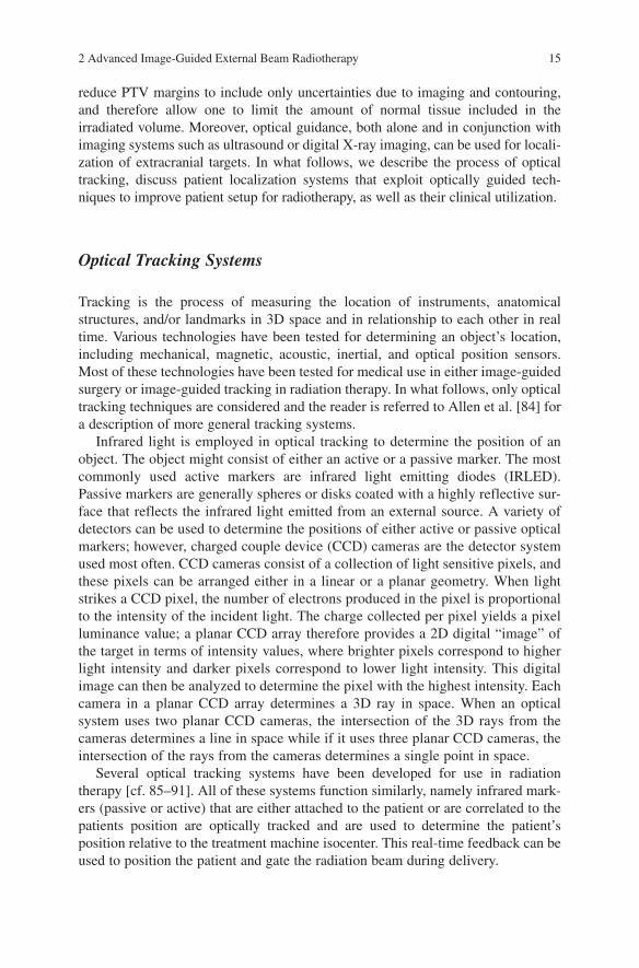
The first commercially available optical tracking system for radiation therapy was developed at the University of Florida [92–97] and is commercially available under the trade name FreeTrack™ (Varian, Inc., Ashland, MA). It can be employed for fractionated stereotactic radiotherapy, intracranial and Head and Neck IMRT [95, 96]. This system employs a hybrid Polaris position sensor unit (Northern Digital, Inc., Waterloo, Ontario), which is an array of two planar CCD cameras surrounded by a ring of infrared light-emitting diodes, to optically track the posi-tion of either active or passive infrared markers arranged in an array to form a fixed rigid body. For the Free Track system, the CCD camera is permanently mounted to the ceiling at a distance of approximately 1.8 m from the isocenter of the treatment machine (cf. Fig. 3). Therefore, the origin of the optical tracking system is located at the ceiling of the treatment room and the axes of its coordinate system are dependent upon the camera’s orientation. The most logical origin for clinical use in radiotherapy is of course the treatment machine isocenter, with the coordinate axes
Fig. 3 A patient undergoing optical-guided head and neck radiotherapy. A camera system moni-tors the translations and rotations of a set of markers that are rigidly fixed to a special bite block
Mehta_Ch02.indd 16Mehta_Ch02.indd 16 8/27/2007 6:43:23 PM8/27/2007 6:43:23 PM