delignification of valuable timbers decayed by india
TRANSCRIPT
Delignification of Valuable Timbers decayed by India Lignicolous fungi
Praveen Kumar Nagadesi1,*, Arun Arya2
1Department of Botany, P.G College, Andhra Loyola College, Vijayawada - 520008,
Andhra Pradesh, India
2Department of Botany, Faculty of Science, The Maharaja Sayajirao University of Baroda,
Vadodara - 390002, Gujarat, India
*E-mail address: [email protected]
ABATRACT
Wood degrading capacity of lignicolous fungi was studied by decay test. In which two methods
were followed, i) wood chips method ii) wood block method. Eight timbers infected by six fungi were
selected for studying percentage of decay and biochemical test was done to know delignification.
After 12 months, 90 % of wood block of T. arjuna was decayed by L. stereoides. In teak wood 16.82
% of decay was due to H. apiaria in 3 months. As the percentage of moisture was less, percentage of
weight loss was also less; this indicated that decay capacity of fungi will depends on % moisture
content in wood. The percentage loss in hot water soluble substrates was more in case of T. crenulata
due to L. stereoides for 5 months, whereas lowest in case of teak wood decayed by H. apiaria for 5
months. The percentage loss in ethanol benzene soluble substrate was more in case of Adina wood
decayed by C. versicolor for 5 months, whereas lowest in case of teak wood infected with L.
stereoides for 3 months. As the incubation period increases, percentage loss in acid soluble lignin was
more in case of infected woods. L. stereoides, C. versicolor, and H. apiaria showed selective
delignification in all infected woods, whereas T. pini showed simultaneous degradation of lignin in all
woods tested. The valuable timber like teak wood was not resistant to wood decay because they loss
50% of lignin. The in vitro wood decay test can’t be taken as absolute evidence for wood decay
behavior of lignin-degrading fungi, so we should conform decay of wood by consider biochemical
test. For rapid evaluation of wood decay the wood chip method was best suitable. For the first time
the wood decay and biochemical test of 8 wood samples infected by white rot fungi like S. commune,
L. stereoides, H. apiaria, C. versicolor, T. pini and soft rot fungi like T. viride was studied.
Keywords: Biochemical; Delignification; Lignicolous fungi; Wood decay; Teak
1. INTRODUCTION
Wood was a very important byproduct, produced by different biological processes in
tree. It was an important natural resource from forests which can contribute greatly to climate
change and biodiversity conservation (Kaimowitz D. 2003). Based on the FAO definition
there are around 3.7 billion ha of forests in the world. Majority of forest was natural forests,
in which more than 50 % in South America and Europe, while plantations cover only 187
million ha, representing 5 % of the total forested area (FAO 2007). Global wood consumption
was increasing but at a relatively low pace. In the last 20 years the average global
International Letters of Natural Sciences Online: 2014-05-30ISSN: 2300-9675, Vol. 16, pp 101-120doi:10.18052/www.scipress.com/ILNS.16.101CC BY 4.0. Published by SciPress Ltd, Switzerland, 2014
This paper is an open access paper published under the terms and conditions of the Creative Commons Attribution license (CC BY)(https://creativecommons.org/licenses/by/4.0)

consumption of wood increased on average only 0.3 % per year, and the estimated annual
wood consumption was now around 3.5 billion cubic meters (ITTO 2006). Out of this total
volume approximately 50 % are classified as industrial logs. The global consumption of
industrial roundwood would achieve around 1.9 billion by the year 2010 (FAO 2007).
The total forest cover in India according to the latest State of Forest Report 2011 is
78.29 million ha and this constitutes 23.81 % of the geographic area. The state of Gujarat was
one of the progressive states in western part of India, with an area of 78,687 sq mi
(203,800 km2). While recorded forest area is 18,962 sq. km. which was 10 % of total
geographical area. The production of fuel and timber was much less than the demand. The
forest area which produces timber and fuel wood was only 63.5 % of the recorded area.
Lignicolous fungi belonging to Aphyllophorales were economically important, as many of
these were pathogens of forest trees and cause serious damage (Natarajan K. and
Kolandavelu K. 1998). The forest wood cell wall was composed of cellulose, hemicelluloses
and lignin. Cellulose was most consistent of structural components varying minimally
between wood species. Lignin and hemicelluloses, however, vary both in composition and
amounts not only between hard woods and conifers but also among hardwoods (Timells T.
1967). Cellulose was a long chain polymer of glucose anhydride units joined by β 1-4
linkages. In general, cellulose components of wood are light in colour, have strong affinity
for water and were soft and tough. Hemicelluloses consist of similar polymers of glucose
joined by other linkages or polymers of monosaccharide’s other than glucose. Lignin was
quite different from celluloses and hemicelluloses and was also most resistant to
biodegradation. It was three dimensional amorphous, branched polymers of phenyl-propane
units joined by a variety of inter-units linkages (Alder E. 1977).
Delignification was deterioration of timber brought about by chemical breakdown and
separation of the cell walls of timber. It was also known as “Defibration of Timber”. Timer
decay was caused by primarily enzymatic activities of microorganisms. Lignicolous fungi
were responsible for decay in timber and those fungi that feed on the cell contents, causing
stains. These fungi seriously weather timber, ultimately rendering it valueless by consuming
cell wall constituents and lead to the disintegration of wood tissue (Desch, H.E. and
Dinwoodie, J. M. 1996). Three general types of decay were recognized (Blanchette, R.A.
1991, Eaton, R. A., and Hale M. D. C. 1993, Zabel, R. A., and Morrell J. J. 1992). In white
rot, all cell-wall constituents was degraded. Two forms of white rot were distinguished. In
selective delignification, polyoses (=hemicelluloses) and lignin were preferentially attacked,
especially in early stages. In simultaneous white rot, carbohydrates and lignin were attacked
more or less uniformly (Blanchette, R.A. 1991). In brown rot, carbohydrates were extensively
removed, but lignin was degraded only to a limited extent (Wilcox, W. W.1968). Soft rot,
most recently described type of wood decay, has proven difficult to define and differentiate
from other decays. It was caused by Ascomycetes and Deuteromycetes, All cell wall
constituents may be degraded during soft rot, but there was usually a preference for
carbohydrates, especially in hardwoods (Eslyn, W. E., Kirk T. K., and Effland M. J. 1975,
Nilsson, T. Daniel T. G., Kirk T. K., and Obst J. R. 1989).
A better understanding of diverse kinds of lignicolous fungi and their decay types will
support efforts to prevent and control wood decay as well as recent efforts to find
biotechnological applications of such fungi in the pulp and paper and other industries (Kirk,
T. K., and H.M. Chang. 1990). Blanchette (1984) mentioned about lignicolous fungi that
remove lignin selectively without appreciable losses of cellulose were extremely attractive
for use in biological pulping processes. Such knowledge may also provide perspective in
considering evolution of wood-decay capability in various groups of fungi. Our objectives
102 ILNS Volume 16

were to 1) survey lesser-known and previously neglected lignicolous fungi for the ability to
cause wood decay; 2) elucidate the wood decay and biochemical features of decayed wood by
such fungi in comparison with known decay types; 3) decay classification of lignicolous
fungi, and; 4) Quick identification of wood decay by different test. In the present paper wood
degrading capacity of lignicolous fungi was studied by decay test like wood chip and wood
block method. Wood of Tectona grandis, Terminalia arjuna, T. bellerica, T. crenulata, Adina
cordifolia, Dalbergia sissoo, Pinus longifolia, and Acacia arabica were selected on the value
of timber. Lignicolous fungi like S. commune, L. stereoides, H. apiaria, C. versicolor, T. pini
and soft rot fungi like T. viride were used to infect wood chips, blocks and logs of above
timbers and these were chemically analyzed.
2. MATERIALS AND METHODS
Wood degrading capacity of lignicolous fungi was studied by decay test. In which two
methods were followed, i) wood chips method ii) wood block method. In biochemical
analyses the degraded wood samples were analyzed for water content, pH of samples,
solubility in hot water, and ethanol-benzene, acid insoluble lignin and chlorite holocellulose
(Dill, I.; Kraepelin, G. 1986).
2. 1. Wooden chips method
A survey was undertaken in forests and sawmills of Gujarat, India, during January 2007
to July 2011, to find out occurrence of lignicolous fungi. These fungi were isolated by PDA
medium from sporophore and decayed wood and were grown on 2 % malt extract agar in
petriplates for 7d prior to inoculation in decay chambers. Seven woods like T. grandis, T.
arjuna, T. bellerica, A. cordifolia, D. sissoo, P. longifolia, and A. arabica wooden chips
(0.5g) were added to decay chamber containing Modified Asthana and Hawker’s medium
‘A’. The composition of medium was 10 g of D – glucose, 3.5 g of KNO3, 1.75 g of KH2PO4,
0.75 g of MgSO4·7H2O and 20 g of Agar. The decay chambers were sterilized at 121 ºC for 1
h. The decay chambers were inoculated with Lignicolous fungi like S. commune, L.
stereoides. H. apiaria and soft rot fungi like T. viride. Three decay chambers were used for
each isolated lignicolous fungi per wood and the fungus inoculated in decay chamber without
wooden chips served as control. Assembled decay chambers were incubated in the dark for
20 and 40 d at 27 ±1 ºC. The wooden chips were filtered, oven dried, and weighed. Percent
weight loss was determined as follows:
Percent weight loss =Weight loss of oven − dried wood after incubation
Weight of oven − dried original wood 𝑥 100
Each set of treatment was run in triplicates and average weight loss was always taken as
standard value for comparison of wood decay. The weight loss results were statistically
analyzed by using the MS office Excel software and the significant values were taken for
study.
International Letters of Natural Sciences Vol. 16 103

2. 2. Wooden block method
Timber decay caused by Lignicolous fungi like S. commune, L. stereoides. H. apiaria
C. versicolor and soft rot fungi like T. viride was observed in wood blocks of T. grandis and
T. arjuna and T. bellerica for 20 days to 1year. These fungi were grown on 2 % malt extract
agar for 7 d prior to inoculation. The PDA plates were prepared by inoculating each isolate of
above fungi and incubated for 7-10 d.
Totally 12 wooden blocks (1 x 1 x 1 cm) per wood per each lignicolous fungi were cut
from the respective logs and soaked in distilled water for 30 min. These wooden blocks were
autoclaved. After completely spreading of above fungus, four blocks of each wood were
placed on the medium per plate and incubated for 3, 6 and 12 months at 27 ±1 ºC. To
maintain moisture in test plates two layers of Whatmen filter paper No 1 were placed on the
surface of blocks. The sterile distilled water was added to it regularly. The un-inoculated
wooden blocks acted as control. After completion of incubation period each block was
cleaned (of the mycelium), oven dried and weighed. The percentage weight loses was
calculated as described above.
2. 3. Spawn preparation
For spawn preparation a media used for developing sporophores of wood rot fungi as
suggested by Etter (1929) was used. The spawn preparation medium consisted of 48 g of
corn-meal, 16 g of corn-starch and 8 g of powdered wood. The spawn was taken in a
polypropylene bag and 2.5 % malt extract was added. The bag was closed by putting the
moist cotton swab to maintain moisture level inside the bag; such bags were sterilized at 121
ºC for 1 h and inoculated with four test fungi like L. stereoides C. versicolor H. apiaria T.
pini. The bags were incubated in dark for 15d at 27 ±1 °C. The fully-grown spawn was used
for artificial inoculation in wooden logs.
2. 4. Wooden log preparation
The wood log of T. grandis, A. cordifolia, T. crenulata and T. arujna were infected
with above lignicolous fungi and used for biochemical analysis. The average size of (4) wood
plank used was 2 x 2 x 30 cm length. The bark was not removed. It helps to maintain
moisture and keeps away the foreign fungi. A 5/16” drill bit was used to make holes in the
logs for insertion of spawn as diamond drilling pattern. After drilling the entire log was
autoclaved for 1 h. After autoclaving spawn was inoculated into the holes. It was then sealed
off with paraffin wax. Logs were covered with cheese cloth to maintain moisture, packed in
polythene bags and incubated in dark for 12 months.
2. 5. Bio – chemical analysis of decayed wood
The chemical composition of sound and decayed wood as determined by previously
described technique (Dill, I.; Kraepelin, G.1986). Decayed wood was dried at 105 ºC and
then ground to pass through a 60 μ mesh screen. It was used for further analysis.
2. 5. 1. Water content
To obtain water present in the sample decayed wood (3 g) was dried at 105 ºC for about
48 h, cooled in a desiccator and weighed. The difference in two weights gave the water
content in milligrams.
104 ILNS Volume 16
2. 5. 2. pH of samples
The pH was determined potentio-metrically after suspension of the samples in distilled
water for about 30 to 45 min. Analysis of decayed wood was made with a few grams of fresh
material and that of corresponding sound wood was made with 1 g of dry wood meal.
2. 5. 3. Solubility in hot water
One gram of dry wood meal was placed in a 250 ml Erlenmeyer flask. After addition of
100 ml of distilled water, the mixture was slowly stirred at 80 ºC for 3 h. The samples were
then filtered by using Whatman filter paper No 1, washed with hot water, dried at 105 ºC for
about 24 h, cooled in desiccator and weighed.
2. 5. 4. Solubility in ethanol - benzene
About 1.5 g of dry wood meal was extracted with ethanol – benzene (1:2 v/v) for 4 h in
a Soxhlet extractor, keeping the liquid boiling briskly. Each extracted sample was washed
with 50 to 100 ml of ethanol and dried at 105ºC. After evaporation of solvent, each extract
was dried at 105 ºC for 24 h, cooled in desiccator and weighed.
2. 5. 5. Acid insoluble lignin (klason lignin)
Flasks containing 1 g of ethanol - benzene extracted wood meal and 20 ml of H2SO4
(72 %) were gently shaken in a water bath at 30 °C for 1 h. The acid was then diluted with
H2O to 4 % (wt/vol), and the samples were autoclaved at 121 °C for 30 min. The lignin that
settled overnight was quantitatively collected by filtration through a Whatman filter paper
No. 1, washed free of acid with hot water, and dried. The lignin content was calculated as a
percentage of oven-dried, non-extracted wood meal.
2. 5. 6. Chlorite holocellulose (CHC)
Chlorite holocellulose was also determined as described by Seifert 91983). Extracted
wood samples of approximately 400 mg were placed in 50 ml Erlenmeyer flasks. Seven
milliliters of buffer solution consisting of 60 ml glacial acetic acid and 1.3 g sodium
hydroxide per 1000 ml distilled water was added to each flask. Three milliliters of 20 %
(w/w) aqueous solution of sodium chlorite was immediately added and the flasks were sealed
with paraffin wax and aluminum foil. The flasks were placed in an orbital shaker at 110 rpm
at 45 °C for 36 to 40 h. after incubation period; flasks were placed in ice bath to stop the
reaction. The contents were then transferred to pre weighed Whatman filter paper No. 1 using
100 ml of 1 % acetic acid. The holocellulose was washed with 5 ml of acetone three times
and oven dried at 105 °C for 4 to 6 h before weighing.
3. RESULTS AND DISCUSSION
3. 1. Wooden chips method
Percentage decay of seven different woods caused by S. commune, L. stereoides. H.
apiaria (White rot fungi) and T. viride (soft rot fungi) was observed. As compared to other
white rot and soft rot fungi, teak and sissoo wood was efficiently degraded by L. stereoides,
where the percentage weight loss was 34.6 and 44.6 % after 40days. Whereas in case of T.
arjuna, T. bellerica, A. cordifolia, A. arabica, and P. longifolia woods were efficiently
International Letters of Natural Sciences Vol. 16 105

degraded by H. apiaria, where the percentage weight loss was 62.0, 66.4, 60.4, 52.5 and 65.4
% respectively in 40 days. When compared to the all wood decayed after 40 days of
incubation, S. commune showed 29.1 % decay in A. arabica, whereas the L. stereoides
showed 54.3 % decay in T. arjuna. The fungus H. apiaria showed 66.4 % decay in T.
bellerica, while T. viride showed 22.3 % decay incase of A. arabica. The minimum amount
of decay was observed in teak wood i.e 3.9 % in 20 days by S. commune (Table 1 and
Histogram 1).
Table 1. Percentage weight loss of wood chips of seven woods by four Lignicolous fungi.
Percentage weight loss
Sch
izo
ph
yllu
m c
om
mun
e
Len
zite
s st
erio
ides
Hex
ag
on
ia a
pia
ria
Tri
cho
der
ma
vir
ide
*2
0 d
ays
pH
*4
0 d
ays
pH
*2
0 d
ays
pH
*4
0 d
ays
pH
*2
0 d
ays
pH
*4
0 d
ays
pH
*2
0 d
ays
pH
*4
0 d
ays
pH
Aca
cia
ara
bic
a
20
±1
.0
5.2
2
29
.1±
1.5
4.9
22
.2±
1.2
3.5
39
.8±
5.6
4.0
47
.3±
2.4
4.0
9
52
.5±
1.3
4.3
9
20
.6±
2.5
8.3
3
22
.3±
2.5
9.0
Ad
ina
cord
ifo
lia
7.3
±1.5
5.6
9
15
.4±
2.7
6.1
39
.8±
1.8
4.3
44
.6±
3.5
4.5
23
.3±
1.8
4.1
5
60
.4±
2.7
4.5
19
.6±
2.6
5.9
1
15
.8±
1.9
5.8
7
Da
lber
gia
siss
oo
14
.0±
2.5
5.9
3
15
.2±
2.6
6.0
39
.9±
2.5
4.9
44
.6±
4.8
5.3
28
.6±
2.6
4.3
2
40
.3±
2.6
4.4
4.7
±3.8
7.6
2
12
.6±
1.4
8.0
8
106 ILNS Volume 16

Pin
us
lon
gif
oli
a
2.8
±1.0
5.4
6
13
.0±
2.3
5.4
13
.4±
2.4
4.2
45
.5±
4.5
3.9
44
.7±
4.8
4.6
2
65
.4±
5.3
5.2
6
14
.8±
2.4
9.0
4
18
.8±
2.6
10
.0
Tec
ton
a
gra
nd
is
3.9
±0.9
5.3
6
12
.9±
2.5
5.2
5.1
±2.5
4.9
34
.6±
2.5
4.0
27
.9±
2.4
4.2
5
28
.3±
2.3
5.2
0
7.7
±1.8
3.3
4
11
.2±
1.8
9.0
0
Ter
min
ali
a
arj
un
a
7.8
±2.5
6.3
9
13
.8±
1.8
6.6
48
.4±
3.2
5.0
54
.3±
3.2
5.1
30
.2±
4.9
4.7
2
62
.0±
5.1
5.5
0
14
.2±
1.7
8.8
6
15
.0±
3.4
8.9
7
T.
bel
leri
ca
5.2
±1.6
5.9
7
17
.6±
1.5
5.8
44
.9±
3.8
4.4
54
.0±
4.3
4.3
36
.2±
1.8
4.3
5
66
.4±
5.6
4.2
6
12
.1±
2.1
7.0
4
16
.0±
2.5
8.2
4
∗ indicates each component values are based on the three replicates. Uninoculated wooden blocks were
incubated for 20 and 40 days to act as a control.
± Results were significant at P < 0.05 level by one way ANOVA
Histogram 1. The % weight loss in different woods chips infected with four lignicolous fungi.
According to ASTM (1969) method of classification of decay resistant classes, teak
wood was very resistant in case of T. viride, and Lenzites sp. up to 3 weeks of incubation.
Moderate resistance was observed in case of H. apiaria and L. stereoides (Nagadesi P. K.,
Arya A., Albert S. 2013). Bakshi et al. (1967) conducted wood decay test with Polyporus
0
10
20
30
40
50
60
70
Tectona grandis Terminalia arjuna T. bellerica Adina cordifolia Acacia arabica Dalbergia sissoo Pinus longifolia
% W
eig
ht
loss
S 20days
S 40days
L 20days
L 40days
H 20days
H 40days
T 20days
T 40days
International Letters of Natural Sciences Vol. 16 107

hirsutus Fr., P. sanguineus L. ex Fr., P. versicolor L. ex. Fr., P. palustris B. & C. and Irpex
flavus Klotzsch. The results showed that in case of teak wood outer heart wood varies in
decay resistance from very resistance to moderate resistance (weight loss 1.98-25.63 %)
(Bakshi et al 1967). In the present study teak wood was resistant to moderately resistant when
different wood rotting fungi were tested in wood chip method.
The Basidiomycetes plus composite inocula caused significantly more weight loss of
inoculated wood chips than members of Basidiomycetes alone after 5 months (Blanchette
R.A. and Shaw C.G. 1978). In the present study the lignicolous fungi like S. commune, L.
stereoides. H. apiaria and soft rot fungi like T. viride alone showed significant decay in wood
chips after 40 days. Wood chips from slash less than 1 and 2 year old were approx. 40 %
decayed by the brown rot fungus P. placenta for 5 months. The decay caused by the white rot
fungus, C. versicolor was approximately half than the P. placenta. Hirschioporus abientinus
caused approximately 20 % weight loss in wood chips in less than 1 year old but 10% in 1
and 2 year old chips (Blanchette R.A. and Shaw C.G. 1978). In the present study the increase
in weight loss by H. apiaria 66.4 % in T. bellerica for 40 days. Whereas soft rot fungi like T.
viride showed 22.3 % weight loss in A. arabica for 40 days.
The wood chips of ten-year old plantation of Pinus caribaea (morelet) were inoculated
separately with two species of white-rot fungi; Coriolopsis polyzona and Pleurotus
squarrosulus, and two species of brown rot fungi; Lentinus lepideus and Gleophyllum
striatum. Wood weight loss due to biodegradation varied from 1.5-48.1 % for Coriolopsis
polyzona, 9.6-58.0 % for Pleurotus squarrosulus, 40.4-78.1 % for Lentinus lepideus and 6.8-
49.2 % for Gleophyllum striatum degrading activities (Emerhi, E. A., Ekeke, B. A. and
Oyebade, B. A. 2008). In the present study the weight loss due to degradation varied from
one to 13% for S. commune, 1 to 45.5 % for L. stereoides, 1-65.4 % for H. apiaria and 1-
18.8% for T. viride in 40days. The highest decay was shown by H. apiaria with 27.9 %
weight loss in 20 days. The lowest decay was showed in case of T. viride with 4.7 % weight
loss in 20 days (Nagadesi P. K., Arya A., Albert S. 2013). In the present study teak and sissoo
woods were efficiently degraded by L. stereoides, whereas, the percentage weight loss was
34.6 and 44.6 in 40 days and T. arjuna, T. bellerica, A. cordifolia, A. arabica, and P.
longifolia these woods were efficiently degraded by H. apiaria whereas the percentage
weight loss was 62.0, 66.4, 60.4, 52.5 and 65.4 % respectively in 40 days only observed.
The molecular structure of wood suggests that cellulose a structural component thereof
could be bio-recycled into glucose, a fermentable sugar (Reddy N. and Yang Y. 2009).
Cellulase, a multi-component enzyme system produced by soft rot fungi such as T. viride and
Aspergillus niger exhibits the ability to saccharify cellulose. These enzymes have been
proved to be effective in the bioconversion of wood products such as wastepaper into
fermentable sugars (van Wyk J. P. H. 2001). In the present paper the ability of soft rot fungi
to degrade lignin was proved by wood chip test. The various sawdust wood samples were
exposed to T. viride cellulase action with the delignified cellulose component bio-converted
into fermentable sugars such as glucose (Bohdan V, Yaser D 2011). In the present study the
T. viride showed significant weight loss in seven woods so it may be used for saccharify
cellulose.
3. 2. Wooden block method
Wood decay caused by four test organisms was observed in wood blocks of T. grandis
and T. arjuna wood blocks after every 20, 40 and 60 days. The maximum decay was shown
by L. stereoides in case of T. arjuna after 60 days. The minimum decay was observed in case
of T. arjuna due to S. commune after 20 days. In initial stages of decay, percentage of
108 ILNS Volume 16

moisture was more whereas in advanced stages of decay the % moisture reduced. As the
percentage moisture was less the percentage weight loss was also less, this indicates that the
decay capacity of lignicolous fungi depends on the % moisture content in wood (Table 2 and
Histogram 2).
Table 2. Moisture loss and weight loss of two different wood blocks caused by three lignicolous
fungi.
Schizophyllum commune
20 days 40 days 60 days
Wood % Moisture
Loss*
% wt.
loss*
% moisture
loss*
% wt.
loss*
%
moisture
loss*
% wt.
loss*
Tectona grandis 28.34±0.7 4.1±0.8 4.1±0.2 4.7±0.7 3.33±0.5 6.96±0.8
Terminalia arjuna 21.25±0.2 1.0±0.5 3.3±0.5 2.0±0.8 5.36±0.8 3.86±0.9
Lenzites sterioides
T. grandis 3.76±0.6 2.3±0.1 3.71±0.7 3.8±0.8 5.48±0.8 6.57±0.3
T. arjuna 5.84±0.8 7.2±0.4 3.16±0.2 11.4±0.5 6.53±0.5 9.25±0.5
Hexagonia apiaria
T. grandis 10.05±0.3 5.85±0.3 7.85±0.7 8.25±0.4 4.89±0.3 9.12±0.6
T. arjuna 16.08±0.8 3.5±0.7 10.24±0.5 4.5±0.2 8.98±0.2 8.05±0.3
Trichoderma viride
T. grandis 25.17±0.5 4.1±0.2 15.87±0.8 4.4±0.5 3.77±0.6 6.41±0.7
T. arjuna 47.08±0.8 1.3±0.3 3.18±0.2 2.2±0.3 2.81±0.7 2.5±0.5
∗ indicates each component values are based on the three replicates. Uninoculated wooden blocks were
incubated for 20 and 40 days to act as a control.
± Results were significant at P < 0.05 level by one way ANOVA
Histogram 2. The % weight loss of two wood blocks infected with lignicolous fungi.
0
2
4
6
8
10
12
S 20
days
S 40
days
S 60
days
L 20
days
L 40
days
L 60
days
H 20
days
H 40
days
H 60
days
T 20
days
T 40
days
T 60
days
Perc
en
tag
e w
eig
ht
loss
Tectona grandis Terminalia arjuna
International Letters of Natural Sciences Vol. 16 109
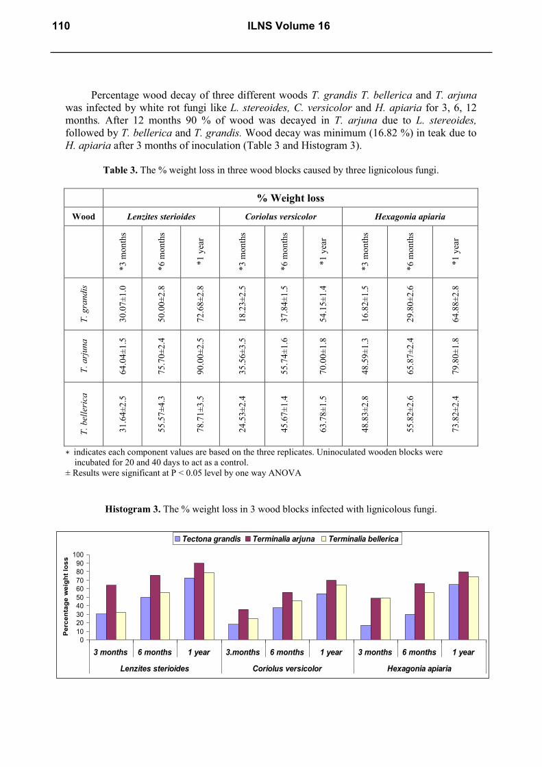
Percentage wood decay of three different woods T. grandis T. bellerica and T. arjuna
was infected by white rot fungi like L. stereoides, C. versicolor and H. apiaria for 3, 6, 12
months. After 12 months 90 % of wood was decayed in T. arjuna due to L. stereoides,
followed by T. bellerica and T. grandis. Wood decay was minimum (16.82 %) in teak due to
H. apiaria after 3 months of inoculation (Table 3 and Histogram 3).
Table 3. The % weight loss in three wood blocks caused by three lignicolous fungi.
% Weight loss
Wood Lenzites sterioides Coriolus versicolor Hexagonia apiaria
*3
mon
ths
*6
mon
ths
*1
yea
r
*3
mon
ths
*6
mon
ths
*1
yea
r
*3
mon
ths
*6
mon
ths
*1
yea
r
T.
gra
nd
is
30
.07±
1.0
50
.00±
2.8
72
.68±
2.8
18
.23±
2.5
37
.84±
1.5
54
.15±
1.4
16
.82±
1.5
29
.80±
2.6
64
.88±
2.8
T.
arj
un
a
64
.04±
1.5
75
.70±
2.4
90
.00±
2.5
35
.56±
3.5
55
.74±
1.6
70
.00±
1.8
48
.59±
1.3
65
.87±
2.4
79
.80±
1.8
T.
bel
leri
ca
31
.64±
2.5
55
.57±
4.3
78
.71±
3.5
24
.53±
2.4
45
.67±
1.4
63
.78±
1.5
48
.83±
2.8
55
.82±
2.6
73
.82±
2.4
∗ indicates each component values are based on the three replicates. Uninoculated wooden blocks were
incubated for 20 and 40 days to act as a control.
± Results were significant at P < 0.05 level by one way ANOVA
Histogram 3. The % weight loss in 3 wood blocks infected with lignicolous fungi.
0
10
20
30
40
50
60
70
80
90
100
3 months 6 months 1 year 3.months 6 months 1 year 3 months 6 months 1 year
Lenzites sterioides Coriolus versicolor Hexagonia apiaria
Pe
rce
nta
ge
we
igh
t lo
ss
Tectona grandis Terminalia arjuna Terminalia bellerica
110 ILNS Volume 16

Biological agar block method allowed wood samples to be evaluated and monitored in
terms of colonization and development of decay by C. versicolor and classified based on
mean mass loss. In this research, the in vitro decay of five commercial woods by C.
versicolor was studied by the agar block method. The selected wood samples were Abies
alba. Populus alba, Fagus orientalis, Platanus orientalis and Ulmus glabra. There was a
high correlation between the mass loss and apparent damage. Therefore biological evaluation
of wood regarding biodegradation and selection of wood types for various applications will
be of high priority (Olfat A.M., Karimi A.N., and Parsapajouh D. 2007). In the present study
also there was a correlation between the weight loss and damage, so different biochemical
test were studied to conform that biological evaluation of wood was necessary.
The Basidiomycetes, Poria carbonica and C. versicolor, caused substantial wood
weight loss over the test period. These results were usually obtained by soil or vermiculite
burial methods (Morrell J.J. and Zabel R.A. 1989). In the present study the wood chip and
block method showed significant weight loss in seven woods infected by white rot and soft
rot fungi. The decreasing weight in studied samples showed that C. versicolor can grow
quickly and may rapidly affect the appearance and degrade the wood (Olfat A. M., and
Karimi A.N., 2005). In the present study the growth of C. versicolor was quick to degrade the
wood very fast when compared to other lignicolous fungi. The lowest weight loss decreasing
was observed in U. glabra and highest value in F. orientalis. This was true for the study of
crude oil and beech wood caused by C. versicolor (Olfat A. M., and Karimi A.N., 2005). In
the present study also C. versicolor degrade T. arjuna wood very fast.
The percentage weight loss in inner heart wood and outer heart woods of New guinea
teak was 44, 54 and 12, 21 for Coniophora olivacea and C. versicolor respectively. The
percentage weight loss in inner heart wood and outer heart wood of Indonesian teak was 54,
55 and 22, 21 for C. olivacea and C. versicolor. The percentage weight loss of inner heart
wood and outer heart wood of Burma teak was 4, 8 and 4, 3 for C. olivacea and C. versicolor
respectively (Guilley et al. 2004). In the present study the heart wood of teak showed 72.68
%, 54.15 %, 64.88 % of weight loss by L. sterioides, C. versicolor and H. apiaria
respectively. Based on percentage weight loss, the American Society for Testing Materials
ASTM (1969) classified resistance of wood. Highly resistant wood showed weight loss of
zero to 10 %, resistant wood shows weight loss of 11 to 24 %, moderately resistant wood
showed 25 to 44 % weight loss, and nonresistant wood showed 45 % or greater weight loss.
In the present study the teak and terminalia wood was moderately resistant to non resistant
when infected with different lignicolous fungi in wood block test. The in vitro decay of five
commercial woods by C. versicolor was studied by the agar block method showed strong
resistance of U. glabra and lowest resistance in F. orientalis (Olfat A.M., Karimi A.N., and
Parsapajouh D. 2007). In the present study teak wood infected with C. versicolor showed
resistant to non resistant. Where as in terminalia wood infected with same fungi showed
moderately resistant to nonresistant. Twelve hundred samples from 31 trees were exposed to
four fungi: Pycnoporus sanguineus, Antrodia sp., Gloephylum trabeum, and Coriolus
versicolor. Tests showed that Antrodia sp. and C. versicolor resulted in <20 % mass loss,
whereas all samples were rated as durable or highly durable with regard to P. sanguineus and
G. trabeum. Inner heartwood was found to be the most resistant to pathogen attack and outer
heartwood the least (Kokutse, et al. 2006). In the present study teak wood infected with L.
stereoides showed moderately resistant to nonresistant, with C. versicolor and H. apiaria
showed resistant to non resistant.
So, the in vitro wood decay test cannot be taken as absolute evidence for the behavior
of lignicolous fungi, they were useful to determine their wood-degrading properties. Weight
International Letters of Natural Sciences Vol. 16 111

loss of yellow-poplar samples incubated with T. versicolor in a soil-block test was
significantly higher than that in an agar-block test. Therefore, the soil-block test was more
sensitive to detect fungal decay in yellow-poplar under the conditions of the experiments
(Schirp A. and Wolcott M. P. 2005). In the present study the most effective method was agar
block method when compared to wood chip method.
3. 3. Biochemical analysis
3. 3. 1. Artificially inoculated wood blocks
The physicochemical analysis of teak, and pine woods infected with wood decay fungi
was done. The details were recorded in Table 4 and 5.
Table 4. Physico-chemical analysis of sound wood of Tectona grandis,
Adina cordifolia and Terminalia bellerica.
Plants
% of dry weight
*Moisture % pH
*Swelling
capacity
%
*Acid
insoluble
lignin
*Holocellulose
*Ethanol-
benzene
soluble
substrate
*Hot water
soluble
substrate
*Ash
(g)
Tectona
grandis 1 6.1±0.26 5.8 45.1±0.7
29.3
±0.9 143.2±1.8 26.7±1.8 7.8±1.0 0.06±0.01
T. grandis 2 6.5±0.25 4.4 40.0±2 42.0
±0.24 112.5±2.5 37.4±1.6 9.5±1.4 0.10±0.03
T. grandis 3 5.1±0.24 5.2 -- 43.2
±1.5 -- 17.2±2.7 5.6±2.5 --
Adina
cordifolia 5.7±0.23 5.3 35.5±1.4
35.0
±2.4 141.5±2.3 32.1±2.4 19.4±0.5 0.21±0.04
Terminalia
bellerica 7.8±0.51 5.2 31.5±1.8
20.3
±1.2 153.0±2.8 28.2±1.8 20.7±0.35 0.06±0.01
Pinus
longifolia 5.8±0.4 5.1 --
42.4
±1.8 -- 11.0±2.5 4.4±0.2 --
∗ indicates each component values are based on the three replicates.
± Results were significant at P <.05 level by one way ANOVA.
Table 5. Chemical analysis of decayed woods of Tectona grandis and
Pinus longifolia by 4 lignicolous fungi.
Plant Fungi days * %
Moisture pH
*Hot water
soluble
substrate
*Ethanol-
benzene
soluble
substrate
*Acid
insoluble
lignin
*Holo -
cellulose
Pine *Control 5.8±0.8 5.10 4.4±0.2 11.0±1.4 42.4±1.5 4.5±0.8
Lenzites
sterioides 2 20 5.9±0.4 5.57 4.3±0.4 8.2±1.8 11.6±1.2 7.5±1.5
L. sterioides 1 20 5.7±0.2 5.16 3.8±0.8 7.1±1.6 25.6±1.8 10.0±1.3
112 ILNS Volume 16

Schizophyllum
commune 20 4.5±0.6 5.30 4.7±0.5 7.3±2.4 19.6±1.6 5.0±1.8
Trichoderma
viride 20 6.2±1.2 6.05 3.5±0.4 7.1±2.6 26.0±2.3 11.5±1.4
H. apiaria 20 5.5±2.5 6.5 4.7±0.6 8.5±1.5 30±2.6 15±1.0
Teak Control 5.1±0.2 5.26 5.6±0.9 17.2±2.2 43.2±2.8 12.5±1.6
L. sterioides 2 20 5.9±0.7 6.01 4.9±0.3 13.8±1.8 13.0±1.5 19.5±1.4
L. sterioides 1 20 5.5±0.6 5.05 4.5±1.5 12.2±1.4 26.8±1.4 10.5±1.8
S. commune 20 5.9±0.1 5.70 5.9±1.8 17.4±2.3 29.2±1.8 19.0±2.0
T. viride 20 6.2±1.5 6.90 4.6±0.4 14.0±2.5 31.0±1.6 14.0±2.5
H.apiaria 20 5.3±1.8 4.89 5.5±0.7 15.6±1.8 18.8±1.9 7.0±1.0
H. apiaria 40 9.6±2.5 5.27 16.0±2.3 18.4±1.5 41.4±2.5 15.5±2.3
∗ indicates each component values are based on the three replicates. Uninoculated wooden blocks were
incubated for 20 and 40 days to act as a control.
± Results were significant at P < 0.05 level by one way ANOVA.
Highest percentage of moisture was shown by teak wood infected with H. apiaria
whereas it was lowest in case of pine wood infected with S. commune. High acidic nature was
shown by teak wood infected with H. apiaria in 20 days. Almost neutral nature was shown
by teak wood infected with T. viride. The percentage loss of ethanol - benzene soluble
substrates was more in case of teak wood decayed by H. apiaria for 40 days, whereas, lowest
was observed in pine wood decayed by L. stereoides1 and T. viride. The highest percentage
loss of acid insoluble lignin was observed in case of teak wood decayed by H. apiaria for 40
days, whereas, lowest in case of teak wood inoculated with L. stereoides 2. Where as highest
percentage loss of Acid insoluble lignin was observed incase of pine woods decayed by H.
apiaria and lowest incase of pine wood inoculated with L. stereoides 2. The percentage loss
of holocellulose was more in case of teak wood infected with L. stereoides 2, whereas, lowest
in case of wood decayed by H. apiaria. The highest loss of holocellulose in pine wood was
observed by H. apiaria decay and lowest in case of S. commune decay
Three white rot fungi Daedalea elegans, Polyporus glaganetus, and L. betulina were
screened for their lignin degrading abilities on rice straw, maize cob, sawdust of Terminalia
superba and sugarcane bagasse at different time intervals (30, 60 and 90 days). All the fungi
demonstrated varying levels of ligninolytic capability with different degrees of lignin
degradation in all the fermented substrates. The highest lignin reduction of 92.9 % was
recorded in maize cob fermented with D. elegans after 90 days (Adejoye O.D. and Fasidi I.O.
2009). But in the present study only 26 % of reduction in lignin was observed in pine and
teak wood blocks infected by L. stereoides, 30 % loss in pine and 18 % loss of lignin in teak
woods infected by H. apiaria, 31 % loss in teak and 26 % loss of lignin in pine woods
infected by T. viride
When a brown-rot-causing fungi Polyporus palustris was infected to Mangifera indica
wood shavings for considerable periods, approximately 40 to 50 % lignin loss was observed
in two years (Ananthanarayanan, S.; Wajid, S.A.; Padmanabhan, S. 1978). The Mangifera
wood blocks were infected with white-rot-causing fungi for 90 days, the utilization of lignin
was 26 % by F. flavus and 20 % by S. commune (Padhiar A., Albert S., Nagadesi P. K. and
International Letters of Natural Sciences Vol. 16 113

Arya A. 2010). But in the present study the maximum lignin loss was recorded in teak than in
pine by L. stereoides. The loss of lignin in teak and pine wood infected by S. commune was
29.2 % and 19.6 % respectively.
3. 3. 2. Artificially infected wood Planks
The artificially infected wood log of Tectona, Adina, T. crenulata and T. arujna were
chemically analyzed. The percentage of moisture loss was highest in case of T. crenulata
wood decayed by L. stereoides, whereas, lowest in case of teak wood decayed by C.
versicolor. The acidic nature was shown by T. crenulata wood decayed by C. versicolor,
whereas, basic nature was shown by teak wood infected with L. stereoides. The percentage
loss in hot water soluble substrates was more in case of T. crenulata due to L. stereoides for
5 months, whereas, lowest in case of teak wood decayed by H. apiaria for 5 months. The
percentage loss in ethanol-benzene soluble substrate was more in case of Adina wood
decayed by C. versicolor in 5months. Whereas, lowest in case of teak wood infected with L.
stereoides for 3 months. The percentage loss of acid soluble lignin was more in case of T.
crenulata wood decayed by L. sterioides for 5 months, whereas, lowest in case of teak wood
decayed by L. stereoides for 3 months. The percentage loss in holocellulose was more in case
of Adina wood decayed by C. versicolor, whereas, least in case of T. crenulata wood infected
with C. versicolor for 5 months and T. pini for 5 months period (Table 6 and Histogram 4).
Table 6. Chemical analysis of three woods decayed by lignicolous for 5 months.
Plant Fungi Months *%
Moisture pH
*Hot
water
soluble
substrate
*Ethanol-
benzene
soluble
substrate
*Acid
insoluble
lignin
*Holo -
cellulose
Tectona
grandis *Control 3 and 5 5.1±0.8 5.26 5.6±0.6 17.2±1.5 43.2±1.3 12.5±0.8
L. sterioides 3 4.0±0.4 9.08 10.5±0.3 17.7±1.3 20.8±1.8 19.5±1.5
C. versicolor 3 3.5±0.6 8.30 10.3±0.7 27.9±1.8 30.4±2.5 14.0±1.4
T. pini 5 4.2±0.2 8.10 5.8±0.8 27.9±1.6 36.4±2.4 18.5±1.7
H. apiaria 5 3.8±0.5 6.87 2.5±0.4 21.7±2.5 27.2±3.0 22.5±3.2
Adina
cordifolia *Control 3 and 5 5.7±0.1 5.30 19.4±0.8 32.1±2.3 35.0±2.5 41.5±3.6
L. sterioides 3 3.6±0.3 5.98 8.6±0.2 19.9±2.8 42.4±2.4 24.0±2.5
C. versicolor 5 5.0±0.4 4.60 15.9±0.5 38.1±2.5 33.6±1.6 48.5±2.7
T. pini 5 4.7±0.6 4.67 18.4±0.6 25.7±1.8 33.8±1.4 25.5±3.4
H. apiaria 3 4.9±0.7 5.60 9.3±0.2 20.7±1.6 29.2±1.8 26.5±1.8
Terminalia
crenulata *Control 5 6.5±0.7 5.20 25.7±0.4 30.0±2.5 40.5±2.5 35.0±1.6
L. sterioides 5 5.5±0.2 5.86 22.2±0.7 23.7±1.9 74.0±2.8 20.0±2.5
C. versicolor 5 4.3±0.4 4.50 9.2±0.3 24.4±2.5 55.0±2.9 10.5±2.5
T. pini 5 4.8±0.8 4.56 16.5±0.8 20.4±2.6 24.8±2.5 28.5±1.8
∗ indicates each component values are based on the three replicates. Uninoculated wooden blocks were
incubated for 20 and 40 days to act as a control.
± Results were significant at P < 0.05 level by one way ANOVA.
114 ILNS Volume 16

Histogram 4. The % loss of Acid insoluble lignin in 2 woods infected with lignicolous fungi.
When S. commune was grown on liquid media containing 14
C-lignin-labeled wood, the
degradation of lignin was low and variable (Boyle, C.D.; Kropp, B.R.; and Reid, I.D. 1992).
S. commune has ability to produce lignin degrading enzymes for degradation of
lignocellulosic materials (Padhiar A., Albert S., Nagadesi P. K. and Arya A. 2010). In the
present study as the incubation period was increase the loss of lignin also increase. After five
months, the highest lignin loss was observed in T. crenulata infected with L. stereoides.
The chemical analysis of artificially inoculated wood blocks for 1 year was studied
(Table 7). As the incubation period increased the percentage loss in acid soluble lignin was
more in case of all infected woods reaching to almost 50 %. Whereas, the percentage loss of
holocellulose was up to 20 % only (Histogram 5).
Histogram 5. The % loss of Acid insoluble lignin in woods infected with lignicolous fungi.
0
10
20
30
40
50
60
70
Control Lenzites
sterioides
Coriolus
versicolor
Control L. sterioides H. apiaria
Tectona grandis Adina cordifolia
% lo
ss
of
Ac
id in
so
lub
le li
gn
in
3 months
1 year
0
10
20
30
40
50
60
70
80
Co
ntr
ol
Tra
me
tes
pin
i
He
xa
go
nia
ap
iari
a
Co
ntr
ol
C.
ve
rsic
olo
r
T. p
ini
Co
ntr
ol
L.
ste
rio
ide
s
C.
ve
rsic
olo
r
T. p
ini
H. a
pia
ria
co
ntr
ol
L.
ste
rio
ide
s
T. p
ini
Tectona grandis Adina cordifolia Terminalia crinulata T. arjuna
% L
os
s o
f A
cid
in
so
lub
le lig
nin
5 months
1 year
International Letters of Natural Sciences Vol. 16 115

Table 7. Chemical analysis of four woods decayed by lignicolous fungi for one year.
Plant fungi %
Moisture pH
Hot water
soluble
substrate
Ethanol- benzene
soluble substrate
Acid
insoluble
lignin
Holo -
cellulose
Tectona
grandis Control 5.1±0.8 5.26 5.6±1.2 17.2±1.2 43.2±2.5 12.5±1.2
L. sterioides 5.7±0.2 5.66 10±1.6 17.86±1.5 50.4±2.8 8.0±1.4
C. versicolor 4.80.4 5.88 6.6±1.2 17.33±1.9 43.0±2.6 12.5±1.6
T. pini 4.8±0.5 5.54 6.0±0.8 18.66±1.3 50.4±2.8 16.5±1.8
H. apiaria 4.5±0.6 5.36 7.4±0.3 15.20±1.7 50.8±2.5 9.0±1.5
Adina
cordifolia Control 5.7±-0.2 5.30 19.4±1.8 32.1±1.4 35.0±2.1 41.5±1.8
L. sterioides 6.1±0.1 4.93 12.8±1.4 18.53±1.6 51.4±2.6 15.5±1.6
C. versicolor 35.0±1.5 4.89 39.6±2.4 28.53±1.8 31.2±2.7 46.2±2.5
T. pini 14.8±1.4 4.99 16.4±1.6 13.80±1.4 56.6±2.4 15.0±1.4
H. apiaria 19.5±1.8 4.95 23.0±1.5 7.20±1.6 57.8±2.8 14.0±1.9
Terminalia
crenulata Control 6.5±0.6 5.20 25.7±1.3 30.0±1.3 40.5±2.6 35.0±2.8
L. sterioides 7.1±1.2 5.42 13.4±1.7 18.60±1.6 41.0±2.7 23.0±2.4
C. versicolor 8.9±1.4 4.91 16.6±1.8 19.20±1.4 47.0±2.4 28.5±2.7
T. pini 14.7±2.5 4.92 19.8±2.6 16.20±1.7 35.8±2.6 6.0±0.8
H. apiaria 10.2±1.5 5.10 13.8±2.8 11.80±1.5 48.2±2.8 7.0±0.6
T. arjuna Control 2.2±0.4 5.50 9.3±2.5 22.5±1.0 50.0±2.5 22.0±2.4
L. sterioides 6.7±1.2 5.50 13.8±2.8 10.60±1.4 41.2±2.2 16.5±2.8
T. pini 7.7±1.8 5.21 13.4±2.4 28.73±1.2 49.0±2.7 14.0±2.7
∗ indicates each component values are based on the three replicates. Uninoculated wooden blocks were
incubated for 20 and 40 days to act as a control.
± Results were significant at P < 0.05 level by one way ANOVA.
The highest percentage of hot water soluble substrates in teak wood was 10 % when
infected with L. stereoides whereas lowest incase of T. pini infected wood (6.0 %). The
highest percentage of hot water soluble substrates in Adina wood was 39.6 % when infected
with C. versicolor whereas lowest incase of L. stereoides infected wood (12.8). The highest
percentage of hot water soluble substrates in T. crenulata wood was 19.8 % when infected
with T. pini whereas lowest incase of L. stereoides infected wood (13.4 %). The highest
percentage of hot water soluble substrates in T. arjuna wood was 13.8 % when infected with
L. stereoides. The highest percentage of ethanol- benzene soluble substrate in teak wood was
18.6 % when infected with T. pini whereas lowest incase of H. apiaria infected wood (15.2
%). The highest percentage of ethanol- benzene soluble substrate in Adina wood was 28.5
%when infected with C. versicolor whereas lowest incase of H. apiaria infected wood (7.2
%). The highest percentage of ethanol- benzene soluble substrate in T. cenulata wood was
116 ILNS Volume 16

19.2 % when infected with C. versicolor whereas lowest incase of H. apiaria infected wood
(11.8 %). The highest percentage of ethanol- benzene soluble substrate in T. arjuna wood
was 28.73 % when infected with T. pini. The highest percentage loss of lignin in teak wood
was 54.8 % when infected with H. apiaria; whereas lowest in case of C. versicolor infected
wood (43.0 %).
The highest percentage loss of lignin in Adina wood was 57.8 % when infected with H.
apiaria, whereas lowest incase of C. versicolor infected wood (31.2 %). The highest
percentage loss of lignin in T. crenulata wood was 48.2 % when infected with H. apiaria,
whereas lowest incase of T. pini infected wood (35.8 %). The highest percentage loss of
lignin in T. arjuna wood was 49.0 % when infected with T. pini. The highest percentage loss
of holocellulose in teak wood was 16.5 % when infected with T. pini. whereas lowest incase
of L. stereoides infected wood (8.0 %). The highest percentage loss of holocellulose in Adina
wood was 46.2 % when infected with C. versicolor, whereas lowest in case of H. apiaria
infected wood (14.0 %). The highest percentage loss of holocellulose in T. crenulata wood
was 28.5 % when infected with C. versicolor. whereas lowest incase of T. pini infected wood
(6.0 %). The highest percentage loss of holocellulose in T. arjuna wood was 16.5 % when
infected with L. stereoides.
White rot fungi F. flavus and S. commune selectively degraded the lignin of Syzygium
cumini rather than the holocellulose component, whereas simultaneous degradation of lignin
occurred in the case of M. indica (Padhiar A., Albert S., Nagadesi P. K. and Arya A. 2010).
In the present study the L. stereoides, C. versicolor, and H. apiaria showed the selective
delignification in all artificially inoculated woods, whereas, T. pini showed simultaneous
degradation of lignin in all woods tested. After 90 days of pretreatment with F. flavus, loss in
lignin content was 25.7 % in M. indica wood. However, 8 % loss of holocellulose was caused
by S. commune in S. cumini wood. (Padhiar A., Albert S., Nagadesi P. K. and Arya A. 2010).
In the present study after 1 year of pretreatment with H. apiaria, loss in lignin content was 58
% in Adina wood.
However 6 % loss of holocellulose was caused by T. pini in T. crenulata wood.
Adaskaveg et al. (1990) observed selective delignification and simultaneous decay in oak
wood infected with Ganoderma isolates. In decay of oak wood, for simultaneous decay, the
ratio of Klason lignin (% KL) to Chlorite Holocellulose (% CHC) obtained was 1:1 by G.
meredithiae; for moderate amount of delignification the ratio was 1.5:1 by G. zonatum; and
for high amount of delignification 2.5 to 5:1 by G. colossum and G. oregonense. After 90
days of incubation, both the white-rot fungi degraded a moderate amount of lignin in M.
indica wooden blocks, while in S. cumini a moderate amount of delignification was shown by
F. flavus and S. commune (Padhiar A., Albert S., Nagadesi P. K. and Arya A. 2010).
In the present study the white rot fungi degraded highest amount of lignin in teak,
adina, terminalia woods. The chemical analysis decayed wood showed highest delignification
(i.e. Loss of Klason lignin) up to 84.71 % by L. stereoides in teak wood (20) but in the
present study showed delignification up to 50.4 % only by L. stereoides in teak wood, but
highest amount of delignification was shown by H. apiaria.
The maximum percentage of lignin loss by D. confragosa & Phellinus pectinatus was
found to be in wood shavings Bamboo clum – PDA + Hydrofluoric acid (41.66 % & 33.33 %
respectively) (Albert S. and Padhiar A. 2012). The highest percentage loss of lignin in teak,
Adina, T. crenulata wood was 54.8 %, 57.8 %, 48.2 % respectively when infected with H.
apiaria where as in T. arjuna wood was 49.0 % when infected with T. pini.
International Letters of Natural Sciences Vol. 16 117

4. CONCLUSIONS
The wood degrading capacity of lignicolous fungi was studied by decay test. In which
two methods were followed, i) wood chips method ii) wood block method. For rapid
detection of decay in wood, the wood chips method was best. When we compare Soil Block
method, Agar Block method and wood chip method, the agar block method was best to study
decay pattern and Biochemical changes in wood. As the fungi required suitable conditions
like suitable temperature, moisture, pH for better growth and also for decay of wood. In the
present paper in initial stages of decay the percentage of moisture was more, whereas in
advanced stages of decay the % moisture was less. As the percentage moisture was less
percentage weight loss was also less, this indicates that the decay capacity of fungi depend on
% moisture content in wood. Based on the weight loss studies different scientists have
explained that the woods were resistant to a particular fungi. But in present study it was
found that in case of lesser weight loss also wood was severely degraded by the lignicolous
fungi. Therefore, on the basis of weight loss studies alone the type of wood decay can not be
certainly decided. As the incubation period increased the percentage loss in acid soluble
lignin was more in case of all infected woods. L. stereoides, C. versicolor, and H. apiaria
showed the selective delignification in all infected woods, whereas, T. pini showed
simultaneous degradation of lignin in all woods tested. The valuable timber like teak wood
was not resistant to wood decay by lignicolous fungi used because they utilized 50% of
lignin. For the second time the ability of wood decay by soft rot fungi like T. viride was
described. For the first time the biochemical changes like loss of klason lignin and
holocellulose from T. viride infected woods was described. For the first time the wood decay
and biochemical changes in T. crenulata, T. bellerica, and T. arjuna woods infected by L.
sterioides, C. versicolor H. apiaria T. pini, S. commune, and T. viride was described.
Acknowledgements
The authors are thankful to the Head, Department of Botany, The Maharaja Sayajirao University of Baroda for
laboratory facilities and to Department of Science and Technology, New Delhi for providing financial support.
References
[1] D. Kaimowitz, XII world Forestry congress. Quebec city Canada. (2003) pp. 10-16.
[2] FAO - Available at: <http://faostat.fao.org/site/381/default.aspx> (2007).
[3] ITTO - STCP Report, in print (2006).
[4] K. Natarajan, K. Kolandavelu, CAS in Botany University of Madras, Chennai. (1998) pp
133.
[5] T. Timells, Wood Science and Technology, (1967).
[6] E. Alder, Wood Science and Technology 11 (1977) 169-218.
[7] H.E. Desch, J. M. Dinwoodie, MacMillan Press Ltd., London, (1996) pp. 306.
[8] R.A. Blanchette, Annual Reviews of Phytopathology 29 (1991) 381-398.
[9] R. A., Eaton, M. D. C. Hale, Chapman & Hall, London, United Kingdom, 1993, pp. 546.
118 ILNS Volume 16
[10] R. A. Zabel, J. J. Morrell, Academic Press, San Diego, California, 1992, pp. 476.
[11] W. W. Wilcox, USDA Forest Products Laboratory (Madison). Research Paper FPL 70
1968, pp. 46.
[12] W. E., Eslyn, T. K., Kirk, M. J. Effland, Phytopathology 65 (1975) 473-476.
[13] T. Nilsson, T. G. Daniel, Holzforschung 43 (1989) 11-18.
[14] T. K., Kirk, H.M. Chang, Butterworth-Heinemann, Boston, Massachusetts, 1990,
pp. 666.
[15] R.A. Blanchette, Applied Environmental Microbiology 48 (1984) 647-653.
[16] I. Dill, G. Kraepelin, Applied and Environmental Microbiology 86 (1986) 1305-1312.
[17] B. E. Etter, Mycologia 21(4) (1929) 197-203.
[18] K. Seifert, Holz rohwerks 26 (1983) 208-215.
[19] ASTM, ASTM designation, D- (1969), 2017-2063.
[20] P. K. Nagadesi, A. Arya, S. Albert, Journal of the Indian Academy of Wood Science 1
(2013) 45-70.
[21] B.K. Bakshi, Y.N. Puri, S. Singh, Indian forester 93 (1967) 305-328.
[22] R.A. Blanchette, C.G. Shaw, Phytopathology 68 (1978) 631-637.
[23] E. A., Emerhi, B. A. Ekeke, B. A. Oyebade, African Journal of Biotechnology 7(10)
(2008) 1512-1515.
[24] N. Reddy, Y. Yang, Bioresearch and Technology 100(14) (2009) 3593-3598.
[25] J. P. H. van Wyk, Trend in Biotechnology 19(5) (2001) 172-177.
[26] V. Bohdan, D. Yaser, International Journal of Energy and Environment 2(3) (2011)
427-446.
[27] A.M. Olfat, A.N. Karimi, D. Parsapajouh, Pakistan Journal of Biological Science 10(6)
(2007) 1073-1077.
[28] J.J. Morrell, R.A. Zabel, Wood and Fiber Science 17 (1985) 132-143.
[29] A. M. Olfat, A.N. Karimi, Pakistan Journal of Biological Science 8 (2005) 1453-1456.
[30] E. Guilley, J. P. Charpentier, Wood Science and Technology 38 (2004) 539-554.
[31] A.D. Kokutse, A. Stokes, Trees 20 (2006) 219-223.
[32] A. Schirp M. P. Wolcott, Wood and Fiber Science 37(4) (2005) 643-652.
[33] O.D. Adejoye, I.O. Fasidi Bioresources 4 (2) (2009) 816-824.
[34] S. Ananthanarayanan, S.A. Wajid, S. Padmanabhan, Wood Science and Technology 4
(1970) 213-215.
[35] A. Padhiar, S. Albert, Journal of Wood Chemistry and Technology 30(2) (2010)
129-139.
[36] C.D. Boyle, B.R. Kropp, I.D. Reid, Applied Environmental Microbiology 58 (1992)
3217-3224.
International Letters of Natural Sciences Vol. 16 119
[37] J.E. Adaskaveg, R.L. Gilbertsonand, Applied Environmental Microbiology 56(6) (1990)
1932-1943.
[38] S. Albert, A. Padhiar, International Journal of Applied Biology and Pharmaceutical
Technology 3(4) (2012) 369-375.
[39] Shivaraj Ninganagouda, Vandana Rathod, Dattu Singh, International Letters of Natural
Sciences 10 (2014) 49-57
( Received 16 May 2014; accepted 23 May 2014 )
120 ILNS Volume 16