degradation of zooxanthellae and regulation of their density
TRANSCRIPT

MARINE ECOLOGY PROGRESS SERIES Mar Ecol Proq Ser 1 Publislled August 29
Degradation of zooxanthellae and regulation of their density in hermatypic corals
E. A. Titlyanovlr*, T. V. Titlyanoval, V. A. ~eletkin' , J. Tsukahara2, R. van Woesik3, K. Yamazato4
'Institute of Marine Biology, Far East Branch of Russian A ~ a d e m y of Sciences, Vladivostok, 690041, Russia 2Department of Biology, Faculty of Science, Kagoshima University, Kagoshima 80, Japan
3Department of Marine Sciences, University of the Ryukyus, Senbary 1. Nishihara, Okinawa 903-01. Japan 'Departnlenl of Biology andTropica1 Biosphere Research Centre, Unil-ersity of the Ryukyus. Nishihara-cho, Okinawa 903-01, Japan
ABSTRACT. This study investigated the process of zooxanthellae degradation in hermatypic corals. The number of degraded zooxanthellae in corals taken fmm rllfferent light conditions amounted to 1 to 6 % a day, which was similar to the number of dividing amxanthellae. Zooxanthellae degradation takes place only at night in the connecting sheet and tentacle but both at night and during the day in the gastroderm of the mesenteries. Zooxanthellae degradation continues for about 6 h. DNA staining with DAPI (4'6-diamidino-2-phenylindole) and light, UV and electron microscopic examinations showed that zooxanthellae under degradation lost DNA, protein of pyrenoids and lipid drops. The degraded zooxanthellae particles contained 'accumulat~on bodies', unpacked thylakoids, starch grains and a pyrenoid starch envelope. Under starvation experirnenls the number of degraded zooxanthellae in Stylophora pistillata increased in the tissue, as did thefi ~ e l e a s e . It is concluded that hermatypic corals are capable of regulating their zooxanthellae popu la t i a by digestion and extrusion of zooxanthellae remnants.
KEY WORDS: Hermatyplc corals . Zooxanthellae . Degradation Digestion Regulation density
INTRODUCTION
Population densities of syn~biotic algae in host inver- tebrates can be maintained for invariably long periods of time (Pardy & Muscatine 1973, Pardy 1974, Pool 1976, 1979, Muscatine & Pool 1979). A possible mech- anism relating to the regulation of algal numbers is that host cells divide in 'synchrony' with the algal populations (Pardy 1974, Muscatine et al. 1985, Trench 1987). However, it was shown by Muscatine et al. (1985) in the hermatypic coral Stylophora pistillata that the growth rate of zooxanthellae was higher than the growth rate of polyps. McAuley (1994) also expressed the opposite opinion: 'in almost every type of algal/ invertebrate symbiosis the maximum growth rate of host cells is much lower than the maximum growth rate of the smaller algal symbionts'. If we accept that host
cells divide in 'synchrony' with the algal population (Trench 1987), algal regulation in the host tissue may involve (1) the host cell eliminating excess algae by exocytosis or digestion (Reimer 1971, Muscatine et al. 1985, Trench 1987, Gates et al. 1992, Buddemeier & Fautin 1993), (2) the host controlling the algal popula- tjon by prohibiting algal division (Trench 1987, Cook & Fit1 1990, Fitt & Cook 1990, McAuley 1994), and for (3) inhibition or stimulation of algal growth by host ceUs changing the supply of nutrients to the symbiont, or the production of a growth-inhibiting or growth- stimulating compound (Muscatine & Pool 1979, Mc- Auley 1985, 1994).
Changes in symbiont densities may be prompted by environmental cues. At times it is a slow process, induced by light adaptation or seasonal changes (Titly- anov et al. 1983, Muller-Parker 1987), at other times a rapid decrease of symbionts is evident for hosts under stress. The main way a host responds to stress is through the expulsion of living zooxanthellae cells
0 Inter-Research 1996 Resale of full article not permitted

Mar Ecol Prog Ser 139: 167-178, 1996
(Muscatine & Pool 1979, Glider 1983, Steen & Musca- tine 1987, Gates et al. 1992). For instance, expulsion of zooxanthelIae was evident for Aiptasia tagetes (sea anemone) under sustained bright light (Steele 1976), and exocytosis took place in Aiptasia pulchella held in total darkness (Muller-Parker 1984) and in darkness with starvation (Steen & Muscatine 1987). In herma- typic corals, unusually high or low water temperatures induce zooxanthellae release (Yamazato 1981, Coles & Fadlallah 1990, Gates 1990, Glynn 1990, Williams & Bunkley-Williams 1990, Brown et al. 1995), as do low salinities (Goreau 1964, Coles & Jokiel 1978, Van Woesik et al. 1995).
It is generally regarded that under normal conditions symbiont density can be regulated by expulsion of zooxanthellae (Taylor 1969, Steele 1977, Trench 1987). However, to date this regulatory mechanism has been found only in sea anemones (Steele 1976, Steen & Muscatine 1987). Hermatypic corals, under normal circumstances, can expel 0.1 to 1.0% of their algal standing stock every day (Hoegh-Guldberg et al. 1987, Stimpson & Kinzie 1991). Corals with this rate of zoo- xanthellae expulsion are not likely to regulate their symbiont population by expulsion because the release rate of healthy zooxanthellae is almost an order of magnitude lower than the replenishment rate (mitotic index from 0.5 to 10%) (Muscatine et al. 1985, Hoegh- Guldberg & Smith 1989, and the present paper). On this basis we can suppose that in hermatypic corals the expulsion process of healthy zooxanthellae is not the mechanism that regulates the algal population density.
The digestion of symbionts is a possible mechanism that regulates symbiont density in invertebrates under normal conditions (Muscatine & Pool 1979, Muscatine et al. 1985, Hoegh-Guldberg et al. 1987, Trench 1987). The sea anemone Phyllactis flosculifera (Act~niaria) is capable of 'farming' its zooxanthellae which it uses as a nutritive source (Steele & Goreau 1977). Recently, Fitt & Cook (1990) found 'moribund' zooxanthellae in digestive cells of Myrionema ambionense (marine hydroid) which were subjected to lysosomal attack, mainly at night when phagolysosomes are common in the digestive cells. Cassiopea xamachana (jellyfish) in the polyp (scyphistoma) stage is capable of digesting a small portion of phagocytotically intaken algae (Symbio- dinium microadn'aticum) (Colley & Trench 1985).
Symbiont digestion was first found m scleractinian corals by Boschma (1925), who observed different stages of zooxanthellae digestion in the mesenterial filaments of Astrangia danae and concluded that zoo- xanthellae are a part of their normal diet. However, after a series of experiments on the Great Barner Reef of Australia, Yonge (1931) and Yonge & Nichols (1931) concluded that the Madreporaria (Scleractinia) do not utilize plants as food and stated that zooxanthellae
are only commensals, without any nutritive value for corals. In the vast literature devoted to interactions of zooxanthellae and polyps (Srnlth et al. 1969, Taylor 1%9, Trench 1987, Sorokin 1990, McAuley 1994) it is usual to find statements like 'the question of digestion of algae by host remains controversial and problem- atic' (e.g. Trench 1987).
Our interests were directed towards the study of zooxanthellae degradation in hermatypic coral tissue when recent investigations on the adaptation of algae population densities under different light regimes revealed significant amounts of degraded zooxan- t h d a e . In this paper we will describe a series of ex- periments conducted on 8 hermatypic corals (mainly Sp1ophora pistillata), and outline observations on zoo- xanthellae regulation in terms of proliferation, diges- tion and expulsion by the host.
MATERIALS AND METHODS
Collection and maintenance of specimens. From March to June 1995, colonies of Millepora tenella Ort- mann 1892, ~Montipora digitata (Dana 1846), Pocillo- p r a damicornis (Linnaeus 1758), Porites cylindrica (Dana 1846), Porites horizontalata (Hoffmeister 1925), Seriatopora caliendrum (Ehrenberg 1834), Seriatopora hystnx (Dana 1846) and Stylophora pistillata (Esper 1797) were collected from different shallow water habitats near Sesoko Station, Tropical Biosphere Research Centre, University of Ryukyus (Okinawa, Japan). Coral colonies were removed from the sub- strate and immediately placed in a running sea water aquarium. Branch tips (3 to 5 cm length) were broken off each colony and left in these aquaria until morning. They were collected at 09:OO h the next day and all branches were either analyzed or used in physiology experiments.
Experimental design. Diurnal patterns o f zooxan- thellae degradation in the gastrodermis (Expt 1): Four branches, from 2 separate colonies, were placed in a 1 1 glass jar. Four replicate jars were maintained under aeration in an outdoor (running sea water) tank. The water temperature was 26 to 28°C during the day and 24 to 26°C at night. Incident photosynthetic active radiation (PAR) (2100 pm01 m-2 S-' at midday) was re- duced to 60 to 80% by grey plastic mesh. Sea water in the jars was changed twice a day at 08:OO h and 19:00 h. Diurnal patterns of zooxanthellae degradation in the pdyp gastrodermis and accumulation of degraded zooxanthellae in the body cavity were studied after the 3rd day in the aquaria. Two branches (1 branch from each colony) were analyzed every 3 h during the day. The experiment was replicated twice with analo- gous results; only 1 set of results is presented.

Tltlyanov et a1 : Zooxanthel llae in hermatyplc corals
Temporal release o f healthy (h.z.) and degraded zooxanthellae (d.z.) b y coral colonies (Expt 2): Small coral colonies (diameters about 10 cm) of Stylophora pistillata, Seriatopora caliendrurn and Pontes horizon- talata were placed separately into 2 1 glass jars and maintamed as described for Expt 1. The released zoox- anthellae were collected and counted twice a day, when sea water was changed. Zooxanthellae release was studied for 10 d . The experiment was replicated 3 times with analogous results; the results of 1 expen- ment are presented.
Diurnal patterns o f accumulation o f degraded zoo- xanthellae particles (d.z.p.) (Expt 2a): On the 5th d of Expt 2, we collected 200 m1 of water from the jars every 2 h (water was subsequently replaced). T h ~ s was fll- tered (with Millepora filter Type J H 0.45 pm), and the number of individual d .z .p , and d.z.p. in pellets was counted.
Diurnal patterns o f degraded zooxanthellae released b y coral branches (Expt 3): Six branches from differ- ent colonies of Stylophoi-a pistillata and Seriatopora caliendrum were placed separately in 2 1 glass jars. Maintenance of the specimens was as described in Expt 1. The release rates of d.z.p. were studied for 3 d . 200 m1 water samples were taken every 3 h and ana- lyzed as in Expt 2a. The experiment was replicated 3 times and results were analogous.
Starvation experiment (Expt 4 ) : External branch tips (4 cm length) were taken from the upper portions of 3 Stylophora pistillata colonies that were found in shaded habitats with 10 to 20% of surface irrad~ance. Six branches were placed in 2 1 jars with glass niicro- fibre filtered (GF/B, 1 pm) sea water. There were 4 replicate jars. Morpho-physiological analyses of the coral branches were conducted on the lst , 4th, 8th, 12th, 24th and 36th days of the experiment. One branch from each colony was analysed during each sampling event The experiment was replicated twice with analogous results.
Measurement techniques. Light microscopy o f living tissues: Small pieces of living tissue were detached from the skeleton using a scalpel and were viewed under a Nikon microphoto-FXA microscope with X 1000 magnif~cation.
Light and electron microscopy investigation o f zoo- xanthellae debris: Individual degraded zooxanthellae particles (d z.p.) were viewed under a light microscope (magnification x400) and photographed directly on the filter paper. They were subsequently washed from the filter and centrifuged. The d.z.p. suspension was used for electron microscopy and prepared as follows: (1) pre-fixed in 3 % glutaraldehyde in 0.1 M phos- phate buffer (pH 7.4) and 0.5 M NaCl for 2 to 3 h at 4'C; (2) nnsed 3 tlmes with the same buffer sallne solution; (3) post-f~xed In 1 % 0,0, In the same buffer saline
solution for 1 h at 0°C; (4) rinsed once with the same buffer saline solution; (5) dehydrated with an alcohol series (50, 70, 90, 95, 99, 100%) for 15 min at each step at 0°C; (6) embedded with Spurr resin; (7) cut into 70 to 80 nm sections; (8) stained with uranyl acetate and lead citrate (according to Keynolds 1963).
Zooxanthellae release from corals, their analyses and photography: Liv~ng tissue was removed with a Water-Plk (Johannes & Wiebe 1970). The samples were taken for determlnining the number of healthy zooxanthellae (h.2.) using a hematocytometer, the de- graded zooxanthellae frequency (DZF), the proliferat- ing zooxanthellae frequency (PZF), the diameters of h.z. and d.z. and the chlorophyll content and for photography. The number of zooxanthellae was cal- culated per polyp and per coral area. First, the num- ber of polyps per branch was counted, second the coral's surface area was determined by a mod~fied wax method (Sorokin 1990). The means for the DZF were calculated on the basis of 30 microscope fields (x400). Dividing cells were any cells between the point of initial appearance of cell walls in the mother cells to the formation of their own cell envelope in the daughter cells. The h.z. and d .z . diameters were mea- sured with an ocular-micrometer and their sizes were calculated using the volume formula for a sphere. Chlorophylls a and c were extracted from zooxanthel- lae or d.z.p. (non-distinctive) w ~ t h a 90% aqueous solution of acetone, and refrigerated for 2 d . Concen- trations were measured by a Hi tach~ U-2000 spec- trophotometer. The amount of chl a and c was calcu- lated using the Jeffrey & Humphrey (1975) formula. Healthy and degraded zooxanthellae, In different stages of degradation, were photographed with a light microscope (X 1000).
Counting h.2. and d.z. numbers in different gastro- derm tissue: Mid portions of coral branches (ca 100 polyps) were placed into 5 % HCL until the skeleton was dissolved, then washed in sea water and exam- ined under x400 magnificat~on. Gastroderm cell layers of the connecting sheet and tentacles were viewed through the ectodermis. Gastroderm cells of the mesenteries were observed from the ~ n n e r part of the polyp tissue. The number of h.z. and d.z. were counted (with a fixed focus) and the diameters of h.z. and d.z. were measured Means were calculated from 30 micro- scopy fields and 2 p ~ e c e s of coral tissue
Determination o f d.z.p. number in the body cavity: The number of d .z .p , were determined by drawing up 0.01 m1 of body cavity liquid (the needle was Inserted into the polyp pharynx). The contents of 40 body cavl- ties, diluted with scd water to 1 ml, were analyzed for d.z.p. concentration using a haemocytometer. The number of d.2.p. per polyp was calculated from these extractions.

170 Mar Ecol Prog Ser 139: 167-178. 1996
Staining of DNA with DAPI: In order to reveal local- ized DNA in the nuclei, a solution of 0.5 mg ml-l DAPI (4'6-diamidino-2-phenylindole; Sigma Chemical Co.) was prepared according to Hull et al. (1982). H.z., d.z. and d.z.p. were derived by centrifugation, flxed and bleached in a mixture of concentrated acetic acid and a 90% aqueous solution of acetone (1:10), and stained with DAPI (15 min, 24°C).
Photosynthetic active radiafion (PAR): The avail- ability of PAR on the water surface, under water in the natural habitat, and in the experimental jars was measured with an underwater photometer LI-Cor, Inc. Model LI-185 B.
(an average 8.6 + 0.1 pm in diameter); a relation consistent for degraded zooxanthellae. In all species ex- amined the average diameter of d.z. amounted to ap- proximately 50°0 of the h.z, diameter
Often, d.z. were in different stages of degradation (Table 1, Fig. 1A). At the flrst stage, the cells differ from h.z., and appear browner and more dense, and support small droplets in the cell (Fig. 1A). At the second stage, the zooxanthellae take on a deep brown colour, the cell wall is partly destroyed, and the chloro- plast looks compressed. At this stage the cell is gener- ally surrounded by droplets. Completely degenerated zooxanthellae appear orange to deep brown, and sup- port starch-like globules. Dividing cells were at times found among the degraded zooxanthellae (Fig. I A ) .
RESULTS
Zooxanthellae proliferation and degradation in Completely degraded zooxanthellae particles (d.z.p.) corals from different light habitats
Released d.z.p. had either a round or irregular shape The hermatypic corals Porites cylindrica, Serllatopora (Fig. l B ) , their diameters were less than 50% of the h.2.
caliendrum and Stylophora pistillata taken from, diameters, and their volume amounted to 1/8 of the shaded habitats had 10 to 100% more zooxanthellae h.2. volume. The d.z.p. were orange to deep brown in than those taken from well lit habitats (Table l ) , al- colour. Intact colonies of Stylophora pistillata, Seriato- though the number of zooxanthellae was approximately pora caliendrum and Pontes horizontalata released not equal in Monfipora digitata and Seriatopora hystrix in only single and double particles (Fig. IC) , but also vari- both habitats. Proliferating zooxanthellae frequency ously shaped pellets, at times containing 1000s of d.z.p. (PZF) fluctuated from 0.3 * 0.1 to 5.9 + 0.9 O/o in all species (Fig. 1B). In Expts 2 and 2a, the pellets consisted of 99 % examined. In most cases it was lower in corals taken from d.z.p., and less than 1 % h.2. The pellets were released shaded habitats. Degraded zooxanthellae frequency irregularly. Sometimes the released pellets had a small (DZF) varied from 1 . l + 0.3 to 7.6 + 0.9% and was lower number of d.z.p. and were largely mucus and h.2. for shaded colonies. The largest zooxanthellae (an aver- Under a light microscope an 'accumulation body', a age 13.8 + 0.3 pm in diameter) were found in Millepora chloroplast and pyrenoid remnants were observed in tenella, and the smallest in both species of Seriatopora d.z.p. Under UV light the yellow 'accumulation body'
Table 1 Density of zooxanthellae (z ) population, daytime degraded zooxanthellae frequency (DZF) and proliferating zoo- xanthellae frequency (PZF), and diameters of healthy and degraded zooxanthellae (h z and d.z.) for corals from different Light
habitats. Values are means * SD of 6 to 8 measurements (branches)
Species Surface 10h z. cm-' 10.' z. per PZF (day- DZF (day- Diameter Diameter irradiance coral surface polyp time %) time %) h.z. (pm) d.2. (pm)
~Millepora tenella 60-80 0.37 k 0 05 11.6 t 1.4 2.0 t 0.8 3.2 * 0.7 13.8 k 0.3 6.4 _t 0 7
Montipora digitata 60-80 1.40 * O 21 2 3 . 0 k 7 4 3.1k1.8 2 .8*0.9 1 1 0 * 0 . 8 6 . 0 t 0 3 15-20 1.38 t 0.19 20.6+2.3 0 .3k0 .1 1 .1k0.3 1 1 . 4 i 0 . 6 6 .0&0.6
Pocillopora damicornis 60-80 1 66 r 0.48 24.6 i 4 . 4 3.2 i 1.3 4 . 6 i 1.7 11.0 k 0 . 3 6.4 r 0.4
Porites cylindrica 60-80 0.66 k 0.13 10.9 r 1.9 5.4 i 2.5 4.3 * 1.2 10.5 0.3 5.6 T 0.5 15-20 1.19 k 0 21 2 2 . 1 t 3 2 4 . l k 1 . 3 3 . 1 i 1 . 0 1 0 6 i 1 . 0 5 .9k0 .7
Seriatopora callendrum 60-80 1.33 k 0 42 2 3 . 3 k 4 9 4.8k1.8 7 . 6 i 0 . 9 1 0 O k 0 . 3 5 . 1 r 0 . 4 5-10 1.89 i 0.29 26.3 * 2.7 2.0 * 0.7 3.8 + 0.6 8.6 + 0.8 4.2 + 0.5
Seriatopora hystrix 60-80 1.20 i 0.24 19.7 i 3.9 5.9 i 0.9 5.3 L 0.4 9.5 + 0.3 4.8 k 0.4 5-10 1.04 i 0.10 11.8 i 2.5 1.3 k 0.4 2.6 i 0.7 8.6 + 0.1 4.7 k 0.6
Stylophora pistillata 60-80 0.68 t 0 09 1 5 . 7 * 2 4 3 . 3 k 1 . 2 3 . 8 r 1 . 2 10.9k0.4 6 . 1 k 0 9 15-25 0.90 i 0.14 21 .8k3 .7 4 .3k1 .2 3 O k 0 . 7 1 0 8 + 0 . 5 5 . 7 + 0 4

Titlyanov et al . : Zooxanthellae in hermatypic corals
Fig. 1 Stylophora pistillata. Light and electron micrographs of degraded zooxanthellae and debris. (A) Healthy and degraded zooxanthellae in coral tissue removed from the skeleton with a Water-Pik: (1) healthy zooxanthella, (2) zooxanthella in the initial stage of degradation, (3) zooxanthella in the degradation process, (4) degraded zooxanthellae (particles). Insert shows dividing zooxanthella with degrading daughter cell. Magnification 1000x. (B) Individual degraded zooxanthellae particles (d.2.p.) and d z p. packed in the pellets (on the millepore filter). In the main picture are finished pellets containing about 200 d.2.p. and indi- vidual d.z.p, under 200x magnification Inserts show (1) the pellet in the process of formation (about 30 d .z .p . ) , magnification 400x, (2 ) the pellet containing more than 4000 d.z.p., magnification 100x, and (3) the pellet containing about 50 d.z.p. , magnifica- tion 200x. (C) Edge of the pellet presented in Fig. lB, insertion 2. Insert shows a double d.z.p. Magn~fication 1000x. (D) Contents of the pellet presented in Fig. l B , insertion 3. (E) Electron microscopy of degraded zooxanthellae particles. AB: 'accumulation
body'; P: pyrenoid, T: thylakoids, MP: mucus particles. Magnification 6000x
was similar to that found in h.z.. however the chloro- plast remnants fluoresced a pale red colour, as op- posed to the bright red fluorescence in h.2. No DNA was observed in the stained d.2.p. (using fluorochrome DAPI). In contrast, nuclei of h.z. were stained by DAPI and fluoresced a bright blue colour. In addition d.2.p. lacked chl c and, compared to h.z., chl a content was reduced by 30 to 50 %. Electron microscopic examina- tions only showed that d.2.p. contained 'accumulation bodies', unpacked thylakoids, starch grains and a pyrenoid starch cover (Fig. 1E). The nuclei, pyrenoid protein and lipid drops were lacking.
Zooxanthellae degradation and proliferation in different types of tissue
The process of zooxanthellae degradation occurs in the gastrodermal cells of the connecting sheet, tenta- cles and mesenteries. DZF reach their maximum at night in Stylophora pistillata (Fig. 2A). The DZF was 5 to 6 times higher in the mesenterles than in the con- necting sheet and 30 times higher than in tentacles. In contrast, in S. pistillata tissue the d.2. density per mm2 of gastrodermal surface was highest in the con- necting sheet and lowest in the tentacles (Fig. 2B). The

172 Mar Ecol Prog Ser 139: 167-178, 1996
T e n t a c l . Con.sh. + Mesent. l 10 1 A
a 15 18 21 24 3 6 9 12
0 T ~ m e of day 15 18 21 24 3 6 9 12
Time of day
Fig. 2. Stylophora pistillata. Number of degraded and dividlng cells in the connecting sheet (con. sh.), tentacles (tentacl.) and mesenteries (mesent.] during a day cycle. (A) Degraded zooxanthellae frequency (DZF) where the left-hand scale is for tentacles and connecting sheet and the right-hand scale is for mesenteries. (B) Degraded zooxanthellae (d.2.) density. (C) Percent d.z. with
diameters 7 to 8 pm. (D) Proliferating zooxanthellae frequency (PZF)
highest density of d.z. was observed at 24:OO h in the connecting sheet and at 03:OO h in the tentacles and mesenteries. In S, pistillata tissue, the presence of 7 to 8 pm diameter zooxanthellae indicated the com.mence- ment of the degradation process (Fig. 2C). In the con- necting sheet and tentacles, the degradation process began at 21:OO h, and no further degradation was initi- ated after 09:OO h. In the mesentenes, degradation began earlier, at 15:OO h, and no further degradation occurred after 09:00 h.
Stylophora pistillata zooxanthellae divided mainly at night in all investigated tissues. PZF varied over the die1 cycle: it was highest between 03:OO and 06:OO h and was lowest from 12:00 to 18:OO h . PZF al.so varied in accordance with location of tissue within the polyp: the highest means were within tentacles and the lowest within the connecting sheet.
Release of d.2.p. and h.z.
Fig. 3 shows the number of d.2.p. and h.z. released per 24 h period in colonies of Stylophora pistillata, S e ~ i a topora caliendrum and Pontes horizon tala ta. Each S. pistillata polyp released 50 to 1000 d.z.p. dur-
1 2 3 4 5 6 7 8 9 1 0
Time (in days) . .
ing 24 h. Each S. caliendrum polyp released 100 to Fig. 3. (A) Stylophora pistillata. (B) Seriatopora caliendrum,
2250 ~ . z . P . and each between and (C) /Jodtes horizontalata. Daily release of degraded zoo- 80 and 3300 d.2.p. during 24 h. Expulsion of h.2. In all xanthellae particles (d.z.p.) and healthv-lookina zooxan- , - - - - 3 species was approximately 2 orders of magnitude thellae (h.z.1
Titlyanov et al.: Zooxanthellae ~n hermatypic corals 173
lower, and amounted to 3 to 8 cells per polyp over 24 h. There was a diurnal rhythm in the release of d.z.p., and generally the quantity of release alternated from high to low on consecutive days.
The d.z.p. were released by polyps elther as separate particles or as pellets (Fig. 4). The accumulation of individual d.z.p. and d.2.p. in pellets significantly dif- fered during the course of the day. The quantity of individual d.2.p. slowly accumulated from 23:OO to 05:OO-07:OO h and then rapidly increased from 07:OO to 11:OO-13:OO h. The number of individual d.z.p. sharply decreased after 11:OO to 13:00 h. Generally, the number of d.2.p. pellets slowly increased from 23:OO to 15:00 h and then rapidly increased from 15:OO to 21:OO h. Therefore, there was a simultaneous de- crease in individual d.2.p. and sharp increase in pellet abundance.
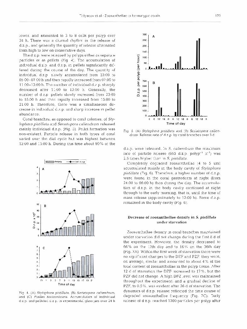
Coral branches, as opposed to coral colonies, of Sty- lophora pistillata and Seriatopora caliendrum released mainly individual d.z.p. (Fig. 5). Pellet formation was non-existent. Particle release in both types of coral varied over the die1 cycle but was highest between 12:00 and 15:00 h. During this time about 90% of the
Pellets + lnd. d.2.p.
23 1 3 5 7 9 11 13 15 17 19 21
Time of day
Fig. 4 . (A ) Stylophora pistillata. ( B ) Seriatopora caliendrum, and ( C ) Porites horizontalata. Accumulation of individual d.2.p. and pelleted d.2.p. in experimental glass jars over 24 h
0 6 12 18 0 6 12 18 0 6 12 18 0
Time of day
Fig. 5 . (A) Stylophora pistillata and ( B ) Seriatopora calien- drum. Release rate of d.2.p. b y coral branches over 3 d
d.2.p. were released. In S. caliendrum the maximum rate of particle release (665 d.z.p. polyp-' h-') was 2.5 tlmes higher than in S. pistillata.
Completely degraded zooxanthellae (4 to 5 pm) accumulated mainly in the body cavity of Stylophora pistillata (Fig. 6 ) . Therefore, a higher number of d.2.p. were found in the coral gastroderm at night (from 24:OO to 06:OO h) than during the day. The accumula- tion of d.2.p. in the body cav~ty continued at night through to the early morning, that is, until the time of mass release (approximately to 12:OO h). Some d.2.p. remained in the body cavity (Fig. 6).
Decrease of zooxanthellae density in S. pistillata under starvation
Zooxanthellae density in coral branches maintained under starvation did not change during the first 8 d of the experiment. However, the density decreased to 66% on the 12th day and to 16%) on the 36th day (Fig. ?A). Within the first week of starvation there were no significant changes to the DZF and PZF; they were, on average, similar and amounted to about 4 O/r, of the total content of zooxanthellae in the polyp tissue. After 12 d of starvation the DZF increased to 17'Y0, but the PZF did not change. A high DFZ level was maintained throughout the experiment, and a gradual decline of PZF, to OS%, was evident after 36 d of starvation. The dynamics of d.2.p. release reflected the time course of degraded zooxanthellae frequency (Fig. ?C). Daily release of d.z.p. reached 1300 particles per polyp after

Mar Ecol Prog Ser 139: 167-178, 1996
DISCUSSION D 2 . in Release +Dz.p. in
gastr. d.2.p. body 1 cavity
Time of day
Fig 6. Stylophora pistillata Relative quant~taties ( ~ n terms of % of the maximum value) of d.z. accumulation In the gastro- derm (gastr.), d.2.p. in the body cavity and rate of d.2.p.
release over 24 h
12 d of the experiment. The expulsion of healthy-look- ing zooxanthellae was insignificant and amounted to, on average, 10 to 20 cells polyp-' d-' throughout the starvation experiment (Fig. ?C).
B OZF + PZF
1 -0 l .p . +H2. I a C I 2. - 25
0 P 20 ; L - aJ
7
Q: l 15
- ' L 0 Y z g 10 U
X - 4 5 ; ; 0 0 P
1 4 8 12 16 20 24 28 32 36
Days of starvation
Flg. 7 . Stylophora pistillata. ( A ) Zooxanthellae population density, (B) degraded (DZF) and proliferating (PZF) zooxan- thellae frequency and (C) release of d.2.p. and h.% , as a func- tion of days of starvation. Values represent the average for
3 coral branches
The proportion of degraded zooxanthellae in living tissue of the corals Millepora tenella, Montipora digi- tata, Pocillopora damicornis, Porites cylindnca, Porites horizontalata, Seriatopora caliendrum, Seriatopora hys- trix and Stylophora pistillata amounted to 1 to 8%. Boschma (1925) also reported on degraded zooxan- thellae in Astrangia danae and detected that 'these algae are always found in the remains of the food in the gastric cavity'. Yonge & Nichols (1931) while study- ing the effect of starvation on the relation between coral host and zooxanthellae also observed degraded zooxanthellae. Degraded zooxanthellae have been ob- served in bleached coral tissue subjected to elevated sea-water temperatures (Glynn et al. 1985, Hayes & Bush 1990). Szmant & Gassman (1990) also found abnormal zooxanthellae in the basal tissue of bleached Montastrea annularis. Recently, Brown et al. (1995) showed that the degradation of zooxanthellae is one of the mechanisms involved in coral bleaching, when water temperatures are higher than average (1 to 2°C higher than seasonal maxima), with a possible syner- getic effect of temperature and irradiance. A lot of disrupted and deformed zooxanthellae were detected in the gastroderm of both partially and fully bleached corals. Unbleached colonies also had degraded zoo- xanthellae in the mesenterial filaments, but not in other gastrodermal tissue (Brown et al. 1995).
In the present study, zooxanthellae in corals from shaded and well lit habitats were in all stages of onto- genesis, even in the stage of cell division (Fig. l A ) , and the degraded zooxanthellae frequencies were similar to proliferating zooxanthellae frequencies. Therefore, under naturally constant conditions, the dividing pro- cess of symbiotic algae is probably in balance with degradation rates. In most cases, shaded habitats caused the accumulation of zooxanthellae, and values of degraded zooxanthellae frequency and proliferating zooxanthellae frequency decreased. In all cases the average diameter of a degraded zooxanthella was approximately half the size of a healthy zooxanthella. It is noteworthy that alga in Zoanthus sociatus (Antho- zoa, Coelenterata), in the final stage of degradation, become reduced to approximately half their origi.na1 diameter (Trench 1974).
Degraded zooxanthellae particles were expelled from the coral as individual particles or as pellets. The pellets contained mucus with over 90% d.z.p., and a small number of normal zooxanthellae. Steele & Goreau (1977) observed the release of pellets consisting of healthy zooxanthellae and zooxanthellae debris from the sea anemone Phyllactis flosculifera. In tropical zoanthids of the genera Polythoa and Zoanthus, healthy zooxanthellae were expelled as a rolled fila-

Titlyanov et al.. Zooxanthellae ~n hermatypic corals 175
ment or 'zooxanthellar body' (Reimer 1971, see also process. Maximum proliferating zooxanthellae fre- Trench 1974), and in the sea anemone Aiptasia tagetes quency was found in the tentacles (3 %) of S1.yJophora pellets are expelled containing almost exclusively pistillata, and was lowest in the connecting sheet healthy zooxanthellae, including dividing forms (Steele (1.6 %). In situ phased division of zooxanthellae is 1976). In Zoanthus sociatus, Trench (1974) described often seen in cnidarian hosts (including hermatypic characteristics of degraded zooxanthellae similar to corals) (Wilkerson et al. 1983, Smith & Hoegh-Guld- those described here in hermatypic corals. In the final berg 1987, Hoegh-Guldberg & Smith 1989). stage of degradation this undigestable fragment con- A comparison of degraded zooxanthellae frequency tained a conglomerate of membranes derived from cell and prohferating zooxanthellae frequency in the mesen- organelles and an 'accumulation body' (Trench 1974). tenes showed that there was approximately 30 % degra- Degraded symbionts of Phyllactis flosculifera and dation during the day, and only 2.5 % division. Taking Zoanthus sociatus are seen only in the free edge in this as a basis, it can be assumed that zooxanthellae are the former species and in the 'digestive-excretory' expelled into the body cavity (by exocytosis of the zone cells of the mesenteries in the latter (Trench 1974, gastrodermal cells in the connecting sheet and tenta- Steele & Goreau 1977), whereas, in hermatypic corals, cles), and then incorporated into the mesenteries (by degradation of zooxanthellae occurs in the gastroderm phagocytosis of the gastroderm cells of the mesentenes). of the mesentenes, tentacles and connecting sheet. This is similar to the process described for Zoanthus
The degradative process is phased and varied over sociatus by Trench (1974). It is noteworthy that the body the die1 cycle in the gastroderm of hermatypic corals. cavity of Stylophora pistillata always contains a small During a 24 h period, approximately 30 % of the zoo- number of healthy zooxanthellae within the polyps. xanthellae degrade in the mesenteries, around 6 % in A schematic reconstruction of the possible pathways the connecting sheet and around 2 % in the tentacles. taken by zooxanthellae in Stylophora pistillata over the Cell degeneration was highest at night and lowest course of a day is depicted in Fig. 8. Degraded zoo- during the day. Zooxanthellae degrad- tion took place only at night in the con- necting sheet and tentacles, but both at night and during the day in the gastroderm of the mesenteries. Fitt & Cook (1990) found a phased cycle of moribund zooxanthellae abundance, with a peak around midnight in the , digestive cells of the marine hydroid Myrionema ambionense. In our ex- , periments on Stylophora pistillata the highest frequencies of degraded zoo- . , xanthelae were found in the mesen- teries, while the highest densities were found in the gastroderm of the con- necting sheet. Considering the high level of degraded zooxanthellae fre- quencies found in the mesenteries, and their extended degeneration period, we are inclined to predict that the mesenterial gastroderm is a special- ized place for zooxanthellae degrada- Flg. 8. Schematic representation of the zooxanthellae degradat~on process in tion. Although the number of healthy Stylophora pistillata during 24 h. (1) Degradat~on of zooxanthellae In the gastro-
and degraded zooxanthellae are higher dermis of the mesenteries (18:OO to 12:00 h). (2 , 3 ) Degradation of zooxanthellae
in the connecting sheet, the proportion in gastrodermis of the connecting sheet and tentacles, respectively (21:OO to 12:OO h). (4) Mass appearance of d.z.p. in the body cavity 6 h later and presence
degraded zOOxanthellae to of abundant d.z.p. in the body cavity from 00:OO to 18:OO h. (5. 6) Formation of zooxanthellae was considerably lower pellets from z ~ o ~ a n t h e l l a e debris, at ;he base of the tentacles and in the connect- than in the mesenteries. ing sheet, respectively (09:OO to 15:OO h ) . (7) Possibility of transfer of d.z.p. from
~ ~ ~ ~ ~ ~ ~ h ~ l l ~ ~ proliferation also had one polyp into another wi th~n the gastrovascular cavity. (8) Release of individual d.z.p. and pellets from the polyp. (9) Occasional migration of d.2.p. and pellets
a phased in gastroderm between polyps. (10) Possible exocytosis of zooxanthellae by gastroderrn cells examined, with a peak a 'ound 03:OO h of the tentacles and connecting sheet. (11) Hypothetical phagocytosis of zoo- in close phase with the degradative xanthellae by the mesenterial cells

Mar Ecol Prog Ser 139: 167-178,1996
xanthellae are extruded from the gastrodermis and accumulate in the body cavity. The appearance of d.z.p. in the body cavity took place at 00:OO-03:OO h, i.e. about 6 h after the first degraded cells appeared in the connecting sheet and tentacles. Therefore, the process of zooxanthellae degradation and extrusion of debris into the body cavity probably takes about 6 h. Forma- tion of pellets occurs mainly at the base of the tentacles and in the connecting sheet between the sclerosepta. The pellets appear to form by the rotation of debris, involving mucus, individual d.z.p. and a few h.z.
Regulation of zooxanthellae population density by coral under starvtion
Symbiont population densities decrease In corals under starvation (Yonge & Nichols 1931, Szmant- Froelich & Pilson 1984). Meyer & Schultz (1985) found that corals exposed to nutrients from fish excretion sup- ported higher densities of zooxanthellae than colonies without resident fish. A comparison of fed and unfed sea anemones also displayed significant differences in algal densities (Clayton & Lasker 1984, Cook et al. 1988).
In our experiments Stylophora pistillata lost 80% of its symbiont population over 36 d of starvation at 60 to 80% of surface irradiance. During the first 8 d, the zooxanthellae densities did not decrease. Similarly, Cook et al. (1988) reported that zooxanthellae densi- ties in Aiptasia pallida did not change during 10 d of starvation. In our experiments, algal density decreased only on the 12th day of starvation, to approximately 70%. On the same day, the degraded zooxanthellae frequency increased from 4 to 17%, and the ratio of proliferating to degraded zooxanthellae frequency dropped from 1 to 0.25 on the 12th day of the experi- ment. The number of released d.z.p. increased an order of magnitude. From the 12th to 36th day of starvation the degraded zooxanthellae frequency and d.z.p. release rate remained high (but variable), and the proliferating zooxanthellae frequency dropped. from 3 4 % to 0.5% In the sea anemones (Cook et al. 1988) the mitotic index decreased from 8 to 1% after 30 d of starvation.
We suggest that Stylophora pistillata used the avail- able resources of limited biogenic elements (N and P) over the first 8 to 10 d of starvation for the growth of animal tissue and for symbiont population replenish- ment, and then began to obtain these elements from zooxanthellae. Limiting the supply of biogenic ele- ments to the zooxanthellae resulted in almost complete cessation of division. Our results with S. pistillata strengthen the widespread opinion that endosymbiotic algae in the invertebrate host utilize host metabolites
as nutrient sources (e.g. Yonge & Nichols 1931, Smith 1939, Cates & McLaughlin 1979, Trench 1979, Steen 1986, Cook et al. 1988, McAuley 1994) and can be limited by N and P when the host is deprived of food (Cook et al. 1988, McAuley 1994). In the degradative process, zooxanthellae lost protein structures, nuclei and lipid drops. At the same time, the coral, which was deprived of food, lost more than ha.lf its zooxanthellae. Loss of zooxanthellae in S. pistillata during the food deprivation experiment was evidenced by zooxan- thellae degradation and release of their remnants. Expulsion of healthy zooxanthellae, over the period starvation, was 2 orders of magnitude less than the d.z.p. release.
On this basis, we consider that the expulsion of h.2. under normal conditions is not important i.n the algal regulation process, and the principal mechanism whereby symbionts are eliminated is by degradation and release of debris. Under normal conditions, the host undoubtedly influences not only the zooxanthel- lae degradation process, but also their reproduction. Reproductive inhibition of zooxanthellae has also been shown to occur in sea anemones (Cook et al. 1988). As did Cook et al. (1988), we can only hypothesize that inhib~tion of coral symbiont reproduction occurs be- cause the supply of nutrients stops. Other possibilities, such as inhibitor secretions, are not expected to be the cause (Muscatine & Pool 1979, Trench 1987).
Do hermatypic corals digest their own zooxanthellae?
We are inclined to call the above process digestion of symbionts by the host because ( l ) the degradation process has a clear die1 rhythm and occurs mainly at night, (2) zooxanthellae degradation occurs inside the animal gastrodermal cells; the gastrodermal cells of the mesenteries appear most specialized in the degra- dation process, ( 3 ) nuclei, protein structures, lipid drops and chl c were not found within the zooxan- thellae debris, and (4 ) the polyps regulate the intensity of algal degradation which is stimulated by a defl- ciency of food, and degradation is rapidly terminated upon reintroduction of food (Titlyanov unpubl.).
We didn't see the fusion of lysosome with zooxan- thellae in polyp as was observed by Colley & Trench (1985) in the polyp stage of the jellyfish Cassiopea xamachana and Fitt & Cook (1990) in the marine hydr0i.d Myrionerna ambionense. However, in our opinion, under norm.al conditions, old or weak cells may be subject to autodegradation. In our case, as was stated above, even in the cell divis~on stage, zooxan- thellae were subjected to degradation. The process of digestion of zooxanthellae in hermatypic corals differs from that described by Trench (1974) in Zoanthus

Titlyanov et al.. Zooxanl .hellae in hermatypic corals 177
sociatus in which only senescent cells breakdown, and only in the digestive-excretory zone (as in a 'grave yard' for old defunct members of the algal population). However, in the present study, we observed digestion of apparently healthy zooxanthellae in the endoderm of the connecting sheet, tentacles and mesenteries. Digestion of coral symbionts is probably similar to the use of zooxanthellae as a nutrition source in the sea anemone Phyllactis flosculi (Steele & Goreau 1977). A similar process of intracellular digestion has also been found in giant clams (Fankboner 1971) and in the marine hydroid Myrionema an~bionense (Fitt & Cook 1990). Infected symbionts are also digested by the protozoan Pararnecium bursaria (Karakashian & Kara- kashian 1973, Weis 1976) and by the polyp stage of the jellyfish Cassiopea xamachana (Colley & Trench 1985). Therefore we conclude that the digestion of the algal symbionts by a coral host is a common process rather than a rare event.
Acknowledgements. The Russian authors thank the President of University of the Ryukyus. Keishin Sunagawa, for the invitation to work at Sesoko Station as visiting foreign researchers together with an accompanying person [T.V.T.) We are grateful to the staff for use of facihties, technical help and the atmosphere of friendship and confidence. Special thanks are due to our friend Prof. Y. Loya (Tel-Aviv Univer- sity, Israel) for discussions on our work
LITERATURE CITED
Boschma H (1925) On the feeding reactions and digestion in the coral polyp Astrangia danae, with notes on its sym- bionts with zooxanthellae. Biol Bull htlar Biol Lab Woods Hole 49:407-439
Brown BE, Le Tissier MDA, Bythell J C (1995) Mechanisms of bleaching deduced from histological studies of reef corals sampled during a natural bleaching event. Mar Biol 122: 655-663
Buddemeier RW, Fauti DG (1993) Coral bleaching as an adap- tive mechanism. Bio Sci 43:320-326
Cates N. McLaughlin JJA (1979) Nutrient availability for zooxanthellae derived from physiological activities of Condulactus spp. J Exp Mar Biol Ecol 37:31-41
Clayton WS Jr. Lasker HR (1984) Host feeding reglme and zooxanthellae photosynthesis in the anemone, Aiptasia pa l l~da (Verrill.) Biol Bull Mar B101 Lab Woods Hole 167: 590-600
Coles SJ, Fadlallah YH (1990) Reef coral survival and mortal- ity a t low temperatures in the Arabian Gulf: new species- specific lower temperature limits. Coral Reefs 9:231-237
Coles SJ , Jokiel PL (1978) Synergistic effects of temperature, salinity and light on thr Rermatypic coral Montlpora ver- rucosa. Mar Biol49:187-195
Colley NJ, Trench RK (1985) Cellular events in the re- establishment of a symbiosis between a marine dinoflagel- late and coelenterate. Cell Tissue Res 239:93-103
Cook CB, D'Elia CF, Muller-Parker G (1988) Host feeding and nutrient sufficiency for zooxanthellae in the sea anemone Afptasia pallida. Mar Biol 98:253-262
Cook CB, Fitt WK (1990) Some effects of dissolved inorganic nutnents in the hydroid Myrionema amblonense. In: Nar-
don P, Gianinazzi-Pearson V. Grenier AM, Margulis L, S m ~ t h DC (eds) Endocytobiology. INRA, Paris, p 285-288
Fankboner PV (1971) 1:ntracellular digestion of symbiot~c zooxanthellae by host amoebocytes in giant clams (Bi- valvia: Tridacnidae), with a note on the nutritional role of the hypertrophical siphonal epidermis. B101 Bull Mar Biol Lab Woods Hole 141:222-234
Fitt WK, Cook CB (1990) Some effect of host feeding on growth of zooxanthellae in the marine hydroid Myrionema amblonense in the laboratory and in nature. In- Nardon P, Gian~nazzi-Pearson V, Grrnier AM, Margulis L, Smith DC (eds) Endocytobiology IV. INRA. Paris. p 281-284
Gates RD (1990) Seawater temperature and sublethal coral bleaching in Jamaica. Coral Reefs 8:193-198
Gates RD, Baghdasarian G , Muscatine L (1992) Temperature stress causes host cell detachment in symbiotic Cnidari- ans: implications for coral bleaching. Biol Bull 182:324-332
Glider WV (1983) The biology of association of Symbiodjnium microadrjaticum with Aiptasia pallida an anemone-alga symbiosis. PhD thesis, University of Nebraska, Lincoln
Glynn P (1990) Coral mortality and disturbances to coral reefs in the tropical eastern Pacific. In: Glynn 13W (ed) Global ecological consequences of the 1982-83 EL-Nino South- ern Oscillation. Elsevier, Amsterdam, p 55-126
Glynn PW, Peters EC, Muscatine L (1985) Coral tlssue micro- structure and necrosis: relation to catastrophic coral n~ortality in Panama. Dis Aquat Org 1:29-37
Goreau TF (1964) Mass expulsion of zooxanthellae from Jamaican reef communit~es after Hurricane Flora Science 145:383-386
Hayes RL, Bush PG (1990) Microscopic observations of re- covery in the reef-building scleractinian coral, Montastrea annularis after bleaching on a Cayinan reef. Coral Reefs 8:203-209
Hoegh-Guldberg 0, Mc!'loskey LR, Muscatine L (1987) Ex- pulsion of zooxanthellae from synlbiotic Cnidarians from the Red Sea. Coral Reefs 7:113-116
Hoegh-Guldberg 0. Smith GJ (1989) Influence of the popula- tion density of zooxanthellae and supply of ammonium on the biomass and metabolic character~stlcs of the reef corals Seriatopora hystrix and Stylophora p~sti l lata. Mar Ecol Prog Ser 57: 173- 186
Hull HM, Hoshaw RW, Wang JC (1982) Cytofluorometric determination of nuclear DNA in living and preserved algae Stain Technol 57:273-282
Jeffrey SW, Humphrey GP (1975) New spectrophotometric equations for determining chlorophylls a, b, c, and c:! In higher plants, algae and natural phytoplankton. Biochem Physiol Pflanj.cn 167:191-194
Johannes RE , \t\/~ebe \\'J (1970) A method for determination of coral tissue biomass and composition. Limnol Oceanogr 21:540-547
Karakashian MW, Karakashian SJ (1973) Intracellular diges- tion and symbiosis in Parameciun~ hursaria. Expl Cell Res 81:111-119
McAuley PJ (1985) The cell cycle of symblotlc Chlorella. I. The relationship between host feeding and algal cell growth and div~sion. J Cell Sci 77:225-239
McAuley PJ (1994) Interactions between host and symblonts in algal invertebrate intracellular symbioses. Bot J Scotl 47(1):97-112
Meyer JL , Schultz ET (1985) Tissue condition and growth rate of corals associated with schooling fish. Limnol Oceanogr 30:157-166
Muller-Parker G (1984) Photosynthesis-irradiance responses and photosynthetic period~city in the sea anemona Aipta- sia pulchella Mar Biol 82.225-232

178 Mar Ecol Prog Ser 139: 167-178, 1996
Muller-Parker G (1987) Seasonal variation In light-shade adaptation of natural populatlons of the symbiotic sea anemone Aiptasia pulchella (Carlgren 1943) in Hawaii. J Exp Mar Biol Ecol 112: 165-183
Muscatine L, McCloskey LR, Loya Y (1985) A comparison of the growth rates of zooxanthellae and animal tissue in the Red Sea coral Stylophora pistillata Proc 5th Int Coral Reef Symp 6:119-123
Muscatine L, Pool RR (1979) Regulation of numbers of intra- cellular algae. Proc R Soc Lond B 204:131- 139
Pardy RL (1974) Some factors affecting the growth and distri- bution of the algal symbionts of Hydra viridis. Biol Bull mar Biol Lab Woods Hole 147:105-118
Pardy RL, Muscatine L (1973) Recognition of symbiotic algae of Hydra viridis. A quantitive study of the uptake of living algae by aposymbiotic H. viridis. Biol Bull Mar Biol Lab Woods Hole 145:565-579
Pool RR (1976) Symbiosis of Chlorella and Chlorohydra viridissima. PhD dissertation, University of California, Los Angeles
Pool RR (1979) The role of algal antlgenic determinants in the recognition of potential algal symbionts by cell of Chlorohydra. J Cell Sci 35:367-379
Reimer AA (1971.) Observations on the relationships between several species of tropizoanthids (Zoanthidea, Coelentei-- ata) and their zooxanthellae. J Exp Mar Biol Ecol 7. 207-214
Reynolds ES (1963) The use of lead nltrate at high pH as an electron opaque stain in electron microscopy. J Cell Biol 17.208-212
Smith DC, Muscatine L, Lewis DH (1969) Carbohydrate movement from autotrophs to heterotrophs in parasitic and mutualistic symbiosis. Biol Rev 44:17-90
Smith GJ, Hoegh-Guldberg 0 (1987) Variation in the growth rate of zooxanthellae with coral host colony size is not con- trolled by changes in the duration of cytokinesis. EOS (Trans Am Geophys Un) 68:1724
Smith HG (1939) The significance of the relationship between actinians and zooxanthellae. J Exp Biol 16:334-345
Sorokin YI (1990) Coral reef ecosystems. Nauka. Moscow Steele RD (1976) Light intensity as a factor in the regulation of
the density of symbiotic zooxanthellae in Aiptasia tagetes. J 2001 179:387-405
Steele RD (1977) The significance of zooxanthellae- containing pellets extruded by sea anemones. Bull Mar Sci 27.591-594
Steele RD, Goreau NI (1977) The breakdown of symbiotic zooxanthellae in the sea anemone Phyllactis (= Oulactls) flosculifera (Actiniaria). J Zoo1 Lond 181:421-437
Steen RG (1986) Evidence for heterotrophy by zooxanthellae in symbiosis with Ajptasia pulchella. Biol Bull 170.267-278
Steen RG, Muscatine L (1987) Low temperature evokes rapid
exocytosis of symbiotic algae by a sea-anemone. Biol Bull 172:246-263
Stimpson J , Kinzie RA (1991) The temporal pattern and rate of release of zooxanthellae from the reef coral Pocillopora damicornis (Linnaeus) under nitrogen-ennchrnent and control conditions. J Exp Mar Biol Ecol 153.63-74
Szmant A, Gassman NJ (1990) The effects of prolonged 'bleaching' in the tissue biomass and reproduction of the reef coral Montastrea annularis Coral Reefs 8:217-224
Szmant-Froelich A , Pilson EQ (1984) Effects of feeding fre- quency and symbiosis with zooxanthellae of nitrogen metabolism and respiration of the coral Astrangia danae. hlar Biol81:153-162
Taylor DL (1969) On the regulation and maintenance of algal members zooxanthellae-coelenterate symbiosis, with a note on the nutritional relationship in Anemonia sulcata. J Mar Biol Ass UK 49:1057-1065
Titlyanov EA, Zvalinsky VI, Leletkin VA, Shaposhnikova MG (1983) Zooxanthellae photosynthesis of reef-building corals in different light conditions. In: Krasnov EV (ed) Investigations on the Phantom Bank (Timor Sea). Far East Sci Center USSR, Vladivostok, p 51-74
Trench RK (1974) Nutritional potentials in Zoanthus sociatus (Coelenterata, Anthozoa). Helgol Wiss Meeresunters 26: 174-216
Trench RK (1979) The cell biology of plant-animal symbiosis. A Rev Plant Physiol30:485-531
Trench RK (1987) Dinoflagellates in non-parasitic symbiosis. In: Taylor FJR (ed) The biology of dinoflagellates. Black- well Scientific Press, Oxford, p 531-570
Van Woesik R, De Vantier LM, Glazebrook JS (1995) Effects of cyclone 'Joy' on nearshore coral communit~es of the Great Barrier Reef. Mar Ecol Prog Ser 128 261-270
Weis DS (1976) Digestion of added homologous algae by Chlorella-bearing Paramecium bursaria. J Protozool 23(4): 527-529
Wilkerson RP, Muller-Parker G, Muscatine L (1983) Temporal patterns of cell division in natural populations of endosym- biotic algae. Limnol Oceanogr 28:1009-1014
Williams EH Jr, Bunkley-Williams L (1990) The world-wide coral reef bleaching cycle and related sources of coral mortality. Atoll Res Bull 335:171
Yamazato K (1981) A note on the expulsion of zooxanthellae during summer, 1980 by the Okinawan reef-building corals. Sesoko Mar Sci Lab Tech Rep 8 9-18
Yonge CM (1931) Studies on the physiology of corals. 111. Assimilation and excretion Scient Rep Gt Barner Reef Exped 1:l.l-91.
Yonge CM, Nichols AG (1931) Studies on the physiology of corals. V. The e f f ~ c t of starvation In light and in darkness on the relationship between corals and zooxanthellae. Scient Rep Gt Barrier Reef Exped 1 177-211
This artjcle was submitted to the editor Manuscript first received: November 18, 1995 Revised version accepted: March 21, 1996