ddc - dtic.mil plotted in the same way as the following drawingi as a unit was accepted a ratio of...
TRANSCRIPT
%m»m\m^ \J^ I
<3r? 5 '.~:c:
£#
... ^X
/
m' '£*<• »r_
AIR FORCE SYSTEMS COMMAND
WXIGHT-PAHERSON AIR FORCE BASE
OHIO
%#
DDC _C0PY_^-— HARD COPY 11. / Co
±4- ^rp
/3f
r-ffi* « ; »■
i&th a
;'i
-;U_>
/ w
L
This translation was made to provide the users with the basic essentials of the original document in the shortest possible time. It has not been edited to refine or improve the grammatical accuracy, syntax or technical terminology.

FTD-TT- 65-36/1+2
UNEDITED ROUGH DRAFT TRANSITION
OPPOSITE CHANGES IN ELECTROPHYSILOGICAL REACTIONS OF THE HUMAN BRAIN IN ADOPTING THE FREQUENCY OF LIGHT FLASHES AT VESTIBULAR AND OPTOKINETIC STIMULI
BY: V. G. Samsonova
English pages: 10
SOURCE: Fiziologicheskiy Zhumal SSSR im. I M. Sechenova (Russian), Vol. 50, No. 6, 1964, pp. 649-654.
S/0239-064-050-006
(Ace. No. TP5000109)
THIS TRANSLATION IS A tlNMTtOM OP TMI ORIOL HAL PORIMN TIXT WITHOUT ANY ANALYTICAL 0« ■MTOtlAL COMMNT. STATRMINTS OR TNIQRIIS ADVOCATIDORINPLIIDARtTHOSIOPTHt SOURCt AND DO NOT NRCISSAMLY RIPLICT TNI POSITION OR OPINION OP TNI PORItON TICHN0LO0Y M> VISION.
PRtPARID IYI
TRANSLATION OIYIUON PORtlON TICNNOLOOY DIVISION WP'APt, OHIO.
FTD-TT- 65-36/1+2 Ölte 13 May 1965
B

OPPOSITE CHAIGBS IH ILBWB0PBT3IL0GXCAL BtACT 10» 0» TBB BOMal BRAU IH ADOPTWQ TBB rHKJÖESCT OF UWS FLASH» Af VS8TIBÜLAR
AID OPTOKUBflC STimi
?. 0« Samsonova
Inst. of Higher lervous Activity ud Seuropbyaiolo.iy Academy of Sciences USSR, Moscow
The reaction of cortical structures, oonnaottd witli analysis of light Sig-
nale, on the irritationa of other nodalites appears to be a substantial link in
the problem of convergence, reaction and overlapping of impuleation, determining
the state of excitability and analytical activity of the central eeotiona of
analyser systeme.
In role of one of excitation level indioatora can he used a summary elec-
tric activity of the brain-enoephalograa, which ia used ■ore often aa an indic-
ator of the functional atata of the brain»
The investigation by V. A« Ilyanek (1962) showed» that the adoption by
the brain of a light flashes rhytha, «pressed in a rise ia HO oscillations
amplitude, synchronous to the frequency of a pulsating light» appears to he
even further demonstrative proof of the state of excitability of the stimulated
structures. At the same time V« A. Ilyaaak (1959* 1961a, 1961b) discovered
FTD-TT-65-36/1 + 2 1

mm?
that the amplitude of the light pulsation frequencies reproduced by the brair is
due to the characteristic of the light signal - its intensity, duration and fre-
quency« I (Samsenova, 1961) hare revealed that in this reaction is found ade-
quate reflection also for the properties of daily and dusk vision of the human
being. It is essential, that its change is correlated quite well with the data
of psyohophysiologioal experiments concerning tne differentiation of these or
any other properties of light signals.
Consequently, the reaction of adopting by the brain a rhythm of light
flashes reflexes the activity of these cortical structures of the brain, which
participate direotly in the analysis of light stimulif consequently by its
change oan be judged the state of excitability and about the analytical activ-
ity of brain structures, reacting to light stimulation*
On the basis of these prerequisites was set up the problem of investiga-
ting, with the aid of this electrical reaction, in adopting by the brain of the
rhythm of light flashes, the excitability of visual oenters under conditions of
their reaction with vestibular-optomotorial system. The biological importance
of this reaction is exclusively grsat, because it determines the position and
orientation of human being and animal in space. It was assumed, that the ampli-
tude of light flash frequencies reproduced by the brain will change monodireo-
tionally as during vestibular, and as during optokinetio stimulations, because
anatomioally, and functionally direct and numerous averaged relationships have
been long established between vestibular and optioomotorial systems.
According to oertain data, the human encephalogram at vestibular stimula-
tions changes slightly. We have not suooeeded in discovering functions, in
which would have been investigated changes in amplitudes of light flash fre-
quencies reproduced by the brain at vestibular and optokinetio stimulations.
FTD-TT-65-36/1 + 2

vWNtöKHfcrm:
HETHOD
The examined at the time of experimenting sat in a Barani ohair, situated
in a screened chamber. The experimentor with the aid of remote control, situa-
ted out side of the ohan-bcr, oould regulate the rate of rotation of the system,
rotating the chain from 1 to 18 per ,nin,, and change the direction of rotation.
Vestihular irritations were produced by rotating the chain at slow speed, which
was then increased gradually.
Around the ohain was fastened a circular, uniformly illuminated white
screen, by which in the experiments with optoklnetio stimuli were moved black
strips at a rate of 1, 2 per 1 see.
During experiment with long lasting rotation ( in case of the bands, in
other case with the human rotating in the chain) to the round screen was per-
iodically fed an achromatic flashing light of fixed frequency ( in a range
from 7 to 33 c) from a Soneckle pulse photostimulator.
For arifaotless recording of EEO in the process of human rotation A* A«
Marinichev created in our lab a special transient device, allowint to register
w/o distortions at greater amplification the biopotentials of the brain at the
moment of the very rotation.
In experiments were used unipolar sections occiput-ear, sinoiput-earf
EEO recording was realized on an ink-writing oscillograph from the "APvar Co")
registered were also the frequency speotra of EEO of these areas of the brain,
separated by the two-channel Walter analyser, dynamics of EEO frequency changes,
corresponding to the rhythm of light flashes, obtained by separating these fre-
quencies by analyser filters, as well as the frequency marker of band rotations,
Barani chair and light flashes.
The experiment began with the recording of background EEO under conditions
when the examined had his sight fixed on the uniformly illuminated screen, and

«JF'Sfc , ■- -■.;«»*',
than under action of the flashing light, Then a rotation was imparted eithsr
to the chair, or to ths hands, which remained stationary tw this moment. In
the process of their rotation within eaoh 3-10 min, was registered the EEC w/o
flashing light and at light flashes, lasting for 40 sec,.
The amplitudes of EEC frequencies, corresponding to frequency of flashing
light, were calculated by analyser data, averaged by 40 sec,, immediately prior
to application of rhythmical light stimulation and during the period of such
irritation. Their changes were judged by the magnitude ratios of these ampli-
tudes. Such a method of analyzing results was used because at vestibular-
optokinetic irritations the EEG frequency spectra under conditions of our ex-
periments experienced in turn certain changes,
RESULTS OP INVESTIGATIONS
In the experiments was detected a change in amplitudes of flashing light
frequencies reproduced by the brain at vestibular and optokinetic stimulations.
Human rotation in the ohair caused a reduotion in amplitudes of EEO oscilla-
tions, corresponding to rhythms of light flashes, which is illustrated by the
fragment of rcoording the experiment (fig, 1 B),
notation of bands led to an opposite effect - it was accompanied by a rise
in amplitude of rhythmic light stimulation frequency reproduced by the brain
in comparison with oontrol EEO (Fig, 1, C),
A reduction in ESO amplitudes, synchronous to frequency of light flashes,
began ordinarily immediately after beginning of human rotation and rose some-
what in the prooess of its further rotation (Fig, 2,A),
An inorease in these amplitudes, which took place during th* rotation of
bands, was frequently intensified in the prooess of their rotation (Fig, 2,3),
but in many investigated it was very considerable from the first moments of ro-
tation, and later on it either remained as it was, or began relatively decreasing.

v-r
The contrast in ohage of amplitudes in EEO oscillations, coinciding in
frequency with the rhythm of light flashes, at vestibular and optokiir-tic irri-
tations was detected in conditions of separating EEO from oooiputal area of the
brain on all eleven examined. Individual differences were expressed only in
the degree of described ohanges-in some expressed more and in other less sharply.
The frequency of light flashes also produced a certain effect on the mag-
nitude of the detected effect. This is illustrated by the diagram in fig. 3»
plotted in the same way as the following drawingi as a unit was accepted a
ratio of amplitudes of adopted by the brain rhythm of light flashing to the
amplitude of the very same frequency in background SSO, measured priot to ap-
plying vestibular and optokinetic stimulants.
A 6 a i «WI^MV^VM^V^VV» /%v**'tyvVY*»,*,vV*J*> 1*fi*<**fi™j»/*»4*w* 2 A^^//4^*Wv**jfaA tf^^jfv^^^^ t*/^Mf^^¥J^M^ 3 HiiiiiüiKtTiia *-~. j~ v .. 3 j^t^X---^.--»-«-«™.^-,
tmint rji wflN^lÜO^""^!^ ♦.
Fig. 1. Change in reactions of adopting by the brain of light flashing rhythms at bestibular (ß (B)) and optokinetic ( B(£)) stimulants. A - control record- ing. Examined K. K. Experiments of Aug. 3, 1962. 1-EBG at oooiputal deriva- tion) 2- at parietal) 3- stimulation marker) a-rotation of chair) b-rotatien of beads) o-flashing light) 4-frequenoy of EBG osoillations ?o* separated by analyser filter at occipital) 5- at parietal derivation) 6- recording of ESO frequency spectrum with two channel Walter analysers I-occipital, II- parietal removal.
A change in this electrical reaotion of the brain during rotation of the
human or bands is expressed by average value analogous to the ratio of ampli-
tudes for all measurements in each experiment, then in all experiments on all
sfteBs li,, _ ,s. ,-.
investigated In comparison with the oontrol ratio of amplitudes.
* * t$ H 33 3$ «5 S* MEM it 20 23 28 .15 <a <4 Umm.
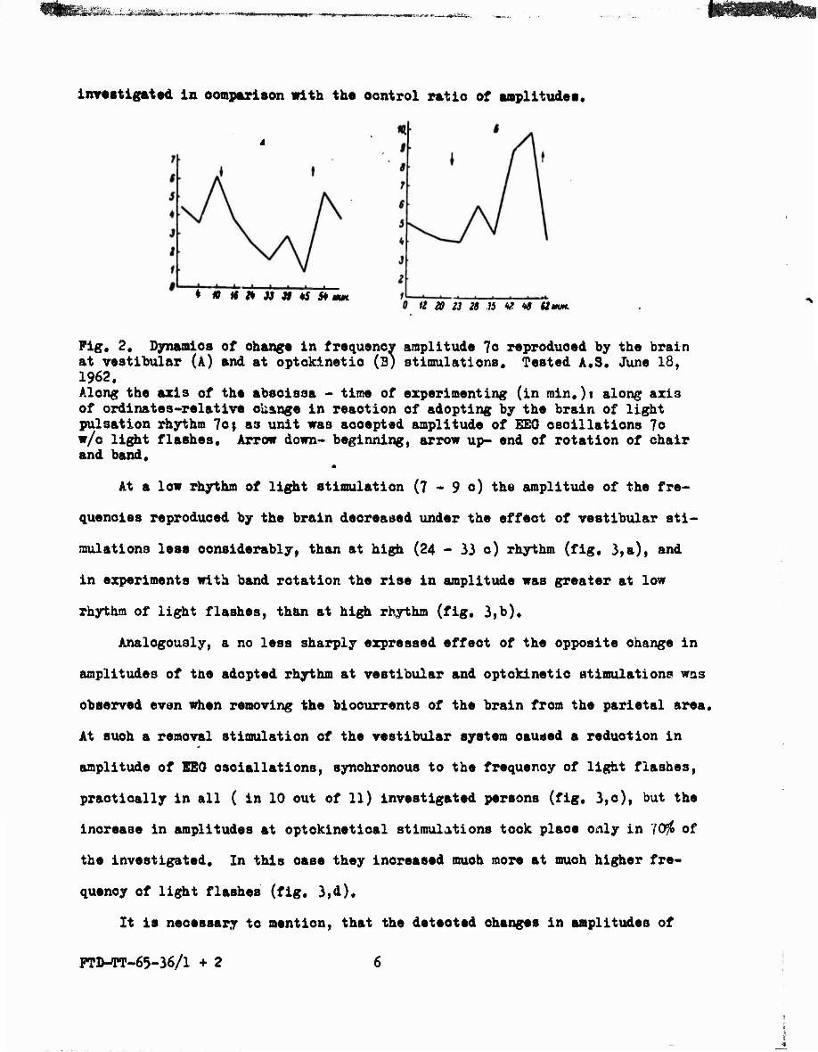
Fig« 2. Dynamics of ohanga in frequency amplitude 7o reproduced by the brain at vestibular (A) and at optokinetio (B) stimulations. Tested A.S. June 18, 1962. Along the axis of the absoissa - time of experimenting (in min.)t along axis of ordinatcs-relative okange in reaction of adopting by the brain of light pulsation rhythm 7cf as unit was aooepted amplitude of EEG oscillations 7c w/o light flashes. Arrow down- beginning, arrow up- end of rotation of chair and band.
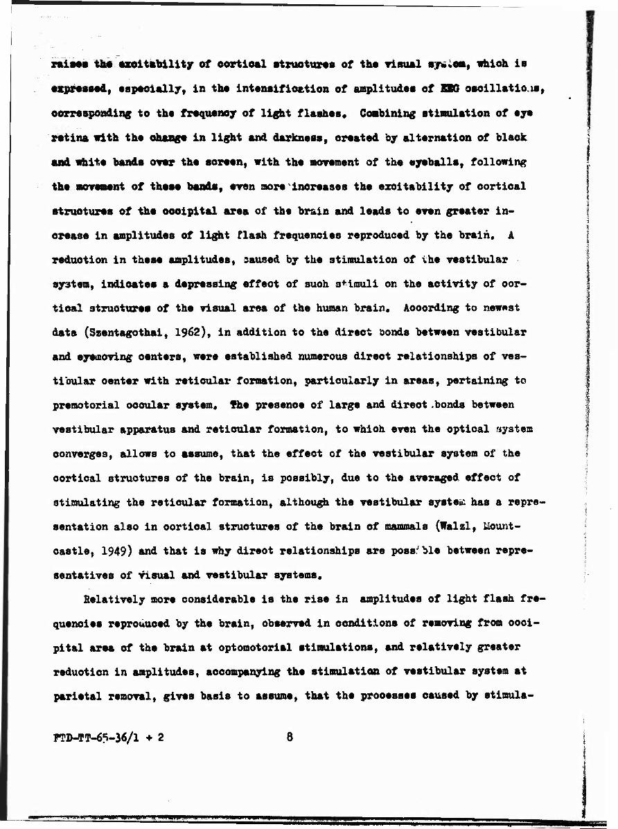
At a low rhythm of light stimulation (7 - 9 o) the amplitude of the fre-
quencies reproduced by the brain decreased under the effect of vestibular sti-
mulations less considerably, than at high (24 - 33 c) rhythm (fig, 3,a), and
in experiments with band rotation the rise in amplitude was greater at low
rhythm of light flashes, than at high rhythm (fig, 3,b),
Analogously, a no less sharply expressed effect of the opposite change in
amplitudes of the adopted rhythm at vestibular and optokinetio stimulations was
observed even when removing the bioourrents of the brain from the parietal area.
At such a removal stimulation of the vestibular system oaused a reduction in
amplitude of EEO osoiallations. synchronous to the frequency of light flashes,
practically in all ( in 10 out of 11) investigated persons (fig. 3,o), but the
inorease in amplitudes at optoklnetioal stimulations took place only in 70$ of
the investigated. In this oase they increased much more at much higher fre-
quency of light flashes (fig. 3,d),
It is necessary to mention, that the deteoted changes in amplitudes of
pnMT-65-36/1 + 2

SA I • * • •
light flash frequencies reproduced by the brain was observed in the sbaeiice of
noticeable symptoms of vegetative and Tostibular disorders among all of the in-
vestigated parsons«
As «as said, by tha change in reaction
of adopting tha brain of light flashing
rhythms oan «a Judge about tha atata of
azoltability of dafinita cortical brain
structures, raaoting to light stimula-
tions, Tha praaantad materials indicate
that it experiences changes under the
effect of veatlbular and optokinetio
stimulations. In first oase it drops
immediately after the beginning of apply-
ing stimulants, and in second - it in-
creases«
This difference in change of azolt-
ability oan have in its bases features
of anatomophyalologioal bonds of vmsti-
bular and optomotorial systems with high-
er level of visual analyser. At present
time (JUNO 1962) it «as established that in optokinetio reactions the selective
and prevailing importance is played by the visual system, and the veetibular
system sffeote only in a modulating manner the visual change in optomotorism«
Ths great aot of visual distinction is inseparably connected in a human
being with the movement of the eyeballs, i.e., excitation of muscular appara-
tus of the eyes, the activity of which la thus always combined with the activ-
ity of higher levels of ths optical analyser« Application of light stimuli
Fig« 3« Diagram of change in ampli- tudes of light flash frequencies re- produced by the brain (average data of experiments on all investigated, experiments 1962)« a-at vestibulart b-at optokinetio stimulations in conditions of re- moving from occipital areaf o- and d-r?speotlvely at very same stimuli under conditions of removing from parietal area of the brain« Along axis of ordinateai ratio of ampli- tudes of frequencies produced by the brain Xrt background KEG prior to applying vestibular and opto- kinetio stimuli«
FMMPT-65-36/1 ♦ 2
raisaa the excitability of cortical structures of tha visual sr^em» which is
«xpressed, especially, in the intensification of amplitudes of SBQ oacillatiois,
corresponding to the frequency of light flashes. Combining stimulation of eye
Mtins with the change In light and darkness, oraatod by altarnation of black
and «hit« bands over the soraanf with ths movement of the eyeballs, following
tha movement of these bands, even sore increases the excitability of cortical
structures of the occipital area of the brain and leads to even greater in-
crease in amplitudes of light flash frequencies reproduced by the brain. A
reduction in theaa amplitudes, caused by the stimulation of ihe vestibular
system, Indicates a depressing effeot of such stimuli on the activity of cor-
tical structures of ths visual area of the human brain• According to newest
data (Szentagothai, 1962), in addition to the direot aonds between vestibular
and syemoving oenters, were established numerous dirsct relationships of ves-
tibular center with retioular formation, particularly in areas, pertaining to
premotorial oooular system, the presence of large and direot .bonds between
vestibular apparatus and retioular formation, to which even the optical system
converges, allows to assume, that the effect of the vestibular system of the
cortical structures of the brain, is possibly, due to the averaged effect of
stimulating the retioular formation, although the vestibular system has a repre-
sentation also in cortical structures of the brain of mammals (Walzl, Mount-
castle, 1949) and that is why direot relationships are possible between repre-
sentatives of visual and vestibular systems,
Relatively more considerable is the rise in amplitudes of light flash fre-
quencies reproduced by the brain, observed in conditions of removing from occi-
pital area of the brain at optomotorial stimulations, and relatively greater
reduotion in amplitudes, accompanying the stimulation of vestibular system at
parietal removal, gives basis to assume, that the processes caused by stimula-
FTD-*T-6S-36/l +2 8
;g^«gggwgp«gigggg ^wgPjgPB» m^mmm i nn uwu

Fij,4. Change in amplitudes of light, flash frequencies repro- duced by the brain (average data of experiments on investigated in 19<'>2)« a-veatibular, b-opto- kinetic stimulations» c-isimul- tn«©ou3 use of both stimuli» Ho^rziuiK.'» designations 3ame as i-i fi,% },
tion of the ooulowotori&l system, are more
connected with the specific visual area
of the brain, and the processes, develop-
ing in the parietal area, are uore con-
jugated with the vestibular 3yme-,
In this way, the basic result, ob-
tained in this investigation, is iae con-
tra3tne38 in the reaction of adjusting
the cortical rhythm to the stiiaulation
of various links of the vestibuiar-ovo-
motorial systems stimulation of the vosti-
bular link and stimulation of one eys-
motorial center changes the functional
state of the cortical structures, con-
nected with the analysis of li#ht stimuli
in opposite directions.
The opposite of the effect of vestibular and eyeraot-,?ial links of thi.i
system on the adoption of li*ht flashes by the brain, reflecting the level of
activity of cortical structures of the visual analyzer, indicates a oonsidsra;>l s
soTvrlaxity in the interaction between three systems - visual, vestibular and
optomotorial, which determin* orientation of human and animal in space.
An is known, the vegetative and eyemotorial t> jparatuses represent a single
veil coordinated system, but the results of our investigation show, that their
interaction with the visual system is in no way synonymous - decrease in excit-
ability of the latter, caused by vestibular stimulations, is levelled to a known
de :rae by optomotorial stimulations,
k known confirmation of this assumption is the latter series of ezperi-
FTD-TT-65-36/1 ♦ 2 C Ja-
rail a
©eats, is which vestibular and optokineticel stimuli ware applied simultaneeusly-
during human rotation in one direotion and rotation of bands in opposite direc-
tion. The results of these experiments are presented in fig« 4* While the am-
plitude of the frequent light flashed reproduced by the brain at vestibular sti-
mulations decreased considerably, and rose considerably at optokinetio stimula-
tions, under conditions of combining both stimulations it changed only by
several percentages,
CONCLUSIONS
lesults of this investigation give bases to assume, that the vestibular-
oculoraotorial system does not appear to be a simple reflectorial system, synony-
mously reacting to the stimulation of its various links. The nature of the in-
terraotions between these links i3 such, that the stimulation of vestibular and
optomotorial links affeots in an opposite manner the level of excitability of
the cortical structures, connected with the analysis of light signals. The
effect of the vestibular system of the visual one is expressed in its depressing
effect on the cortical structures of the visual analyzer, and the eyemotorial
system exerts an opposite activating effeot on the very same cortical structures.
Literature
1, V. A, Ilyanok, Doklady Akademii Nauk SSSR, 129, No, 1, 228, 1959| Biofizika, 6, No, 1, 68, 1961aj No, 6, 711, 196lbf Frequency Spectra of BEG and Their Change under the Effeot of light stimulation. Author ref, dissert, Moscow,1962.
2, V, 0, Samsonova, These of Conference on the Physiology of Analyzers 60, Izdat, Akad, Nauk SSSR, Leningrad, 1961,
3, R. Jung, XXII Internat, Oongr. Fhysiol, Soi,, 1, 2, 518, Leningrad, 1962.
4, J. Szentagothai, XXII Internat, Congr. Fhysiol. Sei,, 1, 485, Leningrad, I70Z»
5, 0, If. Walzl and v, Mountoastle. Am, Jour, Fhysiol«, 159, No, 3, 593, 1949.
Submitted April 7, 1963
FTD-TT-65-36/1 +2 10
/ «ggrmüywjiuumii mnm^mmmfmmemmmmn' —mmmmmmmmmm