cytolethal distending toxin sequence and activity in the ... · enterohepatic pathogen helicobacter...
TRANSCRIPT

INFECTION AND IMMUNITY,0019-9567/00/$04.0010
Jan. 2000, p. 184–191 Vol. 68, No. 1
Copyright © 2000, American Society for Microbiology. All Rights Reserved.
Cytolethal Distending Toxin Sequence and Activity in theEnterohepatic Pathogen Helicobacter hepaticus
VINCENT B. YOUNG,1,2 KIMBERLY A. KNOX,3 AND DAVID B. SCHAUER1,3*
Division of Bioengineering and Environmental Health1 and Division of Comparative Medicine,3 Massachusetts Instituteof Technology, Cambridge, Massachusetts 02139, and Infectious Diseases Unit, Department of Medicine,
Massachusetts General Hospital, Boston, Massachusetts 021142
Received 27 July 1999/Returned for modification 10 September 1999/Accepted 14 October 1999
Little is known about the molecular pathogenesis of hepatitis and enterocolitis caused by enterohepaticHelicobacter species. Sonicates of the murine pathogen Helicobacter hepaticus were found to cause progressivecell distension, accumulation of filamentous actin, and G2/M cell cycle arrest in HeLa cell monolayers. Thegenes encoding this cytotoxic activity were cloned from H. hepaticus. Three open reading frames with closesthomology to cdtA, cdtB, and cdtC from Campylobacter jejuni were identified. Sonicates of a laboratory strain ofEscherichia coli carrying the cloned cdtABC gene cluster from H. hepaticus reproduced the cytotoxic activitiesseen with sonicates of H. hepaticus. Cytolethal distending toxin activity is a potential virulence determinant ofH. hepaticus that may play a role in the pathogenesis of Helicobacter-associated hepatitis and enterocolitis.
Enterohepatic Helicobacter species (EHS) are emerging asimportant pathogens in the genus Helicobacter (15). In contrastto Helicobacter pylori and other gastric Helicobacter species, theEHS colonize the lower gastrointestinal tract, including theileum, cecum, colon, and biliary tree. However, in a mannersimilar to the gastric Helicobacter species, the EHS cause per-sistent infections associated with chronic inflammation andepithelial cell hyperproliferation that can lead to neoplasticdisease (15, 19, 36).
H. hepaticus is an EHS that causes chronic active hepatitisand typhlocolitis in immunocompetent mice (18, 37). In malemice of susceptible strains, H. hepaticus infection leads tochronic active hepatitis and liver cancer (16, 18, 20, 36). Inaddition, H. hepaticus infection is sufficient to induce inflam-matory bowel disease in certain immunodeficient mice (9, 23,34, 35).
Little is known about the molecular pathogenesis of theEHS. Urease, which has been demonstrated to be a virulencefactor in the gastric Helicobacter species H. pylori and H. mus-telae, is not present in all of the EHS. H. hepaticus strains dopossess urease activity (16), but the role of urease in coloni-zation or disease is not clear. Genes homologous to vacA,encoding the vacuolating cytotoxin (Vac) (13), and cag, thecytotoxin-associated genes that are part of a pathogenicity is-land in H. pylori (1, 10), have not been definitively identified inany EHS.
A cytolethal distending toxin (CDT) has been described in anumber of mucosal pathogens, including Campylobacter jejuni(21) and other Campylobacter species (27), certain Escherichiacoli strains (7, 22), Shigella dysenteriae (26), Haemophilus du-creyi (12), and Actinobacillus actinomycetemcomitans (31).CDT causes progressive cell enlargement and eventual death.The mechanism of CDT activity is reported to involve G2/Mcell cycle arrest in target cells, possibly by preventing activationof cdc2 (11, 38). Additionally, the appearance of abnormalaccumulations of polymerized actin has been reported in Chi-
nese hamster ovary cells treated with the CDT from E. coli9142-88 (5).
Campylobacter species are closely related to members of thegenus Helicobacter. Members of both genera are microaerobic,motile, spiral- to curved-shaped, gram-negative bacteria thatcolonize the mucus of the gastrointestinal tract. C. jejuni andother Campylobacter species are an important cause of acutegastroenteritis (3). EHS have also been recognized to causegastroenteritis, and similarities between these two groups oforganisms have resulted in misidentification of some EHS asCampylobacter species in clinical and epidemiologic studies (6,8, 28, 29). It has been suggested that CDT plays a role in thepathogenesis of C. jejuni-induced gastroenteritis (38). Giventhe similarities between campylobacters and helicobacters,particularly the EHS, we examined H. hepaticus for nucleotidesequence homology to the cdtABC gene cluster and for CDTactivity.
MATERIALS AND METHODS
Bacteria and cell lines. H. hepaticus ATCC 51449 was obtained from theAmerican Type Culture Collection (ATCC), Manassas, Va., and was cultured ontryptic soy agar plates supplemented with 5% sheep blood. A microaerobicenvironment was maintained in vented GasPak jars which were evacuated to 220mm Hg and then equilibrated with a gas mixture consisting of 80% N2, 10% H2,and 10% CO2. An incubation temperature of 37°C was used for growth. Long-term storage of bacteria was at 270°C in tryptic soy broth with 30% glycerol.
E. coli XL-1 Blue and SOLR were obtained from Stratagene (La Jolla, Calif.)and maintained on Luria-Bertani broth agar plates with the appropriate antibi-otic selection.
HeLa cells (CCL-2) were obtained from the ATCC and cultured in Dulbecco’smodified Eagle’s medium supplemented with glutamine and 10% fetal calf se-rum.
PCR and DNA sequence determination. Genomic DNA from plate-grownbacteria was isolated by using a Qiagen QIAamp kit for small-scale preparationsor a Qiagen genomic G-100 kit for large-scale purification (Qiagen Inc., SantaClarita, Calif.). Kits were used in accordance with the recommendations of themanufacturer.
The degenerate primers VAT2 and WMI1, originally used to identify the cdtBgene in C. jejuni (27), were synthesized (IDT, Coraville, Iowa) and used for PCR.PCR was performed by using Pharmacia Ready-To-Go PCR beads (AmershamPharmacia Biotech Inc., Piscataway, N.J.). Reactions were set up with 1 ml(approximately 100 ng) of template DNA, 20 pmol of each primer, and enoughwater for a total volume of 25 ml. This yielded a reaction containing 1.5 U of Taqpolymerase, 10 mM Tris-HCl (pH 9.0 at room temperature), 50 mM KCl, 1.5mM MgCl2, 200 mM each nucleotide, and stabilizers, including bovine serumalbumin (BSA). The reaction mixtures were overlaid with 50 ml of mineral oiland subjected to amplification in a DNA thermal cycler (Perkin-Elmer model
* Corresponding author. Mailing address: MIT, Rm. 56-787, Cam-bridge, MA 02139. Phone: (617) 253-8113. Fax: (617) 258-0225. E-mail: [email protected].
184
on March 24, 2019 by guest
http://iai.asm.org/
Dow
nloaded from

FIG. 1. Nucleotide sequence of the cdtABC gene cluster from H. hepaticus. The three presumed ribosomal binding sites (underlined) for each open reading frameare shown along with the deduced amino acid sequences. Also indicated (bold) are the sites at which the degenerate PCR primers VAT2 and WMI1 bind. Forwardprimer VAT2 and the predicted site of reverse primer WMI1 are expected to yield a 505-bp amplicon. The actual binding site of the reverse primer WMI1* along withforward primer VAT2 produced an observed product of 768 bp.
185
on March 24, 2019 by guest
http://iai.asm.org/
Dow
nloaded from
480; PE Biosystems, Foster City, Calif.). The cycling conditions were as follows:initial denaturation at 94°C for 4 min, followed by 30 cycles of denaturation at94°C for 1 min, annealing at 42°C for 1.5 min, and extension at 72°C for 1.5 min.A final extension at 72°C for 4 min was performed. Ten microliters of eachreaction mixture was analyzed by electrophoresis in a 1.0% agarose gel andvisualized after staining with ethidium bromide.
Bands of interest were excised from the gel and purified by using a gel bandpurification kit (Amersham Pharmacia Biotech) in accordance with the recom-mendations of the manufacturer. Purified fragments were ligated into the Pro-mega pGEM T-easy vector (Promega, Madison, Wis.).
DNA sequencing on an ABI model 377 PRISM automated DNA sequencerwas performed by the core laboratory at the Massachusetts General Hospital(Boston). DNA sequence analysis was performed on a Macintosh G3 computerusing the MacVector 6.5 software package (Oxford Molecular, Campbell, Calif.).
Preparation of bacterial sonicates. H. hepaticus cultures grown for 48 h onthree 100-mm-diameter plates were harvested into 1 ml of phosphate-bufferedsaline (PBS). The bacteria were disrupted by six 30-s pulses on ice with aVirSonic 50 sonicator (Virtis, Gardiner, N.Y.). Debris was removed by centrif-ugation at 16,000 3 g in an Eppendorf model 5415 centrifuge (EppendorfScientific, Westbury, N.Y.), followed by filtration through a 0.2-mm-pore-sizefilter. Aliquots of the preparations were stored at 270°C.
Cultures of E. coli harboring the cloned cdtABC gene cluster from H. hepaticuswere grown for 18 h in 5 ml of Luria-Bertani broth supplemented with ampicillinat 100 mg/ml. Bacteria were harvested by centrifugation and then suspended in1 ml of PBS. Preparation of sonicates was then performed as described above.
Tissue culture assay for CDT activity. HeLa cells were seeded onto 13-mm-diameter circular glass coverslips in 24-well tissue culture plates at a density of2 3 103 per well. Twenty microliters of bacterial sonicate was added to each well,and the plates were incubated in 5% CO2 at 37°C. At appropriate time points,coverslips were washed with PBS and then stained with Diff-Quik modified
Wright stain (Baxter Healthcare, Miami, Fla.) and mounted for visualization bylight microscopy.
Immunofluorescence microscopy. Coverslips were washed with PBS and thenfixed with a solution of 3.7% formaldehyde in PBS for 10 min at room temper-ature. After washing with PBS, cells were permeabilized with a solution of 0.1%Triton X-100 in PBS for 10 min at room temperature. The coverslips werewashed again with PBS and then stored at 4°C in PBS with 0.5% BSA until theywere stained.
Polymerized actin was stained with phalloidin labeled with Texas red (Molec-ular Probes, Eugene, Oreg.), and the nuclei were stained with Hoechst 33342 asdescribed previously (39). Photographs were taken on a Nikon Labophot micro-scope (Nikon, USA, Melville, N.Y.) with T-Max 100 film (Kodak, Rochester,N.Y.).
Flow cytometry. Twenty-five-square-centimeter tissue culture flasks wereseeded with 3 3 105 HeLa cells. One hundred microliters of bacterial sonicatewas added to each flask, and then the flasks were incubated in 5% CO2 at 37°C.After 24, 48, and 72 h, cells were removed by trypsinization and transferred to a1.5-ml microcentrifuge tube. Cells were pelleted by centrifugation at 735 3 g for3 min and resuspended in 3% polyethylene glycol 8000–2.5 mg of propidiumiodide per ml–9 U of RNase per ml–0.1% Triton X-100–0.001% BSA in 4 mMsodium citrate. The cells were incubated for 20 min at 37°C and then mixed withan equal volume of 3% polyethylene glycol 8000–2.5 mg of propidium iodide perml–0.1% Triton X-100–0.001% BSA in 0.4 M NaCl. Cells were incubated at 4°Cfor at least 1 h before performance of DNA content analysis on a FACScan flowcytometer (Becton Dickinson, Franklin Lakes, N.J.) using the Cell Quant soft-ware for data acquisition. Data analysis was performed by using the ModFitprogram on 104 cells for each experiment.
Genomic library construction and screening. Two genomic H. hepaticus li-braries were constructed by using the insertion vector lZAPII (Stratagene).Briefly, genomic DNA from H. hepaticus ATCC 51449 was partially digested with
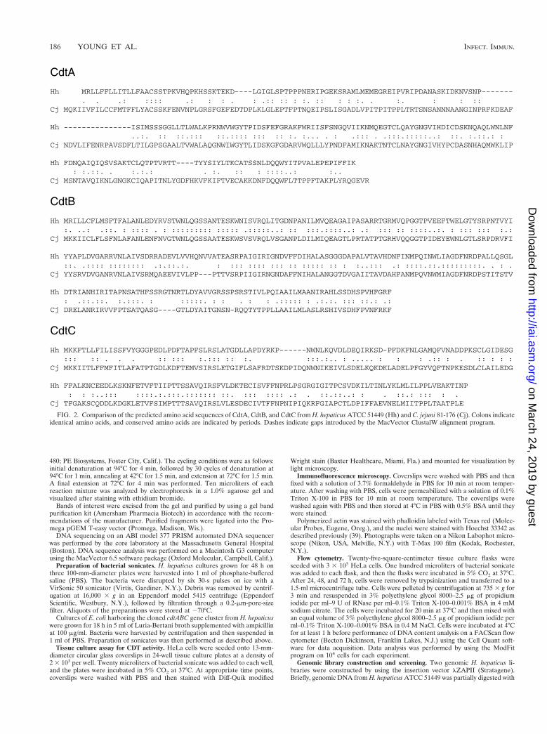
FIG. 2. Comparison of the predicted amino acid sequences of CdtA, CdtB, and CdtC from H. hepaticus ATCC 51449 (Hh) and C. jejuni 81-176 (Cj). Colons indicateidentical amino acids, and conserved amino acids are indicated by periods. Dashes indicate gaps introduced by the MacVector ClustalW alignment program.
186 YOUNG ET AL. INFECT. IMMUN.
on March 24, 2019 by guest
http://iai.asm.org/
Dow
nloaded from

the restriction enzyme Tsp509I. DNA with a length of $5 kb was ligated into theEcoRI site of the vector. The libraries were screened by DNA hybridization usingthe PCR amplicon generated by amplification of H. hepaticus genomic DNA withthe primers VAT2 and WMI1 (27). An [a-32P]dCTP-labeled probe was gener-ated by using a random primer DNA labeling kit (Ready-To-Go DNA labelingbeads; Amersham Pharmacia Biotech). The radioactive probe was used to screen105 recombinant bacteriophage. Probe-positive plaques were identified and sub-cloned into the plasmid vector pBluescript SK(2) using the in vivo excision andrecircularization features of the lZAPII vector. E. coli bacteria carrying theserecombinant plasmids were further characterized by restriction mapping and byscreening for CDT activity.
Nucleotide sequence accession number. The nucleotide sequence of the H.hepaticus cdtABC gene cluster has been entered in the GenBank database underaccession no. AF163667.
RESULTS
H. hepaticus possesses cdtABC nucleotide sequence homol-ogy. The degenerate primers VAT2 and WMI1 (27) amplify a494-bp fragment of the cdtB gene from C. jejuni. Amplificationof H. hepaticus genomic DNA with these primers produced alarger than expected amplicon of approximately 750 bp (datanot shown). The complete nucleotide sequence of this ampli-con was determined, and the deduced amino acid sequenceexhibited significant homology to the published CdtB sequenceof C. jejuni. The predicted H. hepaticus peptide fragment ex-hibited 57% identity and 72% similarity to the C. jejuni geneproduct. The larger than expected size of the amplicon was aconsequence of the WMI1 primer annealing to a site 264 bpdownstream of the anticipated target site (Fig. 1).
By using this PCR product, lambda clones containing ho-mology to cdtB were isolated and corresponding plasmidclones were generated. DNA sequence analysis of these clonesrevealed that H. hepaticus has three closely linked open read-ing frames corresponding to the entire cdtABC gene cluster(Fig. 1). The deduced amino acid sequences had the closesthomology to the proteins encoded by the cdtABC gene clusterfrom C. jejuni (Fig. 2). The amino acid homologies were asfollows: CdtA, 30% identity and 44% similarity; CdtB, 56%identity and 72% similarity; CdtC, 40% identity and 55% sim-ilarity.
Sonicates of H. hepaticus cause morphologic changes inHeLa cell monolayers. Because the presence of cdtABC nucle-otide sequence homology was demonstrated in DNA from H.hepaticus, we sought to determine whether sonicates of thisorganism exhibited CDT activity. HeLa cells treated with soni-cates of H. hepaticus showed marked cellular distension (Fig.3B). The distended cells also exhibited nuclear enlargement,and approximately 15% of the affected cells were found to bemultinucleated. Occasionally, nuclear irregularities and frag-mentation were also seen in HeLa cells treated with sonicatesof H. hepaticus.
The cytoplasmic distension induced by sonicates of H. he-paticus was readily apparent, but the extent of cellular marginswas underestimated by using light microscopy following theapplication of this modified Wright stain. To further examinethe cellular structure of treated cells and to detect associatedcytoskeletal changes, monolayers were stained with fluores-cently labeled phalloidin to visualize filamentous actin (F-ac-tin). Phalloidin staining revealed the full extent of the enlarge-ment of cells in monolayers treated with sonicates of H.hepaticus (Fig. 4). There also appeared to be an increase in theamount of F-actin present in cells in affected monolayers. Nu-clear staining with the fluorescent DNA stain Hoechst 33342confirmed the nuclear enlargement, multinucleation, and nu-clear fragmentation visualized by light microscopy.
Sonicates of H. hepaticus cause G2/M cell cycle arrest incultured cells. To examine whether the morphologic changesobserved in cells treated with sonicates of H. hepaticus werealso associated with cell cycle arrest, the DNA content oftreated cells was determined by flow cytometry. In untreatedcell monolayers, the fraction of cells with a DNA content of 4Nwas consistently 8 to 10% (Fig. 5A). In monolayers treatedwith sonicates of H. hepaticus, an increase in the fraction ofcells with a DNA content of 4N was seen 24 h after sonicateaddition (Fig. 5D). The fraction of cells with a DNA content of4N increased at 48 h and reached a maximum by 72 aftersonicate addition (Fig. 5G and J). In addition, by 72 h, therewas a significant fraction of cells with a DNA content of 8Namong cells treated with H. hepaticus sonicates. Examinationof the size of these cells, as judged by fluorescent width, indi-cates that these are probably multinucleated cells, as opposedto cellular aggregates. This is consistent with the observationthat multinucleated cells were present in treated monolayers(Fig. 3).
Cytopathic activity of E. coli carrying the cdtABC gene clus-ter from H. hepaticus. Sonicates of the 15 E. coli clones har-boring the recombinant plasmids generated from the genomiclibrary screen were examined for a cytopathic effect on HeLacell monolayers. Sonicates of 3 of the 15 clones produced acytopathic effect on cultured HeLa cell monolayers which wasindistinguishable from that produced by sonicates of H. hepati-cus. Representative clones were selected for further character-ization. HeLa cell monolayers treated with sonicates of E. colicarrying the H. hepaticus cdt locus were examined for cytoskel-etal and nuclear rearrangements over time by fluorescence
FIG. 3. Cytopathic effect of H. hepaticus CDT on HeLa cells. Compared tountreated cells (A), cells treated with sonicates from H. hepaticus (B) exhibitedmarked cytoplasmic distension along with nuclear enlargement, multinucleation,and nuclear fragmentation. Magnification, 3250.
VOL. 68, 2000 HELICOBACTER HEPATICUS CDT 187
on March 24, 2019 by guest
http://iai.asm.org/
Dow
nloaded from

microscopy. At 24 h after sonicate addition, nuclear fragmen-tation was observed (Fig. 5F) but the majority of cells still hada normal size and actin ultrastructure. By 48 h, more nuclearabnormalities were observed (Fig. 5I) and cell distension be-came apparent, along with an increase in the amount of F-actin(Fig. 5H). At 72 h, the cells in monolayers treated with soni-cates of an E. coli CDT clone (Fig. 5K and L) were indistin-guishable from those treated with H. hepaticus sonicates (Fig.3).
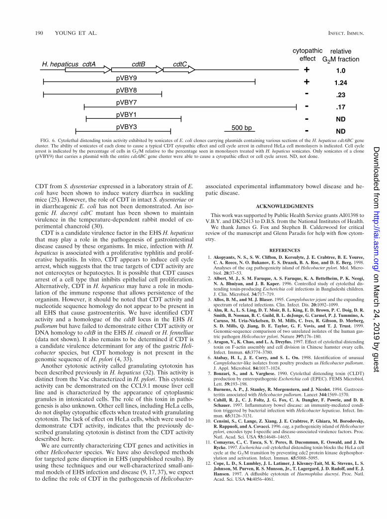
Cell cycle analysis demonstrated that the cytopathic effectproduced by the E. coli clones was also accompanied by G2/Mcell cycle arrest (Fig. 6). Mapping of the inserts from a numberof clones demonstrated that induction of a cytopathic effectand cell cycle arrest required the presence of the entirecdtABC gene cluster (Fig. 6).
DISCUSSION
In this study, we identified and characterized CDT activity inthe EHS H. hepaticus. The term CDT was coined to describean activity in culture supernatants of certain E. coli strains, aswell as C. jejuni, that causes progressive distension and even-tually death of cultured mammalian cells (21, 22). We demon-strate here that sonicates of H. hepaticus induce G2/M cell
cycle arrest in cultured HeLa cells and cause progressive cel-lular enlargement of HeLa cells, accompanied by the appear-ance of abnormal accumulations of F-actin. Coupled with thepresence of DNA sequences homologous to the cdtABC genecluster from C. jejuni and the cytopathic activity of E. colistrains carrying the cloned H. hepaticus genes, these resultsindicate that H. hepaticus possesses a toxin that is a novelmember of the CDT family.
The predicted CDT gene products from H. hepaticus had theclosest homology to the CDT from C. jejuni. As reportedpreviously (27, 31), the greatest homology is seen in the CdtBamino acid sequence. This may indicate a conserved functionfor the CdtB subunit; however, our results are in agreementwith others indicating that all three gene products are neces-sary for cytotoxic activity in laboratory strains of E. coli.
The exact role of CDT in pathogenesis has not been clearlydetermined. It has been proposed that CDT plays a role in thepathogenesis of diarrheal illness. CDT activity in E. coli wasoriginally described in clinical isolates associated with gastro-enteritis (22). A study of children with acute diarrhea showeda trend toward an increased rate of isolation of CDT-produc-ing E. coli among children with diarrhea compared to controls,but this did not reach statistical significance (2). It should benoted that several different virotypes of E. coli are associated
FIG. 4. Cytoplasmic and nuclear distension of HeLa cells treated with sonicates of H. hepaticus. Photomicrographs of untreated cells (A and B) and cells treatedwith sonicates of H. hepaticus (C and D) for 72 h are shown. Fixed and permeabilized cells were double labeled for epifluorescence microscopy with Texas red-labeledphalloidin (A and C) and Hoechst 33342 (B and D). Treated cells exhibited increased cell size and prominent stress fiber-like structures (C) as well, as increased nuclearsize (D). Magnification, 3500.
188 YOUNG ET AL. INFECT. IMMUN.
on March 24, 2019 by guest
http://iai.asm.org/
Dow
nloaded from

with diarrheal illness, each with distinct virulence determinants(24). The presence of CDT in a particular E. coli strain mayrepresent only one of several virulence factors required forgastrointestinal pathogenesis. Conversely, CDT has been dem-onstrated in all C. jejuni isolates, as well as in other membersof the genus Campylobacter (14, 27). Demonstration of a role
for CDT in gastrointestinal pathogenesis mediated by C. jejunihas not been reported. The well-characterized colonizationmodels for C. jejuni may not be optimal for establishing thecontribution of CDT to disease outcome, since they do notreproduce the clinical syndrome of gastroenteritis associatedwith C. jejuni infection. Partially purified preparations of the
FIG. 5. Time dependence of DNA content and cytopathic effect of HeLa cells treated with bacterial sonicates with CDT activity. Compared to control cells (A),cells treated with sonicates from H. hepaticus (D, G, and J) showed a progressive increase in the fraction of cells with a DNA content of 4 N, with a maximal effectbeing reached 72 h after sonicate addition (J). Compared to control cells (B and C), cells treated with sonicates from an E. coli strain harboring the cloned H. hepaticuscdt locus showed a progressive increase in size with accumulations of polymerized actin (E, H, and K) and progressive nuclear abnormalities (F, I, and L). These changesin morphology mirrored the progressive cell cycle block observed by flow cytometry. Magnification, 3400. PI, propidium iodide.
VOL. 68, 2000 HELICOBACTER HEPATICUS CDT 189
on March 24, 2019 by guest
http://iai.asm.org/
Dow
nloaded from
CDT from S. dysenteriae expressed in a laboratory strain of E.coli have been shown to induce watery diarrhea in sucklingmice (25). However, the role of CDT in intact S. dysenteriae orin diarrheagenic E. coli has not been demonstrated. An iso-genic H. ducreyi cdtC mutant has been shown to maintainvirulence in the temperature-dependent rabbit model of ex-perimental chancroid (30).
CDT is a candidate virulence factor in the EHS H. hepaticusthat may play a role in the pathogenesis of gastrointestinaldisease caused by these organisms. In mice, infection with H.hepaticus is associated with a proliferative typhlitis and prolif-erative hepatitis. In vitro, CDT appears to induce cell cyclearrest, which suggests that the true targets of CDT activity arenot enterocytes or hepatocytes. It is possible that CDT causesarrest of a cell type that inhibits epithelial cell proliferation.Alternatively, CDT in H. hepaticus may have a role in modu-lation of the immune response that allows persistence of theorganism. However, it should be noted that CDT activity andnucleotide sequence homology do not appear to be present inall EHS that cause gastroenteritis. We have identified CDTactivity and a homologue of the cdtB locus in the EHS H.pullorum but have failed to demonstrate either CDT activity orDNA homology to cdtB in the EHS H. cinaedi or H. fennelliae(data not shown). It also remains to be determined if CDT isa candidate virulence determinant for any of the gastric Heli-cobacter species, but CDT homology is not present in thegenomic sequence of H. pylori (4, 33).
Another cytotoxic activity called granulating cytotoxin hasbeen described previously in H. hepaticus (32). This activity isdistinct from the Vac characterized in H. pylori. This cytotoxicactivity can be demonstrated on the CCL9.1 mouse liver cellline and is characterized by the appearance of cytoplasmicgranules in intoxicated cells. The role of this toxin in patho-genesis is also unknown. Other cell lines, including HeLa cells,do not display cytopathic effects when treated with granulatingcytotoxin. The lack of effect on HeLa cells, which were used todemonstrate CDT activity, indicates that the previously de-scribed granulating cytotoxin is distinct from the CDT activitydescribed here.
We are currently characterizing CDT genes and activities inother Helicobacter species. We have also developed methodsfor targeted gene disruption in EHS (unpublished results). Byusing these techniques and our well-characterized small-ani-mal models of EHS infection and disease (9, 17, 37), we expectto define the role of CDT in the pathogenesis of Helicobacter-
associated experimental inflammatory bowel disease and he-patic disease.
ACKNOWLEDGMENTS
This work was supported by Public Health Service grants AI01398 toV.B.Y. and DK52413 to D.B.S. from the National Institutes of Health.
We thank James G. Fox and Stephen B. Calderwood for criticalreview of the manuscript and Glenn Paradis for help with flow cytom-etry.
REFERENCES1. Akopyants, N. S., S. W. Clifton, D. Kersulyte, J. E. Crabtree, B. E. Youree,
C. A. Reece, N. O. Bukanov, E. S. Drazek, B. A. Roe, and D. E. Berg. 1998.Analyses of the cag pathogenicity island of Helicobacter pylori. Mol. Micro-biol. 28:37–53.
2. Albert, M. J., S. M. Faruque, A. S. Faruque, K. A. Bettelheim, P. K. Neogi,N. A. Bhuiyan, and J. B. Kaper. 1996. Controlled study of cytolethal dis-tending toxin-producing Escherichia coli infections in Bangladeshi children.J. Clin. Microbiol. 34:717–719.
3. Allos, B. M., and M. J. Blaser. 1995. Campylobacter jejuni and the expandingspectrum of related infections. Clin. Infect. Dis. 20:1092–1099.
4. Alm, R. A., L. S. Ling, D. T. Moir, B. L. King, E. D. Brown, P. C. Doig, D. R.Smith, B. Noonan, B. C. Guild, B. L. deJonge, G. Carmel, P. J. Tummino, A.Caruso, M. Uria-Nickelsen, D. M. Mills, C. Ives, R. Gibson, D. Merberg,S. D. Mills, Q. Jiang, D. E. Taylor, G. F. Vovis, and T. J. Trust. 1999.Genomic-sequence comparison of two unrelated isolates of the human gas-tric pathogen Helicobacter pylori. Nature 397:176–180.
5. Aragon, V., K. Chao, and L. A. Dreyfus. 1997. Effect of cytolethal distendingtoxin on F-actin assembly and cell division in Chinese hamster ovary cells.Infect. Immun. 65:3774–3780.
6. Atabay, H. I., J. E. Corry, and S. L. On. 1998. Identification of unusualCampylobacter-like isolates from poultry products as Helicobacter pullorum.J. Appl. Microbiol. 84:1017–1024.
7. Bouzari, S., and A. Varghese. 1990. Cytolethal distending toxin (CLDT)production by enteropathogenic Escherichia coli (EPEC). FEMS Microbiol.Lett. 59:193–198.
8. Burnens, A. P., J. Stanley, R. Morgenstern, and J. Nicolet. 1994. Gastroen-teritis associated with Helicobacter pullorum. Lancet 344:1569–1570.
9. Cahill, R. J., C. J. Foltz, J. G. Fox, C. A. Dangler, F. Powrie, and D. B.Schauer. 1997. Inflammatory bowel disease: an immunity-mediated condi-tion triggered by bacterial infection with Helicobacter hepaticus. Infect. Im-mun. 65:3126–3131.
10. Censini, S., C. Lange, Z. Xiang, J. E. Crabtree, P. Ghiara, M. Borodovsky,R. Rappuoli, and A. Covacci. 1996. cag, a pathogenicity island of Helicobacterpylori, encodes type I-specific and disease-associated virulence factors. Proc.Natl. Acad. Sci. USA 93:14648–14653.
11. Comayras, C., C. Tasca, S. Y. Peres, B. Ducommun, E. Oswald, and J. DeRycke. 1997. Escherichia coli cytolethal distending toxin blocks the HeLa cellcycle at the G2/M transition by preventing cdc2 protein kinase dephosphor-ylation and activation. Infect. Immun. 65:5088–5095.
12. Cope, L. D., S. Lumbley, J. L. Latimer, J. Klesney-Tait, M. K. Stevens, L. S.Johnson, M. Purven, R. S. Munson, Jr., T. Lagergard, J. D. Radolf, and E. J.Hansen. 1997. A diffusible cytotoxin of Haemophilus ducreyi. Proc. Natl.Acad. Sci. USA 94:4056–4061.
FIG. 6. Cytolethal distending toxin activity exhibited by sonicates of E. coli clones carrying plasmids containing various sections of the H. hepaticus cdtABC genecluster. The ability of sonicates of each clone to cause a typical CDT cytopathic effect and cell cycle arrest in cultured HeLa cell monolayers is indicated. Cell cyclearrest is indicated by the percentage of cells in G2/M relative to the percentage seen in monolayers treated with H. hepaticus sonicates. Only sonicates of a clone(pVBY9) that carries a plasmid with the entire cdtABC gene cluster were able to cause a cytopathic effect or cell cycle arrest. ND, not done.
190 YOUNG ET AL. INFECT. IMMUN.
on March 24, 2019 by guest
http://iai.asm.org/
Dow
nloaded from

13. Cover, T. L. 1996. The vacuolating cytotoxin of Helicobacter pylori. Mol.Microbiol. 20:241–246.
14. Eyigor, A., K. A. Dawson, B. E. Langlois, and C. L. Pickett. 1999. Cytolethaldistending toxin genes in Campylobacter jejuni and Campylobacter coli iso-lates: detection and analysis by PCR. J. Clin. Microbiol. 37:1646–1650.
15. Fox, J. G. 1997. The expanding genus of Helicobacter: pathogenic and zoo-notic potential. Semin. Gastrointest. Dis. 8:124–141.
16. Fox, J. G., F. E. Dewhirst, J. G. Tully, B. J. Paster, L. Yan, N. S. Taylor, M. J.Collins, Jr., P. L. Gorelick, and J. M. Ward. 1994. Helicobacter hepaticus sp.nov., a microaerophilic bacterium isolated from livers and intestinal mucosalscrapings from mice. J. Clin. Microbiol. 32:1238–1245.
17. Fox, J. G., X. Li, L. Yan, R. J. Cahill, R. Hurley, R. Lewis, and J. C. Murphy.1996. Chronic proliferative hepatitis in A/JCr mice associated with persistentHelicobacter hepaticus infection: a model of helicobacter-induced carcino-genesis. Infect. Immun. 64:1548–1558.
18. Fox, J. G., L. Yan, B. Shames, J. Campbell, J. C. Murphy, and X. Li. 1996.Persistent hepatitis and enterocolitis in germfree mice infected with Helico-bacter hepaticus. Infect. Immun. 64:3673–3681.
19. Fox, J. G., L. L. Yan, F. E. Dewhirst, B. J. Paster, B. Shames, J. C. Murphy,A. Hayward, J. C. Belcher, and E. N. Mendes. 1995. Helicobacter bilis sp.nov., a novel Helicobacter species isolated from bile, livers, and intestines ofaged, inbred mice. J. Clin. Microbiol. 33:445–454.
20. Hailey, J. R., J. K. Haseman, J. R. Bucher, A. E. Radovsky, D. E. Malarkey,R. T. Miller, A. Nyska, and R. R. Maronpot. 1998. Impact of Helicobacterhepaticus infection in B6C3F1 mice from twelve National Toxicology Pro-gram two-year carcinogenesis studies. Toxicol. Pathol. 26:602–611.
21. Johnson, W. M., and H. Lior. 1988. A new heat-labile cytolethal distendingtoxin (CLDT) produced by Campylobacter spp. Microb. Pathog. 4:115–126.
22. Johnson, W. M., and H. Lior. 1988. A new heat-labile cytolethal distendingtoxin (CLDT) produced by Escherichia coli isolates from clinical material.Microb. Pathog. 4:103–113.
23. Kullberg, M. C., J. M. Ward, P. L. Gorelick, P. Caspar, S. Hieny, A. Cheever,D. Jankovic, and A. Sher. 1998. Helicobacter hepaticus triggers colitis inspecific-pathogen-free interleukin-10 (IL-10)-deficient mice through an IL-12- and gamma interferon-dependent mechanism. Infect. Immun. 66:5157–5166.
24. Nataro, J. P., and J. B. Kaper. 1998. Diarrheagenic Escherichia coli. Clin.Microbiol. Rev. 11:142–201.
25. Okuda, J., M. Fukumoto, Y. Takeda, and M. Nishibuchi. 1997. Examinationof diarrheagenicity of cytolethal distending toxin: suckling mouse response tothe products of the cdtABC genes of Shigella dysenteriae. Infect. Immun.65:428–433.
26. Okuda, J., H. Kurazono, and Y. Takeda. 1995. Distribution of the cytolethaldistending toxin A gene (cdtA) among species of Shigella and Vibrio, andcloning and sequencing of the cdt gene from Shigella dysenteriae. Microb.Pathog. 18:167–172.
27. Pickett, C. L., E. C. Pesci, D. L. Cottle, G. Russell, A. N. Erdem, and H.Zeytin. 1996. Prevalence of cytolethal distending toxin production in Campy-
lobacter jejuni and relatedness of Campylobacter sp. cdtB gene. Infect. Im-mun. 64:2070–2078.
28. Stanley, J., D. Linton, A. P. Burnens, F. E. Dewhirst, S. L. On, A. Porter,R. J. Owen, and M. Costas. 1994. Helicobacter pullorum sp. nov.-genotypeand phenotype of a new species isolated from poultry and from humanpatients with gastroenteritis. Microbiology 140:3441–3449.
29. Steinbrueckner, B., G. Haerter, K. Pelz, S. Weiner, J. A. Rump, W. Deissler,S. Bereswill, and M. Kist. 1997. Isolation of Helicobacter pullorum frompatients with enteritis. Scand. J. Infect. Dis. 29:315–318.
30. Stevens, M. K., J. L. Latimer, S. R. Lumbley, C. K. Ward, L. D. Cope, T.Lagergard, and E. J. Hansen. 1999. Characterization of a Haemophilusducreyi mutant deficient in expression of cytolethal distending toxin. Infect.Immun. 67:3900–3908.
31. Sugai, M., T. Kawamoto, S. Y. Peres, Y. Ueno, H. Komatsuzawa, T. Fujiwara,H. Kurihara, H. Suginaka, and E. Oswald. 1998. The cell cycle-specificgrowth-inhibitory factor produced by Actinobacillus actinomycetemcomitansis a cytolethal distending toxin. Infect. Immun. 66:5008–5019.
32. Taylor, N. S., J. G. Fox, and L. Yan. 1995. In-vitro hepatotoxic factor inHelicobacter hepaticus, H. pylori and other Helicobacter species. J. Med.Microbiol. 42:48–52.
33. Tomb, J. F., O. White, A. R. Kerlavage, R. A. Clayton, G. G. Sutton, R. D.Fleischmann, K. A. Ketchum, H. P. Klenk, S. Gill, B. A. Dougherty, K.Nelson, J. Quackenbush, L. Zhou, E. F. Kirkness, S. Peterson, B. Loftus, D.Richardson, R. Dodson, H. G. Khalak, A. Glodek, K. McKenney, L. M.Fitzegerald, N. Lee, M. D. Adams, and J. C. Venter. 1997. The completegenome sequence of the gastric pathogen Helicobacter pylori. Nature 388:539–547.
34. von Freeden-Jeffry, U., N. Davidson, R. Wiler, M. Fort, S. Burdach, and R.Murray. 1998. IL-7 deficiency prevents development of a non-T cell non-Bcell-mediated colitis. J. Immunol. 161:5673–5680.
35. Ward, J. M., M. R. Anver, D. C. Haines, J. M. Melhorn, P. Gorelick, L. Yan,and J. G. Fox. 1996. Inflammatory large bowel disease in immunodeficientmice naturally infected with Helicobacter hepaticus. Lab. Anim. Sci. 46:15–20.
36. Ward, J. M., J. G. Fox, M. R. Anver, D. C. Haines, C. V. George, M. J.Collins, Jr., P. L. Gorelick, K. Nagashima, M. A. Gonda, and R. V. Gilden.1994. Chronic active hepatitis and associated liver tumors in mice caused bya persistent bacterial infection with a novel Helicobacter species. J. Natl.Cancer Inst. 86:1222–1227.
37. Whary, M. T., T. J. Morgan, C. A. Dangler, K. J. Gaudes, N. S. Taylor, andJ. G. Fox. 1998. Chronic active hepatitis induced by Helicobacter hepaticus inthe A/JCr mouse is associated with a Th1 cell-mediated immune response.Infect. Immun. 66:3142–3148.
38. Whitehouse, C. A., P. B. Balbo, E. C. Pesci, D. L. Cottle, P. M. Mirabito, andC. L. Pickett. 1998. Campylobacter jejuni cytolethal distending toxin causes aG2-phase cell cycle block. Infect. Immun. 66:1934–1940.
39. Young, V. B., S. Falkow, and G. K. Schoolnik. 1992. The invasin protein ofYersinia enterocolitica: internalization of invasin-bearing bacteria by eukary-otic cells is associated with reorganization of the cytoskeleton. J. Cell Biol.116:197–207.
Editor: P. E. Orndorff
VOL. 68, 2000 HELICOBACTER HEPATICUS CDT 191
on March 24, 2019 by guest
http://iai.asm.org/
Dow
nloaded from