cultivation of mesophilic soil crenarchaeotes in
TRANSCRIPT

University of Northern Iowa University of Northern Iowa
UNI ScholarWorks UNI ScholarWorks
Faculty Publications Faculty Work
2005
Cultivation of Mesophilic Soil Crenarchaeotes in Enrichment Cultivation of Mesophilic Soil Crenarchaeotes in Enrichment
Cultures from Plant Roots Cultures from Plant Roots
Holly M. Simon
Courtney E. Jahn
See next page for additional authors
Let us know how access to this document benefits you
Copyright ©2005 American Society for Microbiology (ASM). The copyright holder has granted
permission for posting.
Follow this and additional works at: https://scholarworks.uni.edu/bio_facpub
Part of the Biology Commons
Recommended Citation Recommended Citation Simon, Holly M.; Jahn, Courtney E.; Bergerud, Luke T.; Sliwinski, Marek K.; Weimer, Paul J.; Willis, David K.; and Goodman, Robert M., "Cultivation of Mesophilic Soil Crenarchaeotes in Enrichment Cultures from Plant Roots" (2005). Faculty Publications. 11. https://scholarworks.uni.edu/bio_facpub/11
This Article is brought to you for free and open access by the Faculty Work at UNI ScholarWorks. It has been accepted for inclusion in Faculty Publications by an authorized administrator of UNI ScholarWorks. For more information, please contact [email protected].

Authors Authors Holly M. Simon, Courtney E. Jahn, Luke T. Bergerud, Marek K. Sliwinski, Paul J. Weimer, David K. Willis, and Robert M. Goodman
This article is available at UNI ScholarWorks: https://scholarworks.uni.edu/bio_facpub/11

APPLIED AND ENVIRONMENTAL MICROBIOLOGY, Aug. 2005, p. 4751–4760 Vol. 71, No. 80099-2240/05/$08.00�0 doi:10.1128/AEM.71.8.4751–4760.2005Copyright © 2005, American Society for Microbiology. All Rights Reserved.
Cultivation of Mesophilic Soil Crenarchaeotes in Enrichment Culturesfrom Plant Roots
Holly M. Simon,1* Courtney E. Jahn,1 Luke T. Bergerud,1† Marek K. Sliwinski,1‡ Paul J. Weimer,2,3
David K. Willis,1,3 and Robert M. Goodman1,4
Department of Plant Pathology,1 Department of Bacteriology,2 and Gaylord Nelson Institute for Environmental Studies,4
University of Wisconsin—Madison, Madison, Wisconsin 53706, and Agricultural Research Service,U.S. Department of Agriculture, Madison, Wisconsin 537063
Received 10 November 2004/Accepted 14 February 2005
Because archaea are generally associated with extreme environments, detection of nonthermophilic membersbelonging to the archaeal division Crenarchaeota over the last decade was unexpected; they are surprisinglyubiquitous and abundant in nonextreme marine and terrestrial habitats. Metabolic characterization of thesenonthermophilic crenarchaeotes has been impeded by their intractability toward isolation and growth inculture. From studies employing a combination of cultivation and molecular phylogenetic techniques (PCR–single-strand conformation polymorphism, sequence analysis of 16S rRNA genes, fluorescence in situ hybrid-ization, and real-time PCR), we present evidence here that one of the two dominant phylotypes of Crenarchaeotathat colonizes the roots of tomato plants grown in soil from a Wisconsin field is selectively enriched in mixedcultures amended with root extract. Clones recovered from enrichment cultures were found to group phylo-genetically with sequences from clade C1b.A1. This work corroborates and extends our recent findings,indicating that the diversity of the crenarchaeal soil assemblage is influenced by the rhizosphere and thatmesophilic soil crenarchaeotes are found associated with plant roots, and provides the first evidence for growthof nonthermophilic crenarchaeotes in culture.
The hallmark of the Archaea is their remarkable specializa-tion; isolates have been recovered almost exclusively from ex-treme environments and specialized niches. Nevertheless, re-sults from recent studies using molecular approaches toexamine the phylogenetic breadth, ecological range, and abun-dance of members of the Archaea have challenged the assump-tion that these microorganisms are absent from, or only minorcomponents of, nonextreme marine and terrestrial habitats(including mesophilic soils and rhizospheres). The accumula-tion of extensive sequence-based evidence by cultivation-inde-pendent approaches has made the broad ecological distribu-tion and considerable abundance of archaea in thesenonextreme habitats indisputable (9–11, 49). One intriguinggroup identified in this manner consists of low-temperatureand mesophilic members of the division Crenarchaeota(termed “nonthermophilic”) (5) whose small subunit (SSU)rRNA gene sequences diverge deeply from those of culturedcrenarchaeotes (3, 8, 15, 22, 51). Phenotypic properties ofnonthermophilic crenarchaeotes are also presumed to be dif-ferent from those of previously cultured members of this divi-sion isolated exclusively from thermophilic environments.
Their ubiquity, in combination with a high abundance in avariety of habitats (12, 27, 31, 38, 44, 45), suggests that theyhave major roles in one or more biosphere processes. How-
ever, to date, the physiological properties and functions innature of these unforeseen archaea are largely the subject ofspeculation, in part because they have been recalcitrant towardisolation and growth in culture.
In an attempt to learn more about the ecophysiology ofthese organisms, we exploited our recent complementary dis-coveries that (i) mesophilic soil crenarchaeotes are foundclosely associated with plant roots (44) and (ii) the plant rhi-zosphere influences the diversity of the crenarchaeal assem-blage in soil (47). A number of reports from other researchgroups are consistent with our findings and describe, for ex-ample, the recovery of SSU rRNA gene sequences of meso-philic crenarchaeotes from the rice rhizosphere (19), washedroots of maize (6), and mycorrhizospheres of pine seedlings(4). Here we extend our previous work by demonstrating thattwo phylogenetic types (phylotypes) of mesophilic crenarchae-otes from the monophyletic clade C1b (11) are predominantlyassociated with the tomato (Esculentum lycopersicon) rhizo-plane of plants grown in soil collected from the West MadisonAgricultural Research Station (referred to hereafter asWMAD soil) (3) and that the C1b.A1 phylotype (44, 46) isselectively enriched in culture amended with root extract as aputative nutrient source. Our results provide further evidencefor a biologically relevant association of distinct members ofthe mesophilic soil Crenarchaeota with plant roots.
MATERIALS AND METHODS
Sample preparation and manipulation. Soil collection and growth of tomatoeswas carried out as previously described (44). Soil from the West Madison Agri-cultural Research station (Plano silt loam containing 61% sand, 23% silt, and16% clay, with 1.7% organic matter, pH 7.0) (3) was collected between 2 and 8cm below the surface using sterile implements. Inoculum was prepared by son-ication for 30 s in a Branson 2200 Ultrasonic cleaner to displace attached
* Corresponding author. Present address: Department of Environ-mental and Biomolecular Systems, OGI School of Science and Engi-neering, Oregon Health and Science University, 20000 NW WalkerRd., Beaverton, OR 97006. Phone: (503) 748-1873. Fax: (503) 748-1464. E-mail: [email protected].
† Present address: Human Genome Sciences, Rockville, MD 20850.‡ Present address: Department of Botany, University of Wiscon-
sin—Madison, Madison, WI 53706.
4751
microorganisms from gently and thoroughly rinsed roots of tomato cultivar M82athat were harvested after germination and growth of the plants in WMAD soil.
Preparation of tomato root extract. Tomatoes were grown in a mixture ofvermiculite and potting soil (1:3) and watered every 3 days with full-strengthHoagland’s nutrient solution (2). Roots were harvested after 60 days by removingall of the soil mixture and rinsing thoroughly. Roots were weighed, frozen inliquid nitrogen, ground into a fine powder, and stored at �80°C. To prepareextract, powdered root material was measured into Milli-Q H2O and vortexed for5 min. Coarse filtration was used to remove larger particles, followed by steril-ization through a 0.22-�m filter.
Enrichment culture treatment and incubation. In our enrichments for ar-chaea, we used a combination of chemical and physical treatments and inhibitorsto restrict the growth of bacteria. The inoculum was treated with lysozymeaccording to the method of Repaske (40). Freeze-thaw cycles were performed bythree consecutive rounds of quick-freezing in a dry ice-ethanol bath for 5 min,followed by a 5-min thaw at 37°C. The inoculum (10% by vol) was incubated forgrowth in batch culture in a basal salts medium [5 mM KH2PO4, 5 mMNa2HPO4, 1 mM (NH4)SO4, 2 mM KCl, pH 7.0], with the addition of 1%Daniels’ mineral elixir (7), 1% vitamin solution (43), 2.5 mM NaHSO3, 0.2� rootextract (1� root extract is 25 g [wet root weight]/liter H2O), 100 mg/liter (each)carbenicillin and streptomycin, 200 mg/liter rifampin (in methanol), 6.7 mg/litercephalothin, and 1.7 mg/liter clindamycin. Culture volumes were 1/10 to 1/5 ofthe incubating flask volume and were incubated on a rotary shaker at 200 rpm inlow light.
DNA isolation and quantification. Inoculum and culture samples were ex-tracted using the FastDNA spin kit for soil (BIO101, MP Biomedicals, Irvine,CA) and further purified using Sepharose microspin columns as described pre-viously (46). Plasmid DNA was isolated from cell culture using a standardalkaline lysis technique, with additional purification performed by phenol-chlo-roform extraction and concentration by precipitation with 0.1 volume of 3 MNaCl, pH 5.2, and 3 volumes of ethanol. Linearized plasmid DNA was quantifiedusing the PicoGreen double-stranded DNA quantitation kit (Molecular Probesof Invitrogen, Eugene, OR) according to the manufacturer’s protocol.
PCR, rRNA gene cloning, and sequencing. Total enrichment DNA was iso-lated as previously described (44) or by a modified agarose plug method (41).PCR was performed in separate reactions with the SSU rRNA gene-specificprimer sets, including the archaea-biased primers 133f, (44) and 915r (48) andthe nonspecific cross-domain primer 1492r (29). PCR products were cloned byligation using the pGEM-T vector system (Promega Corp., Madison, WI). Re-striction enzymes used to generate amplified ribosomal DNA restriction analysispatterns were AluI, RsaI, and Sau3A1. Partial sequences were generated by bidi-rectional sequencing of cloned products using the vector primers SP6 and T7.Sequences were analyzed on an ABI Prism 377 DNA sequencer (University ofWisconsin Biotechnology Center) and tested for possible chimeric artifacts using theCHECK_CHIMERA (30) and Bellerophon (24) programs. Sequences were ana-lyzed using BLAST (1) searches to determine the closest sequences available fromthe database. These sequence data have been submitted to the DDBJ/EMBL/GenBank databases (see “Nucleotide sequence accession number” below).
Phylogenetic analyses of sequence data. SSU rRNA gene sequences werealigned in the ARB database (30a). Regions of ambiguous positional homologywere removed from the data set, with additional manual corrections, wherenecessary, using the Seqlab editor within the GCG Wisconsin Package (Acce-lyrys). Phylogenetic analyses were performed using PAUP*, version 4.0b10 (Al-tivec; D. L. Swofford, Sinauer Associates, Sunderland, Mass.), and ARB. A dataset of 39 sequences containing 1,080 homologous nucleotide positions was usedto generate trees using the maximum-likelihood (ML), maximum-parsimony(MP), and evolutionary distance (ED) methods. To assess branch support, boot-strap resampling was performed in PAUP* with a total of 2,000 resamplings inall cases. MP trees were constructed using the default settings in PAUP*. EDanalyses were conducted using the Kimura two-parameter and general time-reversible substitution model corrections with and without rate correction. Rateheterogeneities were corrected using a gamma distribution model (the shapeparameter, �, was estimated to be 0.66 using a parsimony-based approximationin PAUP*). ML trees were constructed in ARB using the fastDNAml program(13, 35). Additional sequences with fewer than 1,080 homologous nucleotidepositions were added to base trees using the parsimony addition function inARB. All analyses were performed using multiple different out-group species.
Fluorescence in situ hybridization (FISH), cell counts, and microscopy. Cellfixation, probe synthesis, hybridizations, microscopy, and image analysis weredone as previously described (44), except that an Optronics “Magnafire” cooledcharge-coupled device camera with motorized color wheel and RGB dichroicfilters (resolution, 1,280 by 1,024 with 10 bits/pixel) was used in combination withImage-Pro Express, version 4.0, software. The following probes were hybridized
simultaneously: Cren113a, Cren745a, Cren1209 (44), Bact338 (48), and Bact927(17). DAPI (4�,6-diamidino-2-phenylindole) was used to stain for DNA. Cellswere visualized using a BX-60 microscope (Olympus America) equipped forepifluorescence with an HBO 100-W mercury arc lamp and an OlympusUPlanF1 100� objective/1.3 NA, with a field area of 0.035 � 0.002 mm2.
PCR-SSCP. PCR–single-strand conformation polymorphism (PCR-SSCP) wasconducted as described previously (46) using the crenarchaeote biased primer set133FN6F/248R5P. Briefly, PCR mixtures were desalted with Sephadex G50microspin columns packed in a UniFilter GF/B 800 microplate (Whatman, Inc.,Ann Arbor, MI). Following purification, each PCR product was mixed (1:1,vol/vol) with SSCP loading buffer (60% deionized formamide, 25% ROX GeneFlo625 standard [ChimerX, Milwaukee, WI], 7.5% 100 mM NaOH, 7.5% bluedextran EDTA [50 mg/ml blue dextran, 50 mM EDTA]). Samples were dena-tured at 94°C for 2 min and then immediately transferred onto ice. A 0.5-�laliquot of each sample was spotted onto a membrane comb. PolyacrylamideSSCP gels and 1� MDE (BMA, Rockland, ME) were cast according to themanufacturer’s instructions. The gel was loaded onto an ABI PRISM 377XLDNA Sequencer connected to an external cooling water bath to maintain the geltemperature at 23°C. The GS RUN 60W CHILLER module was selected tomaintain constant wattage. PCR-SSCP data were analyzed using GeneScan soft-ware, version 3.1 (Applied Biosystems, Foster City, CA). Peak height cutoffsranged from 5 to 50 relative fluorescence units depending on signal intensity.Samples were aligned to the internal size standard present in each lane.
Primer design and real-time PCR. Real-time PCR was performed using theiCycler iQ thermal cycler with a 96- by 0.2-ml reaction module and iQ software(Bio-Rad Laboratories, Inc., Hercules, CA). In each experiment, three indepen-dent master mixes were prepared from each inoculum and culture sample andtwo independent master mixes for standard DNA were prepared and tested induplicate within a single microtiter plate. Negative-control reactions (withoutadded template) were also included in each experiment. Reactions were per-formed in a volume of 20 �l, using iQ SYBR green supermix (Bio-Rad Labo-ratories, Inc., Hercules, CA) and 0.2 �M concentrations of each primer and wereoptimized for magnesium concentration (2.5 mM MgCl2, final concentration,achieved by adding 0.5 mM EDTA) and annealing temperature. Reactions wereperformed in the iCycler (Bio-Rad Laboratories, Inc.) using the two-step ampli-fication plus melting curve protocol as follows. Enzyme activation and well-factordetermination were at 95°C for 3 min, followed by 40 cycles of 95°C for 10 s(denaturation) and 61.3°C for 45 s (annealing and elongation). The melt curveprotocol began immediately after amplification and consisted of 1 min at 95°C,followed by 1 min at 55°C, and 80 10-s steps with a 0.5°C increase in temperatureat each step. Threshold values for threshold cycle determination were generatedautomatically by the iCycler iQ software. Lack of variation in PCR products andthe absence of primer dimers were ascertained from the melt curve profile of thePCR products. Slopes, regression coefficients, and PCR amplification efficiencycurves were calculated using iCycler iQ software; efficiency (E) was calculatedaccording to the equation E � 10(�1/slope) (39). Mean values and standard errorsof mean values were calculated using Microsoft Excel, version 11.0.0.
Primer sets were designed using the Beacon Designer software program(PREMIER Biosoft International, Palo Alto, CA), the ARB database, andsequences recovered in our studies from cultures, roots, and soil. The predictedspecificity of primers was examined by performing BLAST searches (1) againstthe GenBank database. The primer sequences designed for this study are asfollows: 599F, 5�-GTA-GCC-GGT-TCT-ACA-AGT-C-3�; 669F, 5�-CGA-CGG-TGA-GGG-ATG-AAA-G-3�; 703R, 5�-ACT-GGT-GGT-CTT-CAA-TGG-ATC-3�; 886R, 5�-CCA-GGC-GGC-AAA-CTT-AAC-3�. The predicted bias ofthe 599F/703R primer set is toward C1b sequences (as is also the 133F/248Rprimer set), while the 669F/886R primer set is predicted to hybridize broadly tosequences across most crenarchaeal sequence groups recovered from nonther-mophilic environments (i.e., C1, C2, and C3). Melting curve analysis (transitionrate, 0.5°C s�1), together with separation of PCR DNA fragments performed byelectrophoresis on 3% agarose gels, yielded a single product of the predicted sizefrom both the enrichment culture and the plasmid standard DNA for eachprimer set used.
Nucleotide sequence accession numbers. Sequence data have been submittedto the DDBJ/EMBL/GenBank databases under accession numbers AY487100 toAY487118.
RESULTS
PCR-SSCP analysis of the soil and rhizoplane microbialassemblage before and after incubation with root extract. Us-ing Crenarchaeota-biased primers (see Materials and Methods)
4752 SIMON ET AL. APPL. ENVIRON. MICROBIOL.

and PCR-SSCP (20, 36), an electropherogram consisting of 9different peaks was generated representing the crenarchaealassemblage in WMAD bulk soil (free of any visible root ma-terial) and on the rhizoplane of tomato plants grown inWMAD soil (Fig. 1). The PCR-SSCP profiles from theWMAD soil samples were dominated by a single phylotype ofthe Crenarchaeota, represented by peak 1 (Fig. 1A), with arelative abundance based on peak area of 75% of the totalpeak area (Table 1). The rhizoplane PCR-SSCP profile (Fig.1B) differed from the WMAD soil profile in that the phylotyperepresented by peak 8 was present in approximately equal, orgreater, relative abundance to the phylotype represented bypeak 1 (Table 1). Results from a different experiment exam-ining the crenarchaeal assemblage on the tomato rhizoplanefrom plants grown in WMAD soil were similar; the relativeabundance of peak 1 ranged from 28% to 39% of the totalpeak area (mean, 36%), while that of peak 8 ranged from 51%
to 66% of total peak area (mean, 57%) (n � 6). Peaks repre-senting other crenarchaeal phylotypes were detected at 5%relative abundance.
After 30 days of incubation under air at 24°C with rootextract, the crenarchaeal assemblage detected in cultures thatwere inoculated with sonicate from the rhizoplane (details inMaterials and Methods) consisted almost exclusively of thephylotype represented by peak 8 (Fig. 2B and Table 1). Therelative abundance of the peak 8 phylotype was similar incultures that had been incubated for an additional month (60days total). The treatment group (either no treatment ortreated with lysozyme and incubated with antibiotics), ex-plained in greater detail below, did not show consistent effects,although there was a trend in the rhizoplane samples towardslightly higher relative abundance of the peak 8 phylotype inthe lysozyme-treated cultures. Similar results were obtainedfrom enrichment cultures in two other experiments: (i) 30-daycultures (n � 4), peak 1 ranged from 15% to 24% of total peakarea (mean, 18%) and peak 8 ranged from 76% to 82% of totalpeak area (mean, 79%); (ii) 60-day cultures (n � 8), peak 1ranged from undetectable to 5.4% of total peak area (mean,2.4%) and peak 8 ranged from 87% to 100% of total peak area(mean, 94%).
Upon incubation of WMAD soil inoculum in culture withroot extract, the phylotype represented by peak 8 increased inrelative abundance, while that represented by peak 1 de-creased in relative abundance (Fig. 2A). This result was similarto that observed for cultures starting with root inoculum, inthat a shift occurred in the crenarchaeal assemblage toward ahigher percentage of the phylotype represented by peak 8.After 60 days of incubation in root extract culture, the relativeabundance of the peak 8 phylotype was greater than that of thepeak 1 phylotype in 3 of 4 samples (Table 1).
Phylogenetic analysis of root extract enrichment cultureclones. Crenarchaeota-biased primers were used in PCR toamplify and clone rRNA genes from root extract cultures intwo experiments. Three unique restriction patterns were ob-served from amplified ribosomal DNA restriction analysis of
FIG. 1. PCR-SSCP electropherograms of independent soil (A) andrhizoplane (B) inoculum samples (also referred to as T0 samples).Abbreviations: NT, no treatment; LA, lysozyme and antibiotic treat-ment. For comparisons between samples, individual electrophero-grams were aligned on the basis of a common internal lane standard.The y axis represents relative fluorescence intensity, and the x axisrepresents the relative migration distance of the PCR-SSCP DNAfragment. Arrows indicate peaks representing different crenarchaealphylotypes.
TABLE 1. Results from PCR-SSCP analysis of WMAD soil (T0),rhizoplane (T0), and root extract enrichment cultures
(30-day, 60-day)
Timepoint(days)
WMADsoil
samplea
Relativeabundance (%)
of peak:Rhizo-plane
samplea
Relativeabundance (%)
of peak:
1 8 1 8
0 NT-1 75.9 11.4 NT-5 47.1 37.00 NT-2 81.4 13.6 NT-6 41.0 44.80 LA-3 79.4 9.7 LA-7 24.6 65.90 LA-4 81.0 8.8 LA-8 42.8 57.2
30 NT-9 36.4 40.6 NT-13 12.1 81.230 NT-10 85.2 17.4 NT-14 0.0 88.830 LA-11 49.0 16.0 LA-15 5.1 93.230 LA-12 45.5 17.3 LA-16 6.6 93.4
60 NT-17 29.1 59.1 NT-21 5.4 87.960 NT-18 31.4 53.1 NT-22 7.7 82.860 LA-19 12.2 82.2 LA-23 2.1 97.960 LA-20 20.4 9.1 LA-24 2.0 91.9
a NT, not treated; LA, lysozyme plus antibiotics.
VOL. 71, 2005 ENRICHMENT CULTURE OF MESOPHILIC SOIL CRENARCHAEOTES 4753

46 randomly chosen clones. Clones representative of the threepatterns were chosen for sequence analysis, which revealedfour identical and 15 nonidentical but highly similar sequences(�98.4% identity over approximately 600 nucleotides [nt]).Longer regions of the SSU rRNA gene from five of the des-ignated TREC (tomato root enrichment Crenarchaeota) cloneswere sequenced, and those sequences were 98.5% to 99.1%identical over 1,200 nt. Phylogenetic placement into cladeC1b.A1 indicated that sequences from the crenarchaeotes inthese cultures were most closely related to crenarchaeal se-quences recovered most frequently by culture-independentmethods directly from the tomato rhizoplane (44, 46) (Fig. 3);the near full-length TRC (tomato root Crenarchaeota) andTREC sequences share �98.9% sequence identity over ap-proximately 1,200 nt positions. Consistent with their growthand function in mesophilic soil habitats, the range of G�Cnucleotide content for SSU rRNA gene sequences from thecrenarchaeotes in our enrichment cultures ranged from 54.5%to 55.7%. By comparison, the G�C nucleotide content forSSU rRNA gene sequences from other cultured crenarchae-otes, all thermophiles or hyperthermophiles, is in the range of58% to 60% (16).
PCR-SSCP analysis of crenarchaeal clones. Two of theTREC clones recovered from enrichments, TREC89-17 and
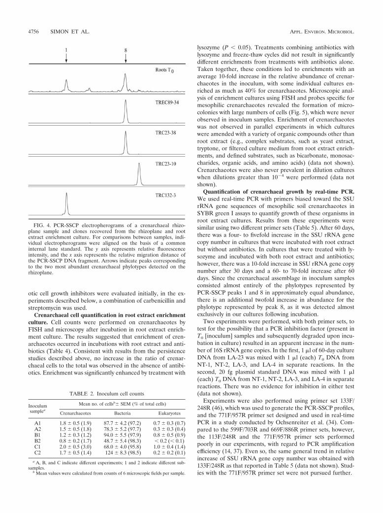
TREC89-34, were selected for PCR-SSCP analysis. The PCR-SSCP profiles of those clones contained a peak that comi-grated with peak 8 from total tomato rhizoplane DNA (repre-sentative profile shown by TREC89-34 in Fig. 4). TRC23-30and TRC23-38, two clones that were recovered directly fromthe rhizoplane (44) and that clustered with the TREC cloneswithin clade C1b.A1, were also analyzed by PCR-SSCP, andeach revealed a similar peak (representative profile shown byTRC23-38 in Fig. 4). Two additional TRC clones, TRC23-10and TRC132-3, and a clone from WMAD soil, MWS38 (46),chosen in part because they did not place within clade C1b.A1(clones TRC132-3 and MWS38 clustered together in cladeC1b.B1, while clone TRC23-10 placed in clade Clb.B2) (46),were analyzed and found to generate peaks migrating at dif-ferent positions from peak 8 on PCR-SSCP electrophero-grams. The peaks generated by clones TRC132-3 and MWS38comigrated instead with peak 1 from WMAD soil and rootsamples (representative profile shown by TRC132-3 in Fig. 4).Although the phylogenetic relationships of the clones de-scribed here were predictive of their PCR-SSCP profiles, this isnot always the case (46).
Crenarchaeal cell quantification in inoculum samples sub-jected to different treatments. We quantified the number ofcrenarchaeotes relative to total microorganisms present in rootextract enrichment cultures. The inoculum was prepared in amanner similar to that for the PCR-SSCP experiments, by soni-cation from gently rinsed tomato roots that were harvestedfrom plants grown in WMAD soil in the growth chamber. Cellnumbers were estimated with FISH probes designed to selec-tively detect mesophilic Crenarchaeota or organisms within thedomain Bacteria. Crenarchaeal cell numbers in untreated in-oculum samples from three experiments ranged from 1.2% to2.1% of total probe-positive cells (mean, 1.8%) (Table 2).These values are similar to those from our previous study inwhich we quantified crenarchaeal cells attached directly torinsed, nonsenescent tomato roots (44).
In addition to the use of root extract as a growth substrate,treatments were chosen to select against bacteria that werepresent in the starting inoculum. The inoculum was treatedwith lysozyme or lysozyme combined with freeze-thaw cyclesbefore incubation for growth. Lysozyme hydrolyzes glycosidiclinkages of peptidoglycan (which is present in cell walls ofbacteria but not in those of archaea) and freeze-thaw treat-ment should enhance lysis of weakened cell walls. Both ly-sozyme (mean, 4.6%) and lysozyme combined with freeze-thaw (mean, 3.4%) treatments had a significant effect (anapproximate twofold increase, P 0.05) on the proportion ofcrenarchaeotes compared to total cells in inoculum samplesprior to growth. This suggests that the treatments lysed a por-tion of bacterial cells in the inoculum.
Persistence of crenarchaeotes in root extract enrichmentcultures. As an additional selection, cultures were incubatedwith antibiotics that target bacteria but not archaea. We mon-itored the presence of crenarchaeotes in root extract culturesby PCR of SSU rRNA gene sequences with Crenarchaeota-biased primer sets. Results indicated that, while crenarchae-otes were initially present in all inoculated samples, over time,Crenarchaeota-specific PCR products were detected only incultures subjected to treatments with particular combinationsof antibiotics (Table 3). While various antibiotics and eukary-
FIG. 2. Representative PCR-SSCP electropherograms of the cren-archaeal assemblage in soil (A) and rhizoplane (B) samples afterincubation with root extract. For comparisons between samples, indi-vidual electropherograms were aligned on the basis of a commoninternal lane standard. The y axis represents relative fluorescenceintensity, and the x axis represents the relative migration distance ofthe PCR-SSCP DNA fragment. Arrows indicate peaks representingdifferent crenarchaeal phylotypes.
4754 SIMON ET AL. APPL. ENVIRON. MICROBIOL.

FIG. 3. Inferred phylogenetic (ML) tree of archaeal SSU rRNA gene sequences cloned from rhizoplane enrichment cultures. The habitat ofeach environmental sequence and sequences from this study is indicated before the clone name. Tomato root enrichment culture clones are shownin boldface type. Clone number prefixes 16 and 89 designate different experiments. Tomato root clones from direct extraction of tomato rhizoplaneDNA are shown on a shaded background. GenBank accession numbers are listed parenthetically. Branch points supported by bootstrap values of90% in all MP and ED methods are indicated by filled circles. Circles with a slash indicate bootstrap support values of 70%, open circlesindicate bootstrap support values of 50%, and branches without circles were not resolved (bootstrap support values of 50%). Bar, 0.1 changeper nucleotide.
VOL. 71, 2005 ENRICHMENT CULTURE OF MESOPHILIC SOIL CRENARCHAEOTES 4755
otic cell growth inhibitors were evaluated initially, in the ex-periments described below, a combination of carbenicillin andstreptomycin was used.
Crenarchaeal cell quantification in root extract enrichmentculture. Cell counts were performed on crenarchaeotes byFISH and microscopy after incubation in root extract enrich-ment culture. The results suggested that enrichment of cren-archaeotes occurred in incubations with root extract and anti-biotics (Table 4). Consistent with results from the persistencestudies described above, no increase in the ratio of crenar-chaeal cells to the total was observed in the absence of antibi-otics. Enrichment was significantly enhanced by treatment with
lysozyme (P 0.05). Treatments combining antibiotics withlysozyme and freeze-thaw cycles did not result in significantlydifferent enrichments from treatments with antibiotics alone.Taken together, these conditions led to enrichments with anaverage 10-fold increase in the relative abundance of crenar-chaeotes in the inoculum, with some individual cultures en-riched as much as 40% for crenarchaeotes. Microscopic anal-ysis of enrichment cultures using FISH and probes specific formesophilic crenarchaeotes revealed the formation of micro-colonies with large numbers of cells (Fig. 5), which were neverobserved in inoculum samples. Enrichment of crenarchaeoteswas not observed in parallel experiments in which cultureswere amended with a variety of organic compounds other thanroot extract (e.g., complex substrates, such as yeast extract,tryptone, or filtered culture medium from root extract enrich-ments, and defined substrates, such as bicarbonate, monosac-charides, organic acids, and amino acids) (data not shown).Crenarchaeotes were also never prevalent in dilution cultureswhen dilutions greater than 10�4 were performed (data notshown).
Quantification of crenarchaeal growth by real-time PCR.We used real-time PCR with primers biased toward the SSUrRNA gene sequences of mesophilic soil crenarchaeotes inSYBR green I assays to quantify growth of these organisms inroot extract cultures. Results from these experiments weresimilar using two different primer sets (Table 5). After 60 days,there was a four- to fivefold increase in the SSU rRNA genecopy number in cultures that were incubated with root extractbut without antibiotics. In cultures that were treated with ly-sozyme and incubated with both root extract and antibiotics;however, there was a 10-fold increase in SSU rRNA gene copynumber after 30 days and a 60- to 70-fold increase after 60days. Since the crenarchaeal assemblage in inoculum samplesconsisted almost entirely of the phylotypes represented byPCR-SSCP peaks 1 and 8 in approximately equal abundance,there is an additional twofold increase in abundance for thephylotype represented by peak 8, as it was detected almostexclusively in our cultures following incubation.
Two experiments were performed, with both primer sets, totest for the possibility that a PCR inhibition factor (present inT0 [inoculum] samples and subsequently degraded upon incu-bation in culture) resulted in an apparent increase in the num-ber of 16S rRNA gene copies. In the first, 1 �l of 60-day cultureDNA from LA-23 was mixed with 1 �l (each) T0 DNA fromNT-1, NT-2, LA-3, and LA-4 in separate reactions. In thesecond, 20 fg plasmid standard DNA was mixed with 1 �l(each) T0 DNA from NT-1, NT-2, LA-3, and LA-4 in separatereactions. There was no evidence for inhibition in either test(data not shown).
Experiments were also performed using primer set 133F/248R (46), which was used to generate the PCR-SSCP profiles,and the 771F/957R primer set designed and used in real-timePCR in a study conducted by Ochsenreiter et al. (34). Com-pared to the 599F/703R and 669F/886R primer sets, however,the 113F/248R and the 771F/957R primer sets performedpoorly in our experiments, with regard to PCR amplificationefficiency (14, 37). Even so, the same general trend in relativeincrease of SSU rRNA gene copy number was obtained with133F/248R as that reported in Table 5 (data not shown). Stud-ies with the 771F/957R primer set were not pursued further.
FIG. 4. PCR-SSCP electropherograms of a crenarchaeal rhizo-plane sample and clones recovered from the rhizoplane and rootextract enrichment culture. For comparisons between samples, indi-vidual electropherograms were aligned on the basis of a commoninternal lane standard. The y axis represents relative fluorescenceintensity, and the x axis represents the relative migration distance ofthe PCR-SSCP DNA fragment. Arrows indicate peaks correspondingto the two most abundant crenarchaeal phylotypes detected on therhizoplane.
TABLE 2. Inoculum cell counts
Inoculumsamplea
Mean no. of cellsb� SEM (% of total cells)
Crenarchaeotes Bacteria Eukaryotes
A1 1.8 � 0.5 (1.9) 87.7 � 4.2 (97.2) 0.7 � 0.3 (0.7)A2 1.5 � 0.5 (1.8) 78.3 � 5.2 (97.7) 0.3 � 0.3 (0.4)B1 1.2 � 0.3 (1.2) 94.0 � 5.5 (97.9) 0.8 � 0.5 (0.9)B2 0.8 � 0.2 (1.7) 48.7 � 5.4 (98.3) 0.2 ( 0.1)C1 2.0 � 0.5 (3.0) 68.0 � 4.0 (95.8) 1.0 � 0.4 (1.4)C2 1.7 � 0.5 (1.4) 124 � 8.3 (98.5) 0.2 � 0.2 (0.1)
a A, B, and C indicate different experiments; 1 and 2 indicate different sub-samples.
b Mean values were calculated from counts of 6 microscopic fields per sample.
4756 SIMON ET AL. APPL. ENVIRON. MICROBIOL.

Based on a calibration curve (32, 37) generated in real-timePCR with purified plasmid DNA from clone TRC23-30 (44)and the 599F/703R primer set, we estimated the copy numberof crenarchaeote SSU rRNA genes in 60-day cultures to be 4.1� 106 gene copies per ml of culture. The slope of the plasmidstandard curve was �3.51, the correlation coefficient was0.99, and the PCR amplification efficiency for the standardDNA was 1.926. The slope of the curve based on a fivefolddilution series of culture DNA (from no dilution to 1.6 � 10�3)was �3.50, the correlation coefficient was 0.95, and the PCRamplification efficiency for the culture DNA was 1.932. Thereproducibility of the data at higher dilutions was poor, corre-sponding to a limit of detection of approximately 25 copies ofthe target gene in these experiments. This number is similar todetection limits obtained by others (21, 28). The estimateddoubling time for crenarchaeotes in root extract cultures wasapproximately 8 days, with about 8 generations in 60 days.
DISCUSSION
Plants are known to deposit up to 20% of their total photo-synthate into the rhizosphere, allowing microorganisms thereto achieve much greater abundance and activity than they can
in habitats of lower nutrient availability, such as bulk soil (re-viewed in references 53 and 54). Microscopic and molecularphylogenetic studies of archaea in soil (3, 46), in the rhizo-sphere (47), and on the rhizoplane (44) led us to the firstdescription of the colonization of plant surfaces by mesophiliccrenarchaeotes and the influence of plant roots on the diversityof the crenarchaeal assemblage in soil. It was our predictionthat the root environment selects for microorganisms that areactively metabolizing root exudates and/or other root materialsand that crenarchaeotes associated with the root surface wouldtherefore be more amenable to growth in culture than thoseresiding in bulk soil. Consistent with this hypothesis, we dem-onstrated growth of one of the two dominant crenarchaealphylotypes found associated with the tomato rhizoplane, rep-resented by PCR-SSCP peak 8, in enrichment cultureamended with root extract. These results provide additionalsupport for a biological role for soil crenarchaeotes in rhizo-sphere microbial ecosystems.
FIG. 5. Fluorescence micrographs of crenarchaeotes from enrich-ment cultures. Representative color micrographs showing inoculumfrom the rhizoplane before (A) and after (B) incubation in root extractenrichment culture and indicating crenarchaeotes (panels labeled 1,orange) and bacteria (panels labeled 2, green). Crenarchaeal cells canalso be seen in panel B2 (red) with the filter set used to visualizebacteria. The arrow indicates the location of a crenarchael cell doublet.
TABLE 3. Persistence of crenarchaeotes in root extract enrichment cultures
Culturedesignation Treatmenta Antibioticsb
PCR product detectede on day after inoculation:
8 12 45 115 166 178 197 208c 238 378
143d None None � � � � � � �144 None None � � � � � � �145 L None � � � � � � �146 L S, C � � � � — � � � � �147 L S, C, R � � � � � � �148 L S, C, Cl, Ce � � � � � � � � � �149 FTL None � � � � � � �150 FTL S, C � � � � � � � � � �151 FTL S, C, R � � � � � � �152 FTL S, C, Cl, Ce � � � � � � � � � �
a L, lysozyme; FTL, freeze-thaw lysozyme (details in Materials and Methods).b C, carbenicillin; Ce, cephalothin; Cl, clindamycin; R, rifampin; S, streptomycin.c After 208 days, samples were taken only from cultures that were positive for crenarchaeotes.d Uninoculated control.e —, not sampled; �, PCR product was detected; �, no PCR product was detected.
TABLE 4. Enrichment of crenarchaeotes in root extract culturese
Treatmenta Antibioticsb Meanc %crenarchaeotad
None � 1.6 AL, FTL � 2.6 ANone � 16.7 BL � 22.3 CFTL � 20.2 B, C
a L, lysozyme; FTL, freeze-thaw and lysozyme (details in Materials and Meth-ods).
b Antibiotic additions (�, added; �, not added) are described in Materials andMethods.
c Means were calculated from counts of 6 microscopic fields per sample.d Mean values with different letters (A, B, and C) differ significantly (P
0.05).e Analysis of variance was performed using the general linear model, and mean
values were compared using Duncan’s multiple-range test (42). Degrees of free-dom: model, 13; error, 250; total, 263; pooled standard error, 9.82. The averagetime of incubation for cultures in each treatment was 22 to 24 weeks.
VOL. 71, 2005 ENRICHMENT CULTURE OF MESOPHILIC SOIL CRENARCHAEOTES 4757

Colonization of tomato roots by mesophilic soil crenarchae-otes was previously described (44) and was based on (i) therecruitment of these microorganisms to roots from soil, (ii)consistent recovery of crenarchaeal sequences and cells fromroots, (iii) a tight association with roots, i.e., cells remainedattached even after thorough rinsing, and (iv) broad distribu-tion of cells and microcolonies on roots. Although the tomatorhizoplane is not exclusively colonized by the peak 8 phylotype,the work presented here suggests that this particular phylotypecomprises a dominant, if not the dominant, crenarchaeal pop-ulation on tomato roots grown in WMAD soil. Detection ofthis phylotype in WMAD soil itself, however, appears to bemore variable. For example, although it was detected at verylow levels in the WMAD soil sampled in experiments pre-sented here, the peak 8 phylotype has been noted in greaterrelative abundance in PCR-SSCP profiles of WMAD soil gen-erated in other research (46). From our sampling thus far, thephylotype represented by peak 1 appears to be the dominantmember of the crenarchaeal assemblage in WMAD soil (inaddition to results presented in this paper, another study bySliwinski and Goodman [46] demonstrated that the relativeabundance of the peak 1 phylotype was higher than that ofother phylotypes, ranging between 43% and 53% of the total).The fact that the peak 8 phylotype consistently colonizes therhizoplane in high relative abundance compared to other phy-lotypes, yet is detected variably in WMAD soil, suggests that itsgrowth requirements are met more consistently on the rhizo-plane than in bulk (WMAD) soil habitats. More investigationis required to define the parameters that influence the distri-bution and abundance of this and other crenarchaeal phylo-types in soil.
We have demonstrated the association of mesophilic cren-archaeotes with plant roots using several approaches. Previousstudies described the direct extraction of crenarchaeal DNAfrom the rhizoplane (44) and rhizosphere soil (47) and visual-ization of crenarchaeote cells directly on the root surface usingphylogenetic probes. We have now provided additional evi-
dence for association by a specific phylotype of soil crenarchae-otes with plant roots. The peak 8 phylotype is one of twodominant phylotypes recovered directly from roots and theonly phylotype recovered from cultures after 60 days of incu-bation with root extract. In two different studies, the majorityof clones recovered directly from the rhizoplane and all of theclones recovered from root extract enrichment cultures placedwithin clade C1b.A1; sequences from the recovered clonescomigrated on PCR-SSCP gels with peak 8 from total DNAextracted directly from the rhizoplane. Furthermore, four linesof evidence, taken together, provide strong support for growthof the peak 8 phylotype when it is incubated in root extractenrichment culture: (i) persistence for 1 year in culture, (ii)the increase in proportion of crenarchaeotes to total cells, (iii)the increase in SSU rRNA gene copy number of crenarchae-otes per unit volume of culture, and (iv) the formation ofmicrocolonies containing large numbers of crenarchaeal cells.In addition, the fact that three different primer sets, eachdesigned to be specific for the SSU rRNA genes of mesophilicsoil crenarchaeotes and one also used in PCR-SSCP analyses,yielded similar results in real-time PCR assays strengthens theconclusion that growth of these microorganisms occurred inroot extract enrichment cultures. Although we achieved en-richment in cultures amended with root extract, we were not assuccessful using a variety of common organic compounds, in-cluding many known to be present in plant exudates. Theseresults may suggest that the actual substrates supportinggrowth are unusual exudate compounds produced either by theplant roots or by associated bacteria. Identification of the ac-tual substrates for growth will be critical for growing the cre-narchaeotes in axenic culture.
Crenarchaeal enrichment was dependent upon the presenceof antibiotics in the medium, although there was also a small(four- to fivefold) increase in crenarchaeal 16S rRNA genecopies in cultures incubated without antibiotics. The antibioticsused in this study were chosen because they are known toinhibit bacteria but not archaea. It was surprising, therefore,that the addition of rifampin to the cultures resulted in the lossof the crenarchaeal PCR signal sooner than it occurred in theabsence of antibiotics. While it is currently unknown whetherthe inhibition was due to a direct or an indirect effect, RNApolymerases from other archaea are not targets for the antibi-otic. On the other hand, sensitivity to rifampin has been ob-served in a number of different archaea, including some cren-archaeotes, at concentrations similar to that used in this study(23, 52). A detergent-like machanism of cell lysis has beensuggested for the inhibitory effect of rifampin on Halobacte-rium halobium (55). Possible indirect effects of rifampin onenrichment of crenarchaeotes include the inhibition of an ob-ligate bacterial partner; involvement by the crenarchaeotes ina symbiotic partnership(s) with other microbial species has notyet been explored. Consistent with this idea is the failure ofserial dilutions to lead to enrichment. Another possible expla-nation for the effect of rifampin could be that certain bacteriapresent in the enrichments were able to metabolize the smallamount of methanol used as a solvent that was introduced intothe culture along with the antibiotic and, by doing so, were ableto overgrow the crenarchaeotes. Bacteria that were presentand growing in the enrichments may have developed resistanceto rifampin (and to other antibiotics used), or, alternatively,
TABLE 5. Results from real-time PCR analysis of crenarchaeaotesin rhizoplane inoculum and root extract enrichment culture
Primer pair Treatment and templatea Enrichment culture/inoculum ratio � SEM
None (untreated)599F/703R Inoculum 6.6 � 0.8599F/703R 30-day culture 3.6 � 0.5599F/703R 60-day culture 33.3 � 3.6669F/886R Inoculum 3.3 � 0.3669F/886R 30-day culture 2.3 � 0.4669F/886R 60-day culture 12.2 � 0.8
LAb
599F/703R Inoculum 1.8 � 0.2599F/703R 30-day culture 16.9 � 3.4599F/703R 60-day culture 129.0 � 5.9669F/886R Inoculum 1.8 � 0.1669F/886R 30-day culture 17.9 � 2.7669F/886R 60-day culture 107.0 � 5.4
a The results given in each row represent the combined average results fromPCR assays of two individually prepared cultures, with the exception of inoculumsamples, in which results from one of the pair of individually prepared inoculumsamples are used as the basis for comparison to the other samples and the secondis compared to the first.
b LA, treatment with lysozyme and antibiotics.
4758 SIMON ET AL. APPL. ENVIRON. MICROBIOL.

diffusion of the antibiotics may have been impeded by extra-cellular polysaccharide that was produced in the cultures.
Our research represents a significant advance in the study ofthe mesophilic Crenarchaeota, first, because it reveals a biolog-ical relationship between members of the C1b.A1 clade andplants and, second, because neither isolation nor growth ofthese organisms in culture has been reported in the decade orso since nonthermophilic crenarchaeotes were initially discov-ered in mesophilic soils (3, 51) or their planktonic counterpartswere discovered in marine waters (8, 15). Our result of amodest increase (102) in SSU rRNA gene copy number over an8-week incubation period is in line with results from a numberof other studies that have attempted enrichments and isola-tions of archaea and bacteria identified first by molecular phy-logenetic methods (that had not been previously detected fromculture-dependent work). Examples include isolates from thebacterial divisions Acidobacteria, Verrucomicrobia, Gemmati-monadetes, Actinobacteria, and Proteobacteria that were ob-tained only after extended incubations (25, 26, 50) and enrich-ments of anaerobic methane-oxidizing archaea from marinesediments, with apparent 2- to 100-fold (or greater) increasesin SSU rRNA gene copies after 24 weeks of incubation (al-though the latter data are somewhat inconclusive because nominimum detection level was established for the target gene insediment cores prior to incubation (18).
Based on evidence from thermophilic and hyperthermo-philic crenarchaeotes that have been isolated in pure cultureand contain a single rRNA operon in each of their genomes, itis not unreasonable to make the assumption that individualgenomes of mesophilic crenarchaeotes also contain only onerRNA operon. Using this assumption and comparing resultsfrom real-time PCR of enrichment culture DNA with plasmidstandard DNA, we estimate that our enrichment cultures con-tained 4.1 � 106 crenarchaeal cells per ml after 60 days ofincubation and, by extrapolation, �104 crenarchaeal cells perml in the rhizoplane sonicates used for the inoculum. Anothergroup investigated crenarchaeal populations in the rhizosphereof the grass Festuca ovina s.l. using real-time PCR (34) andfound a relatively low abundance of crenarchaeal to bacterial16S rRNA gene copies in the rhizosphere versus bulk soil.Based on the numerous differences that exist between the twostudies (e.g., soil type, plant species, growth chamber versusfield study, and units of measurement), direct comparisonsbetween them are not valid. It is worth noting, however, thatstudies by Sliwinski and Goodman, and others, examining cre-narchaeal assemblages in a variety of soils (46) and in therhizosphere of diverse plant species (33, 47) suggests that theabundances of crenarchaeal populations differ in different soiland rhizosphere habitats.
In earlier work (44), we found a 10-fold increase in theabundance of crenarchaeotes on senescent roots over nonse-nescent roots of tomato. One intriguing possibility raised bythat result and by our studies here is the idea that plants thatare stressed, for example, as they might be when grown inunfertilized soil within a growth chamber, harbor larger pop-ulations of crenarchaeotes on their roots than their unstressedcounterparts. This possibility and other factors potentially af-fecting the abundance, distribution, and diversity of crenar-chaeotes in soil and rhizosphere habitats are the subjects ofongoing investigations in our laboratories.
ACKNOWLEDGMENTS
We thank John Helgeson for the generous use of his OlympusBX-60 microscope, Ena Urbach and Brian Manske for advice withphylogenetic analyses, Doreen Gillespie for discussions, and GaryRoberts and Peter Zuber for comments on the manuscript.
A National Science Foundation grant (no. MCB-0136441) and con-tributions from the McKnight Foundation supported this work.
REFERENCES
1. Altschul, S. F., W. Gish, W. Miller, E. W. Myers, and D. J. Lipman. 1990.Basic local alignment search tool. J. Mol. Biol. 215:403–410.
2. Baker, A. 1981. Accumulators and excluders—strategies in the response ofplants to heavy metals. J. Plant Nutr. 3:643–654.
3. Bintrim, S. B., T. J. Donohue, J. Handelsman, G. P. Roberts, and R. M.Goodman. 1997. Molecular phylogeny of Archaea in soil. Proc. Natl. Acad.Sci. USA 94:277–282.
4. Bomberg, M., G. Jurgens, A. Saano, R. Sen, and S. Timonen. 2003. NestedPCR detection of archaea in defined compartments of pine mycorrhizos-pheres developed in boreal forest humus microcosms. FEMS Microbiol.Ecol. 43:163–171.
5. Buckley, D. H., J. R. Graber, and T. M. Schmidt. 1998. Phylogenetic analysisof nonthermophilic members of the kingdom Crenarchaeota and their diver-sity and abundance in soils. Appl. Environ. Microbiol. 64:4333–4339.
6. Chelius, M. K., and E. W. Triplett. 2001. The diversity of archaea andbacteria in association with the roots of Zea mays L. Microb. Ecol. 41:252–263.
7. Daniels, L., N. Belay, and B. S. Rajagopal. 1986. Assimilatory reduction ofsulfate and sulfite by methanogenic bacteria. Appl. Environ. Microbiol. 51:703–709.
8. DeLong, E. F. 1992. Archaea in coastal marine environments. Proc. Natl.Acad. Sci. USA 89:5585–5689.
9. DeLong, E. F. 1998. Archaeal means and extremes. Science 280:542–543.10. Delong, E. F. 1998. Everything in moderation: archaea as ‘non-extremo-
philes’. Curr. Opin. Genet. Dev. 8:649–654.11. DeLong, E. F., and N. R. Pace. 2001. Environmental diversity of bacteria and
archaea. Syst. Biol. 50:470–478.12. DeLong, E. F., K. Y. Wu, B. B. Prezelin, and R. V. M. Jovine. 1994. High
abundance of archaea in Antarctic marine picoplankton. Nature 371:695–697.
13. Felsenstein, J. 1981. Evolutionary trees from DNA sequences: a maximumlikelihood approach. J. Mol. Evol. 17:368–376.
14. Ferre, F. 1992. Quantitative or semi-quantitative PCR: reality versus myth.PCR Methods Appl. 2:1–9.
15. Fuhrman, J. A., K. McCallum, and A. A. Davis. 1992. Novel major archae-bacterial group from marine plankton. Nature 356:148–149.
16. Galtier, N., N. Tourasse, and M. Gouy. 1999. Relationships between genomicG�C content, RNA secondary structures, and optimal growth temperaturein prokaryotes. Science 283:220–221.
17. Giovannoni, S. J., T. B. Britschgi, C. L. Moyer, and K. G. Field. 1990.Genetic diversity in Sargasso Sea bacterioplankton. Nature 345:60–63.
18. Girguis, P. R., V. J. Orphan, S. J. Hallam, and E. F. Delong. 2003. Growthand methane oxidation rates of anaerobic methanotrophic archaea in acontinuous-flow bioreactor. Appl. Environ. Microbiol. 69:5472–5482.
19. Gro�kopf, R., S. Stubner, and W. Liesack. 1998. Novel euryarchaeotal lin-eages detected on rice roots and in the anoxic bulk soil of flooded ricemicrocosms. Appl. Environ. Microbiol. 64:4983–4989.
20. Hayashi, K. 1991. PCR-SSCP: a simple and sensitive method for detection ofmutations in the genomic DNA. PCR Methods Appl. 1:34–38.
21. Hein, I., A. Lehner, P. Rieck, K. Klein, E. Brandl, and M. Wagner. 2001.Comparison of different approaches to quantify Staphylococcus aureus cellsby real-time quantitative PCR and application of this technique for exami-nation of cheese. Appl. Environ. Microbiol. 67:3122–3126.
22. Hershberger, K. L., S. M. Barns, A. L. Reysenbach, S. C. Dawson, and N. R.Pace. 1996. Wide diversity of Crenarchaeota. Nature 384:420.
23. Huber, R., J. K. Kristjansson, and K. O. Stetter. 1987. Pyrobaculum gen.nov., a new genus of neutrophilic, rod-shaped archaebacteria from continen-tal solfataras growing optimally at 100°C. Arch. Microbiol. 149:95–101.
24. Huber, T., G. Faulkner, and P. Hugenholtz. 2004. Bellerophon; a program todetect chimeric sequences in multiple sequence alignments. Bioinformatics20:2317–2319.
25. Janssen, P. H., P. S. Yates, B. E. Grinton, P. M. Taylor, and M. Sait. 2002.Improved culturability of soil bacteria and isolation in pure culture of novelmembers of the divisions Acidobacteria, Actinobacteria, Proteobacteria, andVerrucomicrobia. Appl. Environ. Microbiol. 68:2391–2396.
26. Joseph, S. J., P. Hugenholtz, P. Sangwan, C. A. Osborne, and P. H. Janssen.2003. Laboratory cultivation of widespread and previously uncultured soilbacteria. Appl. Environ. Microbiol. 69:7210–7215.
27. Karner, M. B., E. F. DeLong, and D. M. Karl. 2001. Archaeal dominance inthe mesopelagic zone of the Pacific Ocean. Nature 409:507–510.
28. Kolb, S., C. Knief, S. Stubner and R. Conrad. 2003. Quantitative detection
VOL. 71, 2005 ENRICHMENT CULTURE OF MESOPHILIC SOIL CRENARCHAEOTES 4759
of methanotrophs in soil by novel pmoA-targeted real-time PCR assays.Appl. Environ. Microbiol. 69:2423–2429.
29. Lane, D. J. 1991. 16S/23S rRNA sequencing, p. 115–175. In E. Stackebrandtand M. Goodfellow (ed.), Nucleic acid techniques in bacterial systematics.John Wiley and Sons, New York, N.Y.
30. Larsen, N., G. J. Olsen, B. L. Maidak, M. J. McCaughey, R. Overbeek, T. J.Maeke, T. L. Marsh, and C. R. Woese. 1993. The ribosomal database project.Nucleic Acids Res. 21:3021–3023.
30a.Ludwig, W., O. Strunk, R. Westram, L. Richter, H. Meier, Yadhukumar, A.Buchner, T. Lai, S. Steppi, G. Jobb, W. Forster, I. Brettske, St. Gerber, A. W.Ginhart, O. Gross, S. Grumann, S. Hermannl, R. Jost, A. Konig, T. Liss, R.Lubmann, M. May, B. Nonhoff, B. Reichel, R. Strehlow, A. Stamatakis, N.Stuckmann, A. Vilbig, M. Lenke, T. Ludwig, A. Bode, and K.-H. Schleifer.2004. ARB: a software environment for sequence data. Nucleic Acids Res.32:1363–1371.
31. Massana, R., L. T. Taylor, A. E. Murray, K. Y. Wu, W. H. Jeffrey, and E. F.DeLong. 1998. Vertical distribution and phylogenetic characterization ofmarine planktonic Archaea in the Santa Barbara channel. Limnol. Ocean-ogr. 43:607–617.
32. Morrison, T., J. J. Weis, and C. T. Wittwer. 1998. Quantification of low-copytranscripts by continuous SYBR Green I monitoring during amplification.Biotechniques 24:954–962.
33. Nicol, G. W., L. A. Glover, and J. I. Prosser. 2003. The impact of grasslandmanagement on archaeal community structure in upland pasture rhizopheresoil. Environ. Microbiol. 5:152–162.
34. Ochsenreiter, T., D. Selezi, A. Quaiser, L. Bonch-Osmolovskaya, and C.Schleper. 2004. Diversity and abundance of Crenarchaeota in terrestrialhabitats studied by 16S RNA surveys and real time PCR. Environ. Microbiol.5:787–797.
35. Olsen, G. J., H. Matsuda, R. Hagstrom, and R. Overbeek. 1994. fastDNAmL:a tool for construction of phylogenetic trees of DNA sequences using max-imum likelihood. Comput. Appl. Biosci. 10:41–48.
36. Orita, M., H. Iwahana, H. Kanazawa, K. Hayashi, and T. Sekiya. 1989.Detection of polymorphisms of human DNA by gel electrophoresis as single-strand conformation polymorphisms. Proc. Natl. Acad. Sci. USA 86:2766–2770.
37. Pfaffl, M. W. 2001. Development and validation of an externally standardisedquantitative insulin like growth factor-1 (IGF-1) RT-PCR using LightCyclerSYBR Green I technology, p. 281–291. In S. Meuer, C. Wittwer, and K.Nakagawara (ed.), Rapid cycle real-time PCR, methods and applications.Springer Press, Heidelberg, Germany.
38. Preston, C. M., K. Y. Wu, T. F. Molinski, and E. F. DeLong. 1996. Apsychrophilic crenarchaeon inhabits a marine sponge: Crenarchaeum sym-biosum gen. nov., sp. nov. Proc. Natl. Acad. Sci. USA 93:6241–6246.
39. Rasmussen, R. 2001. Quantification on the LightCycler, p. 21–34. In S.Meuer, C. Wittwer, and K. Nakagawara (ed.), Rapid cycle real-time PCR,methods and applications. Springer Press, Heidelberg, Germany.
40. Repaske, R. 1956. Lysis of gram-negative bacteria by lysozyme. Biochim.Biophys. Acta 22:189–191.
41. Rondon, M. R., S. J. Raffel, R. M. Goodman, and J. Handelsman. 1999.Toward functional genomics in bacteria: analysis of gene expression in Esch-erichia coli from a bacterial artificial chromosome library of Bacillus cereus.Proc. Natl. Acad. Sci. USA 96:6451–6645.
42. SAS Institute. 1985. SAS user’s guide, version 5. SAS Institute, Cary, N.C.43. Schaefer, D. M., C. L. Davis, and M. P. Bryant. 1980. Ammonia saturation
constants for predominant species of rumen bacteria. J. Dairy Sci. 63:1248–1263.
44. Simon, H. M., J. A. Dodsworth, and R. M. Goodman. 2000. Crenarchaeotacolonize terrestrial plant roots. Environ. Microbiol. 2:506–515.
45. Sinninghe Damste, J. S., W. I. C. Rijpstra, E. C. Hopmans, F. G. Prahl, S. G.Wakeham, and S. Schouten. 2002. Distribution of membrane lipids of plank-tonic Crenarchaeota in the Arabian Sea. Appl. Environ. Microbiol. 68:2997–3002.
46. Sliwinski, M. K., and R. M. Goodman. 2004. Spatial heterogeneity of cren-archaeal assemblages within mesophilic soil ecosystems as revealed by PCR–single-stranded conformation polymorphism profiling. Appl. Environ. Mi-crobiol. 70:1811–1820.
47. Sliwinski, M. K., and R. M. Goodman. 2004. Comparison of crenarchaealconsortia inhabiting the rhizosphere of diverse terrestrial plants with those inbulk soil in native environments. Appl. Environ. Microbiol. 70:1821–1826.
48. Stahl, D. A., and R. I. Amann. 1991. Development and application of nucleicacid probes in bacterial systematics, p. 205–248. In E. Stackebrandt and M.Goodfellow (ed.), Nucleic acid techniques in bacterial systematics. JohnWiley and Sons, New York, N.Y.
49. Stein, J. L., and M. I. Simon. 1996. Archaeal ubiquity. Proc. Natl. Acad. Sci.USA. 93:6228–6230.
50. Stevenson, B. S., S. A. Eichorst, J. T. Wertz, T. M. Schmidt, and J. A.Breznak. 2004. New strategies for cultivation and detection of previouslyuncultured microbes. Appl. Environ. Microbiol. 70:4748–4755.
51. Ueda, T., Y. Suga, and T. Matsuguchi,. 1995. Molecular phylogenetic anal-ysis of a soil microbial community in a soybean field. Eur. J. Soil Sci.46:415–421.
52. Watrin, L., E. Corre, and D. Prieur. 1996. In vivo susceptibility of sulfother-mophilic archaea to antimicrobial agents. J. Mar. Biotechnol. 4:215–219.
53. Whipps, J. M. 1990. Carbon economy, p. 59–97. In J. M. Lynch (ed.), Therhizosphere. John Wiley and Sons, Chichester, United Kingdom.
54. Whipps, J. M., and J. M. Lynch. 1985. Energy losses by the plant in rhi-zodeposition. Annu. Proc. Phytochem. Soc. 26:59–71.
55. Zillig, W., K. O. Stetter, W. Schulz, and D. Janekovic. 1980. Comparativestudies of structure and function of DNA-dependent RNA polymerases fromeubacteria and archaebacteria, p. 159–178. In P. Mildner and B. Rieds (ed.),Enzyme regulation and mechanism of action. Pergamon Press, Oxford,United Kingdom.
4760 SIMON ET AL. APPL. ENVIRON. MICROBIOL.