ctcl management: lessons learned - cutaneous …cutaneouslymphoma.stanford.edu/documents/y kim...
TRANSCRIPT

CTCL Management:
Lessons Learned
Youn H Kim, MD
Department of Dermatology
Multidisciplinary Cutaneous Lymphoma Program
Stanford Cancer Center & School of Medicine
Stanford, CA

Disclosure statement
Youn Kim, MD
• Steering Committee
– Eisai, Millennium
• Consultant or Advisory Board
– Kyowa, Celgene, Galderma, Medicis
• Investigator
– Allos, Kyowa, Merck, Millennium, Seattle Genetics, Shape,
Ceptaris/Yaupon, Eisai, Genentech

Clinical Issues in CTCL Management
• How can we optimize our diagnostic ability?
• How do we make optimal treatment decisions with
available therapies?
• How can we improve therapeutics and outcome?

Lesson #1
Clinical-pathologic correlation is essential
for optimal diagnosis & management
Challenge of so many
histopath and clinical mimics

Differential diagnosis of CD30+ atypical
lymphoid infiltrates in the skin
Reactive
• Lymphomatoid drug
reaction (e.g., amlodipine,
carbamazepine, cefuroxime,
valsarten)
• Arthropod reaction
• Infection (esp. viral)
• Misc. inflammatory
dermatoses
Neoplastic
• pc CD30+ LPD
– Lymphomatoid papulosis
– pc CD30+ ALCL
• MF (esp. Large cell
transformation, Woringer-
Kolopp)
• Other CTCLs
• Secondary skin
involvement of sALCL,
HD or other sLPD
Clinico-pathologic correlation is essential

PC CD30+ lymphoproliferative disorder spectrum
LyP === borderline === pc CD30+ ALCL
Lymphomatoid papulosis
• 100% spontaneous regression
• Papules >> nodules
• Crops of lesions, +/- grouped
• Multiple histologic subtypes
(types A-D, other); type A most
common, type B MF-like (low
CD30), type C ALCL-like, type
D mimics CD8+ AETCL
pc CD30+ ALCL
• < 25% spontaneous regression
• Mostly nodules/tumors
• Single, grouped, multifocal
• Usu. sheets of anaplastic large
cells
CLINICAL-PATHOLOGIC CORRELATION IS ESSENTIAL


Type D CD8+ LyP vs. CD8+ aggressive
epidermotropic cytotoxic TCL
LyP type D
Courtesy T Subtil
CD8+ aggressive
epidermotropic
cytotoxic TCL CD8

Differential diagnosis of epidermotropic
process with CD8+ lymphoid infiltrates
Reactive
• Lymphomatoid drug
reaction
• Misc. inflammatory
dermatoses (esp. actinic
reticuloid)
• Infections
Neoplastic
• CD8+ AETCL
• Lymphomatoid papulosis,
type D
• CD8+ MF (hypopig variant)
• SubQ panniculitis-like TCL
• CD8+ LPD of ear/face
• PTCL NOS
• Secondary skin
involvement of PTCL
Clinico-pathologic correlation is essential

Am J Surg Pathol 2007;31:1887
Multicenter Case Series of Indolent Small/Medium-
sized CD8+ Lymphoid Proliferations with
Predilection for the Ear and Face
Janet Y. Li1, Joan Guitart2, Melissa P. Pulitzer1, Antonio Subtil3, Uma
Sundram4, Youn Kim4, Janyana Deonizio2, Patricia L. Myskowski1
Alison Moskowitz1, Steven Horwitz1, Christiane Querfeld1
1Memorial Sloan Kettering Cancer Center and Weill Cornell Medical College,
New York, NY, 2Northwestern University, Chicago, IL, 3Yale University, New
Haven, CT, 4Stanford University, Stanford, CA Am J Dermatopathol, in press 2013

Querfeld, MSKCC
Stanford case
Indolent Small/Med-sized CD8+ Lymphoid Proliferations
with Predilection for the Ear and Face

Am J Surg Pathol 2013;37:1-13

Angioinvasive, aggressive
NK/T-cell lymphoma, nasal-type

J Am Acad Dermatol 2013;68:809

Folliculotropic
Mycosis Fungoides
Clinico-pathologic correlation is essential

Too many clinical, path variants & mimics
leading to more confusion in diagnosis

• Hypopigmented/vitiligenous
MF
– Children, African American,
Asian
• Pagetoid reticulosis
(Woringer-Kolopp type only)
• Folliculotropic MF (+/- FM)
– Head and neck
• Granulomatous MF
– Granulomatous slack skin
• Bullous MF
• PPE-like MF
• Interstitial MF
• Icthyiosiform MF
• Palmar plantar MF
• Hyperkeratotic/verrucous MF
• Papular MF
• Invisible MF
Mycosis Fungoides - the greatest masquerader Clinical & Histologic Variants/Subtypes
• Spongiotic MF
• Lichenoid MF
• CD8+ MF
• Large cell (transformed) MF

Lesson #1: importance of clin-path correlation
Take Home Message
• Numerous mimics of clinical OR path features exist
• Correlation of clinical AND pathologic information is
essential for optimal diagnosis
=> appropriate work-up, prognostication, and management

Lesson #2
“OK” to be noncommittal with diagnosis Impact of a “lymphoma” label

CD4+ sm/med-sized pleomorphic T-cell
“lymphoma”
• Mostly benign/indolent course, especially in kids
• A lymphoid proliferation of undetermined
significance vs. “lymphoma”
• CD4+ sm/med-sized pleomorphic T-cell
lymphoproliferative disorder (LPD)?

Am J Dermatopathol 2009;31:317-322
• Solitary/localized disease with benign outcome
• Majority of H/N
• Rare multifocal presentation with worse outcome

Am J Dermatopathol 2009;31:317-322
CD4+ sm/med pleomorphic T-cell “lymphoma” vs “LPD”?

Querfeld, MSKCC
Stanford case
J Cutan Pathol 2010;37:81-84
Indolent sm/med-sized CD8+ lymphoid
proliferation of the ear/face
Spectrum of lymphoid proliferation:
If clinical course is indolent, ok to
follow and manage conservatively

Lesson #3
Don’t forget to check the blood
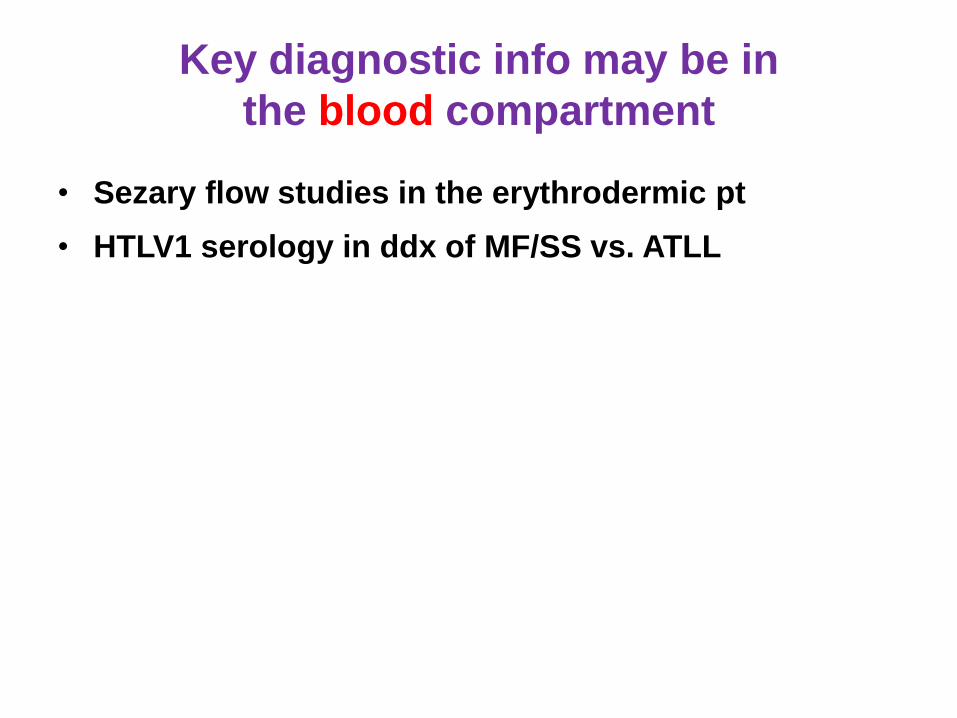
Key diagnostic info may be in
the blood compartment
• Sezary flow studies in the erythrodermic pt
• HTLV1 serology in ddx of MF/SS vs. ATLL


• Neoplastic T-cells are CD3+, CD4+, CD8-, CD25+; epidermotropic
• Endemic in Japan, the Caribbean, S Americas, Central Africa;
• Primarily transmitted by breast feeding

ATLL,
spectrum of skin
presentation
MF-like, smoldering variant Acute, disseminated disease

Courtesy J Brody
ATL can mimic
Sezary syndrome

Acute
ATLL
Coutesy J Guitart

Challenge of the red person

63 F with 4 yr h/o progressive erythroderma
• Itchy scalp and scaly red patches and plaques
– Refractory to topical steroids; pred helps
– Skin biopsy => spong derm
– nbUVB, unable to tolerate
• Progressive erythroderma, keratoderma
– Rebiopsy => psoriasiform derm
– Soriatane => no response
• Immune suppressive therapies
– Cyclosporin x 3 mo => PR
– Humira added => no sig benefit, flares with CSA taper
– Rebiopsy => psoriasiform derm with spong
• No drug etiology

Erythroderma with severe pruritus
DDx- eczematous derm,
psoriasis, drug, PRP,
MF/SS, other

Keratoderma of palms and soles


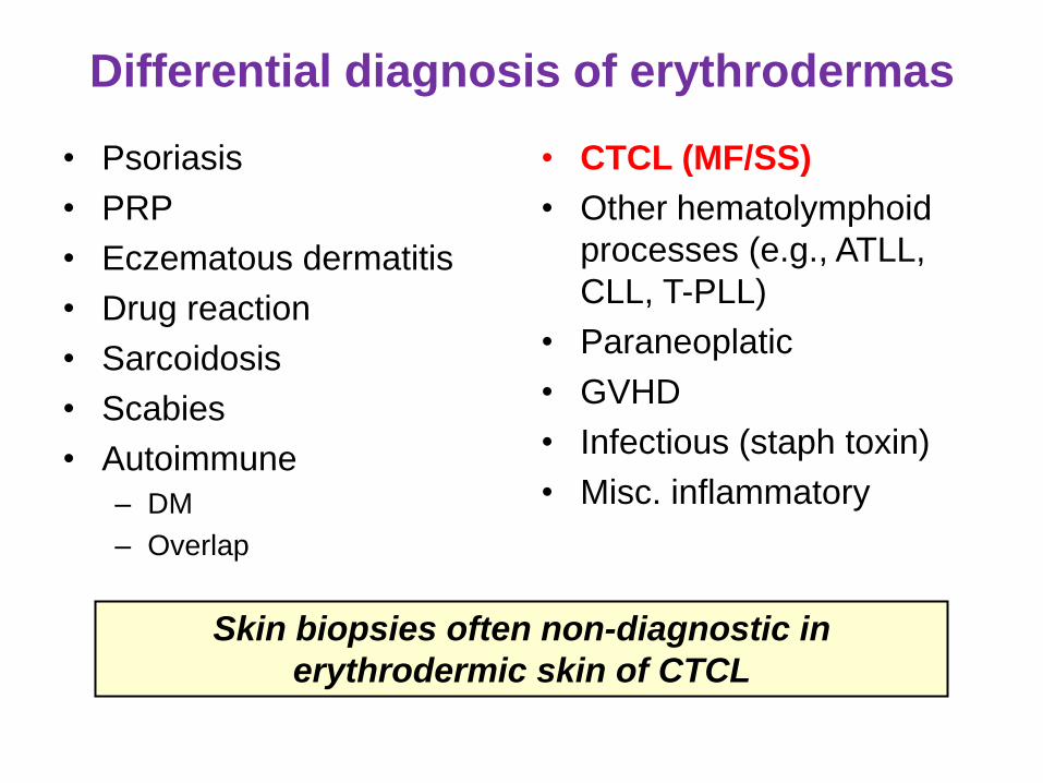
Differential diagnosis of erythrodermas
• Psoriasis
• PRP
• Eczematous dermatitis
• Drug reaction
• Sarcoidosis
• Scabies
• Autoimmune
– DM
– Overlap
• CTCL (MF/SS)
• Other hematolymphoid
processes (e.g., ATLL,
CLL, T-PLL)
• Paraneoplatic
• GVHD
• Infectious (staph toxin)
• Misc. inflammatory
Skin biopsies often non-diagnostic in
erythrodermic skin of CTCL
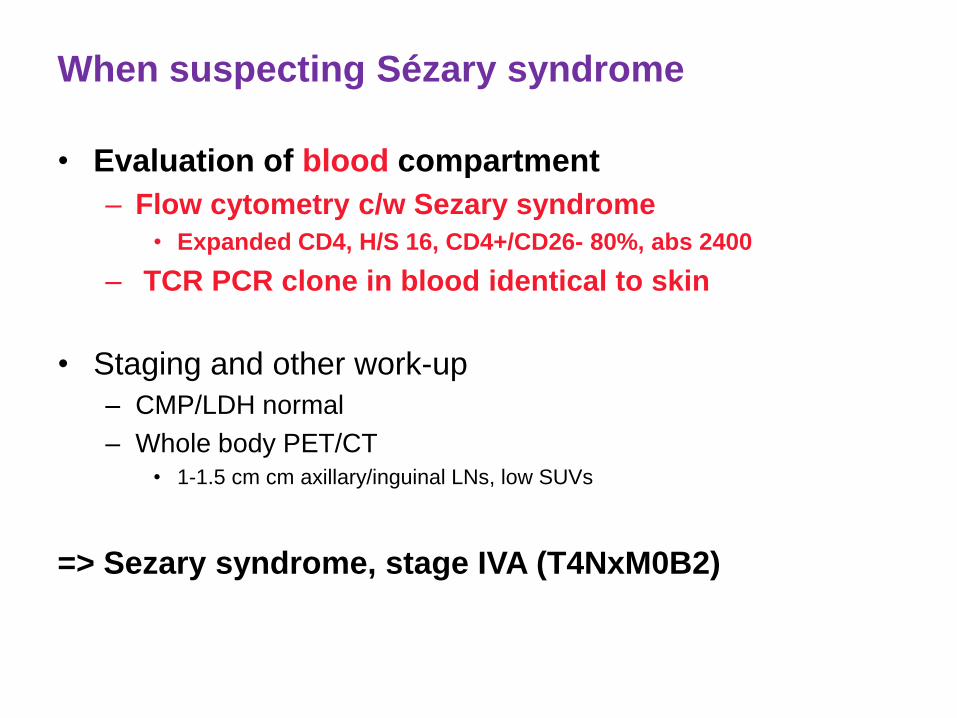
When suspecting Sézary syndrome
• Evaluation of blood compartment
– Flow cytometry c/w Sezary syndrome
• Expanded CD4, H/S 16, CD4+/CD26- 80%, abs 2400
– TCR PCR clone in blood identical to skin
• Staging and other work-up
– CMP/LDH normal
– Whole body PET/CT
• 1-1.5 cm cm axillary/inguinal LNs, low SUVs
=> Sezary syndrome, stage IVA (T4NxM0B2)

Clinical course and management of SS
• ECP + oral bexarotene => mild benefit
• Added IFN-alpha => no response, neutropenia
• MTX 35 mg => minimal benefit
• Anti-CCR4 mab (mogmulizumab)
– Rapid reduction of SCs and pruritus
– Near 3 yrs of great disease control

Case Study: Patient 03-Stanford (SS; Stage IVA; 6 Prior Therapies; 0.3 mg/kg)
Pretreatment
Course 1 Day 1
Post treatment
Post Course 11

Response in Blood: Patient 01-Stanford
(SS; Stage IVA; 6 prior therapies; 0.1 mg/kg)
Pre-treatment
CD3+CD4neg
Normal CD3+CD4+
Lymphoma cells Lymphoma cells
0 10 2
10 3
10 4
10 5
CD4
0
10 2
10 3
10 4
10 5
CD
3
0 10 2
10 3
10 4
10 5
CD26
0
10 2
10 3
10 4
10 5
CD
3
0 10 2
10 3
10 4
10 5
CCR4 1G1
0
10 2
10 3
10 4
10 5
CD
3
0 10 2
10 3
10 4
10 5
CCR4 1G1
0
20
40
60
80
100
% o
f M
ax

Lymphoma cells
undetectable
Response >2 yrs CD3+CD4neg
Normal CD3+CD4+
Lymphoma cells
0 10 2
10 3
10 4
10 5
CD4
0
10 2
10 3
10 4
10 5
CD
3
0 10 2
10 3
10 4
10 5
CD26
0
10 2
10 3
10 4
10 5
CD
3
0 10 2
10 3
10 4
10 5
CCR4 1G1
0
10 2
10 3
10 4
10 5
CD
3
0 10 2
10 3
10 4
10 5
CCR4 1G1
0
20
40
60
80
100
% o
f M
ax
Response in Blood: Patient 01-Stanford
Post-treatment
Phase III RCT in CTCL ongoing
for FDA approval

Challenge of the red person
Take home message
Skin biopsies often non-diagnostic from
erythrodermic skin of CTCL
MUST ASSESS BLOOD if suspect SS
Lesson #4
Advanced MF/SS IS curative

Tumor-directed killing
Road to a CURE Effective tumor killing => lasting responses
by partnering with immune strategies
Immunotherapy
% S
urv
ival
Time
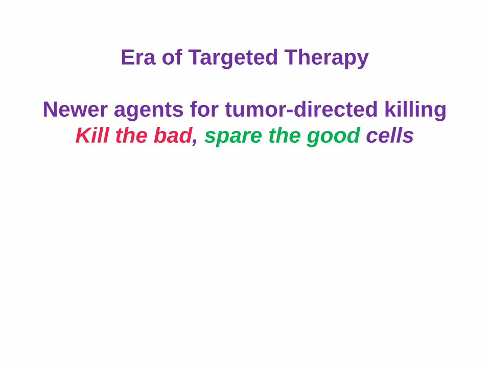
Era of Targeted Therapy
Newer agents for tumor-directed killing
Kill the bad, spare the good cells
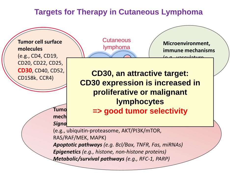
Tumor cell surface molecules (e.g., CD4, CD19, CD20, CD22, CD25,
CD30, CD40, CD52, CD158k, CCR4)
Tumor proliferation, metabolism, survival, progression mechanisms: Signal transduction/transcription activation pathways (e.g., ubiquitin-proteasome, AKT/PI3K/mTOR, RAS/RAF/MEK, MAPK) Apoptotic pathways (e.g. Bcl/Bax, TNFR, Fas, miRNAs) Epigenetics (e.g., histone, non-histone proteins) Metabolic/survival pathways (e.g., RFC-1, PARP)
Microenvironment, immune mechanisms (e.g., vasculature, immune modulation)
Targets for Therapy in Cutaneous Lymphoma
Cutaneous
lymphoma
CD30, an attractive target:
CD30 expression is increased in
proliferative or malignant
lymphocytes
=> good tumor selectivity

Monomethyl auristatin E (MMAE), microtubule-disrupting agent
Protease-cleavable linker
Anti-CD30 monoclonal antibody
ADC binds to CD30
MMAE disrupts
microtubule network
ADC-CD30 complex
is internalized and
traffics to lysosome
MMAE is released
Apoptosis
G2/M cell
cycle arrest
Brentuximab Vedotin Mechanism of Action
Antibody Drug Conjugate
Given IV every 3 wks

Brentuximab vedotin demonstrates clinical
activity in mycosis fungoides / Sézary
syndrome
Krathen M1, Bashey S1, Sutherland K1, Sundram U1,
Nagpal S1, Salva K3, Wood G3, Advani R1, Hoppe RH1,
Reddy S1, Pulitzer M2, Horwitz S2, Kim YH1
1Stanford Cancer Institute, Stanford, CA, USA 2Memorial Sloan-Kettering Cancer Center, New York, NY, USA
3University of Wisconsin, Madison, WI, USA
ASH abstract #797,
presented 12/10/2012

Screening Cycle 6
87 yo M with MF IIB, LCT
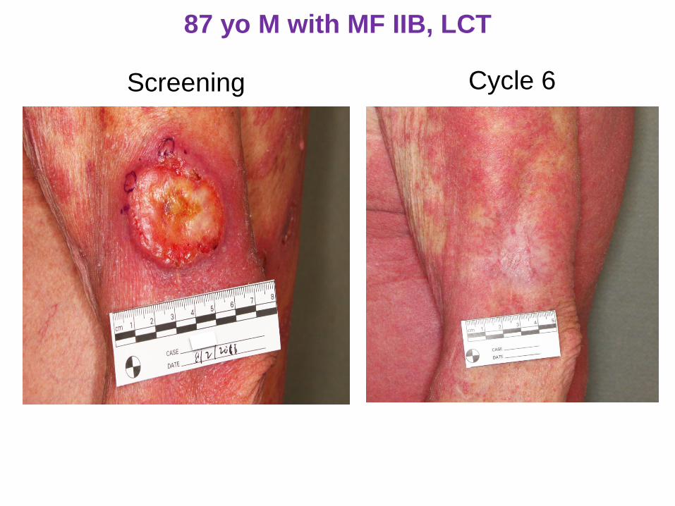
Cycle 6 Screening
87 yo M with MF IIB, LCT

Screening
Subject 12: 66 yo F with MF IVB, LCT w/
oropharyngeal involvement
Cycle 10
20% CD30

Screening Cycle 10

51 yo F stage IVA2 MF with LCT in skin/LNs:
response to brentuximab vedotin
Pre-treatment 12/20/2012 Post 2 cycles 1/29/2013
Phase III RCT in CTCL ongoing
for FDA approval

Tumor-directed killing
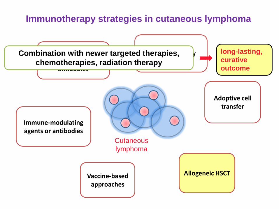
Road to a CURE How do we make the nice responses last?
Partnering with immunotherapy
Immunotherapy
% S
urv
ival
Time

Vaccine-based approaches
Immune-modulating agents or antibodies
Adoptive T-cell transfer
Immunotherapy strategies in cancer
Tumor-specific monoclonal antibodies
Allogeneic HSCT
Cytokine therapy
TILs
lymphoma
M

Vaccine-based approaches
Immune-modulating agents or antibodies
Adoptive T-cell transfer
Immunotherapy strategies in cancer
Tumor-specific monoclonal antibodies
Allogeneic HSCT
Cytokine therapy
TILs
lymphoma
M

1
2
3
Donor Cell Transplant
Replacement of Host Blood System
Lymphocytes
Donor Immune System to
destroy lymphoma cells
Sezary cells
Harnessing the graft-versus-lymphoma effect in
allo HSCT as the ultimate cellular immune therapy

A New Approach in Donor Cell Transplant
Non-Myeloablative Regimen with TLI/ATG
“Protective conditioning”
Mantle
field
Inverted Y
field
Total Lymphoid Irradiation
(TLI)
Anti-Thymocyte Globulin
(ATG, Rabbit anti-T cell antibodies)
Enable Donor Cells to Engraft
aGVHD reduced to 2-5% (vs. 20-65%)
NEJM 353:1321, 2005
Stanford study on going
TSEBT
+

Pre-TSEBT 3.0+ yr (NED, no GVHD)
Mycosis fungoides, stage IVA w/ LCT in skin/LNs: CR

Pre-TSEBT
CD4+/CD26-: 99%, abs 19,780
Sezary syndrome, stage IVA w/ LCT in skin/LNs: CR
2.0+ yr (NED, no GVHD)
CD4+/CD26-: normalized

Pre-transplant 2.0+ yr (NED, no GVHD)
Sezary syndrome, stage IVA w/ LCT in skin/LNs: CR
Vaccine-based approaches
Immune-modulating agents or antibodies
Adoptive cell transfer
Immunotherapy strategies in cutaneous lymphoma
Tumor-specific monoclonal antibodies
Allogeneic HSCT
Cutaneous
lymphoma
Cytokine therapy Combination with newer targeted therapies,
chemotherapies, radiation therapy
long-lasting,
curative
outcome

CTCL Management: Lessons Learned
Take home summary
• Clinical-pathologic correlation is ESSENTIAL for
diagnosis
• “OK” to be noncommittal of the diagnosis
– Follow and reassess; manage according to biologic
behavior
• Check the blood compartment for diagnostic data
– HTLV1 serology for ATLL
– Sezary flow when suspecting SS
• Advanced/refractory MF or SS IS curative
– Must balance risks and benefits of allo HSCT

Stanford Multidisciplinary Cutaneous Lymphoma Group
Holbrook Kohrt
Sunil Reddy
Ron Levy
Wen-Kai Weng
Sally Arai
Katherine Wolpin
BMT partners
Ranjana Advani
Med Onc partners
Rich
Hoppe
Michael
Krathen
Lynn
Million