crystals in brain and hyperoxaluria and...
TRANSCRIPT
J. clin. Path., 1977, 30, 16-18
Crystals in brain and meninges in primaryhyperoxaluria and oxalosisM. T. HAQQANI'
From the Department ofPathology, Stopford Building, University of Manchester
SUMMARY A case of primary hyperoxaluria and oxalosis with chronic renal failure, crystallinemyocarditis, and disseminated calcium oxalate crystal deposition in various tissues including thebrain and meninges is described. Deposition of crystals in brain and meninges is exceptionally rare
in primary oxalosis.
Primary hyperoxaluria and oxalosis is considered tobe a rare autosomal and recessively inherited dis-order (Watts, 1973) in which the rate of oxalateformation is greatly increased (Cochran et al., 1967)and disseminated systemic deposition of calciumoxalate crystals occurs in soft tissues and bones(Scowen et al., 1959). In this report, the occurrenceof calcium oxalate crystals is described in brain andmeninges in an elderly woman with primary hyper-oxaluria and oxalosis, a finding hitherto describedonly once before in the brain of a 5-week-old baby(Hughes, 1959).
Case report
A 50-year-old woman presented with anorexia,vomiting, and anuria of three weeks' duration.Hypertension for the previous 10 years had beencontrolled by hypotensive drugs. On examinationthe blood pressure was 190/95 mmHg; anaemia andsacral oedema were present. Laboratory data showedHb 8 g/dl, RBC 2-8 million/mm3, WBC 5400/mm3with a normal differential count, ESR 42 mm(Westergren), Na 125 mmol/l (125 mEq/l), K 4-1mmol/l (4-1 mEq/l), Cl 81 mmol/l (81 mEq/l), urea2-42 mg/l (242 mg/100 ml), and creatinine 2 mg/l(20 mg/100 ml). Peritoneal dialysis was carried outseveral times without significant clinical improve-ment. An open renal biopsy revealed tubularnecrosis and abundant crystalline debris in thetubules. Later she became hypotensive with brady-cardia and ECG changes of right bundle-branchblock and died the following day. The clinicaldiagnosis was that of renal failure due to essential'Present address: Walton Hospital, Liverpool L9 IAE.Received for publication 24 May 1976
16
hypertension and ischaemic heart disease. There wasno family history of oxalosis or of calcium oxalatestone formation.Postmortem examination revealed moderate
pleural and pericardial effusions, cardiomegaly(heart weight 440 g), left ventricular hypertrophy(LV weight 270 g; RV 70 g; ratio of left to rightventricular weights 4:1), slight to moderate athe-roma of the anterior descending branch of the leftcoronary artery, and coarsely granular contrac-ted kidneys with a gritty cut surface.
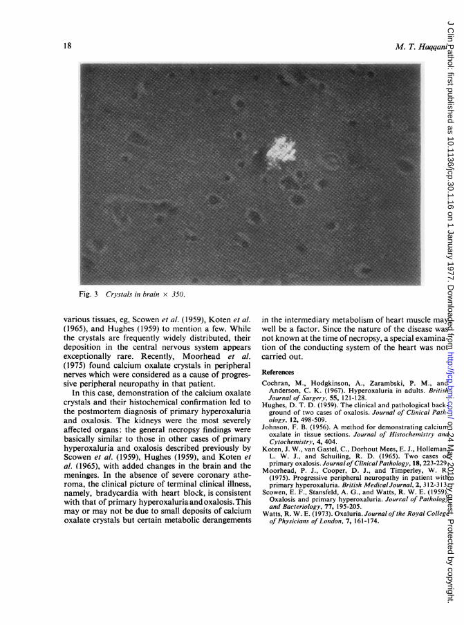
Histologically, all tissues examined revealedcalcium oxalate crystals, shown in sections usingpolarized light as clusters of yellow birefringentbroken plates, or sheaves with a radial rosette-likepattern, or masses of smaller wedge shapes, orisolated deposits. Histochemical confirmation wascarried out in the tissue sections by the techniquedescribed by Johnson (1956). The kidneys showedtubular sludging by the crystals, tubulorrhexis, andcrystals in interstitial tissue (Fig. 1). There werechanges of essential hypertension in the blood vesselswith crystalline deposition in their walls. In theheart, widespread deposition and a picture ofcrystalline myocarditis was noted. Deposition ofcrystals was present in the meninges (Fig. 2) and inwalls of congested meningeal vessels. Scanty butsignificant deposition was seen in the brain (Fig. 3)with focal mild microglial reaction to their presence.
Discussion
Deposition of calcium oxalate crystals in humantissues was first described by Lepoutre (1925) citedby Hughes (1959). A number of accounts have ap-peared in recent years describing their deposition in
on 24 May 2018 by guest. P
rotected by copyright.http://jcp.bm
j.com/
J Clin P
athol: first published as 10.1136/jcp.30.1.16 on 1 January 1977. Dow
nloaded from

Crystals in brain and meninges in primary hyperoxaluria and oxalosis
Fig. 1 Widespread crystalline deposition in the kidney x 150.
Fig. 2 Crystals in meninges x 350.
17
on 24 May 2018 by guest. P
rotected by copyright.http://jcp.bm
j.com/
J Clin P
athol: first published as 10.1136/jcp.30.1.16 on 1 January 1977. Dow
nloaded from
M. T. Haqqani
Fig. 3 Crystals in brain x 350.
various tissues, eg, Scowen et al. (1959), Koten et al.(1965), and Hughes (1959) to mention a few. Whilethe crystals are frequently widely distributed, theirdeposition in the central nervous system appearsexceptionally rare. Recently, Moorhead et al.(1975) found calcium oxalate crystals in peripheralnerves which were considered as a cause of progres-sive peripheral neuropathy in that patient.
In this case, demonstration of the calcium oxalatecrystals and their histochemical confirmation led tothe postmortem diagnosis of primary hyperoxaluriaand oxalosis. The kidneys were the most severelyaffected organs: the general necropsy findings werebasically similar to those in other cases of primaryhyperoxaluria and oxalosis described previously byScowen et al. (1959), Hughes (1959), and Koten etal. (1965), with added changes in the brain and themeninges. In the absence of severe coronary athe-roma, the clinical picture of terminal clinical illness,namely, bradycardia with heart block, is consistentwith that of primary hyperoxaluria and oxalosis. Thismay or may not be due to small deposits of calciumoxalate crystals but certain metabolic derangements
in the intermediary metabolism of heart muscle maywell be a factor. Since the nature of the disease wasnot known at the time of necropsy, a special examina-tion of the conducting system of the heart was notcarried out.
ReferencesCochran, M., Hodgkinson, A., Zarambski, P. M., and
Anderson, C. K. (1967). Hyperoxaluria in adults. BritishJournal of Surgery, 55, 121-128.
Hughes, D. T. D. (1959). The clinical and pathological back-ground of two cases of oxalosis. Journal of Clinical Path-ology, 12, 498-509.
Johnson, F. B. (1956). A method for demonstrating calciumoxalate in tissue sections. Journal of Histochemistry andCytochemistrv, 4, 404.
Koten, J. W., van Gastel, C., Dorhout Mees, E. J., Holleman,L. W. J., and Schuiling, R. D. (1965). Two cases ofprimary oxalosis. Journal of Clinical Pathology, 18, 223-229.
Moorhead, P. J., Cooper, D. J., and Timperley, W. R.(1975). Progressive peripheral neuropathy in patient withprimary hyperoxaluria. British Medical Journal, 2, 312-313.
Scowen, E. F., Stansfeld, A. G., and Watts, R. W. E. (1959).Oxalosis and primary hyperoxaluria. Journal of Pathologyand Bacteriology, 77, 195-205.
Watts, R. W. E. (1973). Oxaluria. Journal of the Royal Collegeof Physicians ofLondon, 7, 161-174.
18
on 24 May 2018 by guest. P
rotected by copyright.http://jcp.bm
j.com/
J Clin P
athol: first published as 10.1136/jcp.30.1.16 on 1 January 1977. Dow
nloaded from