critical care in surgery respiratory system care... · • lung abscess, emphyema • necrotizing...
TRANSCRIPT

CRITICAL CARE MANAGEMENTRESPIRATORY SYSTEM
‐B. SHIVRAJ

Topics
• ARDS• Ventilator and modes• Aspiration• Pneumothorax & hemothorax

Introduction
• Securing the airway.
• ABC of resuscitation
• Most vital organ
• Supplies oxygen‐> energy source for all cells

ARDS
• Non cardiogenic pul edema/ AHMD/ CLS/ SLS/ shock lung.
• Definition
• Aetiology
• Pathology
• Severity scoring
• Management

ARDSDefinition
• Severe, acute lung injury involving diffuse alveolar damage, increased microvascularpermeability and non cardiogenicpulmonary edema
• Acute refractory hypoxemia• High mortality‐ 40%‐60%

ARDS Criteria
• Acute onset of respiratory failure
• Bilateral infiltrate on CXR(some cases do present unilaterally or with pleural effusion
• PCWP <18 or absence of left atrial htn,
• PaO2/FiO2 < 200

ARDS mechanism of lung injury
• Activation of inflammatory mediators and cellular components resulting in damage to capillary endothelial and alveolar epithelial cells
• Increased permeability of alveolar capillary membrane
• Influx of protein rich edema fluid and inflammatory cells into air spaces
• Dysfunction of surfactant

Surfactant


Stages of ARDS
• 1. Exudative (acute) phase - 0- 4 days• 2. Proliferative phase - 4- 8 days• 3. Fibrotic phase - >8 days• 4. Recovery

ARDSTreatment
• Ventilator‐induced lung injury: oxygen toxicity was one of the most important factors in the progression of ARDS and resultant mortality. Recently, it was found that high volume(volutrauma) and high press(barotrauma) are equally if not more detrimental to these pts
• Treatment strategy is one of low volume and high frequency ventilation(ARDSNet protocol)
• Search for and treat the underlying cause• Treat abdominal infection promptly with antibiotics/ surgery• Ensure adequate nutrition• DVT prophylaxis• Prevent and treat any nosocomial infection• Consider short course of high dose steroids (20‐60mg prednisoline) in
patients with chronic unresolving disease.
When all else fails..
• Prone(gravitation atelectasis).
• Inhaled nitric oxide(marginal benefit)
• High frequency oscillation ventillation
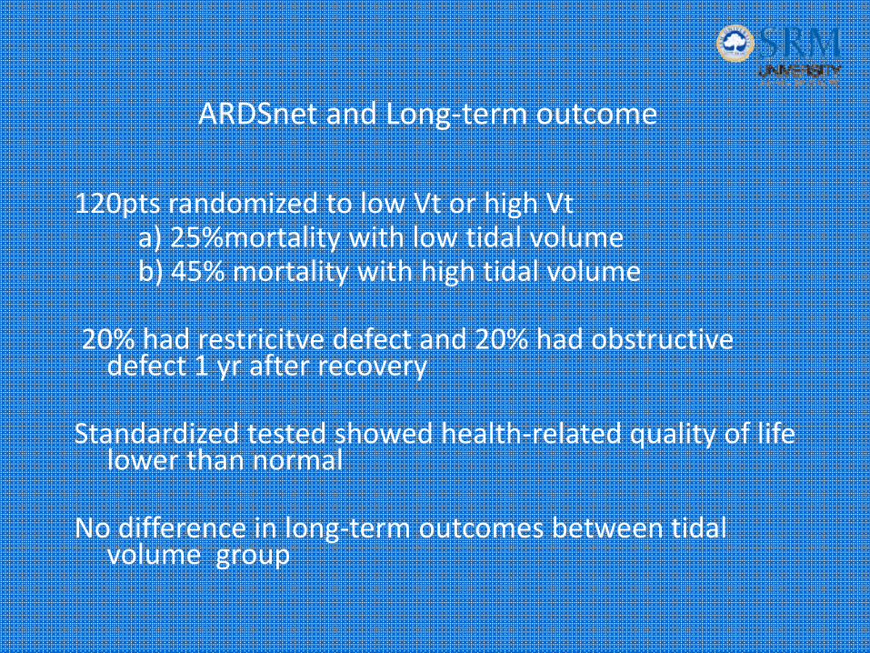
ARDSnet and Long‐term outcome
120pts randomized to low Vt or high Vta) 25%mortality with low tidal volumeb) 45% mortality with high tidal volume
20% had restricitve defect and 20% had obstructive defect 1 yr after recovery
Standardized tested showed health‐related quality of life lower than normal
No difference in long‐term outcomes between tidal volume group

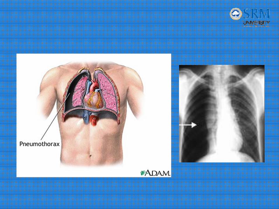
Pneumothorax
• Definition –
“Pneumo”‐ gas“Thorax” – chest cavity
• Pathophysiology –– Pleural space
• Baseline (‐) pressure space
• Parietal Pleura
• Visceral Pleura
– Normal inspiration• Diaphragm
• Transmit (‐) Pressure
– Pathologic inspiration• Excess gas disrupts transmission of (‐) pressure

Types of Pneumothorax
• Spontaneous Pneumothorax– Primary ‐ rupture of subpleural bleb– Secondary ‐ underlying lung/pleural disease
• Emphysema. chronic bronchitis, asthma, TB.
• Traumatic Pneumothorax: – Open
• Chest wall is penetrated : outside air enters pleural space
– Closed• Chest wall is intact ‐> Fractured rib

Types of Pneumothorax
• Tension Pneumothorax– Injury to pleura creates a tissue flapthat opens on inspiration and closes on expiration(ball valve effect).
– Mediastinal shift
• Variations– Hemo‐thorax – Chylo‐thorax
• Injury to thoracic duct
– Empyema• Synpneumonic effusions in community‐acquired pneumonia

Pneumothorax• Signs and symptoms of a pneumothoraxinclude:– Sudden, sharp chest pain
– Shortness of breath
– Chest tightness
– Tachycardia
– Rapid, shallow breaths

Physical Exam ‐ Signs
• Unstable patients vs. Stable patients– Vital Signs
• Asymmetric chest expansion
• Deviated trachea
• Diminished breath sounds unilaterally
• Hyper‐resonance unilaterally
• Decreased tactile fremitus
Treatment
• Small pneumothorax– Resolve over days to weeks– Supplemental oxygen and observation
• Tension pneumothorax– Immediate decompression via chest tube or needle thoracostomy
• Spontaneous pneumothorax– Asymptomatic – outpatient, follow up with serial CXR– Symptomatic – inpatient, chest tube– Recurrent pneumothorax – CT to evaluate need for thoracotomy


Tension PTX
Normal
Tension PTXPTX
R L
heart
Lung
Heart
Airleak

Hemothorax
• Causes of a Spontaneous Hemothorax
– Pulmonary: PE, infarction, Tb, AVM’s
– Pleural: torn adhesions
– Neoplastic: primary, metastatic (melanoma)
– Blood Dyscrasias: thrombocytopenia, hemophilia, anticoagulation
– Thoracic Pathology: ruptured aorta, dissection
– Abdominal Pathology: pancreatic pseudocyst, hemoperitoneum

Hemothorax
• Radiograph showing left sided hemothorax

HemothoraxThe Pathophysiologic Process
• the accumulation of pleural blood forms a stable clot
• ventilation & oxygenation
becomes impaired
• mechanical compression of the lung parenchyma
• mediastinal shift
• flattening of the hemidiaphragm

Hemothorax
The Pathophysiologic Process
• over time, the clot is partially‐absorbed, leaving behind loculated fluid and fibrinous septations
• macro‐fibrin deposition begins to provide a structural framework
• this “peel” slowly contracts to entrap the underlying lung

Hemothorax
• General Management Options
– thoracentesis: bedside / ultrasound‐guided / C.T.‐guided
– thoracostomy drainage: “”the MAINSTAY””
– thoracotomy: massive hemothorax / instability / chronic hemothorax
– local fibrinolytic therapy: urokinase (1000 IU/ml) in 150cc solution
Hemothorax‐ drainage
Undrained hemothorax increases the risk
of empyema & fibrothorax
• Large collections should be drained slowly to minimize the development of re‐expansion‐pulmonary‐edema

ICD insertion
• Arm by the ear
• 4‐5th ICS in the mid‐axillaryline
• Directed upwards
• Position checked with rpt CXR


Aspiration
• Most dreaded complication by anesthetist.• Pre‐disposing factors:
unconscious; loss of gag reflexes; alcoholic; pt on RT feeding; hiatus hernia
• Aspirate‐> food;gastric contents; oropharyngeal secretions

Presentation
• Tachycardia; tachypnea; breathing difficulty; cyanosis.
• R/S‐> crackles, wheeze, pink frothy spuptum
• CXR‐> initially – N ; later – consolidation
• Prevention ‐>1. feeding in propped up position; 2. lying on left pos to prevent asp of contents in unconscious pts.

Treatment
• FB‐> removal (as appropriate)
• O2 support ; propped up
• Antibioic cover to prevent secondary infection.
• Suction (under laryngoscopy/ bronchoscopy/ in ET tube)

Complications
• Aspiration pneumonitis
• Secondary bacterial infections
• Lung abscess, emphyema
• Necrotizing pneumonia

MECHANICAL VENTILATION•Theory
• Ventilation vs. Oxygenation• Pressure Cycling vs. Volume Cycling
•Modes•Ventilator Settings• Indications to intubate• Indications to extubate•Management algorithim•FAQs

Principles (1): VentilationThe goal of ventilation is to facilitate CO2 release and maintain normal PaCO2
•Minute ventilation (VE)• Total amount of gas exhaled/min.• VE = (RR) x (TV)• VE comprised of 2 factors
• VA = alveolar ventilation• VD = dead space ventilation
• VD/VT = 0.33• VE regulated by brain stem, responding to pH and PaCO2.
•Ventilation in context of ICU• Increased CO2 production
• fever, sepsis, injury, overfeeding• Increased VD
• atelectasis, lung injury, ARDS, pulmonary embolism• Adjustments: RR and TV

Principles (2): OxygenationThe primary goal of oxygenation is to maximize O2 delivery to blood (PaO2)
•Alveolar-arterial O2 gradient (PAO2 – PaO2)• Equilibrium between oxygen in blood and oxygen in alveoli• A-a gradient measures efficiency of oxygenation• PaO2 partially depends on ventilation but more on V/Q
matching.
•Oxygenation in context of ICU• V/Q mismatching
• Patient position (supine)• Airway pressure, pulmonary parenchymal disease, small-airway
disease• Adjustments: FiO2 and PEEP

Pressure ventilation vs. volume ventilationPressure-cycled modes deliver a fixed pressure at variable volume.Volume-cycled modes deliver a fixed volume at variable pressure.
•Pressure-cycled modes• Pressure Support Ventilation (PSV)• Pressure Control Ventilation (PCV)• CPAP• BiPAP
•Volume-cycled modes• Control• Assist• Assist/Control• Intermittent Mandatory Ventilation
(IMV)• Synchronous Intermittent
Mandatory Ventilation (SIMV)
Volume-cycled modes have the inherent risk ofvolutrauma.

Pressure Support Ventilation (PSV)Patient determines RR, VE, inspiratory time – a purely spontaneous mode
• Parameters• Triggered by pt’s own breath• Limited by pressure• Affects inspiration only
• Uses• Complement volume-cycled
modes (i.e., SIMV)
• PSV alone• Used alone for recovering
intubated pts who are notquite ready for extubation
• BiPAP (CPAP plus PS)
PSV is most often used together with other volume-cycled modes.PSV provides sufficient pressure to overcome the resistance of the ventilatortubing, and acts during inspiration only.

Pressure Control Ventilation (PCV)Ventilator determines inspiratory time – no patient participation
•Parameters• Triggered by time• Limited by pressure• Affects inspiration only.
•Disadvantages• Requires frequent adjustments
to maintain adequate VE
• Pt with noncompliant lungsmay require alterations ininspiratory times to achieveadequate TV

CPAP and BiPAPCPAP is essentially constant PEEP; BiPAP is CPAP plus PS
•Parameters• CPAP – PEEP set at 5-10 cm H2O• BiPAP – CPAP with Pressure Support (5-20 cm H2O)• Shown to reduce need for intubation and mortality in
COPD pts
•Indications• When medical therapy fails (tachypnea, hypoxemia,
respiratory acidosis)• Use in conjunction with bronchodilators, steroids,
oral/parenteral steroids, antibiotics to prevent/delay intubation
• Weaning protocols• Obstructive Sleep Apnea

Assist/Control Mode
•Control Mode• Pt receives a set number of
breaths and cannot breathebetween ventilator breaths
• Similar to Pressure Control
•Assist Mode• Pt initiates all breaths, but
ventilator cycles in at initiationto give a preset tidal volume
• Pt controls rate but alwaysreceives a full machine breath
•Assist/Control Mode• Assist mode unless pt’s respiratory
rate falls below preset value• Ventilator then switches to control
mode• Rapidly breathing pts can
overventilate and induce severerespiratory alkalosis andhyperinflation.
Ventilator delivers a fixed volume

IMV and SIMV Volume-cycled modes typically augmented with Pressure Support
• IMV• Pt receives a set number of
ventilator breaths• Different from Control: pt can
initiate own (spontaneous) breaths• Different from Assist: spontaneous
breaths are not supported by machine with fixed TV
• Ventilator always delivers breath, even if pt exhaling
•SIMV• Most commonly used mode• Spontaneous breaths and
mandatory breaths• If pt has respiratory drive, the
mandatory breaths are synchronized with the pt’s inspiratory effort

Ventilator settings to improve <oxygenation>
•FIO2• Simplest maneuver to quickly increase PaO2
• Long-term toxicity-• Free radical damage
• Inadequate oxygenation despite 100% FiO2usually due to pulmonary shunting• Collapse – Atelectasis• Pus-filled alveoli – Pneumonia• Water/Protein – ARDS• Water – CHF• Blood - Hemorrhage
PEEP and FiO2 are adjusted in tandem

Ventilator settings to improve <oxygenation>
•PEEP • Increases FRC
• Prevents progressive atelectasis and intrapulmonary shunting
• Prevents repetitive opening/closing (injury)
• Enables maintenance of adequate PaO2at a safe FiO2 level
• Disadvantages• Increases intrathoracic pressure (may
require pulmonary a. catheter)• May lead to ARDS• Rupture: PTX, pulmonary edema
PEEP and FiO2 are adjusted in tandem
Oxygen delivery (DO2), not PaO2, should beused to assess optimal PEEP.

Vent settings to improve <ventilation>
•Respiratory rate• Max RR at 35 breaths/min
• Efficiency of ventilation decreases with increasing RR• Decreased time for alveolar emptying
•TV• Goal of 10 ml/kg• Risk of volutrauma
RR and TV are adjusted to maintain VE and PaCO2
•PIP• Elevated PIP suggests need for
switch from volume-cycled to pressure-cycled mode
• Maintained at <45cm H2O to minimize barotrauma

Alternative Modes
• I:E inverse ratio ventilation (IRV)• In ARDS and severe hypoxemia• Prolonged inspiratory time (3:1) leads to
better gas distribution with lower PIP• Elevated pressure improves alveolar
recruitment
• Prone positioning• Addresses dependent atelectasis• Relief of diaphragmatic pressure from
abdominal viscera• Improved drainage of secretions• Logistically difficult*
• No mortality benefit demonstrated.
• High-Frequency OscillatoryVentilation (HFOV)• High-frequency, low amplitude
ventilation
• Avoids repetitive alveolar open andclosing that occur with low airwaypressures
• Avoids overdistension that occursat high airway pressures
• Well tolerated, consistentimprovements in oxygenation.
• Disadvantages• Potential hemodynamic compromise• Pneumothorax• Neuromuscular blocking agents

Non Invasive Positive Pressure Ventilation– Deliver PS and CPAP via tight fitting mask
(BiPAP: bi‐level positive airway pressure)
– Dyspnea protocol
– May still need sedation

Treatment of respiratory failure
•Prevention• Incentive spirometry• Mobilization• Humidified air• Pain control• Turn, cough, deep breathe
•Treatment• Medications
• Albuterol• Theophylline• Steroids
• CPAP, BiPAP.• Intubation
The critical period before the patient needs to be intubated

Indications for intubation
•Criteria• Clinical deterioration• Tachypnea: RR >35• Hypoxia: pO2<60mm Hg • Hypercarbia: pCO2 > 55mm Hg• Minute ventilation<10 L/min• Tidal volume <5-10 ml/kg• Negative inspiratory force <
25cm H2O (how strong the pt can suck in)
• Initial vent settings• FiO2 = 50%• PEEP = 5cm H2O• RR = 12 – 15 breaths/min• VT = 10 – 12 ml/kg
• COPD = 10 ml/kg (prevent overinflation)
• ARDS = 8 ml/kg (prevent volutrauma)• Permissive hypercapnea
• Pressure Support = 10cm H2O
How the values trend should significantly impact clinical decisions

Extubation
• Weaning– Is the cause of respiratory failure gone or getting better ?
– Is the patient well oxygenated and ventilated ?
–Can the heart tolerate the increased work of breathing ?

• (cont.)–decrease the PEEP (4‐5)
–decrease the rate(<30/min)
–decrease the PIP (as needed)
– “AWAKE PATIENT”
• What you want to do is decrease what the ventilator does and see if the patient can make up the difference….

Indications for extubation
•Clinical parameters• Resolution/Stabilization of
disease process• Hemodynamically stable• Intact cough/gag reflex• Spontaneous respirations• Acceptable vent settings
• FiO2< 50%, PEEP < 8, PaO2> 75, pH > 7.25
•General approaches• SIMV Weaning
• Spontaneous breathing trials• Demonstrated to be superior
No weaning parameter completely accurate when used alone
Numerical Parameters
Normal Range
Weaning Threshold
P/F > 400 > 200
Tidal volume 5 - 7 ml/kg 5 ml/kg
Respiratory rate 14 - 18 breaths/min < 40 breaths/min
Vital capacity 65 - 75 ml/kg 10 ml/kg
Minute volume 5 - 7 L/min < 10 L/min

Spontaneous Breathing Trials
•Settings• PEEP = 5, PS = 0 – 5, FiO2 < 40%• Breathe independently for 30 –
120 min• ABG obtained at end of SBT
•Failed SBT Criteria• RR > 35 for >5 min• SaO2 <90% for >30 sec• HR > 140• Systolic BP > 180 or < 90mm Hg• Sustained increased work of
breathing• Cardiac dysrhythmia• pH < 7.32
SBTs do not guarantee that airway is stable or pt can self-clear secretions
Causes of Failed SBTs
Treatments
Anxiety/Agitation Benzodiazepines or haldol
Infection Diagnosis and tx
Electrolyte abnormalities (K+, PO4-)
Correction
Pulmonary edema, cardiac ischemia
Diuretics and nitrates
Deconditioning, malnutrition
Aggressive nutrition
Neuromuscular disease Bronchopulmonary hygiene, early consideration of trach
Increased intra-abdominal pressure
Semirecumbent positioning, NGT
Hypothyroidism Thyroid replacement
Excessive auto-PEEP (COPD, asthma)
Bronchodilator therapy

Continued ventilation after successful SBT
•Commonly cited factors•Altered mental status and inability to
protect airway•Potentially difficult reintubation•Unstable injury to cervical spine•Likelihood of return trips to OR•Need for frequent suctioning
Inherent risks of intubation balanced against continued need for intubation

Ventilator management algorithimInitial intubation• FiO2 = 50%• PEEP = 5
• RR = 12 – 15• VT = 8 – 10 ml/kg
SaO2 < 90% SaO2 > 90%
SaO2 > 90%• Adjust RR to maintain PaCO2 = 40• Reduce FiO2 < 50% as tolerated• Reduce PEEP < 8 as tolerated• Assess criteria for SBT daily
SaO2 < 90%• Increase FiO2 (keep SaO2>90%)• Increase PEEP to max 20• Identify possible acute lung injury• Identify respiratory failure causes
Acute lung injury
No injury
Fail SBT
Acute lung injury• Low TV (lung-protective) settings
• Reduce TV to 6 ml/kg• Increase RR up to 35 to keep
pH > 7.2, PaCO2 < 50• Adjust PEEP to keep FiO2 < 60%
SaO2 < 90% SaO2 > 90%
SaO2 < 90%• Dx/Tx associated conditions
(PTX, hemothorax, hydrothorax)• Consider adjunct measures
(prone positioning, HFOV, IRV)
SaO2 > 90%• Continue lung-protective
ventilation until:• PaO2/FiO2 > 300• Criteria met for SBT
Persistently fail SBT• Consider tracheostomy• Resume daily SBTs with CPAP or
tracheostomy collar
Pass SBT
Airway stableExtubate
Intubated > 2 wks
• Consider PSV wean (gradualreduction of pressure support)
• Consider gradual increases in SBTduration until endurance improves
Prolonged ventilator dependence
Pass SBT
Pass SBT
Airway stable
Modified from Sena et al, ACS Surgery: Principles and Practice (2005).

Need for tracheostomy
•Advantages• Issue of airway stability can be
separated from issue of readiness for extubation• May quicken decision to extubate
• Decreased work of breathing• Avoid continued vocal cord injury• Improved bronchopulmonary
hygiene• Improved pt communication
•Disadvantages• Long term risk of tracheal stenosis• Procedure-related complication
rate (4% - 36%)
Prolonged intubation may injure airway and cause airway edema
1 - Vocal cords. 2 - Thyroid cartilage. 3 - Cricoidcartilage. 4 - Tracheal cartilage. 5 - Balloon cuff.

Complications
–Oxygen toxicity–Barotrauma/ volutrauma
–Ventilator Associated Pneumonia
–Sinusitis–Risks from associated devices (CVLs, A‐lines)
–Unplanned Extubation

Foreign body
• Acute emergency
• m/c children
• Present with gasping; choking; cyanosis; acute resp difficulty; tacycardia.
• Heimlich manuvoure
• Visualisation ‐>

Magill forceps

Tracheostomy
• Emergency / elective
• 2‐4th trahceal rings
• Indications : securing airway, prolonged ventilation, for extubation.
• Procedure
• Post op care


Laryngoscope with Macintosh blade

ORO PHARYNGEAL AIRWAY
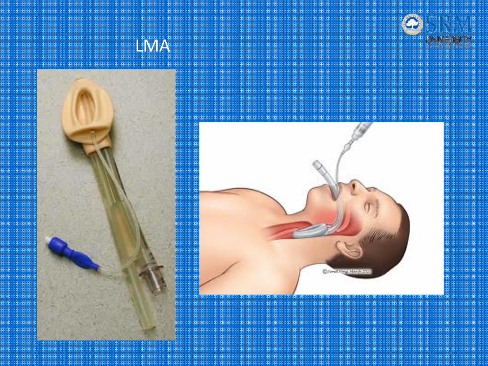
LMA

Combitube

Cricothrodotomy
• Or mini tracheosotomy
• Over the crico‐thyroid membrane
• As an emergency precodure to secure airway


THANK YOU.