creport primary mucosal melanoma of the...
TRANSCRIPT
Case ReportPrimary Mucosal Melanoma of the Stomach
O. Phillips ,1 A. Higdon,2 R. Colaco,2 and H. Fish3
1St. George’s University School of Medicine, West Indies, Grenada2Department of Surgery, Trinitas Regional Medical Center, Elizabeth, NJ, USA3Division of Surgical Pathology, Trinitas Regional Medical Center, Elizabeth, NJ, USA
Correspondence should be addressed to O. Phillips; [email protected]
Received 12 January 2018; Revised 17 June 2018; Accepted 16 July 2018; Published 12 August 2018
Academic Editor: Yoshihiro Moriwaki
Copyright © 2018 O. Phillips et al. This is an open access article distributed under the Creative Commons Attribution License,which permits unrestricted use, distribution, and reproduction in any medium, provided the original work is properly cited.
Considered to be rare, mucosal melanomas are rare type of melanoma that are found on mucosal surfaces and are primary ormetastatic in origin.We report a case of a 66-year-old Hispanic female who presented with vague abdominal pain and upon furtherendoscopic work-up revealed 2 gastric lesions. Endoscopic biopsy results revealed gastric melanoma in the distal lesion. A PET/CTscan indicated it to be suspicious for the primary site of metastasis but was ultimately diagnosed as a benign nevus on biopsy. Anextensive clinical exam showed no other probable sites of origin. The patient underwent a subtotal Billroth II gastrectomy andenterostomy tube placement for temporary feeding. Primary melanoma of the stomach is an exceptionally rare occurrence withlimited cases that can be accounted for in literature; thus we report this case for review.
1. Introduction
Melanoma is the 6thmost common cancer in North America[1]. The incidence of cutaneous melanomas continues toincreases in the United States and is correlated with exposureto ultraviolet radiation in non-Hispanic whites [2]. However,exposure to ultraviolet light is not seen as a risk factor inmucosal melanomas since the mucosa is not exposed to UVlight. No other risk factor for mucosal melanomas has beenclearly identified except for those originating from the oralcavity that has been associated with cigarette smoking [3, 4].Even though melanocytes are predominately cutaneous, theycan be found in various other locations such as the oralpharynx, vulvovaginal, and anorectum [3].
As stated, mucosal melanomas are found to be primaryor secondary in nature. Primary mucosal melanomas areusually seen in older populations with an average age of 70years and accounts for one percent of all melanomas [5–7].Primary sites of mucosal melanomas can also occur alongthe gastrointestinal tract, however; they are more commonlyin the oropharynx [32.8%], anal canal [31.4%], and rectum[22.2%] [3, 8]. They are more rarely found in esophagus[5.9%], stomach [2.7%], and small bowel [2.3%] [3, 8]. Mostlesions found along the gastrointestinal tract are metastatic,with the most frequent locations of metastasis being the
small intestine, followed by the colon and stomach at thetime of presentation [9, 10]. It is rare to find primary gastricmelanomas and specific diagnostic criteria are needed inorder to be conclusive [11]. The pathogenesis and etiologyof mucosal melanomas have not been clearly identified asmelanocytes are not part of the innate cellular constitutionof the stomach epithelium [12]. To date, the knowledge of anycontributing risk factors continues to be elusive [8]. In generalmucosal melanomas are considered to have a poor prognosiswith a five-year survival of twenty-five percent [5].
2. Case Report
A66-year-oldHispanic female presentedwith vague abdomi-nal pain and exertional chest pain. She had a ten-year historyof worsening epigastric pain attributed to gastritis that wastreated with Dexilant. A general physical exam was doneand did not reveal any significant abnormalities; she alsodenied any fevers, chills, weight loss/gain, or change in bowelhabits. Environmental history was noncontributory as shedenied extensive sun-exposure or use of tanning beds andreported that she regularlywould use sunscreenwith a 30 SPF.Still, there was an extensive past medical history for anemia,diabetes mellitus type II, coronary artery disease, asthma,
HindawiCase Reports in Gastrointestinal MedicineVolume 2018, Article ID 6040693, 5 pageshttps://doi.org/10.1155/2018/6040693

2 Case Reports in Gastrointestinal Medicine
Figure 1: Left: gross whole specimen of the subtotal gastrectomy specimen, 14 cm in length. Preoperatively marked on endoscopy with inkalong the greater curvature. Right: proximal lesion near the serosa measured to be 1.1cm x 1 cmwas found to be normal gastric mucosa. Distaland proximal margins of the resection were found to be negative.
Figure 2: Subtotal gastrectomy distal lesion measuring 0.7 cm x0.7 cm x 0.5 cm.The lesion was found to be malignant melanoma ofthe stomach that is invasive into the submucosa. No lymphovascularinvasion was identified. Six negative lymph nodes appreciated withthe specimen.
hypertension, hyperlipidemia, hemorrhoids, and osteoarthri-tis. She also had a broad surgical history, involving a coronaryartery stent in the left anterior descending artery [2016], per-cutaneous transluminal coronary angioplasty [2016], tuballigation [1985], cholecystectomy [2007], bladder prolapserepair [2012], rectal prolapse repair [2012], hysterectomy[2012], and total right knee replacement [2013].
There is no report of primary tobacco use; however, shedoes report an extensive second-hand smoke exposure as aresult of her biological father. Her family history is significantfor multiple cancers, with her father dying of cancer relatedcomplications from a head and neck malignancy at the ageof 65. The patient’s sister died of breast cancer at the age of44 and half paternal sister also died of breast cancer at theage of 38. Lastly, her brother died of colon cancer at the ageof 70 and her mother’s death at the age of 67 was due to amyocardial infarction unrelated to cancer. Interestingly, hereldest daughter was diagnosed with melanoma found in acutaneous lesion, in 2016. Her physical exam did not revealany concerning skin lesions or palpable abdominal lesionsand her vitals were stable.
A near total gastrectomy was performed and multipleadhesions from prior surgeries were divided at the beginning
of the procedure. A thorough surgical exploration of theabdomen demonstrated no lesions of the liver. The gastriclesions, having been preoperatively marked on endoscopy,were inspected.The proximal lesionwas near theGE junctionand another was located more distally closer to the antrum.Figure 1 demonstrates the proximal lesion that was locatednear the greater curvature of the stomach. Figure 2 displaysthe distal lesion marked preoperatively located near theantrum of the stomach with no surrounding necrosis orbleeding but was significant for raised mucosal margins in awheel pattern.
Reconstruction was performed with a Billroth II anasto-mosis. Due to the almost complete removal of the stomach,a pouch was created via a jejunojejunostomy. A temporaryfeeding tube was placed distal to the anastomosis. No bloodwas transfused through this procedure and the estimatedblood loss was 200ml. The pathology associated with thegross specimens is seen in Figures 3 and 4.They are associatedwith the second lesion as the first was found to have normalgastric mucosa.The lesions were positive for three significantimmunological markers for melanoma,Melanin A, HMB-45,and S100 [13]. Though S100 has been found to have highersensitivity but much lower specificity for melanoma [13].
Her immediate postoperative course was unremarkableexcept for a fever of 101 F during the first 24 hours aftersurgery, which returned to 98.7∘F about 9 hours after the ini-tial fever spike in temperature. She remained afebrile for theremainder of her hospital stay. Overall, the patient progressedas expected postoperatively and tolerated her temporary tubefeedings well. An upper GI series on postoperative day 5showed no anastomosis leakage; thus a postgastrectomy dietwas started soon thereafter. After a continued unremarkablepostoperative course shewas discharged onpostoperative day7 to a subacute rehab.
3. Discussion
Mucosal melanomas are a very rare type of melanoma andtheir etiology and pathogenesis continue to remain elusive.Current literature does not have specific staging for primarymucosal melanomas, though the American Joint Committeeon Cancer consider mucosal melanomas of any origin are

Case Reports in Gastrointestinal Medicine 3
(a) (b)
(c) (d)
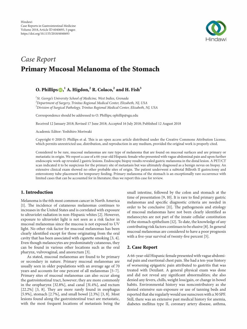
Figure 3: Histological Specimens of Gastric Melanoma. Histological sections of gastrectomy specimen demonstrating primary gastricmelanoma. (a) Right side of the image depicts neoplastic cells that do not invade past the submucosa that still retains normal gastricarchitecture (H&E x100). (b) Primary gastric melanoma positive stain for Melanin A (H&E x100). (c) Primary gastric melanoma positivestain for HMB-45 (H&E x100). (d) Malignant melanocytic neoplasm producing brown melanin pigment with a cherry red nucleoli andmitotic figures (H&E x 400).
Table 1: Basic Classification System of Mucosal Melanoma. AJCC TNM Classification for Mucosal Melanoma.
Primary TumorT3 Mucosal DiseaseT4a Moderately advanced disease. Tumor involving deep soft tissueT4b Very advanced disease
Regional Lymph Node InvolvementNX Regional Lymph nodes cannot be assessedN0 No regional lymph node metastasisN1 Regional lymph node metastases present
Distant MetastasisM0 No distant metastasis presentM1 Distant metastasis
applicable to the current guidelines used for the followingmucosal melanomas: head, neck, and vulva (11). This classi-fication system is depicted in Table 1(11).
Lagoudianakis et al. established specific diagnostic crite-ria for primary malignant melanoma of the gastrointestinaltract [11]. The criteria consist of a lack of any primary sites ofmelanoma; the patient should have no history or concurrentskin or any other organ removal of a melanocytic lesion, alack of any adjacent organ involvement, and finally diseasefree survival for at least 12 months after surgical resection
[11]. The last criterion is of critical importance in order toclearly distinguish between primary and metastatic lesionssince “50% of patients with stage IV melanoma or visceraldisease from unknown primary will have died at 12 monthsfrom diagnosis” [11]. This further confirms our diagnosissince at this point in time the patient is currently thirteenmonths postop.
Additionally, our patient’s lack of physical exam findingswith a positive histopathology for S100, Melanin A, andHMB-45 suggests that this patient has a true primary gastric
4 Case Reports in Gastrointestinal Medicine
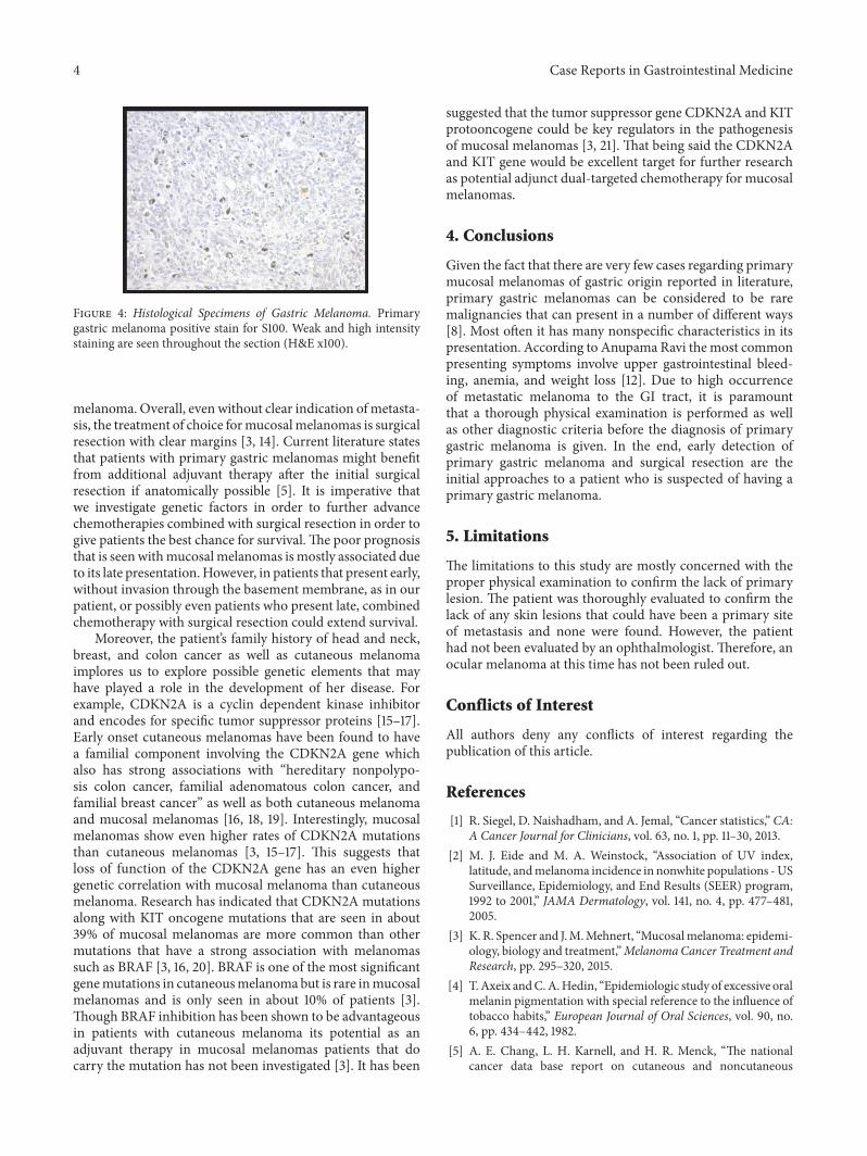
Figure 4: Histological Specimens of Gastric Melanoma. Primarygastric melanoma positive stain for S100. Weak and high intensitystaining are seen throughout the section (H&E x100).
melanoma. Overall, even without clear indication ofmetasta-sis, the treatment of choice formucosalmelanomas is surgicalresection with clear margins [3, 14]. Current literature statesthat patients with primary gastric melanomas might benefitfrom additional adjuvant therapy after the initial surgicalresection if anatomically possible [5]. It is imperative thatwe investigate genetic factors in order to further advancechemotherapies combined with surgical resection in order togive patients the best chance for survival.The poor prognosisthat is seenwithmucosalmelanomas ismostly associated dueto its late presentation.However, in patients that present early,without invasion through the basement membrane, as in ourpatient, or possibly even patients who present late, combinedchemotherapy with surgical resection could extend survival.
Moreover, the patient’s family history of head and neck,breast, and colon cancer as well as cutaneous melanomaimplores us to explore possible genetic elements that mayhave played a role in the development of her disease. Forexample, CDKN2A is a cyclin dependent kinase inhibitorand encodes for specific tumor suppressor proteins [15–17].Early onset cutaneous melanomas have been found to havea familial component involving the CDKN2A gene whichalso has strong associations with “hereditary nonpolypo-sis colon cancer, familial adenomatous colon cancer, andfamilial breast cancer” as well as both cutaneous melanomaand mucosal melanomas [16, 18, 19]. Interestingly, mucosalmelanomas show even higher rates of CDKN2A mutationsthan cutaneous melanomas [3, 15–17]. This suggests thatloss of function of the CDKN2A gene has an even highergenetic correlation with mucosal melanoma than cutaneousmelanoma. Research has indicated that CDKN2A mutationsalong with KIT oncogene mutations that are seen in about39% of mucosal melanomas are more common than othermutations that have a strong association with melanomassuch as BRAF [3, 16, 20]. BRAF is one of the most significantgenemutations in cutaneousmelanoma but is rare inmucosalmelanomas and is only seen in about 10% of patients [3].Though BRAF inhibition has been shown to be advantageousin patients with cutaneous melanoma its potential as anadjuvant therapy in mucosal melanomas patients that docarry the mutation has not been investigated [3]. It has been
suggested that the tumor suppressor gene CDKN2A and KITprotooncogene could be key regulators in the pathogenesisof mucosal melanomas [3, 21]. That being said the CDKN2Aand KIT gene would be excellent target for further researchas potential adjunct dual-targeted chemotherapy for mucosalmelanomas.
4. Conclusions
Given the fact that there are very few cases regarding primarymucosal melanomas of gastric origin reported in literature,primary gastric melanomas can be considered to be raremalignancies that can present in a number of different ways[8]. Most often it has many nonspecific characteristics in itspresentation. According to Anupama Ravi themost commonpresenting symptoms involve upper gastrointestinal bleed-ing, anemia, and weight loss [12]. Due to high occurrenceof metastatic melanoma to the GI tract, it is paramountthat a thorough physical examination is performed as wellas other diagnostic criteria before the diagnosis of primarygastric melanoma is given. In the end, early detection ofprimary gastric melanoma and surgical resection are theinitial approaches to a patient who is suspected of having aprimary gastric melanoma.
5. Limitations
The limitations to this study are mostly concerned with theproper physical examination to confirm the lack of primarylesion. The patient was thoroughly evaluated to confirm thelack of any skin lesions that could have been a primary siteof metastasis and none were found. However, the patienthad not been evaluated by an ophthalmologist. Therefore, anocular melanoma at this time has not been ruled out.
Conflicts of Interest
All authors deny any conflicts of interest regarding thepublication of this article.
References
[1] R. Siegel, D. Naishadham, and A. Jemal, “Cancer statistics,” CA:A Cancer Journal for Clinicians, vol. 63, no. 1, pp. 11–30, 2013.
[2] M. J. Eide and M. A. Weinstock, “Association of UV index,latitude, andmelanoma incidence in nonwhite populations -USSurveillance, Epidemiology, and End Results (SEER) program,1992 to 2001,” JAMA Dermatology, vol. 141, no. 4, pp. 477–481,2005.
[3] K. R. Spencer and J.M.Mehnert, “Mucosalmelanoma: epidemi-ology, biology and treatment,”MelanomaCancer Treatment andResearch, pp. 295–320, 2015.
[4] T.Axeix andC.A.Hedin, “Epidemiologic study of excessive oralmelanin pigmentation with special reference to the influence oftobacco habits,” European Journal of Oral Sciences, vol. 90, no.6, pp. 434–442, 1982.
[5] A. E. Chang, L. H. Karnell, and H. R. Menck, “The nationalcancer data base report on cutaneous and noncutaneous

Case Reports in Gastrointestinal Medicine 5
melanoma: a summary of 84,836 cases from the past decade,”Cancer, vol. 83, no. 8, pp. 1664–1678, 1998.
[6] M. Meleti, C. R. Leemans, R. De Bree, P. Vescovi, E. Sesenna,and I. Van Der Waal, “Head and neck mucosal melanoma:experience with 42 patients, with emphasis on the role ofpostoperative radiotherapy,” Head & Neck, vol. 30, no. 12, pp.1543–1551, 2008.
[7] R. P. Rapini, L. E. Golitz, R. O. Greer, E. A. Krekorian, and T.Poulson, “Primary malignant melanoma of the oral cavity. Areview of 177 cases,” Cancer, vol. 55, no. 7, pp. 1543–1551, 1985.
[8] M. Mihajlovic, S. Vlajkovic, P. Jovanovic, and V. Stefanovic,“Primary mucosal melanomas: a comprehensive review,” Inter-national Journal of Clinical and Experimental Pathology, vol. 5,no. 8, pp. 739–753, 2012.
[9] D.W.Ollila, R. Essner, L. A.Wanek, andD. L.Morton, “Surgicalresection for melanomametastatic to the gastrointestinal tract,”JAMA Surgery, vol. 131, no. 9, pp. 975–980, 1996.
[10] P. J. Capizzi and J. H. Donohue, “Metastatic melanoma of thegastrointestinal tract: a review of the literature,” ComprehensiveTherapy, vol. 1, pp. 20–23, 20.
[11] E. E. Lagoudianakis, E. Genetzakis, andM. Tsekouras, “Primarygastric melanoma: A case report,” World Journal of Gastroen-terology, vol. 12, no. 27, p. 4425, 2006.
[12] A. Ravi, “Primary gastric melanoma: A rare cause of uppergastrointestinal bleeding,” Journal of Gastroenterology and Hep-atology, vol. 4, no. 11, pp. 795–797, 2008.
[13] M. Shinohara, H. Deubner, and Z. B. Argenyi, “S100, HMB-45,andmelan-A negative primarymelanoma,”DermatologyOnlineJournal, vol. 15, no. 9, article no. 7, 2009.
[14] E. Hanna, F. Demonte, S. Ibrahim, D. Roberts, N. Levine, andM.Kupferman, “Endoscopic resection of sinonasal cancers withand without craniotomy,” Archives of Otolaryngology–Head &Neck Surgery, vol. 135, no. 12, p. 1219, 2009.
[15] L. Ai, K. K. Stephenson, W. Ling et al., “The p16(CDKN2a/INK4a) tumor-suppressor gene in head andneck squamous cell carcinoma: A promoter methylation andprotein expression study in 100 cases,” Modern Pathology, vol.16, no. 9, pp. 944–950, 2003.
[16] L. Si, X.Wang, and J. Guo, “Genotyping of mucosal melanoma,”Chinese Clinical Oncology, vol. 3, no. 3, pp. 33-34, 2014.
[17] J. A. Curtin, J. Fridlyand, T. Kageshita et al., “Distinct sets ofgenetic alterations in melanoma,” The New England Journal ofMedicine, vol. 353, no. 20, pp. 2135–2147, 2005.
[18] A. M. Goldstein, M. C. Fraser, W. H. Clark, and M. A. Tucker,“Age at diagnosis and transmission of invasive melanoma in 23families with cutaneous malignat melanoma/dysplastic nevi,”Journal of the National Cancer Institute, vol. 86, no. 18, pp. 1385–1390, 1994.
[19] M. Simons, J. Ferreira, R.Meunier, and S.Moss, “Primary versusmetastatic gastrointestinal melanoma: a rare case and reviewof current literature,” Case Reports in Gastrointestinal Medicine,vol. 2016, Article ID 2306180, 3 pages, 2016.
[20] J. A. Curtin, K. Busam, D. Pinkel, and B. C. Bastian, “Somaticactivation of KIT in distinct subtypes of melanoma,” Journal ofClinical Oncology, vol. 24, no. 26, pp. 4340–4346, 2006.
[21] F. G. Haluska, H. Tsao, H. Wu, F. S. Haluska, A. Lazar, and V.Goel, “Genetic alterations in signaling pathways in melanoma,”Clinical Cancer Research, vol. 12, no. 7, 2006.

Stem Cells International
Hindawiwww.hindawi.com Volume 2018
Hindawiwww.hindawi.com Volume 2018
MEDIATORSINFLAMMATION
of
EndocrinologyInternational Journal of
Hindawiwww.hindawi.com Volume 2018
Hindawiwww.hindawi.com Volume 2018
Disease Markers
Hindawiwww.hindawi.com Volume 2018
BioMed Research International
OncologyJournal of
Hindawiwww.hindawi.com Volume 2013
Hindawiwww.hindawi.com Volume 2018
Oxidative Medicine and Cellular Longevity
Hindawiwww.hindawi.com Volume 2018
PPAR Research
Hindawi Publishing Corporation http://www.hindawi.com Volume 2013Hindawiwww.hindawi.com
The Scientific World Journal
Volume 2018
Immunology ResearchHindawiwww.hindawi.com Volume 2018
Journal of
ObesityJournal of
Hindawiwww.hindawi.com Volume 2018
Hindawiwww.hindawi.com Volume 2018
Computational and Mathematical Methods in Medicine
Hindawiwww.hindawi.com Volume 2018
Behavioural Neurology
OphthalmologyJournal of
Hindawiwww.hindawi.com Volume 2018
Diabetes ResearchJournal of
Hindawiwww.hindawi.com Volume 2018
Hindawiwww.hindawi.com Volume 2018
Research and TreatmentAIDS
Hindawiwww.hindawi.com Volume 2018
Gastroenterology Research and Practice
Hindawiwww.hindawi.com Volume 2018
Parkinson’s Disease
Evidence-Based Complementary andAlternative Medicine
Volume 2018Hindawiwww.hindawi.com
Submit your manuscripts atwww.hindawi.com