covid-19: thrombosis & thromboprophylaxis
TRANSCRIPT

COVID-19: thrombosis & thromboprophylaxisProf Beverley Hunt OBE
Guys & St Thomas Trust/ Kings Healthcare partners , London, UK
Dedicated to my brother Philip who died of COVID-19 this week

COVID-19• Originated in China in Dec 2019
• 101 days since WHO were notified
• Nearly every country of the world affected by April 2020 except for Antartica & Polynesian islands

COVID-19• Originated in China in Dec 2019
• 101 days since WHO were notified
• Nearly every country of the world affected by April 2020 except for Antartica & Polynesian islands
• Aim of this talk
• To look at COVID-19 in the context of what we already know
• Not looking at haemostasis in isolation, for it reflects other organ dysfunction e.g liver disease
• Adhere to hard evidence rather than beliefs
• Recognise that many publications about COVID-19 are poor quality
• Recognition that doctors in their altruism want to help & may over diagnose & over treat in times of stress

The coagulation system evolved as an effector
pathway of the immune response:
laying down fibrin around bacteria to physically entrap them
& prevent their dissemination
Thus the end point of inflammation is thrombosis
e.g Behcets disease, vasculitis
…and anticoagulants do not improve outcome in these states
Instead we treat the inflammatory process
Hypoxia → hypoxia- inducible transcription factors which
leads to a prothrombotic state (affect tissue factor & PAI-1 genes)
A reminder of concepts around haemostasis in sepsis, inflammation &
hypoxia

Admission for COVI-19 @GSTT

What do we find in patients with COVID-19 pneumonia? Changes of acute lung injury
Hyaline membrane formation was observed in some alveoli. The infiltrated immune cells in alveoli were majorly macrophages and monocytes. Moderate multinucleated giant cells, minimal lymphocytes, eosinophils and neutrophils were also observed. Most of infiltrated lymphocytes were CD4-positive T cells. Significant proliferation of type II alveolar epithelia and focal desquamation of alveolar epithelia were also indicated. The blood vessels of alveolar septum were congested, edematousand widened, with modest infiltration of monocytes and lymphocytes. Focal hemorrhage in lung tissue, organization of exudates in some alveolar cavities, and pulmonary interstitial fibrosis were observed. Part of the bronchial epithelia were exfoliated. Coronavirus particles in bronchial mucosal epithelia and type II alveolar epithelia were observed under electron microscope. Immunohistochemical staining showed that part of the alveolar epithelia and macrophages were positive for 2019-nCoV antigen.
Yao et al Zhonhua Bing 2020 March 15th translated from Chinese
Acute lung injury leads to a profound inflammatory state due to a cytokine storm/macrophage/endothelial activation
⇧ IL-1, IL-6, IL-8, TNF-alpha
⇧
• ferritin
• CRP
• D-dimer
• Fibrinogen

What do we find in patients with COVID-19 pneumonia? Changes of acute lung injury
Hyaline membrane formation was observed in some alveoli. The infiltrated immune cells in alveoli were majorly macrophages and monocytes. Moderate multinucleated giant cells, minimal lymphocytes, eosinophils and neutrophils were also observed. Most of infiltrated lymphocytes were CD4-positive T cells. Significant proliferation of type II alveolar epithelia and focal desquamation of alveolar epithelia were also indicated. The blood vessels of alveolar septum were congested, edematousand widened, with modest infiltration of monocytes and lymphocytes. Focal hemorrhage in lung tissue, organization of exudates in some alveolar cavities, and pulmonary interstitial fibrosiswere observed. Part of the bronchial epithelia were exfoliated. Coronavirus particles in bronchial mucosal epithelia and type II alveolar epithelia were observed under electron microscope. Immunohistochemical staining showed that part of the alveolar epithelia and macrophages were positive for 2019-nCoV antigen.
Yao et al Zhonhua Bing 2020 March 15th translated from Chinese
Acute lung injury leads to a profound inflammatory state due to a cytokine storm/macrophage/endothelial activation
⇧ IL-1, IL-6 etc etc
Elevated
• ferritin
• CRP
• Procalicitonin
• Fibrinogen
Also noted in only one of three separate studies that -hyaline thrombi were found in a minority of lung micro vessels. And that degeneration & necrosis of parenchymal cells, formation of hyaline thrombus in small vessels, andpathological changes of chronic diseases were observed in other organs …with no evidence of coronavirus
Tentative conclusion: This is NOT primarily a thrombotic process butthrombosis is occurring secondary to inflammation & hypoxia
But
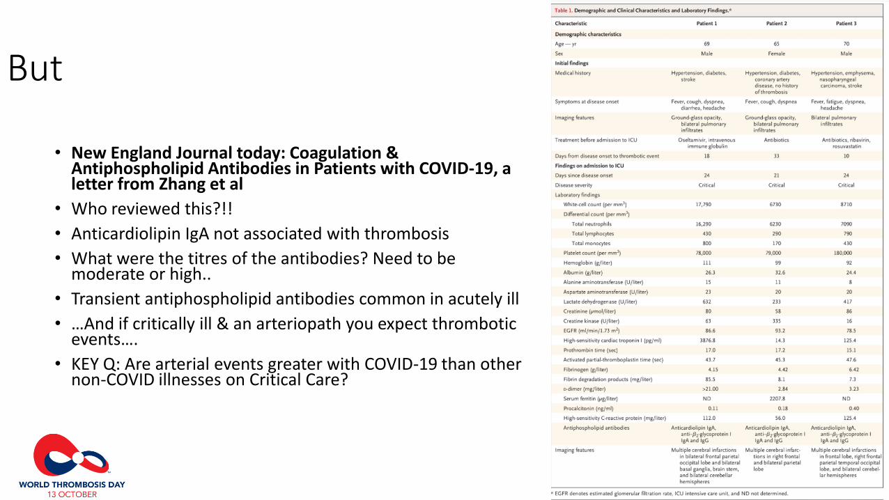
• New England Journal today: Coagulation & Antiphospholipid Antibodies in Patients with COVID-19, a letter from Zhang et al
• Who reviewed this?!!
• Anticardiolipin IgA not associated with thrombosis
• What were the titres of the antibodies? Need to be moderate or high..
• Transient antiphospholipid antibodies common in acutely ill
• …And if critically ill & an arteriopath you expect thrombotic events….
• KEY Q: Are arterial events greater with COVID-19 than other non-COVID illnesses on Critical Care?

D-dimers are increased in inflammatory states, post operatively, pregnancy, going for a run and in ….non COVID pneumoniasTaken from Yin et al JTH 2020 on line 3 4 20
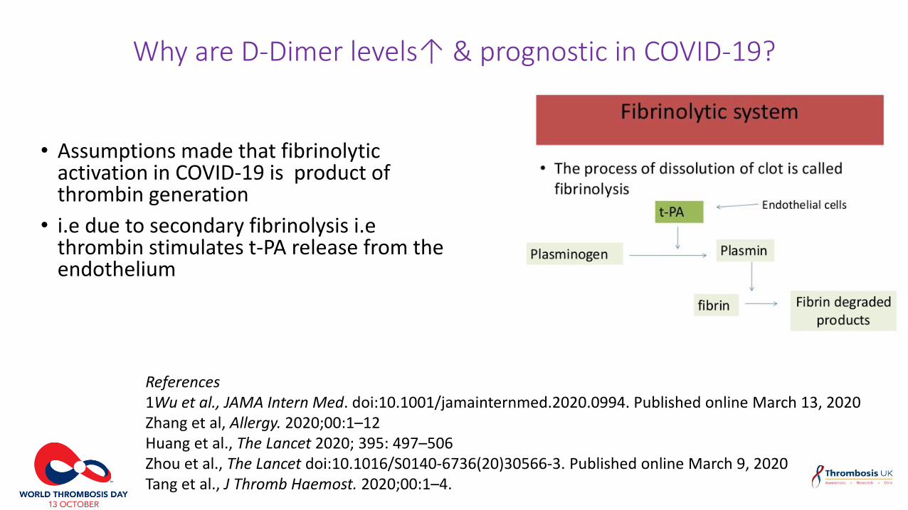
Why are D-Dimer levels↑ & prognostic in COVID-19?
• Assumptions made that fibrinolytic activation in COVID-19 is product of thrombin generation
• i.e due to secondary fibrinolysis i.ethrombin stimulates t-PA release from the endothelium
References1Wu et al., JAMA Intern Med. doi:10.1001/jamainternmed.2020.0994. Published online March 13, 2020Zhang et al, Allergy. 2020;00:1–12Huang et al., The Lancet 2020; 395: 497–506Zhou et al., The Lancet doi:10.1016/S0140-6736(20)30566-3. Published online March 9, 2020Tang et al., J Thromb Haemost. 2020;00:1–4.

Why are D-Dimer levels ↑& prognostic in COVID-19? Hypothesis 1: a result of acute lung injury
• The hallmark of acute lung injury is intra-alveolar fibrin deposition
• (& later remodeling of fibrin → lung fibrosis)
• Urokinase –type plasminogen activator (uPA) produced locally regulates extravascular proteolysis, inhibited by PAI-1
• In SARS -infected mice a dose-dependent ↑ in lung urokinase with dose of SARS injected but locally swamped by PAI-1
• Pathways not clearly understood
Idell, Crit Care Med 2003:S213-20, Gralinski, mBIo 2013; 4:e00271-13

Why are D-Dimer levels ↑& prognostic in COVID-19? Hypothesis 2: produced by activated macrophages
• COVID-19 pneumonia lung histology characterized by many macrophages
• Macrophages generate plasmin & metalloproteinases (MMPs)
• Fibrin degradation also occurs by an alternative pathway- fibrin (ogen) binding to CD11b/CD18 I internalized into the lysosome where cathepsin D degrades it
Loscalzo J. Semin Thromb Hemost 1996; 22:503-6
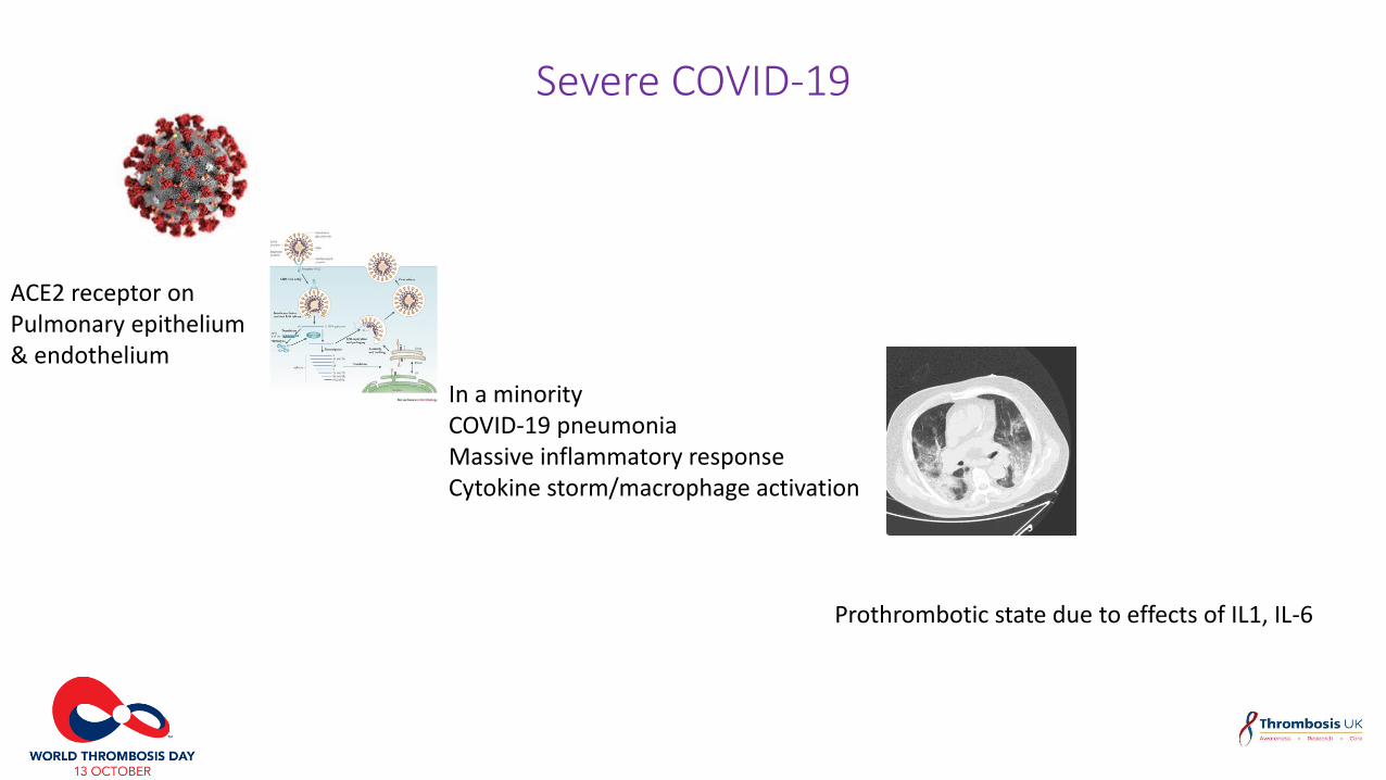
Severe COVID-19
ACE2 receptor on Pulmonary epithelium& endothelium
In a minority COVID-19 pneumoniaMassive inflammatory responseCytokine storm/macrophage activation
Prothrombotic state due to effects of IL1, IL-6

Severe COVID-19
ACE2 receptor on Pulmonary epithelium& endothelium
In a minority COVID-19 pneumoniaMassive inflammatory responseCytokine storm/macrophage activation
Prothrombotic state due to effects of IL1, IL-6Does a pre-existing inflammatory statemake COVID-19 pneumonia more likely?e.g atherosclerosis, diabetes, obesity

Hypertrophic adipocytes (like atherosclerosis & diabetes) induce an
inflammatory state
Obesity is common: BMI > 25 in 75% of UK patients with severe COVID-19 infection(Source @ICNARC)

High background risk of VTE in critically ill patients
• Hospital-associated VTE – VTE occurring in hospital & up to 90 days post discharge
• But lack of current data and variation in severity of illness to qualify for critical care beds across the world
• What is the current VTE risk in critical care?
• 1982 Cade 1982 119 patients DVT in 29% vs 13% UFH 5,000 BD by day 6
• 1999 MEDENOX 1102 pts placebo arm 15% had DVT/PE by day 14
Cade JF, Crit Care Med 1982; 10: 448-50, MEDENOX- Samama New Engl J Med 1999; 341: 793
Risk factors
• Increasing age
• Acute infective illness
• Use of venous lines
• Underlying patient risk factors
• Immobility
• Etc etc

Benefits of heparinoid thromboprophylaxis for 6-14 days over placebo in medical patients
MEDENOX1 63% Placebo
Enoxaparin 40 mg
PREVENT2 49% Placebo
Dalteparin
ARTEMIS3 47% Placebo
Fondaparinux
14.9*
5.5
Study RRR Thromboprophylaxis Patients with VTE (%)
5.0*
2.8
10.5†
5.6
*VTE at day 14; †VTE at Day 15 1Samama MM et al. N Engl J Med 1999;341:793–8002Leizorovicz A et al. J Circulation 2004;110:874–9
3Cohen AT et al. J Thromb Haemost 2003;1 (Suppl 1):P2046
P<0.001
P=0.0015
p=0.029
RRR = relative risk reduction
RRR
63%
45%
47%

Thromboprophylaxis in Critical Care
Which is better?
Lim et al Crit Care Med 212;40: 328
• Compared LMWH vs UFH in same group
• LMWH ↓ DVT & PE> UFH
• For PE RR 0.52: 95% CI 0.28,0.97) p=0.04)
• No difference in bleeding or mortality
Does LMWH/UFH reduce risk?
Alhazzani et al Crit Care Med 2013; 41: 2088
• Systematic review 7,226 pts in RCTs
• ↓sympt/asympt DVT RR 0.51 (95% CI 0.41-0.64); p <0.0001)
• ↓PE RR 0.52 (95% CI 0.28-0.92); p= 0.04)
• No difference in bleeding or mortality

Extended thromboprophylaxis in medical patients
MARINER trial (low dose rivaroxaban) excluded as only measured symptomatic & fatal VTE

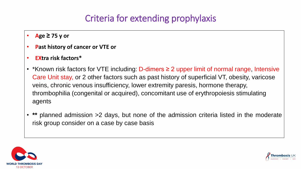
Criteria for extending prophylaxis
• Age ≥ 75 y or
• Past history of cancer or VTE or
• EXtra risk factors*
• *Known risk factors for VTE including: D-dimers ≥ 2 upper limit of normal range, Intensive
Care Unit stay, or 2 other factors such as past history of superficial VT, obesity, varicose
veins, chronic venous insufficiency, lower extremity paresis, hormone therapy,
thrombophilia (congenital or acquired), concomitant use of erythropoiesis stimulating
agents
• ** planned admission >2 days, but none of the admission criteria listed in the moderate
risk group consider on a case by case basis

Anticoagulant treatment is associated with decreased mortality in severe COVID-19 patients with coagulopathy
Tang et al, JTH 2020, preprint
• Retrospective study in Tongji Hospital, 449 patients with severe COVID-19 infection, 99 (22%) received heparin
• d heparin for >7 days
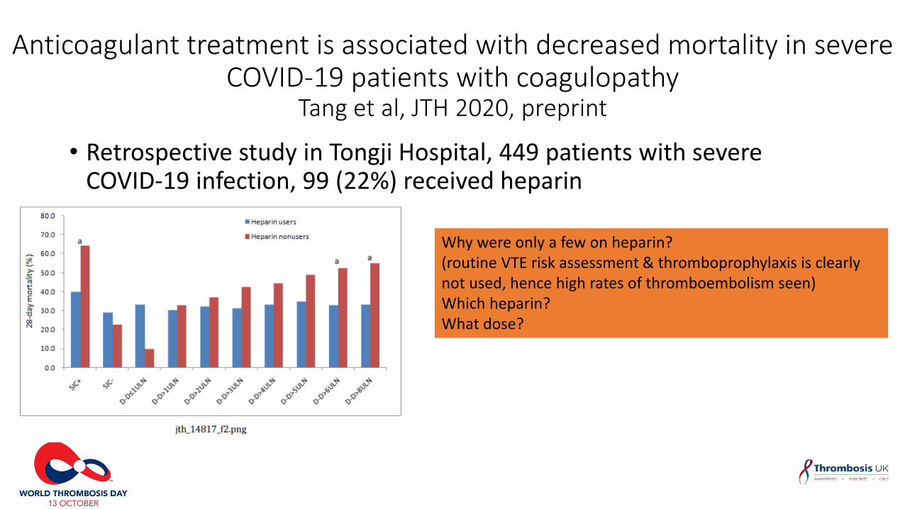
Anticoagulant treatment is associated with decreased mortality in severe COVID-19 patients with coagulopathy
Tang et al, JTH 2020, preprint
• Retrospective study in Tongji Hospital, 449 patients with severe COVID-19 infection, 99 (22%) received heparin
• d heparin for >7 daysWhy were only a few on heparin?(routine VTE risk assessment & thromboprophylaxis is clearly not used, hence high rates of thromboembolism seen)Which heparin?What dose?

Unanswered Qs in thrombosis & thromboprophylaxis in COVID-19 infection
Rates of thromboembolism
• What are the current rates of VTE in critically ill patients?
• Are the rates of thrombosis higher than other patients on critical care especially can we compare with non-COVID-19 viral pneumonia?
Thromboprophylaxis
• Is weight adjusted thromboprophylaxis better than empirical dosing? (Many trial excluded high weight individual AND obesity rates have ⇧ since trials)
• Would a higher dose of thromboprophylaxis be beneficial without significantly increasing bleeding risk?
• Should we add in intermittent pneumatic compression?
• Should we give extended thromboprophylaxis?

Management of fresh VTE in COVID-19
Diagnosis: how to detect PE when cant use D-dimer?
• This is not a new problem!
• Need to maintain high clinical suspicion esp. if becomes more clinically hypoxic suddenly
• Would manage with 3/12 anticoagulation (would start with LMWH while in critical care then switch to DOAC on discharge) according to current guidelines
Catheter-associated thrombosis
• Six weeks Rx according to current guideline
• Expecting to see it frequently
NB despite thromboprophylaxis we will see VTE in at least 7%, probably more, of all critically ill patients whether cOVID -19 or not
