course: opto 328 physiology of vision i units: 2 +...
TRANSCRIPT

OPTO 328 - Physiology of Vision I
1
COURSE: OPTO 328 – Physiology of Vision I
UNITS: 2 + 0 = 2.
TUTOR: Dr. Ali A Abusharha
RECOMMENDED TEXTS:
1) Visual Perception by Cornsweet.
2) Physiology of the Eye by Hugh Davson.
3) OPTOMETRY by Keith Edwards and
Richard Llewelyn
4) Visual Development (2nd
Ed.) by Nigel W.
Daw
OPTO 328 - Physiology of Vision I
2
Topics
1) Glossary of terms
2) Retinal Structure and Organization
3) Photochemical and Electrophysiological Aspects of Vision
4) Sensory aspects of Vision
5) Concepts of Threshold: Frequency of seeing curve
6) Temporal phenomena
I. Flicker
II. Talbot-Plateau law
III. Granit-Harper law
5.1.1 IV. Ferry-Porter law
7) Spatial Phenomena
8) The Electroretinogram
9) The Visual Evoked Potential

OPTO 328 - Physiology of Vision I
1
LECTURE 1
The primary stimulus for vision is LIGHT.
In this course, we will discuss extensively, the chemical processes that take place in the
retina in response to light stimulation, and how this chemical changes are transformed
into electrical impulses for further interpretation in the occipital cortex. All this results in
Visual perception.
Before we begin our discussions, let us revisit some of the terms you may already be
familiar with from the first course in this series.
FLUX The rate of flow of fluid or energy.
LUMINOUS FLUX The rate of flow of light (Lumens).
LUMEN (lm) Unit of luminous flux.
LUMINOUS INTENSITY
In the study of the effects of light on the eye, we are most interested in that quality of
light called 'Brightness'. This brightness (or luminosity) is based on an arbitrary standard
(the Candela).
The luminous intensity of a light source can be vulgarly (commonly) defined as the
degree of illumination (sensation of brightness), which it produces.
INTENSITY OF ILLUMINATION
The intensity of illumination refers the level of illumination on a particular surface. It
depends on
I. The luminous intensity of the source of light.
II. The distance between the light source and the surface in question.
III. The angle the surface makes with the direction of incident light.

OPTO 328 - Physiology of Vision I
2
E = I/r2 where I is the luminous intensity of the light source, E is the intensity
of illumination; and r is the distance between the light source and the
surface.
The equation above is referred to as the Inverse-Square law.
The Inverse square law states: The intensity of illumination at a point, varies inversely as
the square of the distance from that point to the light source.
LUMINANCE
Whereas intensity of illumination describes the amount of light that falls on a surface,
luminance measures the amount of light reflected off a surface from a light source.
A ‘perfect diffuser’ can be imagined as a white surface that reflects all the light that
falls on it.
Thus a perfect diffuser 1cm away (and perpendicular) from a standard light source of
candela has a luminance of 1 Lambert.
This unit of measurement is too large for our purposes. The log micro-Lambert (mL) is
therefore used for studies of light thresholds.
SURFACE INTENSITY
In this modality, the luminance of a surface is defined per unit area of that surface. (e.g.
candelas per square foot or candelas per square centimetre.
EFFECTIVELY POINT SOURCE
An illuminated surface of given area forms an image of a definite area on the retina and
the sensation of brightness, or rather the stimulus, depends on the amount of light falling
on this area of the retina.
If the surface is moved closer to the eye, the amount of light entering the eye is increased
in accordance with the inverse square law, but the surface does not appear brighter
because the size of the image has increased. Therefore the actual illumination of the
retina has not altered.
Thus the subjective brightness of an extended source is independent of its distance from
the observer.
It may be argued that the brightness of a lamp decreases as it is moved away from the eye
and eventually becomes so faint that it disappears if it is moved far enough away. This,
however, is a consequence of the fact that at a certain distance from the eye the lamp

OPTO 328 - Physiology of Vision I
3
becomes an Effectively point source i.e. its image on the retina is of the same order of
size as that of a receptor element.
Consequently, when it is moved further away still, although its image on the retina
becomes smaller, the amount of light, per receptor element, now begins to fall in
accordance with the inverse square law.
RETINAL ILLUMINATION
This modality just has to do with the simple truth that the level of illumination that falls
on a particular retinal area has not only to do with the candlepower per unit area of the
surface being viewed, but also on the pupil size.
The unit of retinal illumination is the Troland. A Troland is that intensity of stimulation
that accompanies the use of a pupillary area of one square millimetre and an external
stimulus surface luminance of one candela per square metre.
LIGHT SENSE
This refers to the appreciation or awareness of light and as such, the modification of its
intensity.
COLOUR SENSE
This enables us to distinguish between the qualities of two or more lights in terms of their
wavelengths.
FORM SENSE
This permits the discrimination of the different parts of a visual image.

OPTO 328 - Physiology of Vision I
4
LIGHT SENSE
Absolute Threshold
Threshold to white light i.e. the minimum light stimulus necessary to evoke the sensation
of light, is the simplest way to study light sense.
To measure thresholds, the subject is placed in a completely dark room and the
illumination of a test screen patch is increased until he is aware of its presence.
The patch may be illuminated all the time and its illumination steadi1v increased or it
may be presented in flashes of varying luminance.
In the fully dark-adapted eye, the minimum stimulus necessary to evoke the sensation of
light is the absolute threshold.
This threshold is not a fixed quantity, but varies from moment to moment, so that if a
subject is presented with a luminous screen in the dark, we may find a range of
luminance over which there will be some probability but not certainty, of his seeing it.
Experimentally, the screen is presented frequently at different levels of luminance. A plot
is then made of the 'frequency of seeing' against luminance to give the frequency of
seeing curve. Threshold is then computed from an arbitrarily chosen frequency.

OPTO 328 - Physiology of Vision I
5
LECTURE 2
Retinal Structure and Organization
Figures 2.1 a and b represent the path through which light information travels to the brain
and back.
Please note the difference between the dotted lines and the solid ones.
Question
What effect on vision would you expect if there were a lesion of the optic tract?
Figure 2.2 is a schematic representation of the retinal layers.
1. Retinal pigment epithelium (RPE)
2. Layer of the rods and cones
3. Outer limiting membrane
4. Outer nuclear layer
5. Outer plexiform layer
6. Inner nuclear layer
7. Inner plexiform layer
8. Ganglion cell layer
9. Nerve fibre layer
10. Inner limiting membrane
The RPE is a single layer of pigmented cells, which is attached to the choroid. Its
main functions are:
It nourishes the photoreceptors from the choroidal blood circulation.
Its melanosomes absorb light photons that pass through the rod and cone
layer but which fail to excite the rods and cones. Normally, these light
photons would be reflected back into the layer of rods and cones and thus,
cause interference with vision. The RPE melanosomes therefore reduce
back-scatter of light into the rod and cone layer which would otherwise
have interfered with the processing of a clear image of the object of
regard.
It digests the outer segment discs shed from the rods and cones.

OPTO 328 - Physiology of Vision I
6
Figures 2.1a and b. a Depicts the general path traveled by nasal and
temporal fibres. b Shows exactly how fibres from each quadrant of the
eye cross the optic chiasma.

OPTO 328 - Physiology of Vision I
7
Figure 2.2 The Structure of the Retina

OPTO 328 - Physiology of Vision I
8
The rod and cone layer contains the outer segment layers of the rods and
cones. The cell bodies/nuclei of the rods and cones actually lie within the
outer nuclear layer.
At the fovea, because there are numerous cones, the cones are very thin
and look like rods.
There are about 110 to 125 million rods, and 6.5 million cones in the eye.
At the central fovea, the cone concentration is maximum and is about
147,000 cones/mm2. About 5mm from the fovea, the concentration of rods
reaches a maximum of about 160,000 per mm2.
Light from an external object passes into the eye and when it reaches the
retina, it first passes through layer 10, then 9, then 8 etc., until it reaches
the photoreceptor layer (rod and cone layer).
The light then bleaches the photoreceptor pigments of the rods and cones.
One such pigment is Rhodopsin – which is the rod pigment.
There are three types of cone pigment in the human retina (red, green, and
blue) and each single cone contains only one type of pigment.
Based on studies of other mammals, the cone distribution is as follows:
o Long Wavelength-Sensitive Cones - 33%
o Medium Wavelength-Sensitive Cones - 13%
o Short Wavelength-Sensitive Cones - 54%
The proportion of blue cones reduces to 3% in the central retina. This finding would at
least partially explain the well known ‘blue-blindness’ (tritanopia) in the foveal region.
Receptor inner segments are depicted in figure 2.3. The receptor inner segment is
easily distinguished into two distinct zones:
o The outer zone, called the ellipsoid, which is packed with mitochondria.
o The inner zone, called the myoid, which contains free ribosomes, granular
and agranular endoplasmic reticulum, vesicles, Golgi membranes, and
neurotubules.

OPTO 328 - Physiology of Vision I
9
Figure 2.3 Photoreceptor Structure

OPTO 328 - Physiology of Vision I
10
o The myoid is responsible for the manufacture of most of the molecules involved
in the production and repair of the outer segment discs of rods and cones.
o Opsin (the visual pigment) is also produced in the myoid, and then it is
transported to the outer segment via the connecting stalk.
Note A thin outer fibre connects the rod’s cell body with its inner segment. The cone’s cell
body on the other hand, is connected directly to its inner segment.
Outer plexiform layer is the layer that contains the connections between the rods and
cones on the one hand, and the horizontal and bipolar cells on the other.
Cones synapse (connect) with:
Midget bipolar cells
Flat midget bipolar cells
Flat bipolar cells
Rods synapse with:
Rod bipolars
o The important points to note about the synapses in the outer plexiform layer are as
follows:
In the fovea only, one cone connects with one midget bipolar cell. Outside the
fovea, this relationship does not exist.
Flat bipolar cells synapse with about six cones each.
One rod bipolar cells synapses with about 45 rods.
o It then becomes clear that the rods bipolar cells receive much more information than the
midget bipolars, or the flat bipolars, both of which synapse with cones.
o The one-to-one relationship between the cone and the midget bipolar cell is necessary for
the preservation of fine detail.
o Horizontal cells also synapse with rods and cones in the outer plexiform layer. One
horizontal cell connects with 6 – 12 cones in the central retina, and 30 – 50 cones in the
periphery. One horizontal cell connects with 80 – 150 rods.
o The likely function of the horizontal cells is to provide communication:
OPTO 328 - Physiology of Vision I
11
Between the cones
Between the rods
o It is unlikely that the horizontal cells provide a pathway for rod-cone
communication.
o The obvious advantage that the feedback mechanism of the horizontal cells
provide is that the response of each cone, and each rod can be modified before
being transmitted to the ganglion cells.
The inner nuclear layer contains the cell bodies of the bipolar, horizontal and amacrine
cells. The amacrine cells also provide a feedback mechanism for the transmission of
signals from the rods and cones to the ganglion cells. This time however, they modulate
the synapses between the bipolar cells, and the ganglion cells (in the inner plexiform
layer).
The inner plexiform layer contains complex interactions between the bipolar and
ganglion cells and amacrine cells. There are many types of amacrine cells that synapse
with multiple bipolar and ganglion cells, as well as with each other.
Interplexiform cells are a distinct type of amacrine cell, which also go back to the
outer plexiform layer and synapse with bipolar and horizontal cells. They
therefore provide a more complex type of feedback between the plexiform layers
rather than within one plexiform layer.
The ganglion cell layer contains the cell bodies of the ganglion cells. There is one-to-one
relationship between a midget bipolar cell and a midget ganglion cell (in the inner
plexiform layer). So, in the central fovea, the information from one cone travels along a
pure path to the visual cortex
Another type of retinal ganglion cell (the diffuse ganglion cell) synapses with
numerous bipolar cells.
Midget ganglion cells are also referred to as beta cells or x-cells.
Diffuse ganglion cells are also referred to as alpha cells or Y-cells.
In the nerve fibre layer the axons of the ganglion cells pass through on their way to the
brain. Whilst passing through this layer, the ganglion cells are sheeted (covered) by
Müller cell processes and astrocytes. These covering of the axons perform the following
functions:
They nourish the nerve fibers.
They provide protection for the axons.
They provide insulation for the fibers so that they conduct electric impulses faster
and more efficiently.

OPTO 328 - Physiology of Vision I
12
LECTURE 3
PHOTOCHEMICAL ASPECTS OF VISION
The visual pigment of rods is known as Visual purple or Rhodopsin. For the study of the
photochemical aspects of vision, we will discuss the process by which Rhodopsin mediates the
sensation of light.
When light photons fall on a rhodopsin pigment molecule, this molecule enters into an
‘excited’ state. This ‘excited state is very unstable and therefore leads to the formation of
new molecules.
These new molecules undergo a series of reactions (called thermal reactions), which
ultimately lead to the sensation of light.
It stands to reason that these reactions should be reversible (at least in part) if not, the eye
would have to produce large amounts of rhodopsin each time the pigment was excited by
light.
As far as the visual process is concerned, a simplified schema of events can thus be
represented as follows:
Rhodopsin Activated state Bleached products
These bleached products are the result of a thoroughly complicated series of events which
involve rhodopsin changing its color from purple to orange to yellow, and finally to
white when it is eventually converted to Vitamin A + Opsin.
Generally then, when the eye is fully dark-adapted we should find only rhodopsin within
the rods. However, when the rods are excited by light, we would find a mixture of
rhodopsin and its bleached products. This mixture will depend on the intensity of the
light and the duration of its exposure.

OPTO 328 - Physiology of Vision I
13
Production And Turnover Of Outer Segment Disc Lamellae
Taking a quick peep at the schematic diagram of rods and cones, we see that there are
numerous Disc Lamellae in the rod and cone outer segments. These lamellae are oriented
at 900 to incident light, an arrangement that maximizes the absorption of incident light.
These lamellae are manufactured in the Myoid portion of the Rod Inner Segment. The
disc lamellae are then moved to the Golgi bodies, which then transport the lamellae into
the Outer Segment. As more and more lamellae are produced, the older lamellae climb
higher up in the Outer Segment until they are secreted into the RPE.
Estimates have been given of the replacement of the disc lamellae at 25 to 36 per day.
It would appear however that the turnover in cones does not undergo a similar process to
that in rods.
Note Purkinje shift refers to the shift of maximum wavelength sensitivity of the eye from
500m during scotopic (rod) vision, to 550m during photopic (cone) vision.
Bleaching of Rhodopsin
Early studies recognized that rhodopsin turned yellow on exposure to light.
The final products of the bleaching of rhodopsin are Retinene, and Vitamin A – into which
retinene is converted if it is exposed to light photons long enough to prevent its re-conversion
into rhodopsin.
Retinene is actually vitamin A aldehyde and is now (more correctly) referred to as Retinal.
Retinal + Opsin = Rhodopsin (Visual purple).
The scheme proposed by Abrahamson and Ostroy (1967) of the chemical intermediaries in the
thermolysis of rhodopsin is still valid today and is as follows:

OPTO 328 - Physiology of Vision I
14
Rhodopsin (250C)
short
Preluminorhodopsin (-1950C)
- 1400C
Luminorhodopsin (-500C)
-400C
Metarhodopsin I (+30C)
-15
0C
Metarhodopsin II (+30C)
-5
0C
Metarhodopsin III (+30C)
H+ + 15
0C
N-retinylidene Opsin (NRO 440 ) NRO 365
H2O
Retinal +Opsin

OPTO 328 - Physiology of Vision I
15
Notes The first step in the reaction above is the only step that requires the absorption of light.
Luminorhodopsin remains stable at temperatures below –400C , above which it breaks
down into a series of metarhodopsin molecules, which vary only in their wavelength of
maximum absorption ( max).
The final conversion (from NRO to Opsin) involves the splitting of retinene from
Opsin. This reaction involves a process known as breaking a Schiff-base link.
Regeneration of Rhodopsin
In the retina, the products of bleaching of rhodopsin (retinene and vitamin A) are re-
combined to form rhodopsin in the dark.
Inasmuch as this combination must take place for the retinene to be recycled into
rhodopsin, the transformation is not straightforward.
Retinene in its combined form with Opsin is in its 11-cis form. But when retinene is
separated from Opsin, it exists in its all-trans form. Therefore, retinene in (retinene +
Opsin) must first be converted (isomerized) into its 11-cis form (or to another form,
which is equally capable of producing rhodopsin – 9-cis retinene). This isomerization
will take place (in the dark) in the presence of an enzyme called Retinene Isomerase, or
by mere exposure to light.
H H H R1
C = C C = C
R R1 R H
Cis Retinene Trans Retinene

OPTO 328 - Physiology of Vision I
16
The Role of the RPE
The RPE’s role in the regeneration of rhodopsin cannot be overemphasized. It has long
since been discovered that rhodopsin would not be regenerated in the absence of the RPE.
The reason for this is now taken for granted to be due to the high concentration of
vitamin A that has been found in the RPE after maximum light adaptation. Conversely,
after full dark adaptation, no vitamin A is found in the RPE indicating that vitamin A, a
vital precursor for rhodopsin, is stored almost entirely in this layer.
Note It is worthy of note that various researchers have reported that the scotopic pigments:
Vary between species.
Could exist in multiple forms in the same animal
Sometimes vary with the period of the year (season)
Sometimes vary according to the habitat of the animal.
OVERVIEW OF ELECTROPHYSIOLOGY OF VISION
When any receptor is stimulated, electrical changes take place that result in the
transmission of electrical current along that receptor’s nerve fibre.
This electrical current takes the form of Action Potentials.
The duration of any one action potential is very short. However, the greater the stimulus
that causes the electrical current, the more action potentials per second there are.
However, the magnitude (size) of the action potential itself never changes.
Therefore, once the stimulus is large enough to generate an action potential, a constant
current is transmitted across the nerve fibre. What changes with the intensity of the
stimulus is the frequency of action potentials. This phenomenon is called the all-or-
nothing-response.
It is important to digress a bit to describe the processes that usually take place when an
action potential is generated in a receptor.
OPTO 328 - Physiology of Vision I
17
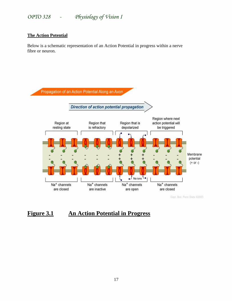
The Action Potential
Below is a schematic representation of an Action Potential in progress within a nerve
fibre or neuron.
Figure 3.1 An Action Potential in Progress

OPTO 328 - Physiology of Vision I
18
The resting membrane potential is –70 mV. When a (local) stimulus causes a reduction of
this RMP to –59 mV, an action potential is generated. A stimulus causing a reduction of
the RMP to less than – 59 mV has no effect. A generator potential then moves this action
potential along the entire length of the membrane.
Once the RMP depolarizes (increases) to –59 mV, Na+
channels are opened and Na+ ions which
normally are more outside the membrane rush into it and cause a massive increase in the
membrane potential to about + 30 mV.
K+ are normally much slower to respond and do so after the membrane potential has reached +
30 mV. The K+ ions then rush out of the membrane and eventually restore the membrane
potential back to (and a little bit greater than) its original resting state of – 70 mV. This is called
Re-polarization of the membrane.
In the eye however, a sufficiently large stimulus causes a hyper-polarization (rather than a
depolarization) of the photoreceptors – so that the membrane potential becomes more negative
instead of more positive. This is how the electrical current carrying vital information to the brain
is generated.
Notes It is pertinent to note at this juncture that the crucial electrical event that leads to a
discharge of electrical current along the nerve fibre is called a Generator Potential. This
potential is a much slower mechanism in which the hyper-polarization of the cell lasts longer.
If the generator potential is maintained long enough, it causes the discharge of electrical
current. The generator potential (as well as the action potential frequency) increases in direct
proportion with the intensity of the light stimulus.
In the photoreceptors, the outer segment is usually more negative than the inner segment in the
dark. This causes a flow of positive current from the inner to the outer segment.
When a sufficient light stimulus excites the photoreceptor, the outer segment increases its
resistance and thus prevents the flow of positive ions into it and thus increases its negativity.
This hyper-polarization reverses the direction of the current and is the first stage of the action
potential that sends visual impulses to the cortex from the photoreceptors.
Nature of Photoreceptor-generated Action Potentials
To understand the nature of these action potentials, we need to discuss a number of terms and
effects.
The optic nerve fibres can be categorized into three groups according to their reaction to light
stimuli.

OPTO 328 - Physiology of Vision I
19
On Fibres These fibres (about 20% of the total) display a strong response
immediately the light impulse is received and stop this response
immediately the light stimulus is removed.
On-Off Fibres These comprise about 50% of the total fibres. They respond to a light
stimulus with an initial burst of activity when the impulse is received, and
with another burst of activity when the impulse ceases.
Off Fibres These comprise the remaining 30% of the fibres and respond only when
the light stimulus stops.
The Electroretinogram is a record of the slow changes of potential that occur in a radial
direction across the retinal surface when light falls on it.
This potential, according to the classic analysis of Granit displays three components:
a) P I This is a slowly developing positive component of the potential which
tends to disappear with strong stimuli or after light adaptation.
b) P II This is a positive component that is turned on rapidly. This component is
strongly associated with the passage of impulses along the optic nerve.
c) P III This is a negative component that comes on rapidly and is responsible for
the return of the membrane potential back to the base line (in short, it is
responsible for the ‘off’ effect). This component is mainly associated with cone
impulses and is believed to function in connection with the inhibition of impulses.
Once the generator potential has set off electrical impulse transmission in the photoreceptor cell,
the following are summaries on how the major fibres involved in this transmission respond:
Horizontal Cell It also hyperpolarizes but its response to the stimulus is much
slower.
Bipolar Cell Response is also slow here. It respsonds to the stimulus in an
opponent (explain) fashion i.e. centre hyperpolarizes, surround
depolarizes.
Amacrine Cells Response here is depolarization. Some receptive fields respond in
an opponent fashion, others in a uniform fashion.
Ganglion Cells Respond with an initial depolarization. Like bipolar cells, centre-
surround antagonism (opponent receptive fields) is very much a
characteristic of their response.