corneal collagen cross-linking for the treatment of progressive keratoconus: 3-year prospective...
TRANSCRIPT

ORIGINAL ARTICLE
Corneal collagen cross-linking for the treatment of progressivekeratoconus: 3-year prospective outcomeYakov Goldich, MD,*,† Yaniv Barkana, MD,* Orly Wussuku Lior, MD,* Arie L. Marcovich, MD,‡
Ami Hirsh, MD,§ Isaac Avni, MD,* David Zadok, MD*
ABSTRACT ● RÉSUMÉ
Objective: To assess the long-term effects of treatment of progressive keratoconus with ultraviolet A-riboflavin collagen cross-linking (CXL).
Design: This was a prospective clinical study.Participants: Seventeen eyes of 17 patients with progressive keratoconus were treated with CXL.Methods: Patients were examined preoperatively, at week 1, months 1, 3, 6, 9, 12, 24, and 36 after treatment. We assessed
uncorrected visual acuity (UCVA) and best spectacle-corrected visual acuity (BSCVA), refraction, biomicroscopy and fundusappearance, intraocular pressure, endothelial cell density (ECD), corneal topography, minimal corneal thickness (MCT), macularoptical coherence tomography, axial length, and corneal biomechanics with the ocular response analyzer.
Results: Comparing the 36-month time point results with pretreatment values, we found that UCVA and BSCVA were unchanged.Steepest meridian keratometry (D) and mean cylinder (D) did not show significant change compared with pretreatment values butshowed a slight increase as compared with the 24-month time point (53.9 vs 51.7 vs 52.5, and 10.5 vs 8.1 vs 9.2 before, at 24months, and at 36 months, respectively). Axial length (mm) showed an elongation trend throughout the follow-up period (24.56 vs24.61 [p ¼ 0.04] vs 24.71 [p ¼ 0.05], before, at 24 months, and at 36 months, respectively). No significant change was observed inECD, corneal hysteresis and corneal resistance factor, MCT, or foveal thickness.
Conclusions: Three-year results after CXL show stable visual acuity, stable corneal thickness, and stable corneal biomechanicalparameters. The decreasing trend in keratometry values that was observed during the first 2 years after CXL was no longer evident.Longer follow-up is needed to decide whether it is a first sign of loss of achieved stability and resumption of keratoconus progression.
Objet : Évaluation des effets à long terme du traitement du kératocône progressif avec la réticulation du collagène par RiboflavineUVA. (RC)
Méthodes : Étude clinique prospective. Dix-sept yeux de 17 patients ayant un kératocône progressif ont été soignés avec la RC. Ilsont été examinés avant l'opération, puis 1 semaine, 1, 3, 6, 9, 12, 24 et 36 mois après le traitement. Nous avons évalué l'AVSC etla MAVC, la réfraction, la biomicroscopie et l'apparence du fond d'oeil, la TIO, la densité cellulaire endothéliale (DCE), latopographie cornéenne, l'épaisseur minimale de la cornée (EMC), la TCO maculaire, la longueur axiale et les biomécaniques dela cornée avec l'analyseur de la réaction oculaire.
Résultats : La comparaison des résultats de point après 36 mois avec les valeurs préopératoires nous a indiqué que l'AVSC et laMAVC n'avaient pas changé. Kmax (D) et Kcyl (D) ne présentaient pas de changement significatif comparativement aux valeursprétraitement, mais montraient une légère augmentation comparativement aux points du 24e mois. (53,9vs51, 7vs52,5, et10,5vs8,1vs9,2; avant, à 24 mois et à 36 mois respectivement). La longueur axiale (mm) montrait une tendance à l'élongationpendant la période de suivi (24,56vs24,61 (P ¼ 0,04) vs 24,71 (P ¼ 0.05); avant, à 24 mois et à 36 mois respectivement). Aucunchangement modificatif n'a été observé concernant la DCE, l'hystérésis de la cornée, le facteur de résistance cornéenne, l'EMCou l'épaisseur de la fovéa.
Conclusions : Les résultats des trois années de suivi de la réticulation du collagène ont démontré une triple stabilité, soitl'acuité visuelle, l'épaisseur de la cornée et les paramètres biomécaniques de la cornée. La tendance à la baisse des valeurskératométriques observée pendant les deux premières années suivant le traitement n'était plus évidente. Il faudra un suiviplus long pour décider s'il s'agit d'un premier signe de perte de stabilité réalisée et de reprise de la progression dukératocône.
Keratoconus is a bilateral, progressive, noninflammatorycorneal degeneration.1 Corneal deformation and thinningcauses irregular astigmatism and leads to visual impair-ment.2 Treatment modalities are based on refractivecorrection with spectacles, contact lenses, and intrastromalcorneal rings to correct astigmatism and restore visualacuity.3 Such modalities do not stop ectatic progression
From the *Department of Ophthalmology, Assaf Harofeh MedicalCenter; †Department of Ophthalmology, Toronto Western Hospital,Toronto, Ont.; and the ‡Department of Ophthalmology, KaplanMedical Center; and §Enaim Refractive Surgery Centers
Originally received Feb. 8, 2013. Accepted Sep. 13, 2013
Correspondence to Yakov Goldich, MD, Department of Ophthalmology,Toronto Western Hospital, University of Toronto, Toronto ON M5T2S8; [email protected]
54 CAN J OPHTHALMOL—VOL. 49, NO. 1, FEBRUARY 2014
and further visual deterioration, which ultimately neces-sitate corneal transplantation in 10% to 20% of patients.4
Corneal collagen cross-linking (CXL) using ultravioletA light (UVA) and riboflavin was introduced by Wollen-sak et al.5 as a method to halt the progression of kerato-conus. Recent clinical studies evaluated the efficacy of thisnew treatment modality.6–10 The aim of this report is to
Can J Ophthalmol 2014;49:54–590008-4182/14/$-see front matter & 2014 Canadian OphthalmologicalSociety. Published by Elsevier Inc. All rights reserved.http://dx.doi.org/10.1016/j.jcjo.2013.09.002

Table 1—Visual acuity and refractive error before and after CXL
Parameter Before CXL 12 mo after CXL 24 mo after CXL 36 mo after CXL
Mean BSCVA � SD (logMAR) 0.18 � 0.1 0.13 � 0.1 0.14 � 0.1 0.14� 0.1 (p ¼ 0.14)Mean UCVA � SD (logMAR) 0.65 � 0.4 0.79 � 0.5 0.82 � 0.5 0.74 � 0.4 (p ¼ 0.73)Mean SE � SD (D) –4.4 � 3.4 –4.0 � 3.3 –4.0 � 3.3 –2.7 � 2.6 (p ¼ 0.01)
CXL, corneal collagen cross-linking; BSCVA, best corrected visual acuity with glasses; UCVA, uncorrected visual acuity; SE, spherical equivalent.
CXL for progressive keratoconus—Goldich et al.
describe the observed changes in keratoconic eyes after3 years of close follow-up after CXL treatment.
METHODS
Patients with keratoconus were prospectively recruitedfrom the cornea outpatient clinic of the Assaf HarofehMedical Center. Included were subjects with progressivekeratoconus confirmed by an increase of at least 1.5 D inastigmatic refraction and/or maximum curvature docu-mented by corneal topography at 3 time points within thepast 12 months. Other inclusion criteria were age olderthan 18 years, no previous ocular surgery, no cornealopacities, minimal corneal thickness (MCT) of 400 μm,and avoidance of contact lens wear for 1 month beforeinitial evaluation and treatment. Patients were treated withUVA-riboflavin CXL under aseptic conditions using top-ical preoperative anesthaesia with oxybuprocaine hydro-chloride 0.4% drops (Localin; Fisher PharmaceuticalLabs). Treatment included 7-mm diameter corneal de-epithelization, instillation of riboflavin 0.1% in dextran20% solution (Peschke Meditrade GmbH, Switzerland)every 5 minutes for 40 minutes and corneal irradiationwith UVA 3 mW/cm2 (UV-X; Peschke MeditradeGmbH, Switzerland) for 30 minutes, 5 cm from thecornea with persistent repeated application of 0.1%riboflavin in 20% dextran solution drops. After theprocedure, patients were treated with a topical antibiotic(ofloxacin 0.3% [Oflox]; Allergan) 4 times a day for 7 daysand a topical corticosteroid (dexamethasone 0.1% [Ster-odex]; Fisher Pharmaceutical Labs) 4 times a day for1 month, and the eye was dressed with a soft therapeuticcontact lens (Ocular Sciences Ltd, Southampton, U.K.)for 3 days. UV irradiance was checked preoperatively ineach case using a UV meter.
Patients were assessed preoperatively and at week 1,months 1, 3, 6, 12, 24, and 36 after treatment. Eachexamination included measurement of uncorrected visualacuity (UCVA), best spectacle-corrected visual acuity(BSCVA), and slit-lamp and dilated fundus examination.Corneal topography, pachymetry, endothelial cell density(ECD), intraocular pressure by Goldmann applanationtonometry (GAT-IOP), central foveal thickness (CFT),and corneal biomechanical parameters according to theocular response analyzer (ORA; Reichert Inc, Buffalo, NY)were assessed as follows. Corneal topography and pachy-metry were assessed preoperatively and at months 6, 9, 12,
C
24, and 36 with Orbscan II (Bausch & Lomb, Claremont,Calif.). ECD was assessed preoperatively and at months 1,6, 12, 24, and 36 with the Konan Noncon Robo SP 6000noncontact specular microscope (Konan Medical Inc,Hyogo, Japan). CFT was assessed preoperatively and atmonths 3, 6, 9, 12, 24, and 36 with Stratus opticalcoherence tomography (OCT; Zeiss Humphrey Instru-ments, Dublin, Calif.). Axial length was assessed preoper-atively and at months 12, 24, and 36 with IOL Master(Carl Zeiss Meditec AG, Jena, Germany). Users of contactlenses were asked to remove them 14 days before eachfollow-up examination.
The study was approved by the institutional ethicscommittee of Assaf Harofeh Medical Center, and a writteninformed consent was obtained from each subject after thenature and intent of the study had been fully explained.The study protocol was consistent with the tenets of theDeclaration of Helsinki.
The data are presented as mean � SD. Paired 2-tailedStudent t test was used to assess differences in respectiveparameters. The distributions of values within each dataset were evaluated graphically. A p value of 0.05 wasselected for the threshold of statistical significance. Anal-yses were performed using Excel (Microsoft Corp,Redmond, WA).
RESULTS
Seventeen eyes of 17 patients (12 males, 5 females) aged27.3� 5.1 years were included. The UCVA, BSCVA, andsubjective spherical equivalent refraction (SE) data aresummarized in Table 1. BSCVA was statistically signifi-cantly better at 12 months compared with the preoperativedata (p ¼ 0.04) and remained better than preoperatively at24 and 36 months, although without statistical signifi-cance (Fig. 1). UCVA did not show a statisticallysignificant change throughout the follow-up period ascompared with baseline. Mean SE decreased continuouslyand significantly during the 36-month follow-up(p ¼ 0.01).
The steepest meridian keratometry (Kmax) and meancylinder (Kcyl ¼ Kmax – Kmin) showed an initialtendency to decrease continuously over the first 24months with changes being statistically significantlydifferent from baseline (Kmax at 12 months: p ¼0.002, at 24 months: p ¼ 0.007; Kcyl at 12 months:p ¼ 0.018, at 24 months: p ¼ 0.007). Evaluation after 36
AN J OPHTHALMOL—VOL. 49, NO. 1, FEBRUARY 2014 55

Fig. 1—Box (mean � SD) and whisker (smallest and largest values) plots showing best corrected visual acuity with glasses(logMAR) before treatment (Preop) and on 12, 24, and 36 months thereafter.
CXL for progressive keratoconus—Goldich et al.
months showed that such a decreasing tendency hasstopped and even reversed (Figs. 2 and 3, Table 2). In3 patients, Kmax increased at 36 months as comparedwith baseline (49.3–52.6 D, 56.1–56.7 D, and 51.9–52.2D). Mean simulated keratometry (simK) did not showstatistically significant changes throughout the follow-upperiod.
Both biomechanical parameters, corneal hysteresis (CH)and CRF, did not show statistically significant changesthroughout the study period (Fig. 4, Table 2).
Axial length measurement increased continuouslyduring the 36-month follow-up, and differences frompretreatment values showed borderline statistical signifi-cance (24 months: p ¼ 0.048, 36 months: p ¼ 0.053;Table 2).
Fig. 2—Box (mean � SD) and whisker (smallest and largest v(D) before treatment (Preop) and 12, 24, and 36 months later.
56 CAN J OPHTHALMOL—VOL. 49, NO. 1, FEBRUARY 2014
Mean GAT-IOP was 10.35� 1.4 mm Hg before cross-linking, 11.06 � 0.9 mm Hg 1 year later, 10.79 �0.8 mm Hg 2 years after treatment, and 11.07 � 1.4 mmHg 3 years after treatment (p ¼ 0.1). There were nostatistically significant differences between preoperativeand postoperative values of ECD, MCT, and CFT atany time point during follow-up (Table 2).
DISCUSSION
In this study, after 3 years of continuous follow-up aftercross-linking treatment, we observed stability in visualacuity and corneal thickness, and decrease in previousbeneficial effects of CXL on corneal curvature.
alues) plots showing steepest meridian keratometry (Kmax)

Fig. 3—Box (mean � SD) and whisker (smallest and largest values) plots showing mean cylinder (Kcyl) (D) before treatment(Preop) and on 12, 24, and 36 months thereafter.
CXL for progressive keratoconus—Goldich et al.
Currently, the CXL treatment is the only conservativemodality aiming to reduce keratoconic progression. Vari-ous studies reported not just halting of progression, but alsosome continuous flattening of corneal curvature.5–7,11 Theaverage reported flattening of the steepest meridian rangedfrom 1.3 to 2.0 D for the first 2 years. In our study group, themean reduction in Kmax was statistically significant duringthe first 2 years at 2.2 D. However, at the 36-month timepoint, the mean Kmax reduction from baseline was only1.4 D and not significant statistically. The behaviour of Kcylwas similar, with initial 2 years of continuous significantflattening and reversal of such tendency compared withpreoperative values at the 3-year examination time point.The nature of the initial continuous flattening still remainsunclear, but plausibly at the longer term, inherent patholog-ical stromal remodeling leads to reversal of achieved cross-linking stabilization effects and renewal of KC progression.
Table 2—Study parameters and their mean change after cross-linking
Parameter Before CXL12 Monthsafter CXL
24 Monthsafter CXL
36 Monthsafter CXL
CH (mm Hg) 7.8 � 1.7 7.3 � 1.6 7.3 � 1.5 7.6 � 1.5CRF (mm Hg) 6.6 � 1.7 6.2 � 1.3 6.6 � 0.8 6.6 � 1.1CFT (mm) 208 � 19 207 � 22 204 � 20 210 � 21ECD (cells/mm2) 2730 � 261 2640 � 266 2541 � 344 2526 � 470MCT (mm) 463 � 38 476 � 50 462 � 43 466 � 50AL (mm) 24.56 � 1.9 24.59 � 1.9 24.61 � 1.8* 24.71 � 1.9*Kmax (D) 53.9 � 5.9 52.1 � 5.0* 51.7 � 5.5* 52.5 � 5.1Kcyl (D) 10.5 � 4.4 9.1 � 3.6* 8.1 � 3.5* 9.2 � 4.5Mean SimK (D) 45.9 � 2.7 45.2 � 2.8 45.6 � 3.7 45.7 � 3.4
Values are mean � SD.
CXL, corneal collagen cross-linking; CH, corneal hysteresis; CRF, corneal resistance
factor; CFT, central foveal thickness; ECD, endothelial cell density; MCT, minimal corneal
thickness; AL, axial length; Kmax, maximal keratometry; Kcyl, mean cylinder; mean SimK,
average simulated keratometry.np r 05.
C
In terms of visual acuity, we observed 3 years of stabilityof BSCVA and UCVA. Similarly, other studies reportedstabilization or even improvement in visual acuity afterCXL.6,8,9,11 It was theorized that corneal flatteningtogether with reduction in total wavefront higher-orderaberrations contribute to improved visual function.6
Considering long-term safety of CXL, we did notobserve significant change in ECD as was assessedthroughout the 3-year study period. We did notice somestatistically nonsignificant reduction in ECD, averaged2.4% a year. Similar reduction, although similar to others,as reported by Vinciguerra et al.11 (2.4% per year) andCaporossi6 (2.0% per year), is still somehow higher thanthe reported physiologic ECD reduction (0.6% per year);therefore, we would recommend further close long-termevaluation of corneal endothelium.6,11,12
Another safety parameter we assessed was central fovealthickness as measured by OCT. Being an indicator foranatomic stability of the retina, it did not show significantchange during the 3-year follow-up, similar to previousreports.6,8,13
CXL supposedly works through increasing corneal stiff-ening. Intuitively, we would expect to observe a change incorneal biomechanics, but several in vivo studies did notshow recordable changes in CH and CRF as measured withORA.14–16 Similarly, in this study, during 3-year follow-up,we did not observe significant change in corneal biome-chanical parameters as presented by CH and CRF. Whetherthe theorized biomechanical changes are too subtle to bemeasured by ORA or have characteristics not measured wellby ORA remains unclear and requires further study.Rehnman et al.17 suggested that cross-linking effect isstronger at the corneal centre and diminishes toward thecorneal periphery. Using Scheimpflug photography, they
AN J OPHTHALMOL—VOL. 49, NO. 1, FEBRUARY 2014 57
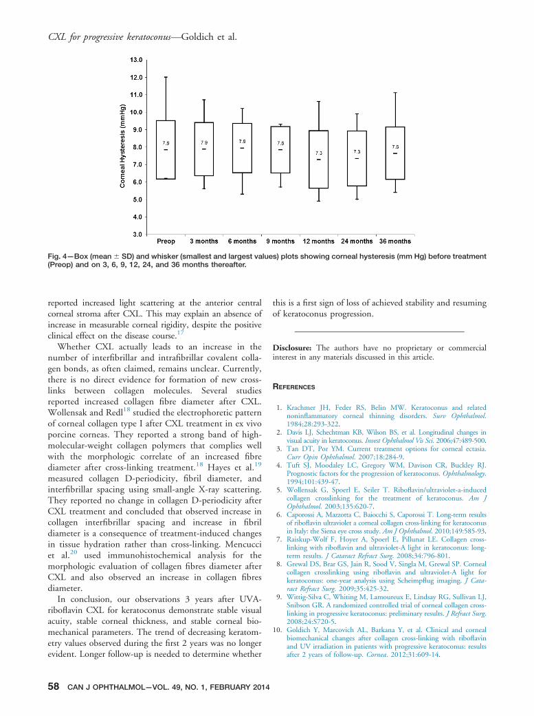
Fig. 4—Box (mean� SD) and whisker (smallest and largest values) plots showing corneal hysteresis (mm Hg) before treatment(Preop) and on 3, 6, 9, 12, 24, and 36 months thereafter.
CXL for progressive keratoconus—Goldich et al.
reported increased light scattering at the anterior centralcorneal stroma after CXL. This may explain an absence ofincrease in measurable corneal rigidity, despite the positiveclinical effect on the disease course.17
Whether CXL actually leads to an increase in thenumber of interfibrillar and intrafibrillar covalent colla-gen bonds, as often claimed, remains unclear. Currently,there is no direct evidence for formation of new cross-links between collagen molecules. Several studiesreported increased collagen fibre diameter after CXL.Wollensak and Redl18 studied the electrophoretic patternof corneal collagen type I after CXL treatment in ex vivoporcine corneas. They reported a strong band of high-molecular-weight collagen polymers that complies wellwith the morphologic correlate of an increased fibrediameter after cross-linking treatment.18 Hayes et al.19
measured collagen D-periodicity, fibril diameter, andinterfibrillar spacing using small-angle X-ray scattering.They reported no change in collagen D-periodicity afterCXL treatment and concluded that observed increase incollagen interfibrillar spacing and increase in fibrildiameter is a consequence of treatment-induced changesin tissue hydration rather than cross-linking. Mencucciet al.20 used immunohistochemical analysis for themorphologic evaluation of collagen fibres diameter afterCXL and also observed an increase in collagen fibresdiameter.
In conclusion, our observations 3 years after UVA-riboflavin CXL for keratoconus demonstrate stable visualacuity, stable corneal thickness, and stable corneal bio-mechanical parameters. The trend of decreasing keratom-etry values observed during the first 2 years was no longerevident. Longer follow-up is needed to determine whether
58 CAN J OPHTHALMOL—VOL. 49, NO. 1, FEBRUARY 2014
this is a first sign of loss of achieved stability and resumingof keratoconus progression.
Disclosure: The authors have no proprietary or commercialinterest in any materials discussed in this article.
REFERENCES
1. Krachmer JH, Feder RS, Belin MW. Keratoconus and relatednoninflammatory corneal thinning disorders. Surv Ophthalmol.1984;28:293-322.
2. Davis LJ, Schechtman KB, Wilson BS, et al. Longitudinal changes invisual acuity in keratoconus. Invest Ophthalmol Vis Sci. 2006;47:489-500.
3. Tan DT, Por YM. Current treatment options for corneal ectasia.Curr Opin Ophthalmol. 2007;18:284-9.
4. Tuft SJ, Moodaley LC, Gregory WM, Davison CR, Buckley RJ.Prognostic factors for the progression of keratoconus. Ophthalmology.1994;101:439-47.
5. Wollensak G, Spoerl E, Seiler T. Riboflavin/ultraviolet-a-inducedcollagen crosslinking for the treatment of keratoconus. Am JOphthalmol. 2003;135:620-7.
6. Caporossi A, Mazzotta C, Baiocchi S, Caporossi T. Long-term resultsof riboflavin ultraviolet a corneal collagen cross-linking for keratoconusin Italy: the Siena eye cross study. Am J Ophthalmol. 2010;149:585-93.
7. Raiskup-Wolf F, Hoyer A, Spoerl E, Pillunat LE. Collagen cross-linking with riboflavin and ultraviolet-A light in keratoconus: long-term results. J Cataract Refract Surg. 2008;34:796-801.
8. Grewal DS, Brar GS, Jain R, Sood V, Singla M, Grewal SP. Cornealcollagen crosslinking using riboflavin and ultraviolet-A light forkeratoconus: one-year analysis using Scheimpflug imaging. J Cata-ract Refract Surg. 2009;35:425-32.
9. Wittig-Silva C, Whiting M, Lamoureux E, Lindsay RG, Sullivan LJ,Snibson GR. A randomized controlled trial of corneal collagen cross-linking in progressive keratoconus: preliminary results. J Refract Surg.2008;24:S720-5.
10. Goldich Y, Marcovich AL, Barkana Y, et al. Clinical and cornealbiomechanical changes after collagen cross-linking with riboflavinand UV irradiation in patients with progressive keratoconus: resultsafter 2 years of follow-up. Cornea. 2012;31:609-14.

CXL for progressive keratoconus—Goldich et al.
11. Vinciguerra P, Albe E, Trazza S, Seiler T, Epstein D. Intra-operative and postoperative effects of corneal collagen cross-linking on progressive keratoconus. Arch Ophthalmol. 2009;127:1258-65.
12. Bourne WM, Nelson LR, Hodge DO. Central corneal endothelialcell changes over a ten-year period. Invest Ophthalmol Vis Sci.1997;38:779-82.
13. Goldich Y, Marcovich AL, Barkana Y, Avni I, Zadok D. Safety ofcorneal collagen cross-linking with UV-A and riboflavin in pro-gressive keratoconus. Cornea. 2010;29:409-11.
14. Goldich Y, Barkana Y, Morad Y, Hartstein M, Avni I, Zadok D.Can we measure corneal biomechanical changes after collagen cross-linking in eyes with keratoconus?—a pilot study Cornea. 2009;28:498-502.
15. Vinciguerra P, Albe E, Mahmoud AM, Trazza S, Hafezi F, RobertsCJ. Intra- and postoperative variation in ocular response analyzer
C
parameters in keratoconic eyes after corneal cross-linking. J RefractSurg. 2010;26:669-76.
16. Sedaghat M, Naderi M, Zarei-Ghanavati M. Biomechanical param-eters of the cornea after collagen crosslinking measured by waveformanalysis. J Cataract Refract Surg. 2010;36:1728-31.
17. Beckman Rehnman J, Janbaz CC, Behndig A, Linden C. Spatialdistribution of corneal light scattering after corneal collagen cross-linking. J Cataract Refract Surg. 2011;37:1939-44.
18. Wollensak G, Redl B. Gel electrophoretic analysis of corneal collagenafter photodynamic cross-linking treatment. Cornea. 2008;27:353-6.
19. Hayes S, Boote C, Kamma-Lorger CS, et al. Riboflavin/UVAcollagen cross-linking-induced changes in normal and keratoconuscorneal stroma. PLoS One. 2011;6:e22405.
20. Mencucci R, Marini M, Paladini I, et al. Effects of riboflavin/UVAcorneal cross-linking on keratocytes and collagen fibres in humancornea. Clin Experiment Ophthalmol. 2010;38:49-56.
AN J OPHTHALMOL—VOL. 49, NO. 1, FEBRUARY 2014 59