copyright © 2010 pearson education, inc. introduction to anatomy & physiology body systems...
TRANSCRIPT

Copyright © 2010 Pearson Education, Inc.
Introduction to Anatomy & Physiology
• Body Systems
• Levels of Organization
• Homeostasis
• Terminology

Copyright © 2010 Pearson Education, Inc.
Anatomy• Anatomy – study of structures of body
– Gross (macroscopic) anatomy - visible to naked eye
• Surface anatomy: superficial features
• Regional “ ”: body areas
• Systemic “ ”: groups of organs
– Microscopic anatomy - cells & molecules
• Cytology: study of cell structure
• Histology: “ ” tissues

Copyright © 2010 Pearson Education, Inc.
Physiology
• Physiology – how organisms perform vital functions
– Cell
– Special : fxns of specific organs
– Systemic : fxns of organ system
– Pathology: effects of diseases on organs/system fxn

Copyright © 2010 Pearson Education, Inc.
Body Systems
• Can you name all the body systems?

Copyright © 2010 Pearson Education, Inc.
Circulatory System
• Transport of materials between all cells of the body
• Heart, Blood Vessels, Blood

Copyright © 2010 Pearson Education, Inc.
Digestive
• Conversion of food into particles that can be transported into the body; elimination of wastes
• Stomach, Intestines, Liver, Pancreas

Copyright © 2010 Pearson Education, Inc.
Endocrine
• Coordination of body function through synthesis and release of regulatory molecules
• Thyroid gland, adrenal gland

Copyright © 2010 Pearson Education, Inc.
Immune
• Defense against foreign invaders
• Thymus, spleen, lymph nodes

Copyright © 2010 Pearson Education, Inc.
Integumentary
• Protection from external environment
• Skin

Copyright © 2010 Pearson Education, Inc.
Muscular
• Allow movement of bones and body parts by working with the nervous and skeletal systems
• Skeletal Muscles

Copyright © 2010 Pearson Education, Inc.
Skeletal
• Support and movement
• Bones

Copyright © 2010 Pearson Education, Inc.
Nervous
• Coordination of body function through electrical signals and release of regulatory molecules
• Brain, spinal cord

Copyright © 2010 Pearson Education, Inc.
Reproductive
• Perpetuation of the species
• Ovaries, uterus, testes
Copyright © 2010 Pearson Education, Inc.
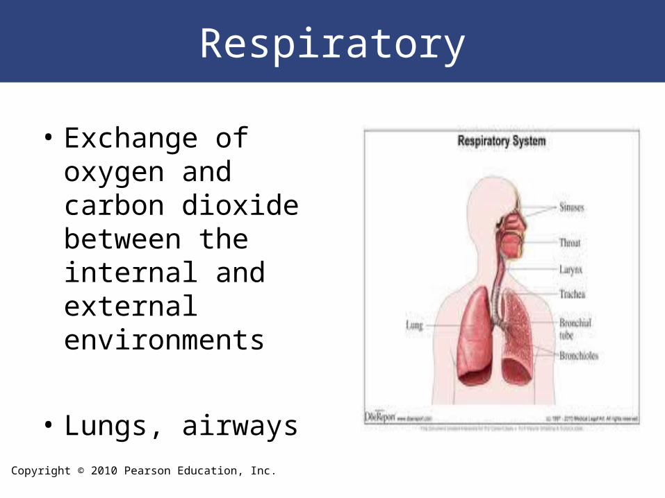
Respiratory
• Exchange of oxygen and carbon dioxide between the internal and external environments
• Lungs, airways

Copyright © 2010 Pearson Education, Inc.
Urinary/Excretory
• Maintenance of water and solutes in the internal environment; waste removal
• Kidneys, bladder

Copyright © 2010 Pearson Education, Inc.
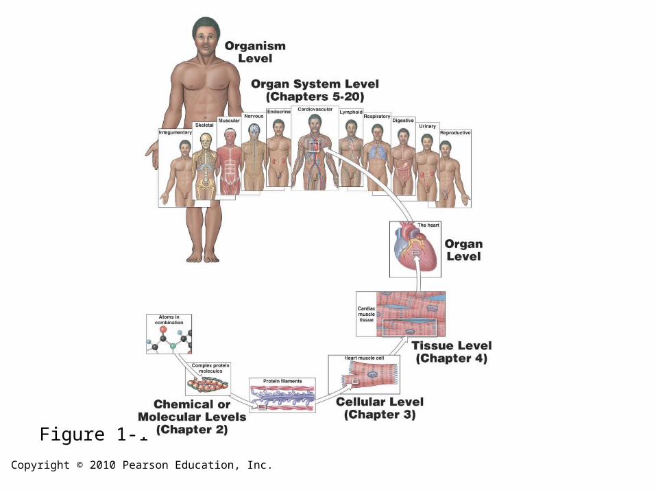
1-3: Levels of Organization
1. Chemical (Molecular) - atoms are smallest chemical units
2. Cellular - smallest unit of structure capable of carrying out life processes
3. Tissue - connections of cells that carry out related functions
4. Organ - Structural and Functional units formed from tissues
5. Organ System - Groups of organs integrating their functions
6. Organism – A living system
*Each level is dependent on the one(s) below it!
Copyright © 2010 Pearson Education, Inc.
Figure 1-1

Copyright © 2010 Pearson Education, Inc.
Homeostasis
• Homeostasis: all body systems working together to maintain a
stable internal environment
1. Receptor – receives stimulus
2. Control Center (Integrator) - processes signal &
sends instructions
3. Effector - carries out response
– failure to restore balance results in illness/disease or death

Copyright © 2010 Pearson Education, Inc.
Figure 1-3

Copyright © 2010 Pearson Education, Inc.
Negative & Positive Feedback
• Negative feedback: variation triggers response that corrects
situation
– response of effector negates stimulus homeostasis
restored
• Positive Feedback: response of the effector reinforces Δ
caused by stimulus body moves away from homeostasis
– Ex: clotting process, childbirth

Copyright © 2010 Pearson Education, Inc.
Figure 1-4

Copyright © 2010 Pearson Education, Inc.
Positive Feedback
Figure 1-5

Copyright © 2010 Pearson Education, Inc.
Anatomy
• Surface Anatomy
– Anatomical position: hands at sides, palms forward
– Supine: lying down, face up
– Prone: “ ”, face down

Copyright © 2010 Pearson Education, Inc.
Anatomical Landmarks. Anterior
Figure 1-6

Copyright © 2010 Pearson Education, Inc.
Anatomical Landmarks. Anterior
Figure 1-6

Copyright © 2010 Pearson Education, Inc.
Anatomical Landmarks. Posterior
Figure 1-6

Copyright © 2010 Pearson Education, Inc.
Anatomical Landmarks. Posterior
Figure 1-6

Copyright © 2010 Pearson Education, Inc.

Copyright © 2010 Pearson Education, Inc.

Copyright © 2010 Pearson Education, Inc.
Abdominopelvic Quadrants
Figure 1-7

Copyright © 2010 Pearson Education, Inc.
Abdominopelvic Regions
Figure 1-7

Copyright © 2010 Pearson Education, Inc.
Abdominopelvic Relationships
Figure 1-7

Copyright © 2010 Pearson Education, Inc.
Abdominopelvic regions

Copyright © 2010 Pearson Education, Inc.
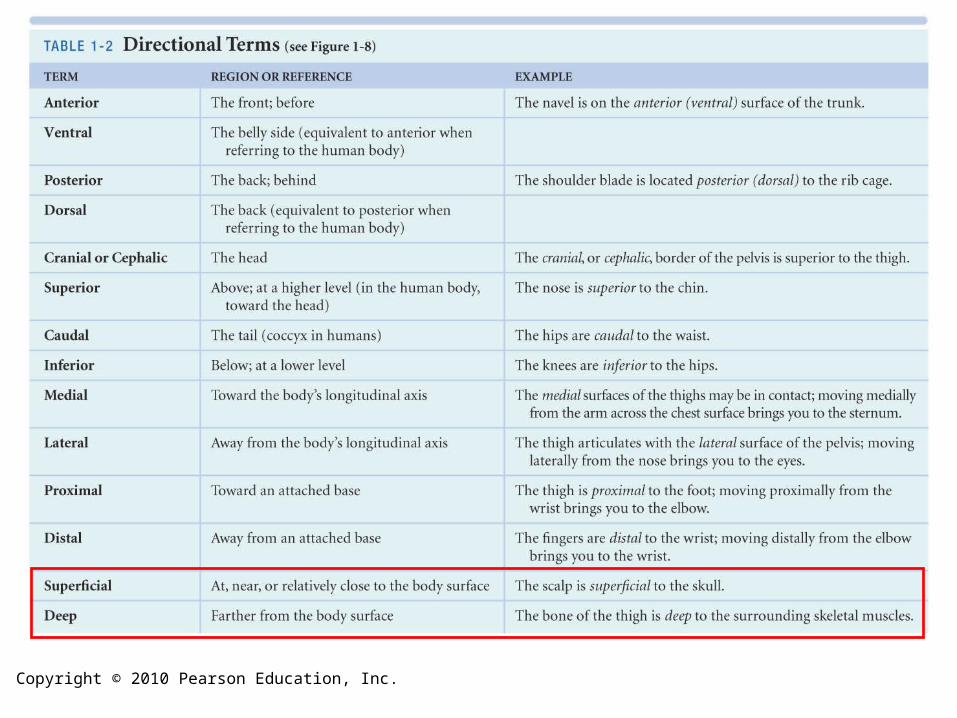
Directional References
Figure 1-8

Copyright © 2010 Pearson Education, Inc.
Figure 1-8
Directional References
Copyright © 2010 Pearson Education, Inc.

Copyright © 2010 Pearson Education, Inc.
Plane of Section
Figure 1-9

Copyright © 2010 Pearson Education, Inc.

Copyright © 2010 Pearson Education, Inc.
Body Cavities
• 2 essential fxns:
1. Protection for organs (shock absorption)
2. Permit Δ’s in size & shape of internal organs
• Serous Membranes - line body cavities & cover organs
– consist of 2 layers:
a) Parietal — lines cavity
b) Visceral — covers organ

Copyright © 2010 Pearson Education, Inc.
Body Cavities• Ventral body cavity (coelom)
– Divided by diaphragm into thoracic & abdominopelvic cavities
– Thoracic cavity
• L & R pleural cavities: contain lungs
• Mediastinum
– contains blood vessels, trachea, esophagus, thymus
– lower portion contains pericardial cavity (holds heart)

Copyright © 2010 Pearson Education, Inc.
Body Cavities– Abdominopelvic / Peritoneal Cavity
• Abdominal cavity — superior portion– contains digestive organs
• Pelvic cavity — inferior portion– contains reproductive organs, rectum, bladder
• Peritoneal cavity — chamber w/in abdominopelvic cavity–Parietal peritoneum lines body wall–Visceral peritoneum covers organs

Copyright © 2010 Pearson Education, Inc.
Figure 1-10

Copyright © 2010 Pearson Education, Inc.
Mediastinum
Figure 14-7a

Copyright © 2010 Pearson Education, Inc.
Figure 16-8b
Copyright © 2010 Pearson Education, Inc.
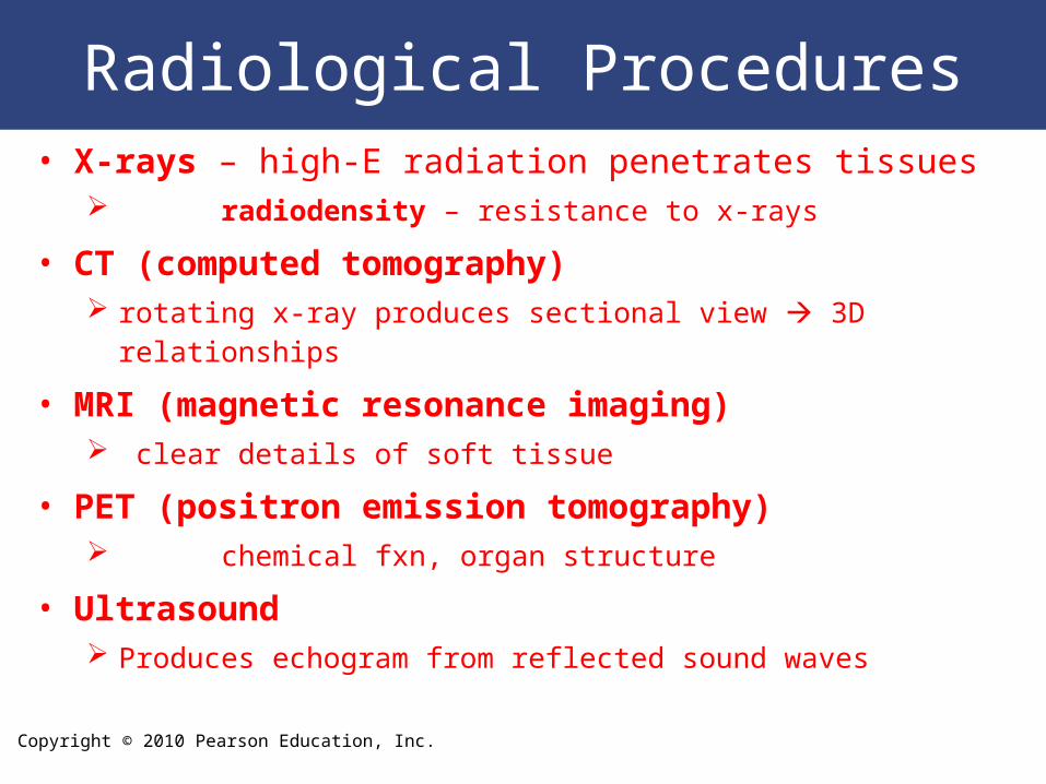
Radiological Procedures• X-rays – high-E radiation penetrates tissues
radiodensity – resistance to x-rays
• CT (computed tomography) rotating x-ray produces sectional view 3D relationships
• MRI (magnetic resonance imaging) clear details of soft tissue
• PET (positron emission tomography) chemical fxn, organ structure
• Ultrasound Produces echogram from reflected sound waves

Copyright © 2010 Pearson Education, Inc.
X-Rays
Figure 1-11

Copyright © 2010 Pearson Education, Inc.
Figure 1-12