copper treatment of peach leaves causes lesion formation
TRANSCRIPT
Copyright © 2019 The Japanese Society for Plant Cell and Molecular Biology
Plant Biotechnology 36, 135–142 (2019)DOI: 10.5511/plantbiotechnology.19.0531b
Copper treatment of peach leaves causes lesion formation similar to the biotic stress response
Fumiyuki Goto1,*,a, Yusuke Enomoto1,2,b, Kazuhiro Shoji1, Hiroaki Shimada2, Toshihiro Yoshihara1
1 Central Research Institute of Electric Power Industry, 1646 Abiko, Abiko-shi, Chiba 270-1194, Japan; 2 Department of Biological Science and Technology, Tokyo University of Science, 6-3-1 Niijyuku, Katsushika-ku, Tokyo 125-8585, Japan* E-mail: [email protected] Tel: +81-952-28-8748 Fax: +81-952-28-8709
Received March 22, 2019; accepted May 31, 2019 (Edited by Y. Kodama)
Abstract Peach (Prunus persica (L.) Batsch) leaves are sensitive to copper (Cu) exposure. The symptoms of Cu exposure are similar to those of bacterial spot disease; however, the mechanism underlying lesion formation caused by Cu exposure is not clear. Here, we investigated whether lesion formation caused by Cu exposure was related to the mechanism underlying plant resistance to microbial pathogens. When Cu was applied to the centre of a pinhole on peach leaves, a two-step process was observed. A pale green section in the shape of a doughnut, located far from a Cu treatment point, first appeared on a leaf treated with 2 mM CuSO4. Next, a yellow–white section gradually spread from the Cu treatment point to the pale green section. Finally, a gap was formed in the middle of the pale green section. The inner part of the pale green section contained 96% of the Cu applied, indicating that Cu is retained in the lesion area. Real-time PCR analysis of the expression of genes encoding pathogenesis-related proteins and enzymes involved in phytoalexin synthesis revealed that three genes (encoding chitinase, pathogenesis-related protein 4, and β-1,3-glucanase-3) of the eight tested were upregulated by Cu treatment. Furthermore, treatment with caspase-1 inhibitors reduced lesion formation. These results show that Cu treatment of peach leaves causes cell death similar to that occurring during the biotic stress response.
Key words: caspase activity, copper, hypersensitive response, peach, programmed cell death.
Introduction
Plants face many stresses, including abiotic stresses such as high temperature, drought, UV exposure, and excess heavy metals, as well as biotic stresses such as infection by viruses, bacteria, and fungi, and attack by insects. The hypersensitive response (HR) is a well-known stress-resistance mechanism in plants. HR consists of programmed cell death (PCD), in which plants kill their own cells in tissues infected with pathogens or damaged by abiotic stress (Beers and McDowell 2001; Kunstler et al. 2016; Pasqualini et al. 2003; Petrov et al. 2015; Tiwari et al. 2002), and the biosynthesis of pathogen-related (PR) proteins and phytoalexins, which suppress the multiplication of microorganisms (Jain and Khurana 2018).
PCD is effective in protecting a host plant from attack by microbes and viruses (De Wit 1997). Caspase activity is thought to be involved in PCD in plants (Coffeen and Wolpert 2004; Filippi et al. 2019; Ge et al. 2016; Lam and del Pozo 2000; Rotari et al. 2005; Sanmartin et al. 2005). For instance, the N gene in tobacco is a known resistance
gene against tobacco mosaic virus (TMV). Transgenic plants with an N background that harbour p35, which encodes a broad-range caspase inhibitor of vaculovirus, show partial inhibition of HR triggered by TMV (del Pozo and Lam 2003). Danon et al. (2004) showed that ultraviolet-C exposure induced caspase-3-like activity and PCD, which could be suppressed by caspase-1, caspase-3, and pan-caspase inhibitors in Arabidopsis. Kim et al. (2003) reported the increased activity of caspase-3-like and caspase-9-like following PCD induced by viral infection. Moreover, Hatsugai et al. (2004) showed that virus-induced PCD is mediated by vacuolar processing enzyme (VPE), which has caspase-1 activity in tobacco; its activity was inhibited by a caspase-1 inhibitor, resulting in the suppression of PCD. These reports indicate that caspase activities play a critical role in PCD induced by both biotic and abiotic stresses (Hatsugai et al. 2015).
A pesticide containing copper (Cu), called Bordeaux mixture, has been used since the 19th century. Bordeaux mixture is a safe pesticide against fungal and bacterial diseases despite its extremely high copper
aPresent address: Faculty of Agriculture, Saga University, 1 Honjo, Saga-shi, Saga 840-8502, Japan bPresent address: Hiroo Gakuen Junior & Senior High School, 5-1-14 Minami Azabu, Minato-ku, Tokyo 106-0047, JapanThis article can be found at http://www.jspcmb.jp/Published online September 7, 2019
Original Paper
136 Cu-induced cell death of peach leaves
Copyright © 2019 The Japanese Society for Plant Cell and Molecular Biology
concentration, and is registered as a pesticide for use in organic cultivation. Small volumes of Cu ions gradually elute from the mixture during crop cultivation. Cu is an essential element for living organisms, but excess Cu is harmful not only to microbes but also to higher plants. Indeed, some plants show symptoms in response to Cu treatment. For example, Pontier et al. (1999) found that cell death markers of the HR (i.e., HIN1 and HSR203J) were induced by Cu treatment in tobacco leaves. In Nicotiana glutinosa, mRNA expression of npRBP (an RNA-binding PR protein) was induced not only by bacterial infection but also by Cu treatment (Naqvi et al. 1998). Additionally, Rakwal et al. (2004) showed that chitinase, another type of PR protein, was induced by Cu, jasmonete, and ethylene in the leaves of rice seedlings. In Phaseolus vulgaris, excess Cu was reported to have an effect on photosynthesis in competition with iron (Fe) and on the accumulation of PR proteins (Cuypers et al. 2005; Patsikka et al. 2002). Furthermore, Cu causes oxidative stress, resulting in an increase in the activities of superoxide dismutase and guaiacol peroxidase and a reduction in catalase activity in Arabidopsis (Drazkiewicz et al. 2004). In recent years, the relationships between PCD and metals such as Cu, aluminium (Al), cadmium (Cd), silver, tungsten, and zinc (Zn) have been reported (Adamakis et al. 2011; Filippi et al. 2019; Huang et al. 2014; Kirisiuk et al. 2016; Xu et al. 2010, 2013).
Spraying Bordeaux mixture onto a peach tree after the fruit’s harvest is effective in preventing the spread of Xanthomonas campestris pv. pruni, which causes the serious disease bacterial spots. However, because peach leaves are sensitive to Cu, they are often severely damaged by the mixture. Leaf symptoms may or may not occur depending on the weather conditions and the Cu concentration in Bordeaux mixture. They include the development of spots and turning yellow and dropping early like leaves infected with X. campestris pv. pruni. Little information is known about the relationship between the two phenomena at the physiological and molecular levels, with the exception of a report describing the anatomical similarity between lesions that develop because of Cu treatment and bacterial infection (Akai 1943). In this study, to test the hypothesis that the lesion formation on peach leaves in response to Cu treatment is related to HR, which is considered a response to biotic stress, we investigated the gene expression of PR proteins thought to be induced by HR after lesion formation was noted. We further tested whether PCD mediated by caspase-1 activity was suppressed by the caspase-1 inhibitor Ac-YVAD-CHO.
Materials and methods
Plant materialTwo-year-old peach (Prunus persica (L.) Batsch cv.
Kawanakajima-Hakutou) and plum (Prunus salicina L. cv. Soldum) trees were grown in 1/2000 Wagner pots in an air-conditioned greenhouse at 25°C. For peach vernalisation, trees whose leaves had started falling and in which small buds had started emerging were kept in a dark room at 4°C for 2.5 months. The trees were then transferred into a greenhouse under conditions of controlled light (16 h per day) and temperature (20–25°C).
Lesion formation from Cu treatmentExpanded leaves located at the top of each branch were used for the lesion formation experiment. A total of 2 µl of various concentrations of CuSO4 (ranging from 0–2 mM) was dropped onto the leaf surface using a mechanical pipette. The leaves were harvested 1 week after Cu treatment and scanned immediately using a GT-9700F scanner (Epson, Suwa, Japan). The lesion area was measured using the NIH Scion Image software program. Detailed analysis of lesion formation after 2 mM CUSO4 treatment was performed using a light microscope (Nikon, Tokyo, Japan) every day for 8 days.
Trypan blue stainingPlant cell death induced by Cu treatment was monitored by lactophenol-trypan blue staining using methods reported by Koch and Slusarenko (1990). A total of 2 µl of 1 mM CuSO4 was dropped on a pinhole on a leaf, and the leaf was harvested at 4 and 5 days after Cu treatment. After staining with lactophenol-trypan blue, the leaf was mounted on a glass slide in 60% glycerol and observed using the SteREO Lumar.V12stereoscopic microscope (Carl Zeiss, Oberkochen, Germany).
Metal extraction and measurementsFive samples for metal extraction were obtained from detached pieces of a leaf treated with 2 µl of 10 mM CuSO4. Samples were dried at 60°C for 1 week, weighed, and mineralised with HNO3 and H2O2 according to the methods reported by Goto et al. (1999). The metal concentration was determined by measuring the absorbance of Cu, Fe, Zn, calcium, magnesium, and manganese (Mn) at 324.754, 259.940, 213.856, 393.366, 279.553, and 257.610 nm, respectively, using inductively coupled plasma spectrometry (P-4000; Hitachi, Tokyo, Japan).
RNA isolation and PCRA total of 2 µl of 2 mM CuSO4 solution or water as a control was dropped onto each of 20 pinholes on the second leaves from the top of each branch, each of which had expanded to a length of approximately 10 cm. The leaves were harvested at 0, 6, and 24 h after treatment, frozen immediately, and stored for reverse transcription (RT)-PCR and real-time RT-PCR analyses. RNA was extracted according to the method reported by Jaakola et al. (2001). Crude RNA was treated with DNase I (Takara, Tokyo, Japan) for 30 min at 37°C. Total RNA (10 µg) was used for reverse transcription using ReverTra Ace (Toyobo, Tokyo, Japan) for 20 min at 42°C, denatured for 5 min at 96°C, and
F. Goto et al. 137
Copyright © 2019 The Japanese Society for Plant Cell and Molecular Biology
kept for 5 min at 4°C. PCR was performed using the reverse-transcribed RNA as a template and ExTaq (TaKaRa) under the following conditions: 94°C for 1 min, then 30 cycles of 94°C for 30 s, 53°C (55°C for actin amplification) for 30 s, and 72°C for 30 s. The target genes for peach were Mn superoxide dismutase (Mn-SOD: AJ238316), basic chitinase (CHI: AF206635), pathogenesis-related protein 4 (PR4: AF362989), phenylalanine ammonia lyase (PAL: AF206634), polyglucanase-inhibiting protein (PGIP: AY352426), β-1,3-glucanase 1, 2, and 3 (Gns1: U49454, Gns2: AF435088, and Gns3: AF435089), and actin (AB046952) as an internal expression control. CHI, PR4, PAL, PGIP, and Gns3 expression was analysed by real-time RT-PCR in a Smart Cycler (Cepheid, Sunnyvale, CA) with SYBR Premix Ex Taq (TaKaRa), according to the manufacturer’s instructions. The accession numbers of template genes and primer sets for PCR are provided in Table 1.
Caspase inhibitor experimentA young leaf grown in a greenhouse after vernalisation was used for the caspase-1 inhibitor experiment. The leaf was rubbed 40 times between fingers, then carborundum (Carborundum 400 mesh; Hayashi Chemicals, Tokyo, Japan) was applied on six spots on the leaf with 10 µl water. Subsequently, the carborundum was washed away, and remaining water was wiped off. A total of 20 µl of 1 mM Ac-YVAD-CHO or leupeptin (Ac-LLR-CHO) dissolved in 1% dimethyl sulfoxide (DMSO) was dropped onto three of the six spots. After 3 h, 20 µl of CuSO4 solution (0, 1, or 2 mM) was dropped onto the spots. As a control, 20 µl of 1% DMSO was dropped onto the remaining three spots.
Results
Lesion formation consisted of two stepsThe lesion area of peach leaves treated with various concentrations of Cu was measured to determine the
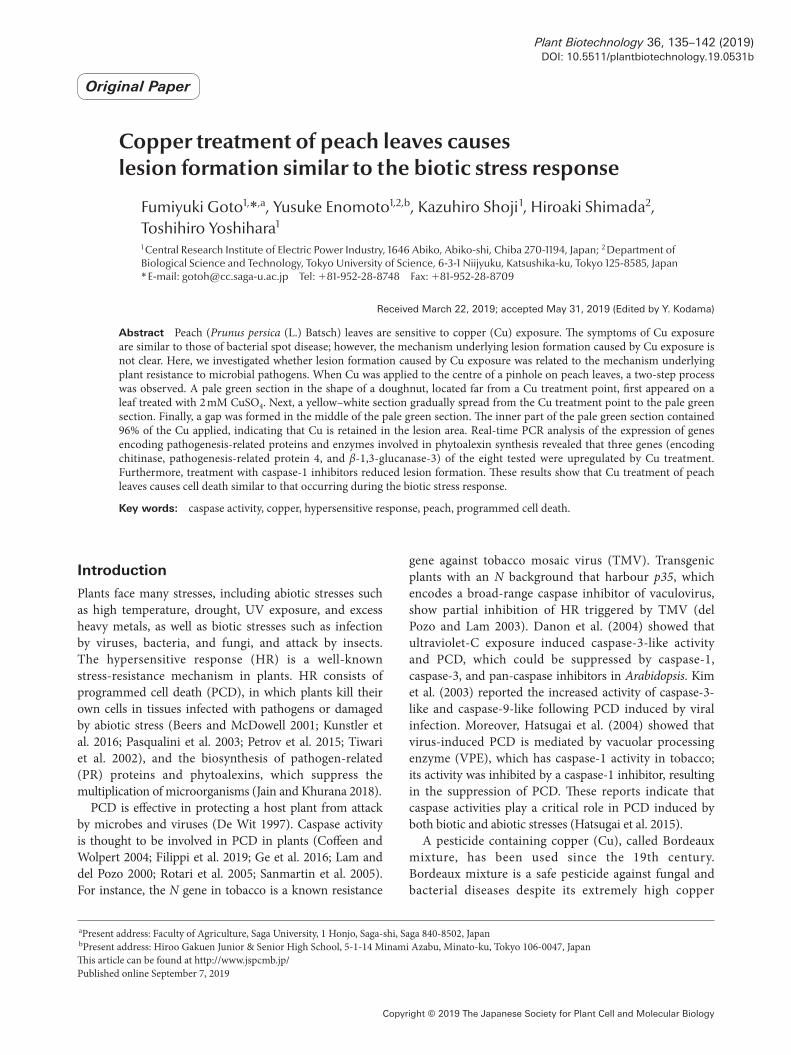
relationship between Cu concentration and lesion severity (Figure 1A). Direct application of 2 µl 0–2 mM CuSO4 onto the leaf had no effect, so we next injured the
Table 1. Primer sequences.
Gene Accession # Forward Reverse
For RT-PCRMn-SOD AJ238316 5′-GCTTGGTATTGATGTTTGGG-3′ 5′-GACGACAAAGCTTATTTCAGG-3′CHI AF206635 5′-GACTTCCCATGAAACTACAGG-3′ 5′-GATCACATCATGGCATGAGG-3′PR4 AF362989 5′-ACATGGGATGCTGATAAGCC-3′ 5′-GCCACAGTCAACAAAGTCGT-3′PAL AF206634 5′-AAAGGTGCATGACTTAGATCC-3′ 5′-CAATTTCAGCACCTTTGAACC-3′PGIP AY352426 5′-CAATACAACCCTCCATTGCC-3′ 5′-AGTCTATGCGGTTGAAGTCC-3′Gns1 U49454 5′-TCGTCATCAGTTGGCAGACT-3′ 5′-CCCAATGTTTCTCAAGCTCT-3′Gns2 AF435088 5′-TCGTCATCAGTTGGCAGATG-3′ 5′-CCCAATGTTTCTCAAGCTCG-3′Gns3 AF435089 5′-AGTCGGTAGAGGCCCTTCAA-3′ 5′-AGCCCCCAATGTTTCTCCAA-3′Actin AB046952 5′-CTTTAATGTGCCTGCCATGT-3′ 5′-TCGCACTTCATGATGGAGTT-3′
For real-time RT-PCRCHI 5′-GGGGTATTGCTATCTCAAGG-3′ 5′-GCAGCACATGGATAATTAGG-3′PR4 5′-ACATGGGATGCTGATAAGCC-3′ 5′-TGTCACCAGTAAGCACTTCC-3′PAL 5′-ATTAGCCATTGCTGCTATTGG-3′ 5′-GCTTCCTGTGAGATTTGAAGG-3′PGIP 5′-CAATACAACCCTCCATTGCC-3′ 5′-AGAGCCTGAGATGTTAGTCC-3′Gns3 5′-ACTTTTGAACCCCATCATCC-3′ 5′-CTGAAATACGGGTACAAATTAACC-3′Actin 5′-ATGCCATCCTTCGTCTTGACC-3′ 5′-AGCAGTAGTGGTGAACATGTAACC-3′
Figure 1. Effect of Cu treatment on lesion formation. (a) The photograph was taken 12 days after Cu treatment at various concentrations (mM) a: 0, b: 0.02, c: 0.2, d: 0.5, e: 0.75, f: 1.0, g: 1.5, h: 2.0. (b) Effect of Cu on lesion formation on a peach leaf. The values provided are the mean values of 15 independent samples. Standard deviations are indicated by vertical bars.
138 Cu-induced cell death of peach leaves
Copyright © 2019 The Japanese Society for Plant Cell and Molecular Biology
leaf surface with a needle (pinhole treatment) before Cu treatment. No lesion was observed on leaves treated with 0, 0.02, or 0.2 mM CuSO4. However, lesion formation following the use of more than 0.3 mM Cu and the lesion area for treatment with over 0.4 mM Cu were almost constant, irrespective of the Cu concentration (Figure 1B). This suggested the existence of a Cu stimulation threshold. Some of the lesion areas fell out several days after Cu treatment. We measured lesion areas of plum and peach leaves to verify that Cu-induced lesion
formation is common in the same genus. However, in contrast to the results for peach leaves, lesions on plum leaves were restricted to the area around the pinhole, regardless of the Cu concentration used (Figure 1A). Moreover, no excision was observed more than 20 days later.
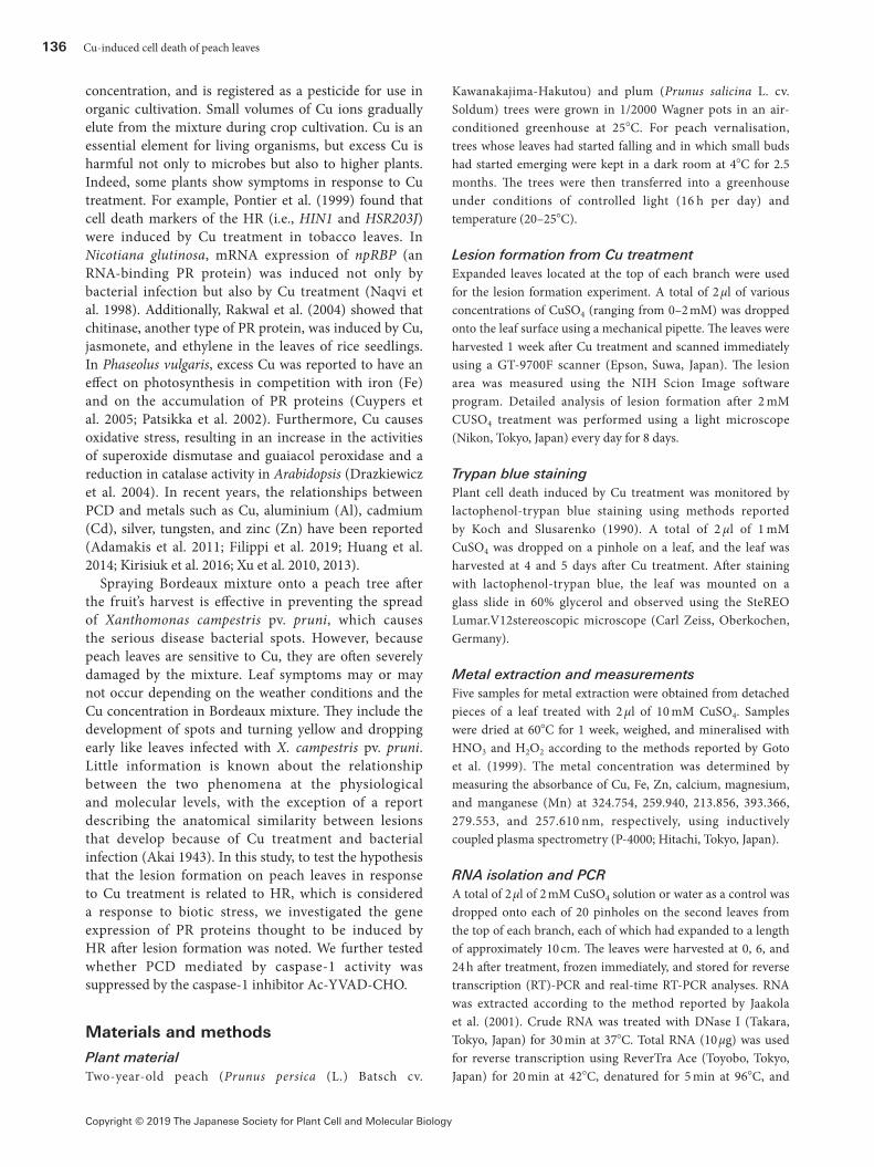
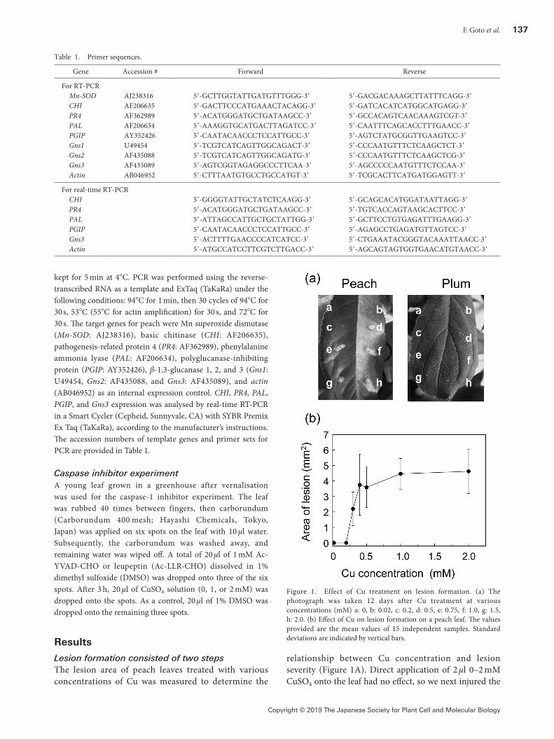
Next, we observed the process of lesion formation on leaves treated with Cu (Figure 2). A pale green doughnut-shaped area 1 mm wide appeared approximately 2 mm from a Cu treatment point 3 days after treatment. Five days after treatment, the inside of the ‘doughnut’ turned yellow–white from the point of treatment to the edge of the doughnut. By day 8, visible cell death had started within an approximately 0.5-mm radius from the doughnut, resulting in gap formation by day 10. The shape of the lesion varied from a circle to an oval with angular parts. Younger leaves showed earlier gap formation after the appearance of the doughnut-shaped area, while some mature leaves formed a reddish-purple doughnut with no gaps. Trypan blue staining results shown in Figure 3 indicate a two-step cell death. Scattered blue staining was observed in the yellow doughnut on days 4 and 5, but a blue line was only detected on day 5. In this case, days 4 and 5 correspond to days 6 and 8 in Figure 2.
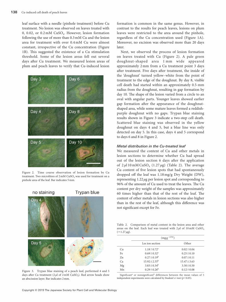
Metal distribution in the Cu-treated leafWe measured the content of Cu and other metals in lesion sections to determine whether Cu had spread out of the lesion section 6 days after the application of 2 µl 10 mM CuSO4 (1.27 µg) (Table 2). The average Cu content of five lesion spots that had spontaneously dropped off the leaf was 1.18 mg/g Dry Weight (DW), representing 1.22 µg per lesion spot and corresponding to 96% of the amount of Cu used to treat the leaves. The Cu content per dry weight of the samples was approximately 60 times higher than that of the rest of the leaf. The content of other metals in lesion sections was also higher than in the rest of the leaf, although this difference was not significant except for Fe.
Figure 2. Time course observation of lesion formation by Cu treatment. Two microlitres of 2 mM CuSO4 was used for treatment on a pinhole area of the leaf. Bar indicates 5 mm.
Figure 3. Trypan blue staining of a peach leaf, performed 4 and 5 days after Cu treatment (2 µl of 2 mM CuSO4). Red arrow heads show an abscission layer. Bar indicates 2 mm.
Table 2. Comparison of metal content in the lesion area and other areas on the leaf. Each leaf was treated with 2 µl of 10 mM CuSO4 (=1.27 µg).
(mg g−1 DW)
Les ion section Other
Cu 1.18±0.72a 0.02±0.04Fe 0.69±0.32a 0.25±0.10Zn 0.27±0.19b 0.07±0.11Ca 15.92±2.55b 15.47±3.63Mg 3.83±0.34b 3.50±0.50Mn 0.29±0.26b 0.12±0.08
Significanta or nonsignificantb differences between the mean values of 5 independent experiments were calculated by Student’s t-test (p<0.05).
F. Goto et al. 139
Copyright © 2019 The Japanese Society for Plant Cell and Molecular Biology
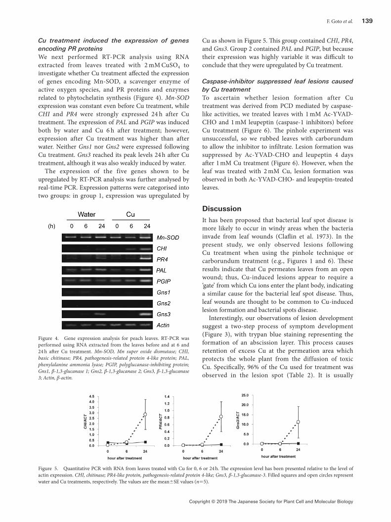
Cu treatment induced the expression of genes encoding PR proteinsWe next performed RT-PCR analysis using RNA extracted from leaves treated with 2 mM CuSO4 to investigate whether Cu treatment affected the expression of genes encoding Mn-SOD, a scavenger enzyme of active oxygen species, and PR proteins and enzymes related to phytochelatin synthesis (Figure 4). Mn-SOD expression was constant even before Cu treatment, while CHI and PR4 were strongly expressed 24 h after Cu treatment. The expression of PAL and PGIP was induced both by water and Cu 6 h after treatment; however, expression after Cu treatment was higher than after water. Neither Gns1 nor Gns2 were expressed following Cu treatment. Gns3 reached its peak levels 24 h after Cu treatment, although it was also weakly induced by water.
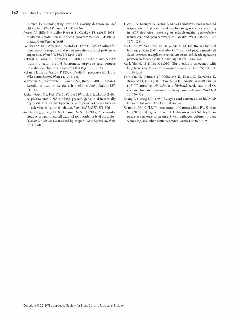
The expression of the five genes shown to be upregulated by RT-PCR analysis was further analysed by real-time PCR. Expression patterns were categorised into two groups: in group 1, expression was upregulated by
Cu as shown in Figure 5. This group contained CHI, PR4, and Gns3. Group 2 contained PAL and PGIP, but because their expression was highly variable it was difficult to conclude that they were upregulated by Cu treatment.
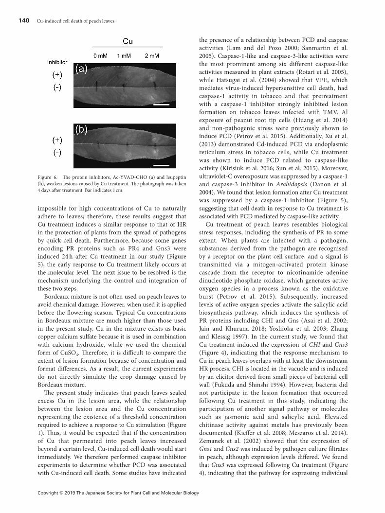
Caspase-inhibitor suppressed leaf lesions caused by Cu treatmentTo ascertain whether lesion formation after Cu treatment was derived from PCD mediated by caspase-like activities, we treated leaves with 1 mM Ac-YVAD-CHO and 1 mM leupeptin (caspase-1 inhibitors) before Cu treatment (Figure 6). The pinhole experiment was unsuccessful, so we rubbed leaves with carborundum to allow the inhibitor to infiltrate. Lesion formation was suppressed by Ac-YVAD-CHO and leupeptin 4 days after 1 mM Cu treatment (Figure 6). However, when the leaf was treated with 2 mM Cu, lesion formation was observed in both Ac-YVAD-CHO- and leupeptin-treated leaves.
Discussion
It has been proposed that bacterial leaf spot disease is more likely to occur in windy areas when the bacteria invade from leaf wounds (Claflin et al. 1973). In the present study, we only observed lesions following Cu treatment when using the pinhole technique or carborundum treatment (e.g., Figures 1 and 6). These results indicate that Cu permeates leaves from an open wound; thus, Cu-induced lesions appear to require a ‘gate’ from which Cu ions enter the plant body, indicating a similar cause for the bacterial leaf spot disease. Thus, leaf wounds are thought to be common to Cu-induced lesion formation and bacterial spots disease.
Interestingly, our observations of lesion development suggest a two-step process of symptom development (Figure 3), with trypan blue staining representing the formation of an abscission layer. This process causes retention of excess Cu at the permeation area which protects the whole plant from the diffusion of toxic Cu. Specifically, 96% of the Cu used for treatment was observed in the lesion spot (Table 2). It is usually
Figure 4. Gene expression analysis for peach leaves. RT-PCR was performed using RNA extracted from the leaves before and at 6 and 24 h after Cu treatment. Mn-SOD, Mn super oxide dismutase; CHI, basic chitinase; PR4, pathogenesis-related protein 4-like protein; PAL, phenylalanine ammonia lyase; PGIP, polyglucanase-inhibiting protein; Gns1, β-1,3-glucanase 1; Gns2, β-1,3-glucanase 2; Gns3, β-1,3-glucanase 3; Actin, β-actin.
Figure 5. Quantitative PCR with RNA from leaves treated with Cu for 0, 6 or 24 h. The expression level has been presented relative to the level of actin expression. CHI, chitinase; PR4-like protein, pathogenesis-related protein 4-like; Gns3, β-1,3-glucanase-3. Filled squares and open circles represent water and Cu treatments, respectively. The values are the mean±SE values (n=5).
140 Cu-induced cell death of peach leaves
Copyright © 2019 The Japanese Society for Plant Cell and Molecular Biology
impossible for high concentrations of Cu to naturally adhere to leaves; therefore, these results suggest that Cu treatment induces a similar response to that of HR in the protection of plants from the spread of pathogens by quick cell death. Furthermore, because some genes encoding PR proteins such as PR4 and Gns3 were induced 24 h after Cu treatment in our study (Figure 5), the early response to Cu treatment likely occurs at the molecular level. The next issue to be resolved is the mechanism underlying the control and integration of these two steps.
Bordeaux mixture is not often used on peach leaves to avoid chemical damage. However, when used it is applied before the flowering season. Typical Cu concentrations in Bordeaux mixture are much higher than those used in the present study. Cu in the mixture exists as basic copper calcium sulfate because it is used in combination with calcium hydroxide, while we used the chemical form of CuSO4. Therefore, it is difficult to compare the extent of lesion formation because of concentration and format differences. As a result, the current experiments do not directly simulate the crop damage caused by Bordeaux mixture.
The present study indicates that peach leaves sealed excess Cu in the lesion area, while the relationship between the lesion area and the Cu concentration representing the existence of a threshold concentration required to achieve a response to Cu stimulation (Figure 1). Thus, it would be expected that if the concentration of Cu that permeated into peach leaves increased beyond a certain level, Cu-induced cell death would start immediately. We therefore performed caspase inhibitor experiments to determine whether PCD was associated with Cu-induced cell death. Some studies have indicated
the presence of a relationship between PCD and caspase activities (Lam and del Pozo 2000; Sanmartin et al. 2005). Caspase-1-like and caspase-3-like activities were the most prominent among six different caspase-like activities measured in plant extracts (Rotari et al. 2005), while Hatsugai et al. (2004) showed that VPE, which mediates virus-induced hypersensitive cell death, had caspase-1 activity in tobacco and that pretreatment with a caspase-1 inhibitor strongly inhibited lesion formation on tobacco leaves infected with TMV. Al exposure of peanut root tip cells (Huang et al. 2014) and non-pathogenic stress were previously shown to induce PCD (Petrov et al. 2015). Additionally, Xu et al. (2013) demonstrated Cd-induced PCD via endoplasmic reticulum stress in tobacco cells, while Cu treatment was shown to induce PCD related to caspase-like activity (Kirisiuk et al. 2016; Sun et al. 2015). Moreover, ultraviolet-C overexposure was suppressed by a caspase-1 and caspase-3 inhibitor in Arabidopsis (Danon et al. 2004). We found that lesion formation after Cu treatment was suppressed by a caspase-1 inhibitor (Figure 5), suggesting that cell death in response to Cu treatment is associated with PCD mediated by caspase-like activity.
Cu treatment of peach leaves resembles biological stress responses, including the synthesis of PR to some extent. When plants are infected with a pathogen, substances derived from the pathogen are recognised by a receptor on the plant cell surface, and a signal is transmitted via a mitogen-activated protein kinase cascade from the receptor to nicotinamide adenine dinucleotide phosphate oxidase, which generates active oxygen species in a process known as the oxidative burst (Petrov et al. 2015). Subsequently, increased levels of active oxygen species activate the salicylic acid biosynthesis pathway, which induces the synthesis of PR proteins including CHI and Gns (Asai et al. 2002; Jain and Khurana 2018; Yoshioka et al. 2003; Zhang and Klessig 1997). In the current study, we found that Cu treatment induced the expression of CHI and Gns3 (Figure 4), indicating that the response mechanism to Cu in peach leaves overlaps with at least the downstream HR process. CHI is located in the vacuole and is induced by an elicitor derived from small pieces of bacterial cell wall (Fukuda and Shinshi 1994). However, bacteria did not participate in the lesion formation that occurred following Cu treatment in this study, indicating the participation of another signal pathway or molecules such as jasmonic acid and salicylic acid. Elevated chitinase activity against metals has previously been documented (Kieffer et al. 2008; Meszaros et al. 2014). Zemanek et al. (2002) showed that the expression of Gns1 and Gns2 was induced by pathogen culture filtrates in peach, although expression levels differed. We found that Gns3 was expressed following Cu treatment (Figure 4), indicating that the pathway for expressing individual
Figure 6. The protein inhibitors, Ac-YVAD-CHO (a) and leupeptin (b), weaken lesions caused by Cu treatment. The photograph was taken 4 days after treatment. Bar indicates 1 cm.

F. Goto et al. 141
Copyright © 2019 The Japanese Society for Plant Cell and Molecular Biology
isoforms of β-1,3-glucanase depends on the stress that the plants are exposed to. PAL is induced by elicitor treatment and plays an important role in the synthesis of phytoalexins (Katz et al. 1998), but it is also induced by other stimuli including those without elicitor activity (Chalker-Scott 1999). Therefore, it is unclear from our current findings whether Cu functions as an elicitor to induce PAL.
Acknowledgements
We thank Ms. Kyoko Tsunokawa (CRIEPI) for her technical assistance and Dr. Maki Kawai (Saitama University) for revising the manuscript. Special thanks are due to Mr. Masahiko Kobayashi (Tokyo University of Science) for his research assistance. We thank Sarah Williams, PhD, from Edanz Group (www.edanzediting.com) for editing a draft of this manuscript.
References
Adamakis IDS, Panteris E, Eleftheriou EP (2011) The fatal effect of tungsten on Pisum sativum L., root cells: Indications for endoplasmic reticulum stress-induced programmed cell death. Planta 234: 21–34
Akai S (1943) Studies on anatomy of the leaves of the genus Prunus suffering bacterial spot disease. Shokubutsu Oyobi Doubutsu 11: 789–792
Asai T, Tena G, Plotnikova J, Willmann MR, Chiu WL, Gomez-Gomez L, Boller T, Ausubel FM, Sheen J (2002) MAP kinase signalling cascade in Arabidopsis innate immunity. Nature 415: 977–983
Beers EP, McDowell JM (2001) Regulation and execution of programmed cell death in response to pathogens, stress and developmental cues. Curr Opin Plant Biol 4: 561–567
Chalker-Scott L (1999) Environmental significance of anthocyanins in plant stress responses. Photochem Photobiol 70: 1–9
Claflin LE, Stuteville DL, Armbrust DV (1973) Wind-blown soil in the epidemiology of bacterial leaf spot of alfalfa and common blight of bean. Phytopathology 63: 1417–1419
Coffeen WC, Wolpert TJ (2004) Purification and characterization of serine proteases that exhibit caspase-like activity and are associated with programmed cell death in Avena sativa. Plant Cell 16: 857–873
Cuypers A, Koistinen KM, Kokko H, Karenlampi S, Auriola S, Vangronsveld J (2005) Analysis of bean (Phaseolus vulgaris L.) proteins affected by copper stress. J Plant Physiol 162: 383–392
Danon A, Rotari VI, Gordon A, Mailhac N, Gallois P (2004) Ultraviolet-C overexposure induces programmed cell death in Arabidopsis, which is mediated by caspase-like activities and which can be suppressed by caspase inhibitor, p35 and Defender against apoptotic death. J Biol Chem 279: 779–787
De Wit PJGM (1997) Pathogen avirulence and plant resistance: A key role for recognition. Trends Plant Sci 2: 452–458
del Pozo O, Lam E (2003) Expression of the baculovirus p35 protein in tobacco affects cell death progression and compromises N gene-mediated disease resistance response to Tobacco mosaic virus. Mol Plant Microbe Interact 16: 485–494
Drążkiewicz M, Skórzyńska-Polit E, Krupa Z (2004) Copper-induced oxidative stress and antioxidant defense in Arabidopsis thaliana. Biometals 17: 379–387
Filippi A, Zancani M, Petrussa E, Braidot E (2019) Caspase-3-like
activity and proteasome degradation in grapevine suspension cell cultures undergoing silver-induced programmed cell death. J Plant Physiol 233: 42–51
Fukuda Y, Shinshi H (1994) Characterization of a novel cis-acting element that is responsive to a fungal elicitor in the promoter of a tobacco class I chitinase gene. Plant Mol Biol 24: 485–493
Ge Y, Cai YM, Bonneau L, Rotari V, Danon A, McKenzie EA, McLellan H, Mach L, Gallois P (2016) Inhibition of cathepsin B by caspase-3 inhibitors blocks programmed cell death in Arabidopsis. Cell Death Differ 23: 1493–1501
Goto F, Yoshihara T, Shigemoto N, Toki S, Takaiwa F (1999) Iron fortification of rice seed by the soybean ferritin gene. Nat Biotechnol 17: 282–286
Hatsugai N, Kuroyanagi M, Yamada K, Meshi T, Tsuda S, Kondo M, Nishimura M, Hara-Nishimura I (2004) A plant vacuolar protease, VPE, mediates virus-induced hypersensitive cell death. Science 305: 855–858
Hatsugai N, Yamada K, Goto-Yamada S, Hara-Nishimura I (2015) Vacuolar processing enzyme in plant programmed cell death. Front Plant Sci 6: 234
Huang W, Yang X, Yao S, LwinOo T, He H, Wang A, Li C, He L (2014) Reactive oxygen species burst induced by aluminium stress triggers mitochondria-dependent programmed cell death in peanut root tip cells. Plant Physiol Biochem 82: 76–84
Jaakola L, Pirttila AM, Halonen M, Hohtola A (2001) Isolation of high quality RNA from bilberry (Vaccinium myrtillus L.) fruit. Mol Biotechnol 19: 201–203
Jain D, Khurana JP (2018) Role of Pathogenesis-Related (PR) proteins in plant defense mechanism. In: Singh A, Singh I (eds) Molecular Aspects of Plant-Pathogen Interaction. Springer, Singapore, pp 265–281
Katz VA, Thulke OU, Conrath U (1998) A benzothiadiazole primes parsley cells for augmented elicitation of defense responses. Plant Physiol 117: 1333–1339
Kieffer P, Dommes J, Hoffmann L, Hausman JF, Renaut J (2008) Quantitative changes in protein expression of cadmium-exposed poplar plants. Proteomics 8: 2514–2530
Kim M, Ahn JW, Jin UH, Choi D, Paek KH, Pai HS (2003) Activation of the programmed cell death pathway by inhibition of proteasome function in plants. J Biol Chem 278: 19406–19415
Kirisiuk Y, Mackievic V, Zvanarou S, Przhevalskaya D, Leschenka Y, Demidchik V (2016) The effect of copper nanoparticles on growth, cell viability and signaling processes of wheat plants. Plant Signalling & Behavior 4th International Symposium Proceedings, p123
Koch E, Slusarenko A (1990) Arabidopsis is susceptible to infection by a downy mildew fungus. Plant Cell 2: 437–445
Künstler A, Bacsó R, Gullner G, Hafez YM, Király L (2016) Staying alive: Is cell death dispensable for plant disease resistance during the hypersensitive response? Physiol Mol Plant Pathol 93: 75–84
Lam E, del Pozo O (2000) Caspase-like protease involvement in the control of plant cell death. Plant Mol Biol 44: 417–428
Mészáros P, Rybanský L, Spieß N, Socha P, Kuna R, Libantová J, Moravčíková J, Piršelová B, Hauptvogel P, Matušíková I (2014) Plant chitinase response to different metal-type stresses reveal specificity. Plant Cell Rep 33: 1789–1799
Pasqualini S, Piccioni C, Reale L, Ederli L, Della Torre G, Ferranti F (2003) Ozone-induced cell death in tobacco cultivar Bel W3 plants. The role of programmed cell death in lesion formation. Plant Physiol 133: 1122–1134
Pätsikkä E, Kairavuo M, Šeršen F, Aro EM, Tyystjärvi E (2002) Excess copper predisposes photosystem II to photoinhibition
142 Cu-induced cell death of peach leaves
Copyright © 2019 The Japanese Society for Plant Cell and Molecular Biology
in vivo by outcompeting iron and causing decrease in leaf chlorophyll. Plant Physiol 129: 1359–1367
Petrov V, Hille J, Mueller-Roeber B, Gechev TS (2015) ROS-mediated abiotic stress-induced programmed cell death in plants. Front Plant Sci 6: 69
Pontier D, Gan S, Amasino RM, Roby D, Lam E (1999) Markers for hypersensitive response and senescence show distinct patterns of expression. Plant Mol Biol 39: 1243–1255
Rakwal R, Yang G, Komatsu S (2004) Chitinase induced by jasmonic acid, methyl jasmonate, ethylene and protein phosphatase inhibitors in rice. Mol Biol Rep 31: 113–119
Rotari VI, He R, Gallois P (2005) Death by proteases in plants: Whodunit. Physiol Plant 123: 376–385
Sanmartín M, Jaroszewski L, Raikhel NV, Rojo E (2005) Caspases: Regulating death since the origin of life. Plant Physiol 137: 841–847
Saqlan Naqvi MS, Park KS, Yi SY, Lee HW, Bok SH, Choi D (1998) A glycine-rich RNA-binding protein gene is differentially expressed during acute hypersensitve response following tobacco mosaic virus infection in tobacco. Plant Mol Biol 37: 571–576
Sun L, Song J, Peng C, Xu C, Yuan X, Shi J (2015) Mechanistic study of programmed cell death of root border cells of cucumber (Cucumber sativus L.) induced by copper. Plant Physiol Biochem 97: 412–419
Tiwari BS, Belenghi B, Levine A (2002) Oxidative stress increased respiration and generation of reactive oxygen species, resulting in ATP depletion, opening of mitochondrial permeability transition, and programmed cell death. Plant Physiol 128: 1271–1281
Xu H, Xu W, Xi H, Ma W, He Z, Ma M (2013) The ER luminal binding protein (BiP) alleviates Cd2+-induced programmed cell death through endoplasmic reticulum stress-cell death signalling pathway in tobacco cells. J Plant Physiol 170: 1434–1441
Xu J, Yin H, Li Y, Liu X (2010) Nitric oxide is associated with long-term zinc tolerance in Solanum nigrum. Plant Physiol 154: 1319–1334
Yoshioka H, Numata N, Nakajima K, Katou S, Kawakita K, Rowland O, Jones JDG, Doke N (2003) Nicotiana benthamiana gp91phox homologs NbrbohA and NbrbohB participate in H2O2 accumulation and resistance to Phytophthora infestans. Plant Cell 15: 706–718
Zhang S, Klessig DF (1997) Salicylic acid activates a 48-kD MAP kinase in tobacco. Plant Cell 9: 809–824
Zemanek AB, Ko TS, Thimmapuram J, Hammerschlag FA, Korban SS (2002) Changes in beta-1,3-glucanase mRNA levels in peach in response to treatment with pathogen culture filtrates, wounding, and other elicitors. J Plant Physiol 159: 877–889